HAL Id: tel-01763670
https://tel.archives-ouvertes.fr/tel-01763670v2
Submitted on 11 Apr 2018
HAL is a multi-disciplinary open access
archive for the deposit and dissemination of sci-entific research documents, whether they are pub-lished or not. The documents may come from teaching and research institutions in France or abroad, or from public or private research centers.
L’archive ouverte pluridisciplinaire HAL, est destinée au dépôt et à la diffusion de documents scientifiques de niveau recherche, publiés ou non, émanant des établissements d’enseignement et de recherche français ou étrangers, des laboratoires publics ou privés.
Mediator and NER factors in transcription initiation
Baptiste Bidon
To cite this version:
Baptiste Bidon. Mediator and NER factors in transcription initiation. Genomics [q-bio.GN]. Univer-sité de Strasbourg, 2017. English. �NNT : 2017STRAJ093�. �tel-01763670v2�
UNIVERSITÉ DE STRASBOURG
ÉCOLE DOCTORALE DES SCIENCES DE LA VIE ET DE LA SANTÉ
IGBMC-CNRS UMR 7104-INSERM U 964
THÈSE
présentée par :Baptiste BIDON
soutenue le : 03 Novembre 2017
pour obtenir le grade de :
Docteur de l’université de Strasbourg
Discipline/ Spécialité
: Biologie/Aspects moléculaires et cellulaires de la biologie
Mediator and NER factors in
transcription initiation
Médiateur et facteurs NER lors de
l’initiation de la transcription
THÈSE dirigée par :
Dr EGLY Jean-Marc DR INSERM, IGBMC, Illkirch
Dr COIN Frédéric DR INSERM, IGBMC, Illkirch
RAPPORTEURS :
Dr DURR Alexandra PUPH, Hôpital Pitié Salpêtrière, Paris
Dr BULAVIN Dmitry DR INSERM, IRCAN, Nice
AUTRES MEMBRES DU JURY :
Dr WEIXLBAUMER Albert CR INSERM, IGBMC, Illkirch
Acknowledgments
I am grateful to Mrs Alexandra Durr, Mr Dmitry Bulavin, Mr Albert Weixlbaumer and Mr Jean-Paul Borg for examining and reviewing my thesis.
I am thankful to Mr Jean-Marc Egly and Mr Frédéric Coin to have given me the opportunity to work on such exciting projects.
I am largely obliged to Mr Jean-Marc Egly and Mr Nicolas Le May for all the scientific work and experiments (functioning or not) that have been achieved and for discussions we had. Thank you…
I do have a special thought to 4-0-2-5 members where I spend the last four years. Not only science is about sharing and this PhD would not have been possible without Lise-Marie, Cathy, Philippe, Emmanuel and Carlos.
I am also thankful to all members of Egly/Coin’s lab for technical support, conversation, time or just the beer we shared. Grazie mille…
Summary
Acknowledgments ... 1
Summary ... 2
Tables and Figures ... 5
Abbreviations ... 6 INTRODUCTION ... 9 I - RNA polymerase II mediated transcription ... 10 1) The RNA polymerase II ... 10 2) Promoter ... 12 1. Organisation ... 12 2. Structure ... 13 2.a. Initiator (Inr) ... 13 2.b. X gene core promoter element (XCPE1/2) ... 14 2.c. TATA box ... 14 2.d. TFIIB Recognition Elements (BRE) ... 14 2.e. Downstream Promoter Element (DPE) ... 15 2.f. Motif Ten Element (MTE) ... 15 2.g. Downstream Core Element (DCE) ... 15 3) Pre-initiation complex ... 16 1. General transcription factors ... 16 1.a. TFIIA ... 17 1.b. TFIIB ... 17 1.c. TFIID ... 17 1.d. TFIIE ... 17 1.e. TFIIF ... 18 1.f. TFIIH ... 18 2. PIC assembly and transcription process ... 19 2.a. PIC formation ... 20 2.b. Initiation ... 20 2.c. Proximal pausing/Elongation ... 20 2.d. Termination/poly-adenylation ... 22 4) Specific transcription factors ... 22 1. E2F transcription factor family ... 23 2. Nuclear receptors ... 24 2.a. Mechanistic classes ... 24
2.b. Retinoic acid receptor ... 25 3. Immediate early genes ... 26 3.a. Primary response ... 26 3.b. Secondary response ... 27 5) Chromatin regulation of transcription ... 27 1. DNA methylation ... 27 2. Chromatin organisation ... 28 2.a. Nucleosome ... 28 2.b. Histone variants ... 30 2.c. Histone post-translational modifications ... 31 2.d. Regulation cross-talk ... 34 3. Chromatin rearrangement ... 35 II - DNA damage response ... 36 1) Nucleotide excision repair ... 36 1. GG-NER ... 36 1.a. Lesions recognition ... 37 1.b. DNA opening ... 37 1.c. DNA incision ... 39 1.d. Synthesis ... 39 1.e. Ligation ... 39 2. TC-NER ... 39 2.a. Stalled Pol II ... 39 2.b. Re-initiation ... 40 2.c. Pol II role in TC-NER ... 40 2) NER associated diseases ... 40 1. Xeroderma pigmentosum ... 40 2. Cockaine syndrome ... 41 3. Trichothiodystrophy ... 41 4. Others ... 42 5. Aetiology ... 42 3) NER in transcription ... 42 III - Mediator complex ... 45 Results ... 73 I - The nucleotide excision repair factor XPC ... 74 II - The Mediator subunit MED12 ... 111 Conclusion ... 134 I - The NER factor XPC regulates transcription ... 135 II - The Mediator subunit MED12 in neurodevelopmental disorders ... 136
Résumé en français ... 138 I - INTRODUCTION ... 139 II - RÉSULTAS ... 142 1) Le facteur de réparation de l’ADN XPC ... 142 2) La sous-unité du Médiateur MED12 ... 144 III - CONCLUSION ... 146 Bibliography ... 147
Tables and Figures
Table 1: RNA Polymerase II ... 11
Table 2: Core promoter elements ... 13
Table 3: General transcription factors ... 19
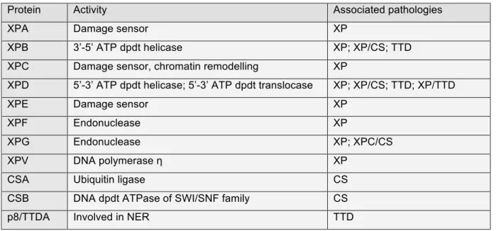
Table 4: NER factors and associated pathologies ... 42
Table 5: Facteurs NER et maladies associées ... 141
Figure 1: Core promoter elements and respective binding GTF ... 16
Figure 2: Transcription mechanism by Pol II ... 21
Figure 3: Nuclear receptor classes (adapted from Mangelsdorf et al., 1995) ... 24
Figure 4: Cytosine methylation and demethylation (from Kang and Hyun, 2017) ... 28
Figure 5: Nucleosome core particle (from Luger et al., 1997) ... 29
Figure 6: Histones post-translational modifications (from Kouzarides, 2007) ... 31
Figure 7: Lysine acetylation ... 33
Figure 8: Arginine and Lysine methylation ... 34
Figure 9: The two NER pathways ... 38
Figure 10: Initiation de la transcription ... 139
Figure 11: complexe Médiateur et maladies associées ... 142
Figure 12: XPC contrôle l’acétylation de H3K9 et la tri-méthylation de H3K4 ... 143
Figure 13: expression des gènes JUN, FOS et EGR1 ... 144
Abbreviations
6-4 PP 6-4 pyrimidine pyrimidone photoproducts AP-1 Activator Protein-1
AR Androgen Receptor
ATAC Ada Two A-Containing
ATP Adenosine TriPhosphate
ATRA All-Trans Retinoic Acid
bp base pairs
ATAC Ada Two A-Containing
BER Base Excision Repair
BRE TFIIB Recognition Elements
CAK CDK-Activating Kinase
CBP CCNC/H CREB-Binding Protein Cyclin C/H CDK Cyclin-Dependent Kinase CETN2 Centrin2
COFS cerebello-oculo-facio-skeletal syndrome
CPD cyclobutane-pyrimidine dimers
CPDD CTCF-Pair-Defined Domain
CPE Core Promoter Elements
CPSF Cleavage and Polyadenylation Specificity Factor
CS Cockaine syndrome
CSA/B Cockaine Syndrome complementation group A/B protein CstF Cleavage stimulatory Factor
CTCF CCCTC-binding protein CTD C-Terminal Domain
DBD DNA Binding Domain
DCE Downstream Core Element
DDB Damage specific DNA Binding protein
DNA Deoxy-riboNucleic Acid
DnmT DNA methyltranferase DP1-3 Dimerization Partner 1-3 DSIF DBR Sensitivity-Inducing Factor
DPE Downstream Promoter Element
E2F Andenovirus E2 gene Factor
EGR-1 Early Growth Response-1 protein
ELL Eleven-nineteen Lysine-rich Leukaemia protein
ER oEstrogen Receptor
ES GCN5 GG-NER
Embryonic Stem
General Control of Nutrition protein 5 Global-Genome NER
GNAT GCN5-related N-Acetyl Transferase
GR Glucocorticoid Receptor
GTF General Transcription Factors
HAT Histone Acetyl Transferase
HDAC Histone De-ACetylases
HNF-4 Hepatocyte Nuclear Factor-4
HPTM Histones Post-Translational Modification
HR Homologous Repair
HR23B Homologue Rad 23 B
HRE Hormone Response Element
HSP Heat Shock Proteins
IEG Immediate Early response Genes
Inr Initiator
KAT2A LBD
Lysine (K) AcetylTransferase 2A Ligand Binding Factor
LMR Low Methylated Regions
LRG Late Response Genes
mRNA messenger RNA
MAT1 Ménage-À-Trois 1 protein
MMR Mis Match Repair
MR Mineralocorticoid Receptor
MTE Motif Ten Element
MYST MOF - Ybf2/Sas3 - Sas2 - Tip60 NCOR Nuclear receptor Co-Repressor NELF Nasal Embryonic LHRH Factor
NER Nucleotide Excision Repair
NHEJ Non-Homologous End Joining PCAF
PCNA
p300/CBP-Associated Factor Proliferating Cell Nuclear Antigen
PIC Pre-Initiation Complex
PR Progesterone Receptor
Pol Polymerase
PPAR Peroxisome Proliferator-Activated-Receptor PRMT aRginine Methyl Transferases
P-TEFβ Positive Transcription Elongation Factor β PTM Post-Translational Modification
RNA RiboNucleic Acid
RAR Retinoic Acid Receptor
RARE Retinoic Acid Response Element
Rpb RNA Polymerase B
RPA Replication Factor A
RXR Retinoid X Receptor
SAGA Spt-Ada-Gcn5 Acetyltransferase Sen1 Senatoxin1
Ser Serine
Sirt Sirtuin
SMRT Silencing Mediator of Retinoic acid and Thyroid hormone receptor snRNA small nuclear RNA
snoRNA small nucleolar RNA
SCC Stem Cell Coactivator
T3R Thyroid hormone Receptor
TAD Trans-Activation Domain
TAF TBP Associated Factors
TBP TATA Binding Protein
TC-NER Transcription-Coupled NER
TFIIA-H/S general Transcription Factor of Pol II A-H/S
TDG TPA
Thymine DNA Glycosylase TetradecanoylPhorbol Acetate
TR2 Testicular Receptor
TRE TPA Responsive Element
TSS Transcription Start Site
TTD Trichothiodystrophy
Tyr Tyrosine
UV Ultra-Violet
VAD Vitamin A Deficiency syndrome
VDR Vitamin D Receptor
XCPE1/2 X Core Promoter Element 1/2
XP Xeroderma Pigmentosum
Transcription is a fundamental process of living cell. It allows the genetic information present in Deoxy-riboNucleic Acid (DNA) to be duplicated on a RiboNucleic Acid (RNA) molecule. A recent study, performed In human cells, has shown that nearly three quarters of the genome can potentially be transcribed(Djebali et al., 2012).
For protein coding genes, transcription is first of two processes that allow protein synthesis. In such case, the molecule is called messenger RNA (mRNA) and is subsequently translated into a functional protein by the ribosomal machinery.
In living cells, a dedicated enzyme called RNA polymerase (Pol) is accomplishing the process of transcription. While there is only one RNA Polymerase in Archea and Bacteria, distinct ones have been isolated and characterised in Eucharyota(Pikaard et al., 2008; Roeder and Rutter, 1970). Pol I is mostly dedicated to the synthesis of ribosomal RNA while Pol III is responsible for the synthesis of transfer RNA and other non-coding RNAs like snRNA, snoRNA or microRNA. Pol IV and V had only been identified in Plantae Kingdom and are dedicated to the synthesis of small interfering RNA. Finally, Pol II is the polymerase eliciting mRNA synthesis. In this manuscript, I will describe the mechanisms of transcription by Pol II and the results we obtained when we investigated it.
I - RNA polymerase II mediated transcription
1) The RNA polymerase II
RNA polymerase II was first isolated by Roeder and colleagues(Roeder and Rutter, 1970). RNA polymerase II also called RNA Polymerase B (Rpb) is a large protein complex composed of twelve subunits named from Rpb1 to Rpb 12 and collectively weighting more than 500 kDa (Table 1). The five subunits Rpb 5, 6, 8, 10 and 12 are in common with the RNA polymerase I and III(Woychik and Young, 1990). Some of them are also conserved with their yeast counterparts(McKune et al., 1995) and to a certain extent with their bacterial counterparts.
Pol II consists of a ten subunits core sub-complex associated with the stalk sub-complex(Armache et al., 2003). The whole complex is first assembled in the
transferred in the nucleus through the nuclear import machinery(Boulon et al., 2010). This assembly starts with the formation of several sub-complexes including Rpb1-Rbp8-HSP90, before the full complex to be assembled. During transcription, the two largest subunits Rpb1 and Rpb 2 are forming a positively charged “cleft” where negatively charged DNA can bind. Rpb1 also forms a mobile “clamp”, which is then close around the DNA-RNA duplex, building a tunnel like structure. Rpb2 forms the “wall” that delineates the extremity of the tunnel(Armache et al., 2003).
Table 1: RNA Polymerase II
Sub-complexes Sub-assemblies Subunits Features Size (kDa)
Core Rpb1 sub-assembly Rpb1 Phosphorylation sites 191.6
Rpb5 Common in Pol I, Pol II, Pol III 25.1 Rpb6 Common in Pol I, Pol II, Pol III 17.9 Rpb8 Common in Pol I, Pol II, Pol III 16;5
Rpb2 sub-assembly Rpb2 NTP binding site 138.8
Rpb9 14.3
Rpb3 sub-assembly Rpb3 Promoter recognition 35.3
Rpb10 Common in Pol I, Pol II, Pol III 8.3
Rpb11 13.6
Rpb12 Common in Pol I, Pol II, Pol III 7.7
Stalk Rpb4 25.4
Rpb7 Unique to Pol II 19.1
The C-Terminal Domain (CTD) of Rpb1 subunit forms a special domain of the polymerase not necessary for its catalytic activity in vitro but allowing the specific control of its function in vivo(Serizawa et al., 1993). It is composed of a hepta-peptide repeated 26 times in yeast and 52 times in human with almost regular sequence of YSPTSPS. Regulation occurs through phosphorylation and dephosphorylation of the tyrosine, threonine and serine residues Tyr1-Ser2-Thr4-Ser5-Ser7, numbered relative to their position in the hepta-peptide. The Pre-Initiation Complex (PIC), including the general transcription factors, has been shown to preferentially associate with the dephosphorylated form of Pol II(Lu et al., 1991; Robinson et al., 2012). Ser5 and Ser7 are phosphorylated by the Cyclin-Dependent Kinase 7 (CDK7), part of the general transcription factor TFIIH, leading to the initiation of transcription and to the escape of the polymerase from the promoter(Wong et al., 2014; Zhou et al., 2000).
Ser 5 is also implicated in the recruitment of the capping machinery at the 5’ extremity of the transcribed gene(Schroeder et al., 2000). Both marks are removed shortly after transcription initiation. Ser2 phosphorylation allows the promoter– proximal pause release(Jonkers and Lis, 2015). This mark is retained on the CTD during elongation and is required for splicing and cleavage machinery to be co-recruited(Gu et al., 2013). In addition to Ser2, Tyr1 and Thr4 phosphorylation are needed to allow the arrival of the poly-adenylation machinery(Harlen et al., 2016; Mayer et al., 2012).
2) Promoter
1. Organisation
Promoter is a region of the gene where the transcription machinery assembled. This region includes the Transcription Start Site (TSS) where the transcription actually initiates.
Two major types of promoter organisation have been described in the past decade, focused and dispersed(Carninci et al., 2006). The first one is characterised by the presence of one TSS, positioned at a specific nucleotide or eventually some contiguous nucleotides. On the other hand, the second promoter type corresponds to several weakly actionable TSS that are spread on a 50 to 100 bp region. Aside from those two main types, a few peculiar promoters have been described to contain a principal TSS surrounded by several weak ones.
If the focused type seems the more frequent in simpler organisms, it represents only a third of human genes. Analysing them has led to the discovery of major Core Promoter Elements (CPE) like TATA box, BRE, XPCE1 or DPE. Focused promoters are mostly present at developmental and highly regulated genes. Dispersed promoters have been less studied even though they represent at least two third of human promoters. They are typically found in CpG regions and are controlling housekeeping or constitutive genes. The manuscript will concentrate on focused promoters.
2. Structure
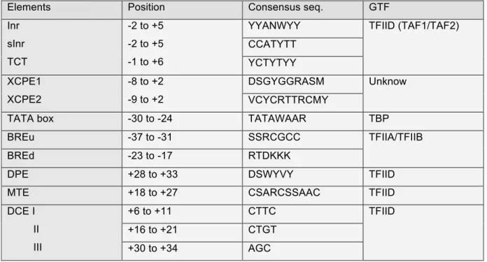
The core promoter is the DNA sequence where the PIC assembles and the transcription initiation occurs. It is organized around the TSS referred to as +1 nucleotide and is generally considered to span 35 to 40 nucleotides upstream and downstream of it. Core promoters contain variable number of elements with various nucleotide sequences; the first identified being the TATA box element by Chambon and colleagues. All sequences are given in accordance with the IUPAC nomenclature and resumed in Table 2.
Table 2: Core promoter elements
Elements Position Consensus seq. GTF
Inr sInr TCT -2 to +5 -2 to +5 -1 to +6
YYANWYY TFIID (TAF1/TAF2)
CCATYTT YCTYTYY XCPE1 XCPE2 -8 to +2 -9 to +2 DSGYGGRASM Unknow VCYCRTTRCMY
TATA box -30 to -24 TATAWAAR TBP
BREu -37 to -31 SSRCGCC TFIIA/TFIIB
BREd -23 to -17 RTDKKK
DPE +28 to +33 DSWYVY TFIID
MTE +18 to +27 CSARCSSAAC TFIID
DCE I II III +6 to +11 CTTC TFIID +16 to +21 CTGT +30 to +34 AGC
2.a. Initiator (Inr)
In 1980, Chambon described Inr, as a sequence enriched around the TSS of Pol II transcribed genes(Corden et al., 1980). Smale latterly defined this sequence encompassing the TSS, as able to drive transcription initiation in vivo and in vitro, without the need of a TATA box. Indeed, the presence of the TATA box or other CPE seems only needed to potentiate the initiation. Pyrimidine rich Inr corresponds to YYANWYY, where the A is defined as the +1 nucleotide(Javahery et al., 1994; Smale and Baltimore, 1989), and the whole sequence is present at nearly half of human genes(Gershenzon and Ioshikhes, 2005). It is recognized by the general transcription
factor TFIID(Kaufmann and Smale, 1994), especially TAF1 and TAF2(Chalkley and Verrijzer, 1999).
Recently, a strict Inr (sInr), with a more restrictive sequence has been found in TATA less promoters where it seems to bypass TATA box by cooperating with the Sp1 sequence(Yarden et al., 2009). CCATYTT sequence was defined as sInr and is present in 1,5% of human genes.
Finally, an alternative pyrimidine rich initiator element called TCT has been described to encompass the TSS of many genes and notably members of the ribosome gene family. Its sequence is YCTYTYY, where C appears to be the TSS(Parry et al., 2010).
2.b. X gene core promoter element (XCPE1/2)
The X Core Promoter Element 1 (XCPE1) has been characterised as able to induce transcription initiation of hepatitis virus X gene and lately found in 1% of human promoter, especially TATA less ones. It spans from -8 to +2 with the sequence DSGYGGRASM, where A is the TSS(Tokusumi et al., 2007).
The XCPE2 core promoter element directs transcription initiation of the second promoter of hepatitis virus X gene and have the sequence VCYCRTTRCMY, where M is the TSS(Anish et al., 2009). It is found at multiple TATA less human promoters.
2.c. TATA box
The TATA box has also been described by Chambon and named after its sequence composition. It was first thought to be a general transcription CPE. Nowadays, it is assumed that it is only present in 10% to 20% of human genes. Its consensus sequence, TATAWAAR, is present upstream to the TSS at nucleotide -28. Inr is associated with around 60% of all TATA box (Gershenzon and Ioshikhes, 2005). The TATA box is well conserved from Archea to human.
2.d. TFIIB Recognition Elements (BRE)
The TFIIB Recognition Elements (BRE) are two sequences located directly upstream (BREu)(Lagrange et al., 1998) and downstream (BREd)(Deng and Roberts, 2005) of the TATA box, but may also be present in TATA less promoters. Both sequences are well conserved from Archea to human and their sequences are
SSRCGCC and RTDKKK respectively. They are recognised by the general transcription factors TFIIA and TFIIB.
2.e. Downstream Promoter Element (DPE)
As its name suggests, the Downstream Promoter Element (DPE) is located downstream the +1 nucleotide between +28 and +33, with the consensus sequence DSWYVY. Its presence is apparently independent of either TATA box or Inr(Burke and Kadonaga, 1997; Gershenzon and Ioshikhes, 2005). The DPE element is notably associated with developmental regulatory genes(Zehavi et al., 2014). It is recognised and bound by the general transcription factor TFIID.
2.f. Motif Ten Element (MTE)
The Motif Ten Element (MTE), named after computational analysis(Ohler et al., 2002), is located downstream of the TSS and directly upstream of the DPE. It spans from +18 to +27 with the sequence CSARCSSAAC. MTE requires association with Inr sequence. Even though it can act independently of TATA box or DPE, their presences strongly reinforce its transcription initiation activity(Lim et al., 2004). MTE is also recognised by TFIID.
More recently, MTE and DPE combination have been found to form a bridge motif and to constitute an independent core promoter element(Theisen et al., 2010).
2.g. Downstream Core Element (DCE)
First observed in β-globin gene, the Downstream Core Element (DCE) is a downstream element alternative to DPE, MTE or Bridge motif(Lewis et al., 2000). It is composed of three sequences, spanning from +6 to +11 (necessary motif CTTC), from +16 to +21 (necessary motif CTGT) and from +30 to +34 (necessary motif AGC). It is recognised by TFIID(Lee et al., 2005).
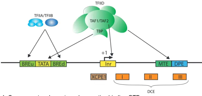
Figure 1: Core promoter elements and respective binding GTF
The diagram shows a promoter with most common CPE and the proteins from the transcription machinery that contact them.
3) Pre-initiation complex
Even if RNA polymerase II alone is able to transcribe DNA templates in vitro, it requires the assembly of a large Pre-Initiation Complex (PIC) for transcription to start at the right nucleotide in a well-coordinated manner in vivo(Weil et al., 1979).
1. General transcription factors
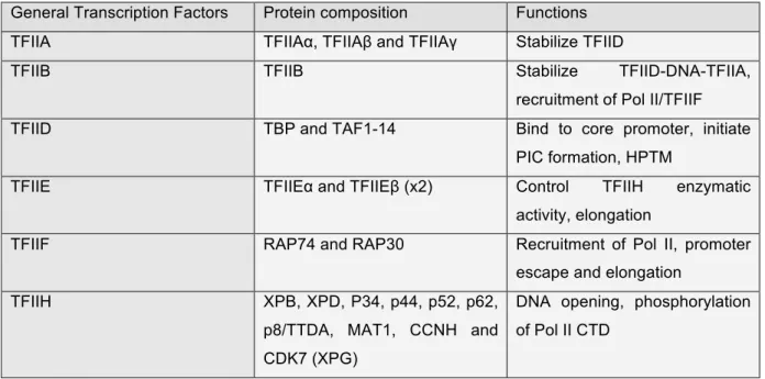
General transcription factors (GTF) have first been purified from human cells in 1980 (Matsui et al., 1980) and further characterized(Reinberg and Roeder, 1987; Reinberg et al., 1987; Samuels et al., 1982). They are named TFIIA, TFIIB, TFIID, TFIIE, TFIIF and TFIIH for basal/general Transcription Factors associated with RNA Polymerase II; the last letter corresponds to purification fractions. When assembled with Pol II on the core promoter, they form the Pre-Initiation Complex. They allow the correct positioning of Pol II on the promoter and the transcription to start at the right nucleotide.
As well as core promoters largely vary in their sequence compositions; nowadays it is assumed that PIC composition may vary, especially for TFIID. Some subunits of general transcription factors seem to be specific to certain set of genes, cell types or even tissues(Akhtar and Veenstra, 2011). Here, we will describe their widely observable composition (Table 3).
TBP TAF1/TAF2 TFIID TFIIA/TFIIB DCE +1 II I III Inr TATA XCPE1 BREd
1.a. TFIIA
The general transcription factor TFIIA is a three subunit complex composed of TFIIAα, TFIIAβ and TFIIAγ. It has been shown to interact with the BRE elements to stabilize TFIID(Buratowski et al., 1989; Lee et al., 1992).
1.b. TFIIB
The general transcription factor TFIIB is a single protein. It stabilizes the TFIID-DNA-TFIIA complex and helps the subsequent recruitment of the RNA polymerase II(Ha et al., 1993; Maldonado et al., 1990) together with TFIIF. TFIIB participates in determining TSS position(Li et al., 1994) and recognizes BRE elements. It regulates itself by auto-acetylation(Choi et al., 2003) and participates in promoter escape(Westover et al., 2004).
1.c. TFIID
The general transcription factor TFIID is the largest complex among all GTF. It is composed of the TATA Binding Protein (TBP) and 14 TBP Associated Factors (TAF). As its name indicates, TBP binds the consensus sequence of the TATA box(Corden et al., 1980; Sawadogo and Roeder, 1985). For either TATA less or TATA containing promoters, the whole TFIID complex binds the core promoter to initiate the PIC formation. TAF1/TAF2 dimer binds Inr element(Chalkley and Verrijzer, 1999). TAF1 is also able to bind DCE(Lee et al., 2005) and TAF6/TAF9 is able to bind both DPE(Burke and Kadonaga, 1996) and MTE(Shao et al., 2005). Some of the subunits are also able to interact with certain nuclear receptors(Lavigne et al., 1999) or to modify histone proteins(Mizzen et al., 1996; Pham and Sauer, 2000), in order to regulate transcription. Furthermore, TFIID interacts with TFIIF(Dikstein et al., 1996).
1.d. TFIIE
The general transcription factor TFIIE is composed of two copies of each subunit TFIIEα and TFIIEβ, forming a hetero-tetramer. It directly interacts with Pol II and DNA promoter as well as TFIID, TFIIF and TFIIH(Maxon et al., 1994; Okuda et al., 2004; Tanaka et al., 2009). It is mostly implicated in the control of TFIIH enzymatic activities and required for Pol II transcription, driving both
initiation(Ohkuma and Roeder, 1994) and transition to elongation(Watanabe et al., 2003).
1.e. TFIIF
The general transcription factor TFIIF is a complex of two proteins, RAP74 and RAP30. It is responsible, especially the small subunit, for the incorporation of Pol II into the PIC and its stable binding on promoter(Flores et al., 1991; Killeen et al., 1992). TFIIF is also essential in promoter escape and proper elongation(Yan et al., 1999; Zhang and Burton, 2004).
1.f. TFIIH
TFIIH is a dual protein complex, as it acts both in transcription and in the Nucleotide Excision Repair (NER) pathway of DNA damage response. It is composed of two sub-complexes, the core and the CDK-Activating Kinase (CAK). The first one is a six subunit ensemble containing Xeroderma Pigmentosum group B protein (XPB), p34/44/52/62 and the lastly discovered p8/TTDA(Giglia-Mari et al., 2004). The CAK is a three subunits complex composed by Ménage-À-Trois 1 protein (MAT1), CDK7 and its associated cyclin H (CCNH). Those two sub-complexes are stabilized and held together by Xeroderma Pigmentosum group D protein (XPD)(Coin et al., 1998; Sandrock and Egly, 2001). In addition, Xeroderma Pigmentosum group G protein (XPG) that is known to participate with TFIIH in NER, have recently been found to directly interact with the core in order to stabilize the whole complex and to control its nuclear receptor phosphorylation activity(Ito et al., 2007).
The XPB protein possesses an ATP-dependent DNA helicase activity. A more recent study enlightened a XPB ATP-dependent translocase activity(Fishburn et al., 2015). TFIIH alters transcription initiation by making the PIC unstable(Plaschka et al., 2016). This block seems to be released by the translocase activity of XPB that would allow the promoter opening(Alekseev et al., 2017).
CDK7 works in collaboration with its partner CCNH and their interaction are stabilised by MAT1, the third member of the CAK(Devault et al., 1995). CDK7 is then able to phosphorylate the CTD Ser5 and Ser7 residues of the Pol II subunit Rpb1(Wong et al., 2014; Zhou et al., 2000). It also controls indirectly the phosphorylation of Ser2 residues by CDK9(Larochelle et al., 2012). Finally, CDK7
also phosphorylates TFIID, TFIIE and TFIIF general transcription factors(Ohkuma and Roeder, 1994).
CDK7 is also implicated in the cell cycle progression. XPD controls CDK7 activity (Chen et al., 2003). XPD notably control the cellular localisation of CDK7 to regulate its mitotic kinase activity and the chromosomal segregation(Li et al., 2010).
Table 3: General transcription factors
General Transcription Factors Protein composition Functions
TFIIA TFIIAα, TFIIAβ and TFIIAγ Stabilize TFIID
TFIIB TFIIB Stabilize TFIID-DNA-TFIIA,
recruitment of Pol II/TFIIF
TFIID TBP and TAF1-14 Bind to core promoter, initiate
PIC formation, HPTM
TFIIE TFIIEα and TFIIEβ (x2) Control TFIIH enzymatic
activity, elongation
TFIIF RAP74 and RAP30 Recruitment of Pol II, promoter
escape and elongation
TFIIH XPB, XPD, P34, p44, p52, p62,
p8/TTDA, MAT1, CCNH and CDK7 (XPG)
DNA opening, phosphorylation of Pol II CTD
Side to its roles in transcription, the TFIIH complex is a major actor of the NER DNA repair pathway, as described in the second part of the introduction.
2. PIC assembly and transcription process
There are several critical steps for transcription to be productive. It starts by the binding of Poll II and general transcription factors on the promoter, which assemble to form the PIC and to initiate the transcription. Then, Pol II performs the elongation along the gene body. Finally, termination occurs and a newly synthesised mRNA is released (Figure 2). While the Mediator complex is largely implicated in the formation of the PIC by helping the recruitment of the transcription machinery and by bridging transcription factors with the transcription machinery, its roles will be detailed in the third part of the introduction.
2.a. PIC formation
Transcription starts with the assemblage of the PIC around the TSS. It can happen in two different manners: the stepwise assembly or the holoenzyme assembly.
The stepwise assembly (aka sequential assembly) starts by the recognition of the promoter by TFIID. TBP first recognizes the TATA box, if it is present. In any case, other TFIID subunits recognise Inr and DPE to interact with the promoter. This interaction is then stabilized and strengthened by the arrival of TFIIA and TFIIB. Subsequently, Pol II joins the promoter in association with TFIIF. TFIIE and TFIIH are the last to be recruited to the PIC(He et al., 2013).
The Pol II holoenzyme consists of a large already assembled complex containing the Pol II, the Mediator complex and most of the general transcription factors except TFIID/TFIIA. In the holoenzyme pathway, it assembles without DNA and comes in almost one single step onto the promoter(Koleske and Young, 1995).
2.b. Initiation
From now on, TFIIH is essential for the opening of DNA. This ATP dependent step allows the transition from a closed conformation to an open conformation PIC. As mentioned above, various scenarios are possible for TFIIH. The DNA opening creates an 11-15 bp bubble and the non-coding strand is then inserted in the active site of Pol II. The first nucleotide synthesis converts the open conformation complex into an initially transcribing complex(Hantsche and Cramer, 2017). This complex enters several abortive initiations that only produces short oligonucleotides of less than 10 bp. If the newly synthetized RNA reaches 10 bp, then Pol II is phosphorylated by TFIIH and dissociates from the initiation complex, in order to start elongation. This step is called promoter clearance. After the RNA reaches a length of 25 bp, the capping machinery is recruited and this step allows productive transcription to start.
2.c. Proximal pausing/Elongation
Pol II encounters a pausing event shortly after starting productive transcription due to DSIF and NELF. Indeed, they bind Pol II and repress its elongation activity. The phosphorylation of NELF by P-TEFβ-associated CDK9 leads to its dissociation.
Pol II is phosphorylated on Ser2. DSIF is also phosphorylated and turn into an activator to promote productive elongation(Bernecky et al., 2017; Yamada et al., 2006).
Figure 2: Transcription mechanism by Pol II
The diagram shows the promoter recognition and the recruitment of the different components of the transcription machinery as well as the cycle of transcription by Pol II.
pol II pol II pol II pol II IIB IIH IIF IIE IID IIA IIH IIF IIE IIA IIB pol II pol II IIB IID IIA IIE pol II IIB IID IIA IIH IIF IIE pol II IIF NELF DSIF CPSF CstF
1-Promoter recognition by TFIID
2-Pre-Intiation Complex assembly
3-Transcription initiation and promoter clearance
4-Promoter proximal pausing follow by elongation
5-Cleavage and termination
IID IIH IIF P-TEFb CDK9 nascent mRNA P P P P P P Ser5 Ser5 Ser2 Ser2 P Ser7 P Ser7 P Ser2 P Tyr1 P Thr4 P Ser2 P Tyr1 P Thr4
Pol II continues to slide down to the 3’ end of the gene, associated with the elongation factors TFIIS, ELL, Elongin and P-TEFβ. These factors sustain the DNA/RNA hybrid, that is necessary for Pol II processivity during elongation(Wind and Reines, 2000).
2.d. Termination/poly-adenylation
The termination occurs when the elongation complex reaches the 3’ end of the gene. There are two main ways to end transcription, depending of the gene sequence(Kuehner et al., 2011).
The first pathway involves the Cleavage and Polyadenylation Specificity Factor (CPSF), which interacts with the Pol II CTD and recognizes a specific poly A signal at the 3’ end of the gene. It induces the elongation to stop. Then, the Cleavage stimulatory Factor (CstF) induces the cleavage of the RNA. Pol II is then released and the newly synthesised RNA is polyadenylated. The remaining part of the RNA, downstream of the cleavage site, is therefore degraded by XRN2. Most of the human genes follow this path.
But for other genes, the termination is achieved through the dissociation of the RNA/DNA complex by Senataxin 1 protein (Sen1). In such case, the resultant RNA is not polyadenylated.
4) Specific transcription factors
A large number of actors are required for transcription to occur in the right full place of the genome, in a well-controlled and coordinated manner. Several proteins and protein complexes are involved to initiate transcription including specific transcription factors, coactivators and the Mediator complex. The last one will extensively be explained in the third part of the introduction.
Specific transcription factors have been selected through evolution by the cells to answer various stimuli and stresses. Their role is to stimulate (activator) or to inhibit (repressor) transcription of specific genes to allow cell division, cell growth and cell death but also cell differentiation, stem cell maintenance or cellular response to stresses.
Specific transcription factors operate through enhancer and silencer regions. These genomic regions are enriched in specific DNA sequences called Response Elements (RE), where transcription factors can bind. Transcription factors are all characterised by a DNA Binding Domain (DBD) to directly contact DNA on specific RE and a Trans-Activation Domain (TAD) to transfer signal to the transcription machinery. The sum of activator and repressor signals finally determines the level of gene activity, ranging from no transcription to high transcribing activity.
Several types of transcription factor have been identified, depending on their structure, their mode of activation and the biological processes in which they are implicated.
1. E2F transcription factor family
The first E2F protein has been discovered as a cellular factor required for adenovirus E2 gene induction(Kovesdi et al., 1986), and lately defined as E2F1. Height different E2F transcription factors have been identified in this family and assigned as activators (E2F1-3) or repressors (E2F4-8). They bind DNA as heterodimer with a Dimerization Partner (DP1-3). This partner is also able to modulate E2F activity(Bandara et al., 1994; Girling et al., 1993; Ingram et al., 2011; Ormondroyd et al., 1995). E2F transcription factors are involved in cell cycle progression and apoptosis, mainly through transcription regulation(DeGregori and Johnson, 2006; DeGregori et al., 1997).
E2F1 is implicated in G1-S phase progression(Yao et al., 2008), where it controls an all-or-nothing process. Division has to occur or cell would enter apoptosis(Wu et al., 2017). E2F1 mainly acts as transcription activator during G1/S phases(Araki et al., 2003; Burke et al., 2014). Indeed, it activates genes responsible for DNA synthesis. Moreover, acetylation of histone H3 and H4 accumulates during G1 phase in an E2F1 dependant manner, in order to open chromatin and allow gene activation as well as S phase entry(Taubert et al., 2004).
Then, it is made inactive during S/G2 phases(Schulze et al., 1995; Zhang et al., 2000). Notably, E2F1 is phosphorylated by TFIIH via an interaction with its p62 subunit. This interaction leads to E2F1 degradation during S phase(Vandel and Kouzarides, 1999).
E2F1 also have some roles in the NER pathway that will be presented in the second part of the introduction.
2. Nuclear receptors
Nuclear receptors are ligand-dependent transcription factors, limited to
Metazoans. They are characterised by a particular Ligand Binding Domain (LBD).
They act either as monomers or dimers but need to bind their specific ligand to be activated and thus modulate transcription(Escriva et al., 2004).
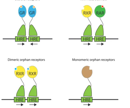
Figure 3: Nuclear receptor classes (adapted from Mangelsdorf et al., 1995) 2.a. Mechanistic classes
There are four main classes of nuclear receptors(Mangelsdorf et al., 1995). The first one includes steroid receptors that bind as homodimers to two repeated but inverted Hormone Response Elements (HRE), separated by several nucleotides. They are activated when associated to their steroid ligand. It includes Estrogen Receptor (ER), Progesterone Receptor (PR), Androgen Receptor (AR), Glucocorticoid Receptor (GR) and Mineralocorticoid Receptor (MR). The second class corresponds to receptors that bind to DNA as a heterodimer complex with Retinoid X Receptor (RXR), to direct repeat HRE. Among many others, it includes
HRE
HRE HRE HRE
RXR RAR HRE HRE RXR RXR HRE GR GR
Steroid receptors RXR Heterodimers
Retinoic Acid Receptor (RAR), Thyroid hormone Receptor (T3R), Vitamin D Receptor (VDR) or Peroxisome Proliferator-Activated-Receptor (PPAR). The third class corresponds to homodimeric nuclear receptors that bind direct repeat HRE. RXR, Testicular Receptor (TR2) or Hepatocyte Nuclear Factor-4 (HNF-4) belongs to this class. Finally, the fourth and last class includes receptors that act either as monomers or dimers but when only one DNA binding domain contacts one half-site HRE. Most of the orphan receptors fall in the last two classes.
2.b. Retinoic acid receptor
Vitamin A is essential for several biological processes like embryogenesis, organ development, homeostasis, immunity and reproduction. Its biological significance is enlightened by fundamental studies but also by clinical observations. Indeed, deficiency in vitamin A can lead to growth retardation, intellectual disability and other symptoms of the Vitamin A Deficiency (VAD) syndrome(Clagett-Dame and DeLuca, 2002; Zile, 2001).
Vitamin A corresponds to a group of organic compound including retinol, retinal and All-Trans Retinoic Acid (ATRA), the last one being the most biologically active form. Its physiological level is tightly regulated(Shannon et al., 2017). The retinoic acid receptor is a ligand-dependent transcriptional regulator. It includes three homologous receptors called RARα, RARβ and RARγ. They form a heterodimer with RXR and activate transcription when they are fixed by their ligand.
The heterodimer RAR/RXR binds to the Retinoic Acid Response Element (RARE) that corresponds to two successive repeats of RGKTCA separated by variable number of nucleotides. When no ligand is associated with RAR/RXR dimer, co-repressors like Nuclear receptor Co-Repressor (NCoR aka NCOR1) or Silencing Mediator of Retinoic acid and Thyroid hormone receptor (SMRT aka NCOR2)(Perissi et al., 2010) are bound to it and serve as a platform for the recruitment of Histone De-ACetylases (HDAC). The latter maintains chromatin in heterochromatin state, blocking transcription.
Upon ligand binding, RAR encounters conformational changes. This process leads to co-repressor release and coactivator arrival(Perissi et al., 2004). It is
accompanied by the recruitment of several Histone Acetyl Transferase (HAT) and Histone Methyl Transferase (HMT), which open chromatin and enable transcription.
Retinoic acid seems able to partially modify the genomic location of certain RAR receptors through a dynamic process and to induce the transcription of previously unbound but very specific genes(Mahony et al., 2011).
3. Immediate early genes 3.a. Primary response
The transcription of certain genes can be activated very quickly and transiently by several stimuli without the need of new protein synthesis. These genes are divided into two classes: the Immediate Early response Genes (IEG) and the delay primary response genes(Tullai et al., 2007). Some of them code for transcription factors that subsequently induce the secondary response(Winkles, 1998).
IEG mRNA appear in the cell few minutes after stimulation. Their transcription is activated by various stimuli like growth factors, mitogens, phorbol esters, immunological and neurological signals or stresses (Ultra-Violet (UV), toxins)(Fowler et al., 2011; Greenberg and Ziff, 1984; Herschman, 1991; Morgan and Curran, 1991). The two most characterised IEG are JUN and FOS(Healy et al., 2013). They are playing roles in cell proliferation, differentiation and survival. They can be regulated by Post-Translational Modifications (PTM), which influence their ability to form a dimer, thus to bind DNA and activate transcription. FOS and JUN form an active heterodimer complex called Activator Protein-1 (AP-1), via a leucine zipper motif. When associated, they form a bipartite DBD. The complex binds DNA at specific responsive element called TetradecanoylPhorbol Acetate (TPA) Responsive Element (TRE).
Early Growth Response-1 protein (EGR1) is also a transcription factor-coding IEG, involved in cell growth and differentiation. It binds DNA on specific consensus sequences and interacts with the transcription machinery(Liu et al., 2001; Zhang et al., 2003).
3.b. Secondary response
The secondary response corresponds to the late induction of a set of genes by the products of the primary response. They are called Late Response Genes (LRGs), as well as secondary response genes.
5) Chromatin regulation of transcription
If DNA is bound by a large variety of protein in different dynamic processes, including transcription, it is rarely naked or freely accessible inside the nucleus. Indeed, DNA can be chemically modified to alter its accessibility. Moreover, it is incorporated into chromatin where it is physically compacted.
1. DNA methylation
DNA methylation is well conserved through evolution. It corresponds to the addition of a methyl group to the 5 position of a cytosine residue (5mC) in the context of a CpG dinucleotide. In mammals, DNA methylation is implicated in transcription regulation but also in X-inactivation, genomic imprinting and the silencing of transposable elements. The genomic pattern of methylation is established during embryonic life by DNA methyltranferase Dnmt3 and is latterly maintained by Dnmt1 during mitosis.
DNA methylation of promoter regions is associated with repression. The presence of methyl groups inside the major groove of DNA is supposed to engendered hindrance that perturbed DNA/TF contacts and block gene activation(Watt and Molloy, 1988). In the opposite, the binding of TF to DNA can also influence its methylation status and generate Low Methylated Regions (LMR)(Stadler et al., 2011).
DNA methylation also participates to gene repression by attracting proteins of the Methyl CpG Binding Domain (MBD) family that are able to repress transcription(Bird and Wolffe, 1999).
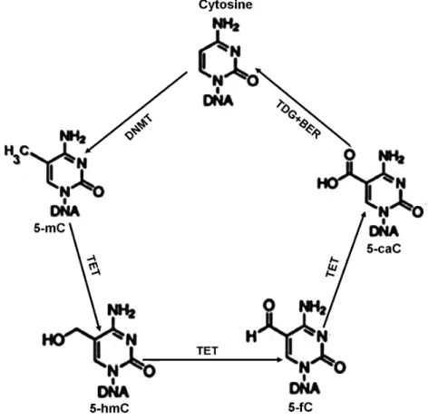
To actively regulate transcription, DNA methylation is dynamically altered in cell. Notably, TET deoxygenase is responsible for 5mC oxidation to hydroxymethylcytosine (5hmC), then to formylcytosine (5fC) and finally to
5-carboxycytosine (5caC). Both 5fC and 5caC can be removed by DNA glycosylase to be restored to regular cytosine by BER (Figure 4)(Weber et al., 2016).
Figure 4: Cytosine methylation and demethylation (from Kang and Hyun, 2017)
2. Chromatin organisation
To protect it from physical damages but also as a transcription regulation mechanism, DNA is incorporated into a nucleoprotein complex called chromatin. The DNA molecule wrap around several histone proteins, in order to form a nucleosome. The chromatin is composed of a succession of nucleosomes and inter-nucleosome linker sequences. It appears as beads regularly positioned along a string. This three-dimensional conformation, called euchromatin, constitutes the first level of a necessary condensation of the DNA molecule into the nucleus. This is also a way to regulate its accessibility and thus transcription.
2.a. Nucleosome
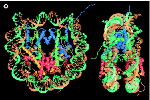
kDa (Figure 5). This octamer is composed of two copies of four histone proteins named H2A, H2B, H3 and H4(Luger et al., 1997). Histones are small proteins weighting from 11 to 22 kDa and well-conserved trough evolution. They are enriched in positive amino acids, like lysine and arginine, to interact with negatively charged DNA. The assembling of the octamer starts by the formation of H3-H4 dimers. Next, two H3-H4 dimers are assembled in a tetramer. Finally, two H2A-H2B dimers are added to the tetramer to form a complete octamer(Arents and Moudrianakis, 1995).This “beads on a string” fibre has a diameter of 11 nm and corresponds to the first level of DNA condensation.
Figure 5: Nucleosome core particle (from Luger et al., 1997)
A ninth “linker” histone named H1 can bind to linker sequences and form a higher ordered and more stable structure with a diameter of 30 nm(Robinson et al., 2006). Each nucleosome then encompasses 160 bp(Syed et al., 2010). Two hypotheses have been proposed for the structure of the 30 nm fibres. The solenoid model predicts that adjacent nucleosome follow a super-helicoidal path with 6 to 8 nucleosome per turn(Widom and Klug, 1985). The zigzag model postulates two nucleosomes with straight linker and successively interleaved(Chen and Li, 2010). Higher structure compactions are less clear but also affect transcription, as it alters access to DNA. This more compacted chromatin is called heterochromatin and does not allow transcription. In the cellular context, chromatin seems heterogeneous
depending on genomic regions or cell cycles phases. It implies dynamic processes to pack and unpack the chromatin and control the access to genetic information.
2.b. Histone variants
Histone proteins are divided into two classes. The canonical one includes the most abundant form of histones. They are produced during the S phase and are the major support for genomic DNA. They are produced along with the newly synthesised DNA and assembled with it. But beside the canonical forms, several histone variants have emerged through evolution(Talbert et al., 2012). They are produced all along the cell cycle and are dedicated to specific functions like transcription initiation and termination, cell division or maintenance of genome integrity. Variants exist for the core histone H2A, H2B, H3 and for the linker histone H1. They differ by few amino acids or additional domains but keep the common histone structure to allow an easy exchange with their canonical counterpart(Venkatesh and Workman, 2015).
Amongst many, the histone variant H3.3 is notably found at enhancer, promoter and in the body of actively transcribed genes, along with elongating Pol II. H3.3 turnover seems to compensate Pol II associated nucleosome displacement(Wirbelauer et al., 2005). In addition, H3.3 deposition at active promoters has been correlated to replication-independent E2F gene activation(Daury et al., 2006).
H2A.Z variant is highly conserved across evolution. It has strong sequence differences with canonical H2A but their similar three-dimensional structures allow them to be exchanged(Talbert and Henikoff, 2010). Subtle structural differences make H2A.Z containing nucleosomes less stable. Indeed, H2A.Z/H2B dimer has an affected interface with H3/H4 tetramer but also with canonical H2A/H2B dimer(Suto et al., 2000).
H2A.Z/H3.3 containing nucleosomes are highly unstable and accumulate at previously considered “nucleosome free region”, around promoters, enhancers and insulators. They maintain an open chromatin state and facilitate transcription factor accessibility(Jin et al., 2009).
2.c. Histone post-translational modifications
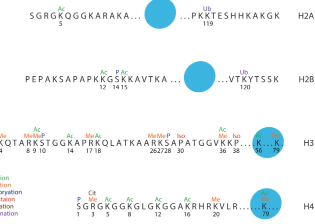
Histones Post-Translational Modifications (HPTM) also impacts transcription. Indeed, unfolded N-terminal tail of histones protrudes outside the nucleosome and is subjected to various enzymatic activities like phosphorylation, methylation, acetylation, ubiquitination, phosphorylation, citrullination, isomerisation and few others more recently discovered (Figure 6)(Kouzarides, 2007). Some modifications have also been identified inside the octamer core(Yu et al., 2012) or on the small protruding C-terminal part of certain histones. Modifications are generally present at specific spatial and/or temporal localisation and associated with either active transcription or repression.
Figure 6: Histones post-translational modifications (inspired from Kouzarides, 2007)
Histone modifications can modify nucleosome electric charges and impact histone/histone or histone/DNA contacts. They can also serve as a recruitment platform for other proteins that will further act on chromatin or transcription.
A specific nomenclature has been established(Turner, 2005): “H3K9ac”, H3 corresponds to the modified histone, K9 describes the amino acid lysine and its
S G R G K Q G G K A R A K A . . . P K K T E S H H K A K G K H2A P E P A K S A P A P K K G S K K A V T K A . . . V T K Y T S S K H2B A R T K Q T A R K S T G G K A P R K Q L A T K A A R K S A P A T G G V K K P . . . H3 S G R G K G G K G L G K G G A K R H R K V L R . . . H4 5 119 12 1415 120 2 3 4 8 910 14 17 18 26 2827 30 36 38 56 79 1 3 5 8 12 16 20 Ac Ac Ac Ac Ac Ac Ac Ac Ac Ac Ac Ac Me Me Me Me Me Me MeMe Me Me Me P P P P P Ub Citrullination Iso Ub Iso Cit Cit Acetylation Methylation Phosphoryation Isomeristaion Ubiquitination . K . . . K . 79 Ac Me . . . K . . .
position in the protein and ac define the chemical modification (ac: acetylation, me: mono-methylation, me2/3: di- or –tri-methylation, ub: ubiquitination, ph: phosphorylation, et caetera).
i. Acetylation
Acetylation is the first described HPTM(Allfrey, 1966), but it also targets other proteins(Choudhary et al., 2009). Histone Acetyl Transferase (HAT) can mediate N-acetylation of lysine residues(Kouzarides, 2007). It uses Acetyl-CoA as acetyl donor to catalyse the reaction. Acetylation suppresses the lysine’s positive charge and diminishes their interaction with the negatively charged DNA. It leads to chromatin decondensation and is generally associated with transcriptionally active genes(Spotswood and Turner, 2002). The reverse reaction is called deacetylation and is catalysed by histone deacetylase, to restore the lysine’s positive charge.
HAT are usually divided into two classes. Type A HAT are located in the nucleus, where they acetylate chromatin-associated histones. Type B HAT are located in the cytoplasm and are responsible for the acetylation of newly synthesised histones.
Type A HAT are subdivided into three families: i) GCN5-related N-Acetyl Transferase (GNAT) that includes General Control of Nutrition protein 5 (GCN5) and p300/CBP-Associated Factor (PCAF), ii) MYST family (MOF - Ybf2/Sas3 - Sas2 - Tip60) and iii) others like CREB-Binding Protein (CBP) and p300(Kimura et al., 2005).
GCN5 also know as Lysine (K) AcetylTransferase 2A (KAT2A) is responsible for H3K14 acetylation alone but it is also included in two different complexes named Ada Two A-Containing (ATAC) and Spt-Ada-Gcn5 Acetyltransferase (SAGA). Both complexes stimulate its acetylation activity, especially ATAC(Riss et al., 2015). In such protein complexes, GCN5 is found to acetylate both H3K9 and H3K14 but on distinct genomic positions. ATAC associated GCN5 markedly targets enhancer regions(Krebs et al., 2011; Nagy et al., 2010). Whether incorporated within the SAGA complex, GCN5 also acetylate H3K18 and H3K23 residues(Rodríguez-Navarro, 2009).
In the context of nuclear receptor activation, GCN5/PCAF and CBP/p300 have been shown to be recruited on the promoter of NR target genes and to be responsible for H3K9ac and H3K18/27ac respectively(Jin et al., 2011). E2F Transcription factors also activate transcription by contacting GCN5 and by bringing it onto promoters. Moreover, E2F1 itself is activated by GCN5/E2F1(Chen et al., 2013; Lang et al., 2001). GCN5 could also be regulated by E2F1(Yin et al., 2015).
HDAC are the enzymes responsible for the removal of acetyl group from lysine residues. They are divided in four classes because of their structural differences,
using Zn2+ as cofactor except for the Sirtuin class (Sirt) that uses NAD+(Thaler and
Mercurio, 2014). HDAC have low specificity and need to be regulated. As example, Sirt1 requires TFIIH to contact and deacetylate the cofactor PGC-1α(Traboulsi et al., 2014).
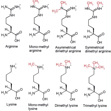
Figure 7: Lysine acetylation ii. Methylation
Histone methylation can have various effects depending on the targeted residue and its position. Methylation targets both lysine and arginine residues but do not alters their charge or the histone structure. Methylated residues seem to serve as a platform for the recruitment of different proteins, able to alter chromatin state or transcription status. Lysine can be mono-, di-, or tri-methylated with variable consequences, but arginine can only be mono- or di-methylated(Bedford and Clarke, 2009; Greer and Shi, 2012). The enzymatic reaction is performed by Histone
Transferase (HMT), which transfers methyl group from S-adenosyl methionine to the targeted residue.
Three families of enzymes have been discovered. The SET-domain-containing proteins and the DOT1-like proteins can both acetylate lysine residues. Protein aRginine Methyl Transferases (PRMT) acetylate arginine residues. H3K4 is notably tri-methylated by SET1/COMPASS complex around the promoter of active genes. H3K36me marks are also enriched along the gene body of highly transcribed genes.
Two families of demethylase are characterised, depending on their active domain; amine oxidases and jumonji-C-domain containing, iron dependant dioxygenases (jJmjC).
Figure 8: Arginine and Lysine methylation 2.d. Regulation cross-talk
Importantly, it has recently been shown that numerous HPTM may influence one another. For example, mono-ubiquitination of H2B promotes methylation of H3K4 and H3K79. Otherwise, repressive H3K9me3 marks need to be actively removed before the active H3K9ac marks to be added by p300 in CD4+ T cells(Ghare et al., 2014).
Histone variants influence the deposition of histone marks. Indeed, H2A.Z containing nucleosomes are more prone to H3K9ac and H3K4me3 marks deposition(Bártfai et al., 2010). DNA methylation status can also influence HPTM. MBD proteins are notably associated with various HDAC(Bird and Wolffe, 1999). DNA methylation is also thought to promote H3K9 methylation.
These interrelations between histone post-translational modifications, histone variants and DNA methylation status constitute a supplemental level of transcription regulation.
3. Chromatin rearrangement
Gene expression can also be regulated through very large distance. Rearrangement corresponds to large chromatin loops that allow to bring closer a specific gene and a distant regulatory element, either upstream or downstream, in order to regulate transcription. When this element is located on the same DNA molecule, it is called cis-regulatory element. On the contrary, when the regulatory element is located on another chromosome, it is called trans-regulatory element.
This has first been enlightened by the discovery of cis-regulation of the β-globin gene over a 150 kb distance(Vernimmen et al., 2007). Two kinds of loops have then been described. The first one allows physical rapprochement between enhancers and promoters(Krivega and Dean, 2012) while the second one brings close the promoters and the terminators(Lainé et al., 2009). These loops have also been described to be highly dynamic. For example, they can be induced under hormonal stimulation(Tan-Wong et al., 2008).
These loop conformations are maintained by a large number of protein. Among many one, CCCTC-binding protein (CTCF) is the strongest and most evolutionary conserved. Two copies of the protein can bind CTCF-Pair-Defined domain (CPD) sequences to gather them. CTCF is implicated in gene activation or repression but also different functions like enhancer blocking, X-chromosome inactivation or genome imprinting. Furthermore, it was found to interact with Dnmt1 to inhibit DNA methylation. CTCF is thought to organise the three-dimensional conformation of the genome and to direct a large transcription regulation system.
II - DNA damage response
All along the life, genomic DNA is exposed to a large number of genotoxic attacks, either endogenous or exogenous. Indeed, biological by-products, UV rays, cigarette smoke and environmental chemicals are able to alter the genomic integrity and to create DNA lesions. These issues have to be properly managed and repaired in order not to perturb essential processes like DNA transcription and replication, which are both crucial for cells. The lack of DNA damage response may lead to cell transformation or death.
Different repair mechanisms have been selected through evolution. They are differentially activated depending on the lesion type, in order to restore the genomic integrity. It includes Nucleotide Excision Repair (NER) that is responsible for the removal of UV-induced lesions and bulky DNA adducts, Base Excision Repair (BER) that is in charge of single strand breaks, the Non-Homologous End Joining (NHEJ) and Homologous Repair (HR) pathways, which are responsible for DNA double strands breaks religation and Mis Match Repair (MMR) that corrects replication issues.
1) Nucleotide excision repair
NER pathways are engaged in the removal of cyclobutane-pyrimidine dimers (CPD) and 6-4 pyrimidine pyrimidone photoproducts (6-4) PP that are induced by UV rays. They are also responsible for the removal of bulky DNA adducts induced by polycyclic aromatic hydrocarbon or anti-cancerous chemicals(Mocquet et al., 2007).
Two different NER pathways have been discovered, depending on the activating signal: the Global-Genome NER (GG-NER) and the Transcription-Coupled NER (TC-NER).
1. GG-NER
As its name indicates, GG-NER acts throughout the genome to search for lesions in order to initiate their reparation.
1.a. Lesions recognition
DNA lesions are recognised all over the genome by the Xeroderma Pigmentosum complementation group C protein (XPC) in complex with its partners Homologue Rad 23 B (HR23B) and Centrin2 (CETN2)(Araki et al., 2001; Masutani et al., 1994). These two proteins are necessary to initiate NER. Indeed, if XPC-HR23B duplex is sufficient for in vitro NER reaction, CETN2 strongly increases in vivo reaction(Araújo et al., 2000; Nishi et al., 2005).
Chemical adducts or photoproducts induce DNA distortions that are recognised by XPC. Nevertheless, only the abnormal DNA structure is recognised by XPC, the presence of a chemical lesion has to be subsequently confirmed(Sugasawa et al., 2001, 2002). Along with this recognition mechanism, less distorting-DNA lesions are not easily identified by XPC/HR23B/CETN2 complex. In such cases, the damage is first recognised by Damage specific DNA Binding protein 2 (DDB2/XPE) in complex with DDB1. This DDB complex enhances the DNA disruption and thus helps to recruit XPC(Fitch et al., 2003). In such case, the DDB-associated Cul4 protein ubiquitinates both DDB2 and XPC to allow NER to initiate(Sugasawa et al., 2005).
1.b. DNA opening
When bound to the damage, XPC slightly distorts the DNA and recruits the TFIIH complex(Volker et al., 2001). As mentioned above, TFIIH is a dual complex. Besides its role in transcription, it is an essential protagonist of NER pathways. Indeed, both XPB and XPD are required for DNA opening around the lesion. The mechanism is not yet fully understood but it seems that XPD helicase activity is responsible for double helix unwinding and opening, when XPB allows the proper TFIIH anchorage on DNA(Oksenych et al., 2009). The CAK sub-complex seems to interfere with the overall repair mechanism and needs to be removed from TFIIH during NER.
When DNA strands are separated around the lesion, the protein RPA binds the undamaged strand to protect it from nuclease activities and to stabilize the open structure(Volker et al., 2001). This step allows the arrival of XPA and the release of the CAK(Coin et al., 2008). RPA is notably acetylated by GCN5/PCAF. This
acetylation promotes XPA/RPA interaction and increases the retention of XPA to the damage site(He et al., 2017; Zhao et al., 2017).
Figure 9: The two NER pathways
XPA XPA XPC XPC IIH XPC RPA IIH RPA RPA RPA RPA IIH RPA RPA RPA XPA XPG XPF ERCC1 RPA RPA RPA RPA XPG XPF ERCC1 CAK XPG RPA RCF PCNA Polδ,ε LigI,IIIα FEN1 pol II pol II pol II pol II CSB pol II pol II CSB IIH
RPA RPA RPA
XPG XPF ERCC1 CSA Cul4 XPA CSB XPA pol II pol II CSB IIH
RPA RPA RPA
XPG XPF ERCC1 CSA Cul4 IIH
RPA RPA RPA
XPG XPF ERCC1 CSA Cul4 Ub Ub Ub pol II pol II XPG RPA RCF PCNA Polδ,ε LigI,IIIα FEN1 IIH XPA XPC XPF ERCC1 PCNA Polδ,ε XPG RPA XPF ERCC1 PCNA Polδ,ε XPG RPA GG-NER TC-NER
1.c. DNA incision
XPA promotes the recruitment of XPF-ERCC1 endonuclease and XPG is recruited on TFIIH, leading to the removal of XPC. XPG incises the damaged strand at the 3’ extremity of the lesion bubble and induces the 5’ end cleavage by XPF-ERCC1. A short 22-30 bp long oligonucleotide is released(Riedl et al., 2003). XPA is then removed from the lesion.
1.d. Synthesis
After incision, only RPA stays on the undamaged strand to protect it from nuclease activities. The double incision allows the recruitment of PCNA and RCF. XPF is then released and the remaining RPA, XPG, RCF and PCNA allow the recruitment of DNA polymerase δ or ε. After the release of XPG and RPA, synthesis of the complementary strand is performed using the undamaged strand as template(Aboussekhra et al., 1995; Moser et al., 2007).
1.e. Ligation
After the synthesis of the complementary strand, a ligase protein performs the necessary ligation of DNA. The implicated Ligase, as well as the responsible DNA polymerase, seems to depend on the cell cycle status. Indeed, the couple Pol δ/Ligase IIIα is in charge of synthesis/ligation throughout the cell cycle while Pol ε and Ligase I are in charge of synthesis/ligation in quiescent cells(Moser et al., 2007).
2. TC-NER
TC-NER is dedicated to the repair of the transcribed strand(Hu et al., 2015). It rapidly arises when the RNA polymerase II is stalled on DNA because of a lesion, during the transcription process. It seems particularly active around promoter and enhancer. Contrary to GG-NER, it removes a specific lesion to ensure the proper termination of a crucial ongoing function. In this context, XPC, XPE and DDB proteins are absolutely dispensable.
2.a. Stalled Pol II
The arrest of Pol II on UV-induced DNA damage strengthens its interaction with Cockaine Syndrome complementation group B protein (CSB)(van den Boom et al., 2004). CSB is further stabilized by its de-ubiquitination by USP7(Schwertman et
al., 2013). From now on, it strongly binds DNA and alters the Pol II/DNA interaction(Beerens et al., 2005). CSB then induces the recruitment of the CSA complex, which notably contains Cul4(Fousteri et al., 2006). CSB also induces the recruitment of TFIIH and the other NER factors RPA, XPA and the endonucleases XPF-ERCC1 and XPG.
2.b. Re-initiation
When NER machinery is set up, the regular repair pathway allows the proper removal of the lesion. After the restoration of genomic integrity, CSA complex re-ubiquitinate CSB to allow its degradation by the proteasome(Groisman et al., 2006). This last step is required for normal transcription re-initiation.
2.c. Pol II role in TC-NER
The role of Pol II during the process of TC-NER is not completely understood. On one hand, the presence of Pol II at the lesion does not prevent dual incision from occurring in vitro(Tremeau-Bravard et al., 2004). On the other hand, it has been shown that Pol II can be ubiquitinated, in order to be degraded(Lee et al., 2002). Finally, CSA also helps the recruitment of chromatin remodelling factors, including HMGN1, which is responsible for H3K14 acetylation. It has been proposed that local chromatin remodelling would help Pol II to get back from the lesion, in order to allow NER(Hanawalt and Spivak, 2008).
2) NER associated diseases
Mutations in NER coding genes are responsible for different syndromes, associated with photosensitivity. Indeed, the genetic mutations lead to partial or total impairment of UV-induced NER pathways (Table 4).
1. Xeroderma pigmentosum
Xeroderma pigmentosum (XP) is the first pathology that has been related to DNA repair deficiency(Cleaver, 1968). Patients were originally described for photosensitivity and for their high rate of fatal skin cancer, with hereditary transmission. Indeed, they have a 1000 increased risk rate for skin but also eye and tongue cancer. Twenty per cent of them also develop progressive neurological degeneration. Furthermore, they suffer from a slightly higher incidence of internal