RESEARCH OUTPUTS / RÉSULTATS DE RECHERCHE
Author(s) - Auteur(s) :
Publication date - Date de publication :
Permanent link - Permalien :
Rights / License - Licence de droit d’auteur :
Bibliothèque Universitaire Moretus Plantin
Institutional Repository - Research Portal
Dépôt Institutionnel - Portail de la Recherche
researchportal.unamur.be
University of Namur
The ambivalent effect of Fe 3 O 4 nanoparticles on the urea-induced unfolding and
dilution-based refolding of lysozyme
Kashanian, F; Habibi-Rezaei, M; Moosavi-Movahedi, A A; Bagherpour, A R; Vatani, M
Published in: Biomedical Materials DOI: 10.1088/1748-605x/aab8d7 Publication date: 2018 Link to publicationCitation for pulished version (HARVARD):
Kashanian, F, Habibi-Rezaei, M, Moosavi-Movahedi, AA, Bagherpour, AR & Vatani, M 2018, 'The ambivalent effect of Fe 3 O 4 nanoparticles on the urea-induced unfolding and dilution-based refolding of lysozyme', Biomedical Materials, vol. 13, no. 4, 045014. https://doi.org/10.1088/1748-605x/aab8d7
General rights
Copyright and moral rights for the publications made accessible in the public portal are retained by the authors and/or other copyright owners and it is a condition of accessing publications that users recognise and abide by the legal requirements associated with these rights. • Users may download and print one copy of any publication from the public portal for the purpose of private study or research. • You may not further distribute the material or use it for any profit-making activity or commercial gain
• You may freely distribute the URL identifying the publication in the public portal ?
Take down policy
If you believe that this document breaches copyright please contact us providing details, and we will remove access to the work immediately and investigate your claim.
Biomedical Materials
PAPER
The ambivalent effect of Fe
3
O
4
nanoparticles on
the urea-induced unfolding and dilution-based
refolding of lysozyme
To cite this article: F Kashanian et al 2018 Biomed. Mater. 13 045014
View the article online for updates and enhancements.
Related content
Modified denatured lysozyme effectively solubilizes fullerene c60 nanoparticles in water
Marialuisa Siepi, Jane Politi, Principia Dardano et al.
-Study on protein conformation and adsorption behaviors in nanodiamond particle–proteincomplexes
Hai-Dong Wang, Catherine Hui Niu, Qiaoqin Yang et al.
-Aqueous ionic liquids and their effects on protein structures: an overview on recent theoretical and experimental results
Jens Smiatek
Biomed. Mater. 13(2018) 045014 https://doi.org/10.1088/1748-605X/aab8d7
PAPER
The ambivalent effect of Fe
3
O
4
nanoparticles on the urea-induced
unfolding and dilution-based refolding of lysozyme
F Kashanian1
, M Habibi-Rezaei1,2
, A A Moosavi-Movahedi3,4
, A R Bagherpour1,5
and M Vatani6,7
1 School of Biology, College of Science, University of Tehran, Tehran, Iran
2 Nano-Biomedicine Center of Excellence, Nanoscience and Nanotechnology Research Center, University of Tehran, Tehran, Iran 3 Institute of Biochemistry and Biophysics, University of Tehran, Tehran, Iran
4 Center of Excellence in Biothermodynamics, University of Tehran, Tehran, Iran
5 Institute of Advanced Magnetic Materials, School of Metallurgy and Materials Engineering, College of Engineering, University of Tehran, Tehran, Iran
6 Terry Fox Cancer Research Laboratory, Department of Biomedical Sciences, Faculty of Medicine, Memorial University of Newfoundland, St John’s, NL, Canada
7 Present address: School of Biology, College of Science, University of Tehran, Tehran, Iran.
E-mail:[email protected]
Keywords: magnetite(Fe3O4) nanoparticles, hen egg white lysozyme, stability, structure ordering/disordering, refolding by dilution
Abstract
Due to the numerous biological applications of magnetite
(Fe
3O
4) nanoparticles (MNPs), it is essential
to identify the influence of these nanoparticles on basic biological processes. Therefore, in this
research, the effect of MNPs on the structure and activity of hen egg white lysozyme
(HEWL) (EC
3.2.1.1) as a model protein was examined using tryptophan intrinsic fluorescence, UV/Vis, and
circular dichroism spectroscopy. Moreover, enzyme activities were analyzed by a turbidometric
approach in the presence of MNPs at concentrations providing MNPs/HEWL ratios in the range of
0.04–1.25. As-synthesized MNPS were characterized by Fourier transform infrared spectroscopy,
x-ray diffraction, scanning electron microscopy, transmission electron microscopy, vibrating sample
magnetometry and the zeta potential of MNPs was measured to be
−29 mV. The goal of this work was
investigating the ordering or disordering effect of MNPs on protein structure at ratios lower or higher
than 0.918 as concentration ratio of threshold
(CRT), respectively, in order to answer the question:
‘How can the denaturation and refolding of a model protein (HEWL) be affected by MNPs?’ As has
been reported recently, the protein folding, helicity, and half-life were improved at
<CRT to make the
protein more ordered and conversely, HEWL was unfolded, and the helicity and half-life were
decreased at
>CRT to make the protein more disordered upon interaction with MNPs. The
disordering effect of urea at
>CRT and even at <CRT in the denaturation buffer (urea 6 M) increased
and at
<CRT the MNPs can provide a significant improvement in the refolding of the unfolded urea
treated protein. These observations provide a new perspective on the growing applications of MNPs in
biotechnology and biomedicine.
Abbreviations
MNPs
Magnetite(Fe3O4) nanoparticlesHEWL Hen egg white lysozyme (EC 3.2.1.1) ML Micrococcus lysodeikticus CRT Concentration ratio of threshold XRD X-ray diffraction VSM Vibrating sample magnetometry SEM Scanning electron
microscopy TEM Transmission
electron microscopy FT-IR Fourier transforms
infrared spectroscopy
RECEIVED 21 October 2017 REVISED 6 March 2018 ACCEPTED FOR PUBLICATION 22 March 2018 PUBLISHED 15 May 2018
DLS Dynamic light scattering
CD Circular dichroism spectroscopy
1. Introduction
Nanotechnology has recently become one of the most exciting forefront fields in analytical chemistry [1].
Magnetite(Fe3O4) nanoparticles (MNPs) with good stability are one of the great points of interest in biotechnological and bio-medicinal applications due to their nontoxicity, biocompatibility, chemical stabi-lity, and substantial potential to reach the target tissue or organ[2]. MNPs have been used in many
applica-tions such as magnetic resonance imaging [3],
hyperthermia for tumor treatment [4], cell labeling
and sorting[5], separation and purification of nucleic
acids[6] or proteins [7], and drug delivery [8].
Nowa-days, approximately 50% of all new medications are biopharmaceuticals, and in comparison with chemi-cal-based drugs, the demand for innovative protein based treatments continues to soar due to their targeted and specific action, in order to achieve better efficacy for fighting against diseases [9].
Determining the nature of the interactions between proteins and MNPs can open a newfield of research rela-ted to the study of MNPs in proteomics[10]. The
func-tional structure of proteins is formed due to the systematic and precise interactions of the protein surface with its aqueous environment. When the balance of interactions between protein molecules and their solvents are disrupted, proteins are unfolded. NPs could poten-tially impact protein–solvent interactions, helping to gain more insight into the function, stability, and dynamics of proteins in water environments[11]. MNPs act as a
dou-ble-edged sword, influencing the structure and function of proteins in a concentration-dependent manner through electrostatic adsorption on NPs. Therefore, NPs can act on proteins either as a friend or foe depending on their concentration[12].
Therefore, an important safety concern for the application of MNPs to biological systems remains that the structure and activity of proteins can be chan-ged upon interaction with MNPs[13,14]. As the
bio-logical activities of proteins depend on their conformations, it is essential to study the structural effects of MNPs on protein conformation for avoiding any threats to their biological activities[15]. Hen egg
white lysozyme(HEWL) (EC 3.2.1.1) is a monomeric globular model protein, comprised of 129 amino acid residues including six tryptophan, three tyrosine, and eight cysteines residues to form four disulfide bonds [16]. It acts to hydrolyze the glycosaminoglycan in
bacterial cell walls. It is a strongly basic protein with an isoelectric point(pI) of 10.7, therefore effectively is absorbed on the negatively charged MNPs[17,18].
Study of the interaction between MNPs and HEWL helps to elucidate the chemical essence of the interactions between bio-macromolecules and MNPs. In this study, HEWL was used as a model protein to investigate the effect of MNPs on protein stability, function and refold-ing(by dilution) after denaturation by urea. It was found that the effect of MNPs on ordering or disordering the protein structure is concentration-dependent.
2. Materials and methods
2.1. Materials
Lysozyme from hen egg white(HEWL) (EC 3.2.1.17, MW: 14.5 kDa) and 9,10-phenanthrenequinone were obtained from Sigma-Aldrich. Other chemicals were from Merck(Darmstadt, Germany) unless otherwise stated. As the substrate, Micrococcus lysodeikticus cell wall was prepared according to Surekha et al[19].
2.2. Magnetic nanoparticles(MNPs) synthesis and characterization
Fe3O4nanoparticles as MNPs were synthesized accord-ing to the Massart method[20,21]. In a typical synthesis
procedure, 10 ml NH3(25%) was added dropwise to the 190 ml solution, containing 16 mmol of FeCl3.6H2O and 8 mmol of FeCl2.4H2O under an N2stream. The precipitates were washed with the mixture of ethanol and distilled water several times and dried at 40°C in a vacuum oven for 1 h. Then the synthesized MNPs were characterized using various methods as follows. Fourier transform infrared(FT-IR) spectra of the MNPs were recorded by a Magna Nicolet 550 spectrophotometer in KBr pellets. Transmission electron microscopy(TEM) images were obtained using a Philips EM208S TEM with an accelerating voltage of 100 kV. Scanning electron microscopy(SEM) with a CamScan MV2300, was used for monitoring MNPs size with 20 000 accelerating voltage for the electrons and a magnification of 70 000 folds. The magnetic property of the sample was measured in a vibrating sample magnetometer(VSM) (Meghnatis Daghigh Kavir Co.; Kashan Kavir; Iran) at room temperature. The zeta potential of MNPs was estimated using dynamic light scattering(DLS) Malvern ZS-Nano series. X-ray diffraction(XRD) patterns were recorded by a Philips-X’PertPro, x-ray diffractometer using Ni-filtered Cu Kα radiation at scan range of 5<2θ<90.
2.3. The enzyme assay
HEWL assay was performed using Micrococcus lyso-deikticus(ML) cell wall as a substrate using a turbido-metric approach[22,23]. In brief, after adding 40 ml
HEWL at 0.2 mg ml−1on 1 ml of ML, the lysis of ML cell wall was monitored upon absorbance decrease at 450 nm. The activity of lysozyme in the absence of MNPs is taken 100, then all resulting values for lysozyme activity in the experiments were reported as the percent of the remaining activity.
2
2.4. The Enzyme treatment by MNPs
Stock solutions of the enzyme were prepared in sodium phosphate buffer (0.1 mM, pH 7.4) and concentrations were adjusted using absorbance at 280 nm[24]. To study the effect of MNPs on HEWL,
the HEWL solutions at 0.2 mg ml−1 were treated in the presence of different concentrations of ultra-sonicated MNPs (0.008–0.24 mg ml−1) to provide MNPs:HEWL ratios in the range of 0.04–1.25 in sodium phosphate buffer 0.1 mM, pH 7.4. A HEWL solution with the same concentration without any MNPs was used as a control. The controls and test samples then were incubated under magnetic stirring for an hour.
2.5. Urea-induced unfolding and dilution-based refolding
To investigate the effect of MNPs on the urea-induced unfolding of HEWL, 0.2 mg ml─1HEWL solutions in sodium phosphate buffer 0.1 mM, pH 7.4 were treated with 6 M urea in the presence of two concentrations of ultra-sonicated MNPs yielding MNPs:HEWL ratios between 0.4 and 1.25. Untreated HEWL(0.2 mg ml─1) in two different states, native protein (Control, −urea), unfolded protein (Control, +urea) and pro-tein treated in the presence of 0.4 and 1.25 MNPs: HEWL were used as controls. The controls and test samples then were incubated in the shaker for an hour. In order to explore the structure ordering effect of MNPs on dilution-based refolding of HEWL, the HEWL solutions(0.2 mg ml−1in sodium phosphate buffer 0.1 mM, pH 7.4) were treated in the presence of MNPs with different ratios to the HEWL. Untreated HEWL(0.2 mg ml−1) in three different states, native protein(Control, −urea), unfolded protein (Control, +urea) and protein treated with 30× diluted urea in the absence of MNPs(Control, diluted) and protein treated with 30× diluted urea and incubated for an hour in the presence of MNPs with a ratio of 0.4 to HEWL.
2.6. Spectroscopic analysis
Tryptophan dependentfluorescence intensities of the HEWL (0.2 mg ml−1) in a sodium phosphate buffer (0.1 M and pH 7.4) were measured in the absence of MNPs as control, and in the presence of different concentrations of MNPs resulting in MNPs:HEWL ratios in the range of 0.4–1.25. The fluorescence spectra were recorded in the wavelength range of 300–400 nm after excitation at 290 nm using a spec-trofluorimeter (BioTek, Synergy™H4 Hybrid micro-plate reader) [25]. Both excitation and emission slits
were set at 5 nm. Afterward, protein and MNPs were dispersed under stirring, for an hour, to establish adsorption–desorption equilibrium. Furthermore, the fluorescence emission of the MNPs in the same buffer at corresponding concentrations was recorded as the blank. Alteration in the helicity of HEWL(0.2 mg ml−1)
in the absence or presence of MNPs was studied using circular dichroism(CD) spectroscopy. The dichrographs were obtained using the AVIV Circular Dichroism Spectrometer Model 215. The far UV region was scanned between 190 and 250 nm and the test was repeated thrice. Thefinal spectra were corrected using the buffer as the blank. The CD data were expressed in terms of mean residual ellipticity in deg cm2dmol−1and the CDNN software was used for deconvolution of the far-CD spectra to measure secondary structure changes and percentage composition of the different secondary structural elements. Each reported data in this paper is the average of three experiments.
3. Results and discussion
The concentration-dependent ordering and disorder-ing effects of MNPs on HEWL as a model protein were investigated through analyzing the protein structure and function in the absence and presence of urea as a denaturing agent. The XRD pattern of the as-synthe-sized MNPs is shown infigure1(a). Bragg’s reflections
are observed for the MNPs in the XRD pattern at values of 30, 36, 43, 54, 58, 63, 71, and 74, representing (2 2 0), (3 1 1), (4 0 0), (4 2 2), (5 1 1), (4 4 0), (6 2 0), and (5 3 3) planes of cubic phase of Fe3O4, respectively, which is in agreement with the JCPDS card No. 19-629 [26]. The FT-IR spectra of MNPs in the range
400–4000 cm−1are shown infigure1(b). The absorp-tion band at 3400 cm−1 is assigned to the ν(OH) stretching mode and the weak absorption band observed at 1633 cm−1is due to the bending vibration of absorbed water[27] which indicates the presence of
physisorbed water molecules linked to MNPs. The most profound impact of biological applications of MNPs with regard to protein adsorption is their surface chemistry and zeta potential (ζ). The zeta potential measurement is essential due to the fact that the changes in the surface of the MNPs are the origins of electrostatic interactions [28, 29]. According to
figure 1(c), the zeta potential of MNPs measured
−29 mV. Carrying a positive charge in a 100 mM PBS solution at pH=7.4, HEWL can adequately be absorbed on the MNPs which carry a negative charge. This alters the pattern of protein–water interactions, in turn, leading to changes in the structure and activity of the protein, in the presence or absence of urea.
According to the SEM and TEM images, the MNPs mainly consisted of spherical particles with the average particle size of 30–40 nm (figures1(d) and (e),
respec-tively). The VSM of MNPs was studied to examine their magnetic properties(figure1(f)). The
magnetiza-tion at saturamagnetiza-tion(Ms) is estimated to be 74 emu g─1at 300 K (the saturation magnetization Ms was deter-mined through extrapolation of the curve of H/M ver-sus H) [30]. Moreover, the size and morphology of
MNPs were examined by SEM and TEM analysis. 3
Intrinsic tryptophan (Trp) fluorescence is effec-tively influenced by the environs of the indole ring and has therefore been recognized as a useful tool to study protein conformational changes. Accordingly, the emission, as the maximumfluorescence intensity (FI), is increased or decreased when buried in a hydro-phobic core or exposed to the polar surface environment,
respectively. In the native HEWL, Trp 62 and Trp 108 are suggested to be dominant emitters and theirfluorescence emission intensity is reduced upon HEWL denaturation [31]. Figure2(a) shows the effect of MNPs on HEWL
structure at different MNPs:HEWL ratios. Accordingly, MNPs have mutual contrast effects on the Trp fluores-cence emission of HEWL to increase or decrease the
Figure 1.(a) X-ray diffraction (b) FT-IR spectra (c) zeta potential (DLS) (d) SEM image (e) TEM images and (f) VSM graph of MNPs.
Figure 2.(a) Effects of MNPs:HEWL ratio on the intrinsic fluorescence of HEWL samples as revealed by fluorescence spectroscopy, (b) far CD spectra of HEWL after treatment with 0.4 and 1.25 MNPs:HEWL ratios. Inset to figure shows the presence of alpha helix structures after treatment with MNPs and(c) assessment of the HEWL remaining activity in the presence of MNPs at low (0.4) and high(1.25) MNPs:HEWL ratios until 64 h.
4
quantum yield offluorescence emission HEWL due to ordering or disordering effects, respectively. As depicted infigure2(a), MNPs exhibit structure ordering or
dis-ordering effects at ratios lower or higher than 0.918, respectively, which is reported as the ratio of threshold (CRT). The value CRT can be obtained according to the intersection of the linear function of Max FI(AU) versus MNPs/HEWL ratio at the corresponding Max FI (AU) value achieved in the absence of nanoparticles. Inspired by the concepts of action of kosmotropic and chaotropic agents, MNPs should most probably improve model protein hydration, and consequently increase protein stability at ratios lower than CRT. Conversely, MNPs diminish HEWL hydration and consequently decrease protein folding at ratios higher than CRT in which a vast number of MNPs are not covered by protein corona that has a negative effect on the protein stability and structure, similar to chaotropic agents. More recently, the stabilizing effect of Fe3O4nanoparticles on HEWL at the ratio range of 0.025–0.75 (<CRT) has been reported[10].
CD spectroscopy was also applied to study the sec-ondary structure of HEWL in the absence of MNPs(as the control), a representative MNPs:HEWL ratio lower than CRT (0.4), and a representative MNPs: HEWL ratio higher than CRT (1.25) (figure 2(b)).
Change in the CD spectra of HEWL (0.2 mg ml−1) after treatment with MNPs at 0.40 and 1.25 MNPs: HEWL ratios are related to change in the secondary structure of the HEWL due to interaction with MNPs in sodium phosphate buffer(0.1 M and pH 7.4). The inset tofigure2(b) shows the α-helix contents before
and after treatment of HEWL with corresponding ratios. Accordingly, the helicity is significantly increased at the ordering region of the MNPs:HEWL ratio(<CRT), but in the disordering region of the MNPs:HEWL ratio (>CRT), the helicity is significantly decreased.
Functional stability of the HEWL as a model pro-tein was investigated to study the mutual effects on HEWL activity under treatment by two representative ordering or disordering MNPs:HEWL ratios for 64 h at 300 K(figure2(c)). As a result, at regions of MNPs:
HEWL ratios lower than CRT or higher than CRT, the functional stability and the half-life of the enzyme were improved or aggravated, respectively. The half-life is defined as the time required for 50% activity loss. The folds of t1/2improvements in the presence of the ordering MNPs:HEWL ratio was found to be 5.5 times. In this regard, a mechanism was proposed about the concentration-dependent effects of MNPs on a model protein; MNPs indirectly influence the structure and dynamics of the water surrounding pro-teins, which in turn affects the structure and function of proteins in a concentration-dependent manner. Therefore, MNPs enhance protein hydration at low concentrations, resulting in an ordering effect. More-over, at higher concentrations, MNPs interfere adversely with protein hydration and present a disordering effect.
Nevertheless, the mechanism has not yet been fully understood and remains to be further explored.
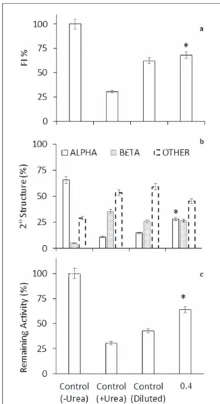
There is a strong demand for the large-scale pro-duction of proteins through heterologous expression of desired genes in bacteria using recombinant tech-nology which often leads to the formation of inclusion bodies through aggregation of unfolded conforma-tions. These inactive aggregates are comprised of almost pure protein, so their recovery is essential in the process of obtaining active proteins from them. The recovery process encompasses solubilization using denaturants, mainly urea, and refolding into an active conformation by reducing the concentration of the denaturant through strategies such as dilution which competes with protein aggregation to form inactive protein species. Hence, we investigated the interference of MNPs in both the disordering effect of urea on folded proteins, and protein refolding by dilu-tion. Urea is a known structure disordering agent which lowers protein hydration upon addition to the protein–solvent interface [32]. Figure 3 reveals the exacerbating effect of MNPs on the disordering effect of urea in a model protein, lysozyme, using intrinsic fluorescence (figure3(a)), CD (figure3(b)), and
func-tional stability (figure 3(c)) analyses. The intrinsic
fluorescence emission spectra of HEWL was decreased in the presence Urea(6 M in sodium phosphate buffer 0.1 mM, pH 7.4) due to the structure disordering effect of urea. Interestingly, the MNPs dramatically exacerbated the disordering effect of urea even at the ordering region of the MNPs:HEWL ratio (ratios lower than CRT) (figure3(a)). Moreover, the observed
exacerbation effect of MNPs was also confirmed at the secondary structure level(figure 3(b)). Accordingly,
the MNPs also dramatically exacerbated the helicity decreasing effect of urea even at 0.4, as a representative for the ordering region of the MNPs:HEWL ratio. Figure3(c) shows the percentages of the remaining
activities of HEWL after 1 h incubation in the absence or presence of urea and MNPs at a ratio that provides ordering or disordering effects on the structure of HEWL in the absence and presence of urea(MNPs: HEWL=0.4), respectively. As expected, the remain-ing activity of the model enzyme was increased 43.11% and decreased 84.7% in the absence and presence of urea (6 M in sodium phosphate buffer 0.1 mM, pH 7.4), respectively. This observation is in good agreement with their observed structural effects using intrinsicfluorescence and CD. Although, the ordering effect of MNPs at ratios lower than CRT is explained by probable increasing in the protein solvation effect, urea disorders both the native and aggregated states of protein which are considered as ordered structures [33, 34]. The concentration-dependent ambivalent
effect of MNPs on the structure and activity of HEWL with a CRT at 0.918 was shown infigure 2. In the absence of urea, the MNPs exhibited ordering and dis-ordering effects on folded native protein species at
5
<CRT and >CRT, respectively. While, in the presence of urea or starting from unfolded denatured protein species, they intensified the disordering effect of urea at>CRT and even at <CRT (0.4, as a representative MNPs:HEWL ratio). The reason for such an interesting observation remains to be elucidated. The schematic presentation of the above mentioned observation has been provided in figure 4. By decreasing the urea concentration using dilution approach, disordered/ unfolded proteins can be ordered through two compet-ing routes to generate active folded or inactive aggre-gated protein species(figure4). Routinely, the addition
of co-solutes is often essential to enhance protein folding and/or suppress protein aggregation [35].
To continue our investigations on the interference of MNPs on the disordering effect of urea on native HEWL(figure3) and protein refolding by dilution, the
interference of MNPs in the dilution process was stu-died at an MNPs:HEWL ratio with proven ordering
effects at <CRT (0.4), by following the structural and functional features of HEWL. The secondary and tertiary structure of HEWL was studied by CD spectro-polarimetry andfluorescence spectroscopy, respectively. Moreover, the interfering effect of MNPs in the diluted urea treated HEWL was examined by determining the remaining activities. Unique and definitive interactions occur during aggregation between molecules[11, 36].
During the refolding process, generation of intermediate structures with partial folding plays a key role in aggre-gate formation[37]. The ability of the MNPs to reduce
aggregate formation is due to interactions with the dena-tured lysozyme, and stabilization of the folded conforma-tion of the protein. Accordingly, figure 5 shows the samples of the native protein(Control, −urea), unfolded protein(Control, +urea), and 30× diluted urea treated protein in the absence of MNPs(Control, diluted) and in the presence of MNPs at thefinal MNPs:HEWL ratio of 0.4, respectively. According to intrinsic fluorescence
Figure 3.(a) Intrinsic fluorescence emission spectra, (b) far CD spectra of HEWL after treating with 0.4 and 1.25 MNPs:HEWL ratios in the denaturation buffer including 6 M urea. Inset to(b) shows the presence of Alpha helix structure and (c) evaluation of the HEWL remaining activity(%) in the presence of MNPs:HEWL ratio of 0.4 in sodium phosphate buffer 0.1 mM, pH 7.4 in the presence or absence of Urea(6 M) after 1 h ambient incubation and 200 rpm shaking.
Figure 4. A hypothetical mechanism of the disordered/unfolded proteins through two competing routes to generate active folded or inactive aggregated protein.
6
results, the achieved refolding efficiency was 31.59% by dilution in the absence of MNPs that was increased to 37.41 by dilution at an MNPs:HEWL ratio of 0.4(<CRT) (figure5(a)). As depicted in the figure5(b), the helicity is
improved in the presence of a 0.4 MNPs:HEWL ratio as established to be the ordering ratio of MNPs:HEWL using CD analysis. As can be seen in thefigure5(c),
mea-surement of the remaining activity of the refolded HEWL shows an increase in activity that causes further destruc-tion of the cell membrane of ML. Therefore, the MNPs at <CRT can provide a significant improvement in the refolding of the unfolded urea treated protein species which is considered to be helpful from a biotechnological point of view.
For increasing efficiency, using small amounts of MNPs is recommended in the dilution process. Eva-luation of secondary structures shows a significant
improvement in the refolding of urea-induced unfol-ded protein species. According to our computations, the proportions of the helical content of HEWL dis-play an increase in the presence of MNPs at a MNPs: HEWL ratio of 0.4(<CRT) (figure5(b)) which is in
accordance with the results provided byfluorescence spectroscopy and the percentage of remaining activity (figures5(a), (c)). Thus, the ordering concentration of
MNPs helps the protein to have a more regular, stable conformation and to display improved activity.
In this work, the dual concentration-dependent effect of MNPs on HEWL was observed in which the concentration threshold shifts between two opposing effects, namely, making and structure-breaking. Ordering and inversely, disordering effects of MNPs on HEWL are reported at lower and higher MNPs/HEWL ratios, respectively. This perspective on the concentration-dependent effect of magnetite nanoparticles on the structure and function of lyso-zyme in solution has not been evaluated with regards to storage conditions and the denaturation and refold-ing of the protein.
4. Conclusion
In conclusion, MNPs impact lysozyme structure and function in a concentration-dependent manner in the absence and presence of urea as a denaturing agent. However, we showed the possibility and necessity of determining a CRT of MNPs for a desired specific protein for biotechnological or biomedical applica-tions with an especial emphasis on biopharmaceuti-cals. Regarding HEWL as a model protein, we determined an MNPs:HEWL ratio threshold at 0.918 which is supported by spectroscopic analysis of the enzyme structure and furthermore by the enzyme activity. Lysozyme shows an increase and decrease in the structure and function at the ordering(<CRT), and disordering(>CRT), a region of the MNPs:HEWL ratio, respectively. The disordering effects of urea were significantly intensified in the presence of MNPs even at ratios lower than the CRT in which ordered structures, both the native and aggregated forms of the protein, exist. MNPs:HEWL ratios lower than the CRT were proven to drive the protein conformation towards more ordered structures, and the MNPs interfered with the dilution process, even at these ratios. It was thus concluded that at these‘ordering’ concentrations, MNPs promote protein activity, as well as stability and regular structure.
Although the mechanism of intercommunications between MNPs and proteins has not been well under-stood and continues to be investigated, we discussed a protein hydration mechanism in the protein corona to support the structure ordering and disordering effects of MNPs at concentrations lower or higher than the resulting CRT, respectively.
Figure 5. Evaluation of the secondary and tertiary structure of HEWL in the absence and presence of MNPs. Untreated HEWL(0.2 mg ml─1) in three different states, native protein (Control, −urea), unfolded protein (Control, +urea) and protein treated with 30× diluted urea in the absence of MNPs (Control, diluted) and protein treated with 30× diluted urea and incubated in the presence of MNPs with a ratio of 0.4 to HEWL were evaluated by(a) intrinsic fluorescence emission spectra,(b) far CD spectra and (c) the percent of remaining activity.
7
Acknowledgments
The support of the University of Tehran and National Institute for Medical Research Development(NIMAD) is gratefully acknowledged.
ORCID iDs
F Kashanian https://orcid.org/0000-0001-5749-1849 M Habibi-Rezaei https://orcid.org/0000-0002-4819-8613 A R Bagherpour https://orcid.org/0000-0003-0560-6899References
[1] Kashanian F, Kokkinis G, Bernardi J, Zand M R, Shamloo A and Giouroudi I 2017 A novel magnetic
microfluidic platform for on-chip separation of 3 types of silica coated magnetic nanoparticles(Fe3O4@SiO2) Sensors Actuators
A270 223–30
[2] Ito A, Shinkai M, Honda H and Kobayashi T 2005 Medical application of functionalized magnetic nanoparticles J. Biosci. Bioeng.100 1–11
[3] Stephen Z R, Kievet F M and Zhang M 2011 Magnetite nanoparticles for medical MR imaging Mater. Today14 330–8
[4] Overgaard J 1989 Hyperthermia in tumor treatment Cancer Ther.(Berlin: Springer) pp 63–70
[5] Zborowski M and Chalmers J J 2011 Magnetic Cell Separation vol 32(Amsterdam: Elsevier)
[6] Berensmeier S 2006 Magnetic particles for the separation and purification of nucleic acids Appl. Microbiol. Biotechnol.73 495–504
[7] Lai Y, Yin W, Liu J, Xi R and Zhan J 2010 One-pot green synthesis and bioapplication ofl-arginine-capped superparamagnetic Fe3O4nanoparticles Nanoscale Res. Lett.5 302
[8] Arruebo M, Fernández-Pacheco R, Ibarra M R and Santamaría J 2007 Magnetic nanoparticles for drug delivery Nano Today2 22–32
[9] Anselment B, Baerend D, Mey E, Buchner J, Weuster‐Botz D and Haslbeck M 2010 Experimental optimization of protein refolding with a genetic algorithm Protein Sci.19 2085–95
[10] Shareghi B, Farhadian S, Zamani N, Salavati-Niasari M and Gholamrezaei S 2016 Stability and enzyme activity of lysozyme in the presence of Fe3O4nanoparticles Mon.hefte Chem.—
Chem. Mon.147 465–71
[11] Kashanian F, Habibi-Rezaei M, Bagherpour A R, Seyedarabi A and Moosavi-Movahedi A A 2017 Magnetic nanoparticles as double-edged swords: concentration-dependent ordering or disordering effects on lysozyme RSC Adv.7 54813–22
[12] Yokomizo T, Higo J and Nakasako M 2005 Patterns and networks of hydrogen-bonds in the hydration structure of human lysozyme Chem. Phys. Lett.410 31–5
[13] Cedervall T, Lynch I, Lindman S, Berggård T, Thulin E, Nilsson H, Dawson K A and Linse S 2007 Understanding the nanoparticle–protein corona using methods to quantify exchange rates and affinities of proteins for nanoparticles Proc. Natl Acad. Sci.104 2050–5
[14] Lynch I, Cedervall T, Lundqvist M, Cabaleiro-Lago C, Linse S and Dawson K A 2007 The nanoparticle–protein complex as a biological entity; a complexfluids and surface science challenge for the 21st century Adv. Colloid Interface Sci.
134 167–74
[15] Aubin-Tam M E and Hamad-Schifferli K 2008 Structure and function of nanoparticle–protein conjugates Biomed. Mater.3 034001
[16] Wu Y L, He F, He X W, Li W Y and Zhang Y K 2008 Spectroscopic studies on the interaction between CdTe nanoparticles and lysozyme Spectrochim. Acta A71 1199–203
[17] Ghosh G, Panicker L, Ningthoujam R S, Barick K C and Tewari R 2013 Counter ion induced irreversible denaturation of hen egg white lysozyme upon electrostatic interaction with iron oxide nanoparticles: a predicted model Colloids Surf. B
103 267–74
[18] Huopalahti R, Anton M, López-Fandiño R and Schade R (ed) 2007 Bioactive Egg Compounds(Berlin: Springer)
[19] Surekha P Y, Dhanya P, Sarath Josh M K, Pradeep S and Sailas B 2016 Micrococcus luteus strain BAA2, a novel isolate produces carotenoid pigment Electron. J. Biol. 12 83–9 [20] Mohapatra M and Anand S 2010 Synthesis and applications of
nano-structured iron oxides/hydroxides—a review Int. J. Eng. Sci. Technol. 2 127–46
[21] Vergés M A, Costo R, Roca A G, Marco J F, Goya G F, Serna C J and Morales M P 2008 Uniform and water stable magnetite nanoparticles with diameters around the monodomain–multidomain limit J. Phys. D: Appl. Phys.41 134003
[22] Li D H and Chi Y J 2002 Facility detection of lysozyme activity China Dairy Ind. 30 128–9
[23] Hoseinifar S H, Safari R and Dadar M 2017 Dietary sodium propionate affects mucosal immune parameters, growth and appetite related genes expression: insights from zebrafish model Gen. Comparative End.243 78–83
[24] Aune K C and Tanford C 1969 Thermodynamics of the denaturation of lysozyme by guanidine hydrochloride: II. Dependence on denaturant concentration at 25 Biochemistry8 4586–90
[25] Lakowicz J R 2006 Principles of Fluorescence Spectroscopy (Boston, MA: Springer)
[26] Varghese S 2012 Magic-angle-spinning solid-state NMR studies of the membrane binding heme protein nitrophorin 7 (NP7) from Rhodnius prolixus PhD Thesis Universitäts-und Landesbibliothek der Heinrich-Heine-Universität Düsseldorf [27] Wang Z, Zhu H, Wang X, Yang F and Yang X 2009 One-pot
green synthesis of biocompatible arginine-stabilized magnetic nanoparticles Nanotechnology20 465606
[28] Patil S, Sandberg A, Heckert E, Self W and Seal S 2007 Protein adsorption and cellular uptake of cerium oxide nanoparticles as a function of zeta potential Biomaterials28 4600–7
[29] Kosmulski M 2002 The pH-dependent surface charging and the points of zero charge J. Colloid Interface Sci.253 77–87
[30] Laurent S, Forge D, Port M, Roch A, Robic C, Vander Elst L and Muller R N 2008 Magnetic iron oxide nanoparticles: synthesis, stabilization, vectorization, physicochemical characterizations, and biological applications Chem. Rev.108 2064–110
[31] Imoto T, Forster L S, Rupley J A and Tanaka F 1972
Fluorescence of lysozyme: emissions from tryptophan residues 62 and 108 and energy migration Proc. Natl Acad. Sci.69 1151–5
[32] Klotz I M 1958 Protein hydration and behavior Science128 815–22
[33] Chakraborti S, Chatterjee T, Joshi P, Poddar A,
Bhattacharyya B, Singh S P, Gupta V and Chakrabarti P 2009 Structure and activity of lysozyme on binding to ZnO nanoparticles Langmuir26 3506–13
[34] Chen P, Seabrook S A, Epa V C, Kurabayashi K, Barnard A S, Winkler D A, Kirby J K and Ke P C 2014 Contrasting effects of nanoparticle binding on protein denaturation J. Phys. Chem. C
118 22069–78
[35] Arakawa T, Ejima D, Tsumoto K, Obeyama N, Tanaka Y, Kita Y and Timasheff S N 2007 Suppression of protein interactions by arginine: a proposed mechanism of the arginine effects Biophys. Chem.127 1–8
[36] Murphy R M and Kendrick B S 2007 Protein misfolding and aggregation Biotechnol. Prog.23 548–52
[37] Fink A L 1998 Protein aggregation: folding aggregates, inclusion bodies and amyloid Folding Des.3 R9–23
8