HAL Id: tel-00831664
https://tel.archives-ouvertes.fr/tel-00831664
Submitted on 7 Jun 2013HAL is a multi-disciplinary open access archive for the deposit and dissemination of sci-entific research documents, whether they are pub-lished or not. The documents may come from teaching and research institutions in France or abroad, or from public or private research centers.
L’archive ouverte pluridisciplinaire HAL, est destinée au dépôt et à la diffusion de documents scientifiques de niveau recherche, publiés ou non, émanant des établissements d’enseignement et de recherche français ou étrangers, des laboratoires publics ou privés.
Ultrastructural, morphological and molecular
characterization of heterotopic cells in the hippocampus
in epileptic Dcx knockout (KO) mice
Reham Khalaf-Nazzal
To cite this version:
Reham Khalaf-Nazzal. Ultrastructural, morphological and molecular characterization of heterotopic cells in the hippocampus in epileptic Dcx knockout (KO) mice. Development Biology. Université Pierre et Marie Curie - Paris VI, 2012. English. �NNT : 2012PAO66514�. �tel-00831664�
THESE DE DOCTORAT DE
L’UNIVERSITE PIERRE ET MARIE CURIE
Spécialité
Génétique et Neurodéveloppement (Ecole doctorale Complexité du vivant)
Présentée par
REHAM KHALAF-NAZZAL
Pour obtenir le grade de
DOCTEUR de l’UNIVERSITÉ PIERRE ET MARIE CURIE
Titre de la thèse :
Caractérisation ultrastructurale, morphologique, et
moléculaire des cellules hétérotopiques dans un modèle
d’épilepsie hippocampique, chez les souris inactivées
pour le gène Dcx
Soutenue le 19 November 2012
Devant le jury composé de: Pr. Rose Katz Président
Dr. Alfonso Represa Rapporteur Dr. Stéphane Auvin Rapporteur
Dr. Catherine Fallet-Bianco Examinateur Pr. Richard Miles Examinateur
TITRE ET RESUME:
Caractérisation ultrastructurale, morphologique, et moléculaire des cellules hétérotopiques dans un modèle d’épilepsie hippocampique, les souris inactivées pour le gène Dcx
Des mutations dans le gène doublecortine (DCX) sont responsables de lissencéphalie de type 1 ou d’hétérotopie laminaire sous-corticale, un spectre de malformations corticales associées à de sévères crises d’épilepsie ainsi qu’à des déficits cognitifs. Les souris invalidées pour le gène Dcx sont épileptiques et présentent des anomalies hippocampiques, avec notamment la présence de deux couches de cellules pyramidales spécifiquement dans l’aire CA3 de cette structure. Le sujet de cette thèse concerne la caractérisation de ces neurones «hétérotopiques ». Cette hétérotopie hippocampique est associée à une hyperexcitabilité susceptible de perturber la fonction du réseau neuronal. Notre but est d’identifier les mécanismes moléculaires et cellulaires responsables de cette hyperexcitabilité chez les souris Dcx. Pendant ma thèse, et grâce à la technique de microdissection au laser suivie d’une étude de transcriptome, j’ai pu isoler les neurones mal-positionnés de la région CA3 de l’hippocampe des souris inactivées pour le gène Dcx, afin de comparer leurs profils d’expression génique à ceux des neurones de la région CA3 des souris contrôles. Les analyses comparatives globales de leurs profils d’expression génique ont permis de révéler des informations importantes sur leur génération et leur migration, nous aidant ainsi à mieux comprendre l’origine de l’hétérotopie. Les résultats au stade P0 (jour de la naissance des souris), montrent que les profils d’expression génique de chacune des deux couches hétérotopiques présentes chez les souris Dcx, diffèrent significativement entre eux ainsi qu’en comparaison avec les profils de la couche pyramidale des souris contrôles. Sur le plan fonctionnel, cette étude indique des perturbations au niveau des organelles intracellulaires, tels que les endosomes, les mitochondries et l’appareil de Golgi. En étudiant séparément les profils associés à chacune des deux couches des souris Dcx, nous avons mis en évidence des différences de degrés de maturité neuronale entre chacune des couches, suggérant des fenêtres temporelles distinctes de production des neurones. L’utilisation de marqueurs moléculaires spécifiques aux couches en combinaison avec des expériences d’injections de bromo-désoxy-uridine (BrdU) à différents moments du
développement suggère une inversion des couches neuronales présentes chez les souris Dcx en comparaison avec les souris contrôles. En complément des données d’expression génique, et en collaboration avec la plateforme de microscopie électronique de notre institut, j’ai également réalisé des analyses morphologiques et notamment des propriétés ultrastructurales des neurones anormaux en comparaison avec celles des neurones contrôles, au stade P0. Ces expériences ont ainsi permis de préciser la nature du défaut de lamination ainsi que les défauts morphologiques des cellules CA3 dans le modèle Dcx. Ces études indiquent que les cellules hétérotopiques des souris Dcx, présentent des anomalies d’organelles intracellulaires, avec notamment des défauts de mitochondries et des modifications de l’appareil de Golgi. Qui plus est, nos données montrent une augmentation significative de la mort cellulaire dans les régions CA1 et CA3 de l’hippocampe. Aussi, nous avons également montré que les couches hétérotopiques étaient hétérogènes, présentant notamment des distributions anormales des précurseurs d’oligodendrocytes et des interneurones exprimant la somatostatine, ce qui n’est pas le cas chez les souris contrôles. L’ensemble de ces données devrait nous permettre de mieux comprendre les dysfonctionnements des neurones pyramidaux chez les souris Dcx adultes. Ces résultats ouvrent donc de nouvelles perspectives pour mieux comprendre la physiopathologie de ces maladies graves associées à des hétérotopies neuronales dans le cerveau, de l’épilepsie et des déficits cognitifs.
TITLE & ABSTRACT:
Ultrastructural, morphological and molecular characterization of heterotopic cells in the hippocampus in epileptic Dcx knockout (KO) mice.
Mutations in the doublecortin gene (DCX) are responsible for type 1 lissencephaly and subcortical band heterotopia, malformations that lead to intellectual disability and epilepsy. This thesis work concerns the characterization of abnormally positioned ‘heterotopic’ neurons present in the Dcx knockout (Dcx KO) mouse model, which exhibits hippocampal dysplasia and epilepsy. The pyramidal cell layer of the hippocampal CA3 region in Dcx KO mutants is divided into two heterotopic cell layers instead of a single layer observed in the wild type (WT) controls. Heterotopic neurons show hyperexcitability, which is likely to lead to a perturbation of network function and subsequent epilepsy. Using the Dcx KO model, our work investigates the molecular and cellular mechanisms leading to hyperexcitability. During my thesis, I was able to isolate abnormally positioned heterotopic neurons using laser capture microdissection, from the CA3 region of the Dcx KO hippocampus, and compare them to WT neurons. Using transcriptome experiments, I aimed to determine the specific gene expression profiles of such cells. Global gene expression analyses of the knockout neurons revealed information concerning their generation and migration, which helps us understand why their positioning is abnormal. Knockout layers were shown to differ from each other and from WT at postnatal day 0. Common perturbed mechanisms affect intracellular organelles including endosomes, mitochondria and Golgi apparatuses. Studying perturbed mechanisms specific to the individual KO layers shows defined but distinct neurogenesis time windows of each layer, that correspond to a different maturity status in early postnatal stages. Layer specific molecular markers and bromo-deoxyuridine (BrdU) birth dating experiments in KO and WT mice, suggest that there is an inversion of the neuronal layers of the Dcx KO CA3 region, compared to wild type. Complementing these gene expression data, and in collaboration with the electron microscopy facility of our institute, I carried out ultrastructural and morphological analyses of abnormal Dcx KO neurons compared to WT, in their tissue environment of the developing brain (postnatal day 0). This revealed the nature of the lamination defect and the state of CA3 cells in this model. Potential organelle abnormalities, including mitochondrial defects and modifications
of the Golgi apparatus, were identified in Dcx KO cells, as well as significantly increased cell death in CA1 and CA3 regions. Oligodendrocyte precursor cells and somatostatin-positive interneurons were found interspersed within the pyramidal cell layers in early postnatal stages, and this was not the case in wild-type. These combined data may provide clues to the abnormal functioning of pyramidal neurons in the adult. These results therefore open up new directions in order to better understand the pathophysiology of a sepectrum of severe disorders associated with heterotopic neurons in the brain, presenting in human patients with severe epilepsy and, developmental delay, and intellectual disability.
ACKNOWLEDGEMENT:
It would not have been possible to write this doctoral thesis without the help and support of the kind people around me, who made this experience an unforgettable one for me, to only some of whom it is possible to give particular mention here.
I would like to start by expressing my gratitude to the thesis jury members. I thank Dr. Alfonso Represa and Dr. Stéphane Auvin for critically reading the thesis manuscript and their constructive comments that had important impact on its improvement. My special thanks to the thesis jury examiners, Dr. Catherine Fallet-Bianco and Pr. Richard Miles, not only for accepting to be examiners members in the jury, but also for following my scientific development in the past four years. My sincere thanks to Pr. Rose Katz, the thesis jury President. This thesis arose in the context of bilateral exchange between the UPMC and the Palestinian medical schools, for which Pr. Katz is a key member in the durable success of this scientific project.
I would like to express my deepest sense of gratitude to my thesis supervisor, Dr. Fiona Francis. I thank you for establishing this distinguished backbone of research theme and so this thesis, combining the clinical and fundamental research to study brain pathologies. I would like to thank you for your continuous advice, supervision, and crucial contribution to the advancement of my work. I deeply thank you for your scholarly inputs and consistent encouragement during the last four years. Thank you for your patience and enthusiasm when I was first initiated in doing scientific experiments, experiments as simple as DNA extraction to the complicated ones. Thank you for your unconditional support and making yourself available during thesis writing, continuously guiding me to improve the text despite my “limited and poor” English writing skills.
I will forever be sincerely thankful to my initiator on developmental neurosciences and neurogenetics, Pr. Anwar Dudin, the dean of An-Najah medical school. Your foremost vision, intelligence, hard work and confidence that you donate to people working with you makes you stand as a role model. As I told you once, I’m deeply proud to be your fellow. And as you suggested once, I will try to be an ambitious big dreamer. My special thanks to Dr. Samar Musmar, dean assistant for clinical affairs. To Pr. Rami Hamdallah, An-Najah University president, I acknowledge my gratitude,
for the heavy load you are bearing, to improve and revolutionize the higher education in Palestine. For your continuous support, and the opening avenues you are offering to establish a clinical research activity in the university. I would like to acknowledge An-Najah National University for the financial support during my thesis, and for the continuous support provided for my near future return project.
I express my gratitude to my thesis committee members. I thank Dr. François Giudicelli, Pr. Eric Leguern, and again Pr. Ricard Miles. I also thank the doctoral school represented by Ms. Elizabeth Clement and Dr. Muriel Umbhauer for their continuous orientation and care.
I thank all the past and present members of Fiona Francis group. Special thanks to Françoise Phan Dinh Tuy and Katia Boutourlinsky, for receiving me when first arrived to Paris. For your unforgettable help in performing the initial experiments, and the valuable support in learning to speak French, I loved your nice French expressions that you articulate from time to time. You made the tough task greatly enjoyable! I am indebted to Audrey Roumegous, your unconditional support in performing the experiments, your unique organization skills, and non-vanishing smile transformed my lab life into a real joy! I vividly thank you for being as great as you are! My very sincere thanks to Richard Belvindrah, with you, hippocampus in utero electroporation was miraculously successful! I deeply appreciated our insightful scientific discussions that tethered me further to the domain of developmental neurosciences. Thank you Richard for your efforts in comprehensively answering any rising question in my mind, and for your constructive feedback for the thesis manuscript. I thank also Elodie Bruel-Jungerman and Sara Bizzotto for your help in writing thesis manuscript, and the moments we shared together.
I express my sincere thanks to Iffat Sumia, the multi-skillful and intelligent M1 student. Your commitment, wisdom, and nice humor turned your rotation into an unforgettable life experience. I express my deep appreciation towards you, and wish you a wonderful future, for which you got all of the needed elements.
I was extraordinarily fortunate in being a member of the Institute du Fer à Moulin. I thank all people I encountered there. I thank Dr. Jean-Antoine Jirault, the director of the institute and Dr. Patricia Gaspar and Dr. Andre Sobel, the co-directors, for the
endless care you are providing to this place making it a unique and rich scientific environment. Some members of the Institute have been very kind enough to extend their help at various phases of my stay, whenever I approached them, and I do hereby acknowledge all of them. I vividly thank Jocelyne Bureau for the joyful experience we had together, studying and reasoning at the electron microscope. Jocelyne: I appreciate your sympathy, commitment, and faith, and I will eternally keep a good memory of you. I thank Mythili Savariradjane, Jean Paul –Rio, Jocelyne Chevallier, Christine Vaillant, Marianne Coutures, Geraldine, Ghislaine, Evelyne, and Sylvie Clain. My field study and my life was made less obstacle-ridden because of the presence of a few individuals, I express my deep gratitude to my two valuable friends Imane Moutkine and Ahlem Assali. I thank 4th
floor people particularly Lucie Viou, Charlotte Plestant, and Sara Devaux. I thank my office colleague Stéphanie Chauvin. I am particularly indebted to animal house facility personnel, without whom this thesis would have never been possible. I thank the IFM Olympics’ team. I really enjoyed it!
I vividly thank all of our collaborators, without whom this thesis would have never seen the day. I sincerely thank Dr. Robert Olaso, Mrs. Sylvie Dumont, Dr. Wassila Carpentier, and Mr. Benoit Albaud. I thank Dr. Sophie Hamelin and Dr. Antoine Depaulis. I acknowledge my gratitude to Pr. Richards’ Miles group and Dr. Nadia Bahi-Buisson.
Collective and individual acknowledgments are also owed to my Parisian friends, who accompanied me in the discovery of this wonderful city. I thank you for the family core that we established here together and for the moral support and the continuous care you surrounded me during my happiness and sorrows. I thank again Ahlem and Imane, but also Manar, Marc and Zeinab, Amanie, Azza, Suhaib, Saleh, Motee’, Nadia, Jawad, and Nasim. I am deeply thankful to Jill, Jane and John for the special holidays we spent together in the UK.
I take this opportunity to express the profound gratitude from my deep heart to my beloved parents, for their love and continuous spiritual support. I thank you for all what you gave to your little spoiled girl. I will never forget these moments, when this eerily strong maternal instincts sense would drive my mother to call, when I’m in my tears, missing you badly.
My greatest sense of gratitude is addressed to my husband whose dedication, love and persistent confidence in me, has taken the load off my shoulder, even when we spent four years, thousands of miles apart. I owe you for being unselfishly loving, temporarily scarifying our couple life to let your passions and ambitions collide with mine. I’m eternally indebted to you!
And not to forget to thank the soul of my beloved uncle. The tender love, courage, and sympathy you provided me, your faith in my strength, and your emphasis on achieving my dreams were the nurture that fed my heart. Unable to be between us in this occasion, I dedicate all of this work to your memory.
Above all, I owe it all to Almighty God for granting me the wisdom, health and strength to undertake this research task and enabling me to its completion.
I dedicate this thesis to my family, my husband, Mahmoud,
and the memory of my beloved uncle for their constant support
and unconditional love.
I love you all dearly
.TABLE OF CONTENTS:
CHAPTER 1: INTRODUCTION...11
PREAMBLE...13
SECTION 1: CEREBRAL CORTEX DEVELOPMENT, PATTERNING AND NEURONAL CELL FATE SPECIFICATION...15
1.1 Intrinsic control of cerebral cortex development through patterning centers and transcription factor (TF) gradients... 16
1.2 Extrinsic signals that control cerebral cortex patterning ... 17
SECTION 2: CEREBRAL CORTEX HISTOGENESIS, NEUROGENESIS AND SPECIFICATION OF NEURONAL PHENOTYPES IN THE DEVELOPING TELENCEPHALON ...19
2.1 NEUROGENESIS : FROM NEUROEPITHELIAL STEM CELLS TO RADIAL GLIAL CELLS ... 23
I. Types of neuronal progenitors in the developing telencephalon ... 24
II. The subventricular zone: a secondary zone of neurogenesis... 29
2.2 SPECIFICATION ... 29
I. Neuronal versus glial cell fate choice in the telencephalon... 29
II. Specification of neuronal phenotypes in the ventral telencephalon ... 31
III. Specification of neuronal phenotypes in the dorsal telencephalon (the developing cerebral cortex), (Figure 8) ... 33
SECTION 3: CELL TYPE DIVERSITY IN THE BRAIN...35
3.1 CAJAL-RETZIUS CELLS ... 35
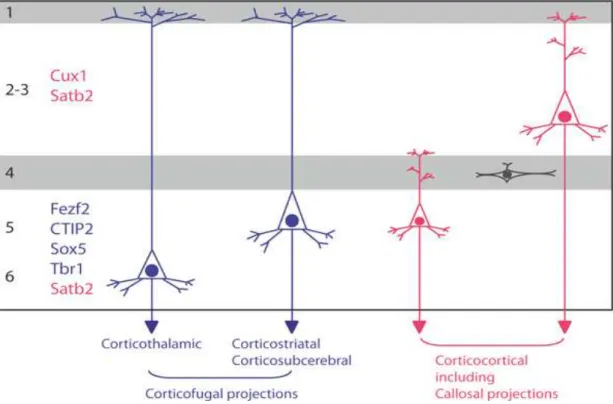
3.2 PROJECTION NEURONS ... 37
3.2.1 Laminar fate specification of projection neurons... 37
3.2.2 Molecular specification of cortical projection neurons ... 38
A Specification of cortical identities... 38
B Temporal specification of cortico-laminar identities... 40
C Subtype specification of projection neurons ... 42
3.2.3 Migration of projection neurons... 45
A Inside-out lamination ... 45
B Cellular and molecular mechanisms involved in neuronal migration... 47
3.3 INTERNEURONS ... 56
3.3.1 Migration of interneurons from the ventral telencephalon to developing cortex57 A : Tangential neuronal migration ... 57
B: Intracortical migration of interneurons ... 59
C: Differentiation and maturation of interneurons ... 60
3.3.2 Major classes of cortical interneurons ... 61
A: Parvalbumin (PV) Interneuron Group ... 61
B: Somatostatin (Sst) Interneuron Group ... 62
C: 5HT3aR Interneuron Group ... 63
3.4 MACROGLIA, ASTROCYTES AND OLIGODENDROCYTES ... 64
I. Migration and functional differentiation of of astrocytes and oligodendrocytes... 67
II. Function of astrocytes and oligodendrocytes in adult brain ... 68
3.5 MICROGLIA... 69
SECTION 4: AXON GUIDANCE, SYNAPTOGENESIS, AND MATURATION OF DEVELOPING NEURONAL CIRCUITS ...71
SECTION 5: HIPPOCAMPAL DEVELOPMENT...81
5.1 PATTERNING EVENTS AND FIELD SPECIFICATION IN THE DEVELOPING HIPPOCAMPUS... 84
5.1.1 Cortical hem boundary specification ... 85
A Molecular markers of the cortical hem ... 86
B Organizer function of the cortical hem ... 87
5.1.2 Field pattern specification in developing hippocampus and the role of cortical hem... ... 88
5.1.3 Field sub divisions and the complex genomic and functional anatomy of the hippocampus ... 90
5.2 CELL HETEROGENEITY AND THE DEVELOPMENT OF NEURONAL TYPE DIVERSITY IN THE ADULT HIPPOCAMPUS ... 93
5.2.1 Hippocampal projection neurons ... 93
A Histology ... 93
B Neurogenesis and neuronal migration of projection neurons in the developing hippocampus ... 94
C Sojourn of newly born cells in the developing hippocampus... 96
D Migration of neuroblasts in the developing hippocampus: ... 97
E En route differentiation ... 100
5.2.2 Hippocampal interneurons ... 102
A Developmental processes culminating in the generation and migration of hippocampal interneurons ... 104
B Maturation of hippocampal interneurons and synaptogenesis: signals from postsynaptic neurons trigger GABAergic synaptogenesis. ... 105
5.3 FACTORS AFFECTING NEURONAL MIGRATION IN THE DEVELOPING
HIPPOCAMPUS... 106
5.4 COORDINATED SYNAPTOGENESIS... 107
SECTION 6: DOUBLECORTIN, A NEURONAL MIGRATION GENE...114
6.1 CLASSICAL LISSENCEPHALY ... 114
6.1.1 Neuropathology ... 115
6.1.2 Genotype-phenotype correlation and molecular diagnosis of classical lissencephaly ... 116
6.1.3 Subcortical band heterotopia ... 119
6.2 DOUBLECORTIN, AN MT ASSOCIATED PROTEIN INVOLVED IN NEURONAL MIGRATION AND DIFFERENTIATION ... 121
6.2.1 The interaction between DCX and microtubules... 121
6.2.2 The role of phosphorylation in regulating DCX function... 125
6.2.3 Dcx’s role in enhancing long-distance cellular transport... 127
6.2.4 DCX’s interaction with LIS1 ... 128
6.2.5 DCX’s interaction with Neurofascin (NF)... 129
6.3 MIGRATION DEFECTS AND SPONTANEOUS EPILEPSY IS A COMMON FEATURE OF DIFFERENT DCX MUTANTS’ MODELS... 130
CHAPTER 2: RESULTS ...133
PREAMBLE...135 ARTICLE 1: ...141 ARTICLE 2: ...193CHAPTER3: DISCUSSION ...249
CHAPTER 4: ANNEX ...273
CHAPTER 5: REFERENCES...341
List of Figures:
Figure 1: Schematic representation of the role patterning centers and secreted morphogens in telencephanlon development
Figure 2: Patterning over a long distance.
Figure 3: The developmental processes that will lead to organization of the neocortex into distinct neuronal layers.
Figure 4: A three-dimensional reconstruction of a migrating neuron along the surface of a radial glial fiber
Figure 5: Neuronal progenitors
Figure 6: Rodent and human neocortical development Figure 7: Neuronal versus glial cell fate specification
Figure 8: Specification of neuronal phenotypes in the developing telencephalon Figure 9: Schematic representation showing multiple origins and distribution of Cajal-Retzius cells in the brain.
Figure 10: Projection neuronal types in the cerebral cortex
Figure 11: Gene expression correlation to axonal projection and cortical layer organization. Figure 12: The two distinct forms of cortical neuron movement during migration.
Figure 13: Adhesion dynamics of migrating neurons leading to the extension and maintenance of the leading process.
Figure 14: Cellular and molecular mechanisms of neuronal migration Figure 15: Origin and migratory routes of cortical interneurons.
Figure 16: Signaling cascade that results in the generation of glial precursors from RGC Figure 17: Competing waves of oligodendrocytes in the developing forebrain
Figure 18: Dynamic calcium oscillations mediate growth cone responses to axon guidance cues, involving cGMP and cAMP.
Figure 19: Cellular response and subsequent calcium oscillations in response to attractive and repulsive cues during axon guidance.
Figure 20: Mechanisms of axon guidance and molecules involved in synapse formation. Figure 21: Successive events leading to contact stabilization and maturation of developing synapses.
Figure 22: Molecular mechanisms involved in synapse growth and stabilization. Figure 23: The organization of hippocampal fields and stratum
Figure 24: The hippocampal network
Figure 25: The organization and overall contribution of the di-synaptic and tri-synaptic circuits in the adult hippocampus
Figure 26: Pattern of expression of TF that will define rostro-caudal bondaries of the cortical hem
Figure 27: Molecular and morphological features of the hem and the nearby choroid plexus epithelium and cortical neuroepithelium.
Figure 28: Embryonic and mature hippocampal pyramidal cells are identified by the expression of field specific markers.
Figure 29: Representation of three-dimensional molecular signatures along the septotemporal axis of the hippocampal CA3 region.
Figure 30: Molecular markers along outer boundary of the radial unit of hippocampla CA3 field.
Figure 31: Summary diagram of waves of neurogenesis and migration of pyramidal cells in the hippocampus.
Figure 32: Structural and morphologic characteristics of migrating neurons at different stages.
Figure 33: Hippocampus-specific gene targeting by in utero electroporation.
Figure 34: Hippocampal interneurons can be classified by their calcium-binding protein content, and by their synapse sites on hippocampal pyramidal cells.
Figure 35: The unique structure and the specificity of synaptic connections in the hippocampus.
Figure 36: Cell autonomous pruning defect of the IPB in Plexin-A3 mutants.
Figure 37: Magnetic resonance images (MRI) at the level of basal ganglia showing different degrees of LIS severity.
Figure 38: Microtubule organization and dynamics in developing neurons.
Figure 39: A proposed model of DCX interaction with MT in the in the leading process of migrating neurons showing cooperative DCX binding to MT and an affinity for growing microtubule.
Figure 40: Ultrastructural features of non-neuronal cell located in the SVZ in Dcx KO CA3 region.
Figure 41: Schematic presentation of the observed birth date and layer fate of BrdU labeled neurons in the WT, and the Dcx KO
Figure 42: Expression pattern of the potassium- chloride cotransporter 2 (KCC2) in the WT and the Dcx KO CA3 region at P0.
List of Abbreviations:
5HT3aR serotonin receptor 5HT3a LIF leukaemia inhibitory factor AIS axon initial segment LIS Classical lissencephaly AIS axon initial segment LMT large mossy fiber terminal
AnkG AnkyrinG LP lateral pallium
ANR anterior neural ridge LTP long term potentiation
AP apical progenitors MAP microtubule associated protein BDNF brain-derived neurotrophic
factor MB
the major bundle, the suprapyramidal bundle BLBP brain lipid binding protein MGE Medial ganglionic eminenece BMPs Bone morphogenetic proteins MRI Magnetic resonance images BrdU Bromodeoxyuridine MT microtubule
C-DC C-terminal conserved DCX
domain MZ marginal zone
CA cornu ammonis N-DC N-terminal conserved DCX domain
CAM L1 cell adhesion molecule NE neuroepithelial CaMKII Ca2+/ calmodulin dependent
proteinkinase II NF Neurofascin
CDK5 cyclin-dependent kinase 5 NKCC1 Na+–K+–2Cl- co-transporter CGE Caudal ganglionic eminence NPY neuropeptide Y
CNS central nervous system NRG1 neuregulin 1 CNTF ciliary neurotrophic factor NT4 neurotrophin4
CoP commissural plate OPC oligodendrocyte precursor cell
CP cortical plate oRG outer radial glial
CR Cajal-Retzius PAFAH1
B1
platelet-activating factor acetylhydrolase 1B a subunit
Crl calretinin PF protofilaments
CSF colony stimulating factor-1 POA preoptic area DC
domain
conserved doublecortin
domain PP preplate
Dcx Doublecortin PP1 protein phosphatase 1
DG dentate gyrus PP2A Protein Phosphatase 2A
DG dentate gyrus PSB pallial–subpallial boundary
EE enriched environment PSC post synaptic current
EM Electron microscopy PV parvalbumin
FGF fibroblast growth factor RGC radial glial cell GABA gamma-aminobutyric acid RP roof plate
GAD glutamic acid decarboxylase SBH subcortical band heterotopia GLAST astrocyte-specific glutamate
transporter Shh Sonic hedgehog
Hes Hairy/Enhancer of Split SP subplate
IN interneuron SsT somatostatin
IP intermediate progenitor SVZ subventricular zone IPB the infrapyramidal bundle TA terminal arborization IPSP inhibitory post-synaptic
potential TA terminal arborization
IS irregular-spiking TF transcription factor
IZ intermediate zone TGF! transforming growth factor ! JIP-1 JNK interacting protein Unc5 uncoordinated 5
JNK c-Jun N-terminal kinase VAMP2 vesicle-associated membrane protein 2
KCC2 potassium- chloride
cotransporter 2 VP ventral pallium LGE Lateral ganglionic eminence VZ ventricuar zonel
PREAMBLE
The behavioral and cognitive tasks performed by the adult brain depends on its structural organization and a correct sequence of developmental events occurring during embryonic and early postnatal life, that will assure correct connectivity between several millions of neurons. This thesis discusses a neurodevelopmental problem and hence different developmental steps are presented here. Despite its complexity, the brain starts as a sheath of non-specified neuroepithelium. Elaborate early processes of patterning events, including neuronal identity specification at the anterior end of the neural plate, and territorial definition and arealization, will result in the formation of the telencephalon. This structure gives rise principally to two major progenitor domains; a dorsally positioned cortical ventricular zone that will generate the majority of glutamatergic excitatory (pyramidal) neurons, and ventrally positioned ganglionic eminences, that will contribute to the generation of inhibitory GABAergic interneurons in the adult cortex and hippocampus. A tightly coordinated program of neurogenesis, neuronal migration, differentiation and maturation of neuronal circuits is fundamental to assure normal brain development and adult functions.
From the earliest stages of neurogenesis, neuronal molecular identity and laminar fate are determined. This fate will be further refined during later processes of neuronal migration and differentiation, during which synapse formation, neurotransmitter release and cell to cell electrical signaling will permit the individual cell to incorporate intrinsic genetic and extrinsic environmental factors, that will allow the cell to interact in synchrony with other cells of the same or distinct types, contributing to the overall brain function.
Disruptions at various stages of cortical development lead to a wide spectrum of disorders presenting frequently in children with developmental delay and epilepsy. In 1996, Barkovich introduced the term Malformation of Cortical Development (MCD) to include a spectrum of cerebral cortical malformations that were detected and classified according to magnetic resonance imaging findings. With the huge advancement of genetic techniques, including most recently high throughput genomic and exome sequencing, the hunt for new disease-causing genetic mutations has largely widened our knowledge in this field. New syndromes have been described, and many new genes and mutations have been identified. In addition, the advancement in molecular and cell biological techniques has helped us to model these
disease-causing mutations in other species, opening the avenue to new discoveries, and better understanding of the role of each of these genes and mutations in cerebral cortical development in humans and model organisms. Therefore, the integration of genetic together with the cellular and molecular data has led to a better understanding and classification of MCDs; for example, we know now that while disorders of neurogenesis give rise to abnormalities in cerebral cortical size, disorders of neuronal migration give rise to aberrantly positioned ‘heterotopic’ neurons, either close to the ventricular surface or within the white matter.
The work of my thesis was focused on the study of perturbed developmental programs and subsequent morphological, molecular and ultrastructural phenotypes of malpositioned, ‘heterotopic’ neurons in the hippocampus of the doublecortin knockout (Dcx KO) mouse model of neuronal migration defects and spontaneous epilepsy. More specifically, my first objective was to characterize molecular profiles of heterotopic cells in the hippocampal CA3 region of Dcx KO mouse during development and in the adult. In my second objective, I aimed at studying the morphological and ultrastructural characteristics, and the maturation program of these heterotopic cells at perinatal period. Therefore, in the introduction of my thesis, I will address the major steps of cerebral cortex development focusing on neuronal phenotype specification, neurogenesis, neuronal migration, generation of cellular type diversity, and synaptogenesis, processes that will culminate in the generation of normal mature neuronal circuits. The development of the hippocampus, a structure with a particular layout of cells and fiber pathways that has historically attracted the attention of neuroscientists, and is particularly affected in Dcx KO mice will be presented. Being attracted by the beauty and the particularity of this structure, I will address in some depth the seminal work that revealed the elegance of developmental programs culminating in its formation. The distinct cellular events and molecular programs that fingerprint the unique hippocampal architecture, development, and connectivity patterns from other cortical areas, will be addressed here. In the last section of the introduction, I will summarize what we know about Dcx protein function, human patient phenotypes mutated for DCX, and I will briefly present the available mouse models of Dcx loss of function, that helped in shedding light on important aspects of its function and interaction with other molecular partners during cerebral cortex development.
SECTION 1: CEREBRAL CORTEX DEVELOPMENT, PATTERNING AND NEURONAL CELL FATE SPECIFICATION
The cerebral cortex is a layered structure derived from the dorsal pallium which is basically a cylindrical layer surrounding fluid-filled ventricles. Other telencephalic structures are derived from pallium including the medial pallium (MP), which gives rise to the archicortex, and the hippocampus; the lateral pallium (LP), which generates the olfactory cortex; and the ventral pallium (VP), from which the claustroamygdaloid complex is generated.
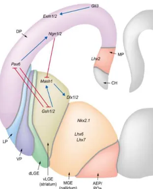
The ventral telencephalon consists of two distinct progenitor domains, the lateral (LGE) and medial (MGE) ganglionic eminences, generating the striatum and pallidum, respectively. These telencephalic subdivisions are traced on the basis of differences in morphology, connectivity and neurochemical profiles, and by distinct patterns of gene expression, reflecting the initial acquisition of regional identity by progenitor populations during early embryonic development.
The neocortex represents the brain structure that has been subjected to a major expansion in its size and complexity during the course of mammalian evolution. The achievement of such a highly complex architecture relies on a precise orchestration of the proliferation of progenitors, onset of neurogenesis, spatiotemporal generation of distinct cell types and control of their migration and differentiation. Complex molecular mechanisms participate in coordinating growth and patterning of the developing cortex
The cerebral cortex is organized into 6 layers of neurons, which are arranged in an in-side-out lamination sequence (Angevine and Sidman, 1961; Rakic, 1972). Cortical layers are not simply stacked one over the other; there exist characteristic connections between different layers and neuronal types, which span all the thickness of the cortex. These cortical microcircuits are grouped into cortical columns and minicolumns, the latter of which have been proposed to be the basic functional units of cortex (Jones and Rakic, 2010). Functional properties of the cortex change abruptly between laterally adjacent points; however, they are continuous in the direction perpendicular to the surface. Functionally distinct cortical columns have been shown in the visual cortex, auditory cortex, and associative cortex (Maduzia and Padgett, 1997; Meyer et al., 2010a, 2010b).
Patterning cues in the developing cerebral cortex are governed by both intrinsic and extrinsic signals of neuronal progenitors. Early anterioroposterior and dorsoventral patterning of the developing embryo, and generation of progenitor domains and neuronal specification into major classes of telencephalic cells, together with the precise coordination of cell cycle control, differentiation and spatiotemporal control of cell fate along both the radial and tangential dimension of the developing cerebral cortex are crucial steps required for the patterning of early cortical territories (regionalization) and of postnatal cortical areas (arealization).
1.1 Intrinsic control of cerebral cortex development through
patterning centers and transcription factor (TF) gradients
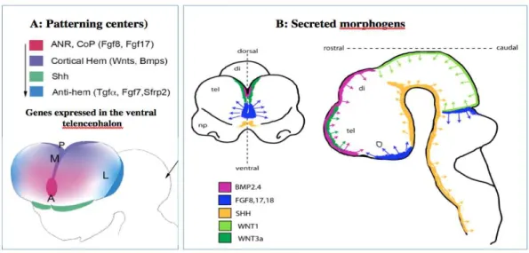
Multiple signaling centers or ‘organizers’ are involved in the induction and patterning of early brain territories through the expression of specific TFs. Secreted molecules (morphogens and growth factors) at signaling centers affect cortical growth and patterning at short range at early developmental stages. Four patterning centers are responsible for early induction and patterning of the telencephalon in the mouse embryo (Figure 1):
1. The anterior neural ridge /commissural plate at the rostral midline forming the border between neural and non-neural ectoderm. During neurulation and closure of the neural tube, Fgf8 is expressed at the rostral midline in the anterior neural ridge, thus defining the anterior patterning center. Later on, at E10.5–E12.5, after the expansion of the telencephalic vesicles, the fibroblast growth factors (Fgf8, Fgf15, Fgf17, Fgf18) are expressed anteriorly in nested domains at the commissural plate (CoP) and septum.
2. The roof plate (RP) and cortical hem at the dorsal/caudal midline and immediately adjacent territories, the Wnts and Bone morphogenetic proteins (Bmps) are localized at the dorsal/caudal midline in the RP and cortical hem. 3. The prechordal plate (PP)/ventral telencephalon signaling units, Sonic
4. The pallial–subpallial boundary (PSB) or anti-hem get involved later to coordinate growth and spatial patterning as growth proceeds and the distance between patterning centers increases (E11.0–E13.5). The PSB localizes at the lateral edge of the pallium and expresses of Fgf7, Sfrp2, and Tgfa (Assimacopoulos et al., 2003).
Figure 1: Schematic representation of the role patterning centers and secreted morphogens in telencephanlon development:
Adapted from (O’Leary and Sahara, 2008; Stevens et al., 2010).
During neurulation and closure of the neural tube, Fgf8 is expressed at the rostral midline in the ANR, Shh is expressed at the ventral midline in the PP, and Wnts/Bmps are localized at the dorsal midline in the RP. After the expansion of the telencephalic vesicles, the Fgfs are expressed anteriorly at the CoP and septum. The Wnts and Bmps are localized at the dorsal/caudal midline in the RP and cortical hem and Shh in the ventral telencephalon in addition to the PP. At this stage the the anti-hem localizes at the lateral edge of the pallium.
Fgfs in telencephalic progenitors and Shh and Wnts in other CNS regions accelerate the cell cycle by shortening the G1 phase, thus enhancing cell cycle exit (Pierani and Wassef, 2009). This represents an intrinsic effect that will ultimately have a crucial role in controlling the area-specific and, possibly, species- specific rates of neuron production.
1.2 Extrinsic signals that control cerebral cortex patterning
Although patterning centers and their secreted morphogens play a crucial role in the initial territorial shaping of the developing telencephalon, the proceeding of growth and the increase in the distance between signaling centers require the addition of other
patterning mechanisms. Diffusion works efficiently at short distances, but alone cannot pace with the expanding size. The emergence of other efficient signals that combine speed and functionality sounds fundamental, and a postmitotic neuronal compartment, together with migrating and differentiating cells seem needed to assure a correct fine-tuning of developing brain.
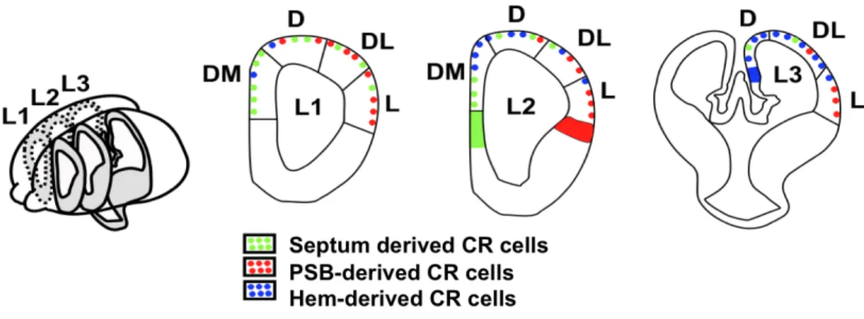
The ventricular fluid, which has been shown to contain signaling molecules like bFGF or Slits, could serve as a vehicle for forebrain growth signals through the formation of gradients mediated by the synchronized beating of cilia (Sawamoto et al., 2006). Additionally, migrating cells could act as mobile patterning cues where through their motility; they can mediate long distance transport of signaling molecules. Cajal-Retzius (CR) cells represent a transient class of earliest born neurons that populate the marginal zone of the developing cerebral cortex from early stages of development when regionalization takes place. CR cells secrete Reelin, an extracellular matrix protein that plays an important role in normal lamination of developing cortex. They are composed of a combination of molecularly distinct subtypes arising from restricted locations at the borders of the developing pallium, which actually represent the major patterning centers, namely the hem, the pallium–subpallium border and the septum (Figure 2) (Bielle et al., 2005; Pierani and Wassef, 2009). After generation, CR cells migrate from sites of origin and distribute in specific cortical regions. Fgf8, expressed at the rostral patterning center, and Tgfb, expressed at the caudal patterning center, induce the production of CR subtypes, which modulate early cortical patterning by transporting signaling molecules over a long distance (Griveau et al., 2010).
Figure 2: Patterning over a long distance.
Adapted from Borello and Pierani, 2010
Fgf8 and Tgfb expressed at the rostral and caudal patterning centers respectively, induce the production of CR subtypes, which modulate early cortical patterning by transporting signaling molecules over a long distance.
Additionally, a new class of migrating projection neurons, cortical plate (CP) transient cells, as well as the meninges and the invading vasculature also play a role in fine-tuning the developing telencephalon (Vasudevan et al., 2008; Siegenthaler et al., 2009).
Altogether, a complementary interplay between intrinsic and extrinsic patterning mechanisms seems pivotal for the early events that help in correctly instruction telencephalon development and patterning.
SECTION 2: CEREBRAL CORTEX HISTOGENESIS,
NEUROGENESIS AND SPECIFICATION OF NEURONAL
PHENOTYPES IN THE DEVELOPING TELENCEPHALON
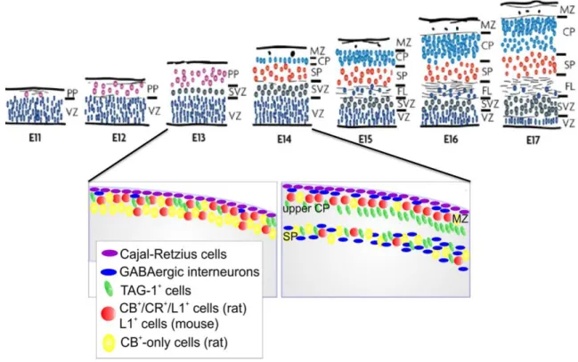
The histogenesis of the cerebral cortex starts from the time when the cerebral hemispheres develop from the wall of the telencephalic vesicle. The neuroepithelial cells initially span the thickness of the wall, and as they continue to undergo cell division, the area of the hemispheres expands. At this early stage of development, the progenitor cells are thought to undergo primarily symmetric cell divisions, and their progeny both remain in the cell cycle. Soon, however, a few cells withdraw from the cycle to develop as the first cortical neurons. These neurons migrate a short distance to form a distinct layer, just beneath the pial surface, known as the preplate (Bayer and Altman, 1990) (Figure 3). The preplate and its derivatives contain projection neurons, collectively named as pioneer neurons, whose axonal arborizations establish the earliest corticofugal projection systems during cortical development (Soria et al., 1999; Soria and Fairén, 2000). The CP develops within the preplate, so that preplate neurons become redistributed between the marginal zone, containing a group of large, stellate-shaped CR cells, and a deeper zone of cells called the intermediate zone (IZ)/SP, where most of the glutamatergic pioneer neurons are located and increasing numbers of incoming axons (Allendoerfer and Shatz, 1994; López-Bendito et al., 2002; Morante-Oria et al., 2003; Espinosa et al., 2009). A minor group of preplate pioneer neurons are not found later in the subplate (SP), they are rather divided into two groups and are found in the marginal zone, and the CP respectively (Espinosa et al., 2009). The two groups of non-SP pioneer neurons show distinct early locations in the preplate and later on, express distinct neurochemical properties (figure 3).
Figure 3: The developmental processes that will lead to organization of the neocortex into distinct neuronal layers.
Adapted from (Dehay and Kennedy, 2007; Espinosa et al., 2009)
Schematic presentation of the sequential layering events in the mouse and rat embryonic cortex. VZ: ventricular zone, SVZ: subventricular zone, PP: preplate, SP: subplate, CP: cortical plate, MZ: marginal zone, FL: fiber layer.
In rats, non-SP pioneer neurons situated in the upper part of the preplate before its partition will express the calcium binding proteins calbindin and calretinin, and the cell adhesion molecule L1. However, a second subtype of non-SP pioneer neurons is located deeper in the preplate. They will specifically express the adhesion molecule TAG-1, and will be transiently located in the upper CP (Espinosa et al., 2009). In mice, similar populations of pioneer non-SP neurons also exist. However, the more superficial cells express the adhesion molecule L1, but not the calcium binding proteins calbindin and calretinin. Interestingly, the two populations of non-SP pioneer neurons show different projection patterns. Axons of L1 neurons project to the ganglionic eminences and the anterior preoptic area, whereas axons of TAG-1 pioneer neurons only project to the lateral parts of the ganglionic eminences at the early stages of cortical histogenesis (Dehay and Kennedy, 2007; Espinosa et al., 2009).
From the earliest stages of cortical development, the processes of the apical progenitor cells span the entire thickness of the cortex (Miyata et al., 2001; Nadarajah et al., 2001). The first cortical neurons that are generated use the predominantly radial
orientation of their neighboring progenitor cells to guide their migration. However, the accumulation of neurons within the CP results in a marked increase in cortical thickness. As a result, the processes of later born cells no longer are able to extend to the external surface of the cortex. Nevertheless, the newly generated cortical neurons still migrate primarily in a radial direction guided by a remarkable set of cells, known as radial glia, which provide a scaffold (Rakic, 1978; Nadarajah et al., 2001; Nadarajah and Parnavelas, 2002).
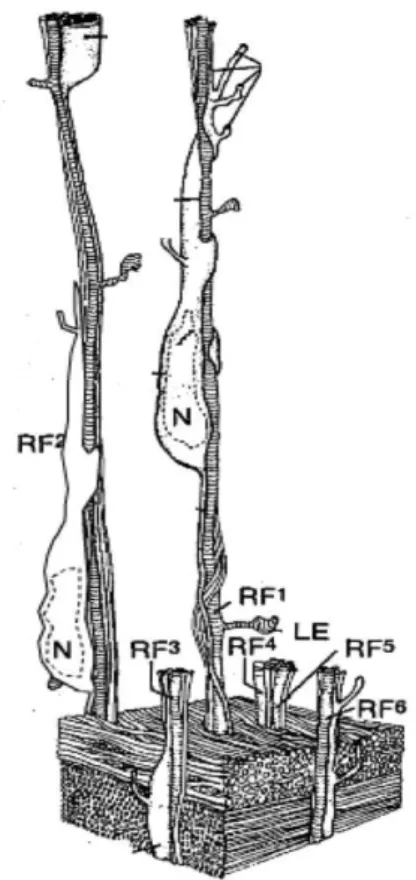
These glial cells have long processes that extend from the ventricular zone all the way to the pial surface. They form a scaffold that neurons migrate along. Serial section electron microscopic (EM) studies by Pasko Rakic first clearly demonstrated the close association of migrating neurons with radial glial cells in the cerebral cortex (Figure 4). The migrating neurons wrap around the radial glial processes, and move in a saltatory way, with migrating neurons frequently starting and stopping along the way. The next phase of cortical histogenesis is characterized by the gradual appearance of defined layers within the CP. As increasing
numbers of newly generated neurons migrate from the ventricular zone (VZ) into the CP, they settle in progressively more peripheral zones. Meanwhile, the earlier generated neurons start to differentiate. Thus, later generated neurons migrate past those generated earlier. This results in an inside-out development of cortical layers (Rakic, 1978; Nadarajah et al., 2001; Nadarajah and Parnavelas, 2002). Richard Sidman used the 3H-thymidine birthdating technique, to mark
newly born neurons from the VZ to their settling point in the CP, and was the first to Figure 4: a three-dimensional reconstruction of a migrating neuron along the surface of a radial glial fiber
Adapted from (Rakic, 2003)
Neurons are generated in the proliferative ventricular zone. They traverse the expanding cerebral wall and pass through the layer of earlier generated neurons in the deep layers, before settling in at the top of the developing cortex. Migrating neuron is attached to the radial glial fiber, which guides the neurons to the appropriate layers of the cerebral cortex. RF: radial fiber, N: neuron.
demonstrate the inside-out pattern of cerebral cortical histogenesis. The neurons labeled in the cortex of pups born from pregnant female rats injected with thymidine on the 13th day of gestation were located in the deeper layers of the cortex, whereas the neurons labeled after a thymidine injection on the 15th day of gestation were found more superficially. This inside-out pattern of cortical neurogenesis is conserved across mammalian species. Thymidine injections at progressively later stages of gestation result in progressively more superficial layers of cerebral cortical neurons being labeled. Each cortical layer has a relatively restricted period of developmental time over which it is normally generated (Angevine and Sidman, 1961; Caviness and Rakic, 1978; Caviness, 1982; Fairén et al., 1986).
Time-lapse imaging of labeled neuroblasts (Nadarajah et al., 2001; Noctor et al., 2004) shows clearly that many of the neuroblasts migrate just as predicted from the EM reconstructions of Rakic. However, direct visualization of the migration process also revealed that many of the neuroblasts move via a very different process, a process termed somal translocation. In this case, in early stages of corticogenesis, the migrating cell has a leading process that extends to the pial surface, while the cell body is still near the VZ. Then, with progressive shortening of the process, the cell soma is drawn to the pial surface (Nadarajah et al., 2001).
In addition to their function in supporting migrating neurons, radial glial cells are also neuronal progenitors. They undergo several cell divisions, and the progeny are not always additional radial glia but migrating immature neurons that migrate along the radial glia that generated them and label for neuron specific markers, while the radial glial cell that generated them expresses proteins typical of radial glia (Noctor et al., 2002; Lui et al., 2011).
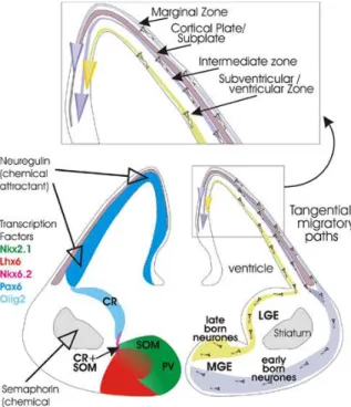
In addition to this predominantly radial migration of the newly generated neurons, it has been shown that other populations of cortical cells migrate tangentially to the cortical surface. These correspond to different populations of GABAergic interneurons, oligodendrocytes, Cajal-Retzius cells and endothelial cells.
Indeed there was an important discovery of a subset of cortical neurons that do not arise in the pallial VZ, but instead, originate in distinct subpallial regions (Wonders and Anderson, 2006). Fate mapping experiments and loss-of-function analyses in rodents have shown that cortical interneurons arise predominantly from the medial
and caudal (CGE) ganglionic eminences, and from the embryonic preoptic area (POA) (Gelman et al., 2009). However, recent observations in fetal human and monkey brains have suggested that, in these two species, a substantial proportion of cortical interneurons may arise from the lateral ventricular epithelium starting from the second half of gestation (Letinic et al., 2002; Yu and Zecevic, 2011).
Abundant evidence indicates that cortical interneurons comprise distinct neuronal subpopulations as defined by their morphological, neurochemical and electrophysiological properties (Butt et al., 2005). It has been suggested that the generation of the different subpopulations is linked to regional differences, defined by the expression of particular combinations of TFs, in the specification of progenitor cells in the subpallium, and these differences are key contributors to the generation of interneuron diversity in the cerebral cortex. These topics will be detailed in this section.
2.1
NEUROGENESIS :
FROM
NEUROEPITHELIAL
STEM
CELLS TO RADIAL GLIAL CELLS
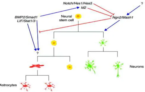
During development, neuroepithelial (NE) cells are the neural stem cells that give rise to all the neurons of the mammalian central nervous system. They are the source of the two types of neurons, cortical projection neurons and interneurons, as well as, astrocytes and oligodendrocytes. NE stem cells are self-renewing for a certain number of cell divisions; they first undergo symmetric, proliferative divisions, each of which generates two daughter stem cells (McConnell, 1995; Rakic, 1995). These divisions are followed by approximately 5-6 asymmetric, self-renewing divisions. Each of these divisions generates a daughter stem cell, plus a more differentiated neuronal precursor.
At the neural tube stage, the NE cell layer is composed of highly polarized pseudostratified epithelial cells, with the nuclei of these cells migrating up and down the apicobasal axis during the cell cycle (interkinetic nuclear migration) (Huttner and Brand, 1997). Tight junctions are present at the most apical end of the lateral plasma membrane and receptors for basal lamina constituents such as integrins contact the basal lamina (Wodarz and Huttner, 2003). With the generation of neurons, the neuroepithelium transforms into a more specialized tissue for neuron production. In
the VZ, the most apical cell layer that contains most of the progenitor cell bodies, neuroepithelial cells switch to neurogenesis and downregulate certain epithelial features (Aaku-Saraste et al., 1996). Astroglial markers appear and a distinct, but related, cell type — radial glial cells — which exhibit residual neuroepithelial as well as astroglial properties appear (Kriegstein and Götz, 2003). Radial glial cells represent more fate-restricted progenitors than neuroepithelial cells and successively replace the latter. As a consequence, all of the projection neurons in the brain are derived, either directly or indirectly, from radial glial cells.
Figure 5: Neuronal progenitors
Taken from (Taverna and Huttner, 2010)
Apical-basal polarity of NE cells (A) and apical progenitors (B). AP mitosis (M) occurs at the apical surface, whereas S phase takes place at a more basal location, with apical-to-basal nuclear migration in G1 and apical-to- basal-to-apical nuclear migration in G2. Mention basal progenitors.
I. Types of neuronal progenitors in the developing telencephalon
Cortical neurons are exclusively generated from progenitors located within the dorsolateral wall of the telencephalon. Heterogeneity within this progenitor population has been shown, with apical progenitors (APs) (some of which are radial glial cells) located along the luminal wall and intermediate “basal” progenitors (IPs) which delaminate from the VZ and divide at a distance from the lumen (Figure 5) (Kriegstein et al., 2006).
1. Apical progenitors (APs)
At the time of corticogenesis (E10 in the mouse), NE cells switch and give rise to other neuronal progenitor populations: radial glial cells (RGCs) and short neural
precursors (SNPs) both still residing in the VZ. APs exhibit apical-basal polarity, with the apical plasma membrane lining the lumen of the ventricle, the primary cilium protruding from the apical plasma membrane, and the interphase centrosomes located at the apical plasma membrane. AP nuclei occupy different positions along the apical-basal axis depending on the phase of the cell cycle and undergo interkinetic movement (Figure 5). Both these cell types will be further detailed here.
a. Radial glial cells:
As NE cells, RGCs exhibit an apico-basal polarity and span the entire cortical wall with an apical process contacting the ventricle and a basal process extending to the basement membrane. RGCs and NE cells also share expression of the intermediate filament protein Nestin, as well as expression of RC2 and the TF Pax6, but RGCs are further delineated by the expression of astroglial markers such as the astrocyte-specific glutamate transporter (GLAST), vimentin and the brain lipid binding protein (BLBP) (Pinto and Götz, 2007). With the onset of corticogenesis, RGCs comprise the predominant progenitor population within the VZ. They play a dual role in providing migratory guides for newly born neurons, and they are able to generate neurons or neuronal progenitors following asymmetric division, thereby giving rise, directly or indirectly, to most projection neurons of the cerebral cortex (Noctor et al., 2004). Like NE cells, RGCs show interkinetic nuclear migration, with their nuclei undergoing mitosis at the apical surface of the ventricular zone and migrating basally for S phase of the cell cycle (Figure 5). However, whereas in neuroepithelial cells the nuclei migrate through the entire length of the cytoplasm, this is not the case in RGCs. This interkinetic nuclear movement underlies the pseudostratification of the VZ. It allows the limited apical space to be used efficiently for AP mitoses, and functions by influencing the AP fate by controlling the exposure of AP nuclei to different, proliferative versus neurogenic, signals localized along the apical-basal axis (Noctor et al., 2004).
Time lapse imaging experiments have shed light on the molecular and cellular mechanisms of interkinetic nuclear movement. This movement appears completely independent of centrosome behavior and occurs along the MT network spanning the entire length of the progenitor cell. RNA interference (RNAi) experiments using in
not basally directed nuclear movement. Basally directed movement, however, was inhibited by RNAi of the unconventional kinesin heavy chain protein, kinesin 3 (Kif1a) (Tsai et al., 2010). Thus, interkinetic nuclear movement is regulated by very specific molecular motors.
b. Short neuronal progenitors SNPs:
The identification of SNPs as a distinct progenitor population added further evidence to the heterogeneity of VZ cells (Gal et al., 2006). RG cells remain the main progenitor population. SNPs can be morphologically characterized by the presence of a short basal process, which does not contact the basal lamina (figure 6), as well as by the activity of the tubulin alpha1 promoter, suggesting that they are restricted neuronal progenitors; their true origin (NE cell- or RGC-derived cells) remains unknown. Despite differing in cellular and molecular features, NE cells, RGCs and SNPs all display strong apico-basal polarity as well as having a specialized membrane domain facing the ventricular lumen that is limited by apical junctional complexes. Remarkably, they all undergo interkinetic nuclear migration. Based on the location of their nuclei during mitosis, all three above-mentioned progenitor cell types can thus be referred to as APs (Fish et al., 2008).
2. Intermediate ‘basal’ progenitors (IPs):
As soon as neurogenesis begins, a different type of progenitor, called intermediate (basal) progenitors, arise from a division event from an AP (e.g. NE cell or RGC), they detach from the VZ to form the subventricular sone (SVZ) at later stages of neurogenesis. In the SVZ, IPs undergo mitosis and contribute to both early and late born neurons (Miyata et al., 2004; Noctor et al., 2004). IPs contribute to neurogenesis by undergoing symmetric cell division and might function to increase the number of neurons generated from an apical division (Haubensak et al., 2004). In contrast to APs, IPs do not exhibit interkinetic nuclear migration, nor display any apparent signs of polarity and typically lack both an apical and a basal process contacting the ventricle and the basal membrane, respectively (Figure 5, 6). Moreover, IPs are molecularly distinct from APs and exclusively express the TF Tbr2, and Cux1; a progressive attenuation of Pax6 expression concomitant with the appearance of Tbr2
transcripts characterizes the transition of APs to IPs (figure 6) (Englund et al., 2005). The non-coding RNA Svet-1 and the TF Cux2 (Nieto et al., 2004) are also expressed in subsets of IPs during the generation of upper layer neurons.
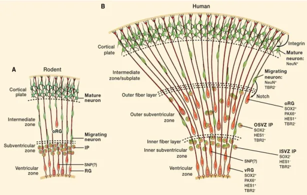
Figure 6: Rodent (A) and human neocortical development (B).
Adapted from (Lui et al., 2011)
In rodents RG cells generate intermediate progenitor (IP) cells that divides to produce pairs of neurons, which use RG fibers to migrate to their final destination. oRG cells exist in the mouse as well as in humans, but they are much less frequent in the mouse. SNPs, are also shown.
APs and IPs produce neurons by distinct mechanisms. During neurogenesis, APs undergo mostly asymmetric, self-renewing divisions, each of which generates an AP and an IP, or an AP and a neuron. IPs (at least in rodents) in most cases (90%) divide once, and nearly always symmetrically to produce two neurons with the same birthdate (Noctor et al., 2004). APs and IPs therefore represent two modes of neuron production, with a self-renewing progenitor pool and a “consumptive”, non-self- renewing pool for neuron production, respectively. For neuron production, the APs act as the self-renewing compartment where asymmetric divisions maintain the pool of progenitors while producing neuronal cells (direct neurogenesis). The self-renewal capacity of APs appears intimately linked to their epithelial-like features such as the apico-basal polarity and apical junctional complexes (Cappello et al., 2006). Mitotic
spindle and cleavage orientations determine if polarity is retained. The loss of these key features seems to be associated with neuronal differentiation and may also be causative for the loss of self-renewal and neurogenic potential at the end of neurogenesis. As for the IPs, these may be considered a neurogenic transit amplifying progenitor population that expands the number of particular neuronal populations (indirect neurogenesis). As both modes of neurogenesis take place simultaneously throughout cortical development, a crucial issue is whether they generate different subtypes of cortical neurons.
3. Outer radial glial cells (oRGCs):
A new class of neuronal progenitors is found in the outer part of the SVZ, termed outer radial glial (oRG) cells. These cells have been described in different species including mouse (Shitamukai et al., 2011; Wang et al., 2011), ferret (Fietz et al., 2010; Reillo et al., 2011), marmoset (García-Moreno et al., 2012; Kelava et al., 2012) and human (Hansen et al., 2010; Lui et al., 2011). Interspecies differences in number and abundance have been described for oRGs, with humans and higher mammals having much more numerous oRGs when compared to rodents (Lamonica et al., 2012). This difference could be one of the evolutionary mechanisms adopted by higher mammals to expand the tangential cerebral cortex surface area and form a gyrencephalic brain. Unlike RG cells, oRG cells are located far from the ventricle, with no apical contact to the luminal surface, but they possess a long basal fiber that often extends to the pial surface (Figure 6). They express Pax6 and Sox2, but not Tbr2 (Hansen et al., 2010; Reillo et al., 2011).
Time lapse imaging experiments have shown common fundamental features between these oRG cells among mammals; they are generated directly from RG cells in the VZ, and migrate away to the superficial SVZ via mitotic somal translocation rather than interkinetic nuclear migration. However, an important difference between species does exist. In mice, oRG cells undergo self-renewing asymmetric division to generate neurons directly, while human oRG cells generate transit-amplifying cells that in turn generate neurons (Wang et al., 2011).
The limited number of oRG cells and their rapid terminal differentiation in mice justifies the fact that the contribution of oRG cells to neurogenesis and cortical layer formation in rodents is small compared to higher mammals, and this may explain why