RESEARCH OUTPUTS / RÉSULTATS DE RECHERCHE
Author(s) - Auteur(s) :
Publication date - Date de publication :
Permanent link - Permalien :
Rights / License - Licence de droit d’auteur :
Institutional Repository - Research Portal
Dépôt Institutionnel - Portail de la Recherche
researchportal.unamur.be
University of Namur
Rapid exclusion of the diagnosis of immune HIT by AcuStar HIT and heparin-induced
multiple electrode aggregometry
Minet, V; Baudar, J; Bailly, N; Douxfils, J; Laloy, J; Lessire, S; Gourdin, M; Devalet, B;
Chatelain, B; Dogné, J M; Mullier, F
Published in:
Thrombosis Research
DOI:
10.1016/j.thromres.2014.01.014
Publication date:
2014
Document Version
Publisher's PDF, also known as Version of record
Link to publication
Citation for pulished version (HARVARD):
Minet, V, Baudar, J, Bailly, N, Douxfils, J, Laloy, J, Lessire, S, Gourdin, M, Devalet, B, Chatelain, B, Dogné, JM
& Mullier, F 2014, 'Rapid exclusion of the diagnosis of immune HIT by AcuStar HIT and heparin-induced multiple
electrode aggregometry', Thrombosis Research, vol. 133, no. 6, pp. 1074-1078.
https://doi.org/10.1016/j.thromres.2014.01.014
General rights
Copyright and moral rights for the publications made accessible in the public portal are retained by the authors and/or other copyright owners and it is a condition of accessing publications that users recognise and abide by the legal requirements associated with these rights. • Users may download and print one copy of any publication from the public portal for the purpose of private study or research. • You may not further distribute the material or use it for any profit-making activity or commercial gain
• You may freely distribute the URL identifying the publication in the public portal ? Take down policy
If you believe that this document breaches copyright please contact us providing details, and we will remove access to the work immediately and investigate your claim.
Regular Article
Rapid exclusion of the diagnosis of immune HIT by AcuStar HIT and
heparin-induced multiple electrode aggregometry
V. Minet
a,⁎
, J. Baudar
b, N. Bailly
b, J. Doux
fils
a, J. Laloy
a, S. Lessire
c, M. Gourdin
c, B. Devalet
d, B. Chatelain
b,
J.M. Dogné
a, F. Mullier
a,ba
Department of Pharmacy, Namur Thrombosis Hemostasis Center (NTHC), Namur Research Institute for LIfe Sciences (NARILIS), University of Namur, Namur, Belgium b
Hematology Laboratory, Namur Thrombosis and Hemostasis Center (NTHC), Namur Research Institute for LIfe Sciences (NARILIS), CHU Dinant Godinne - UCL Namur, Belgium c
Anesthesiology Department, Namur Thrombosis and Hemostasis Center (NTHC), CHU Dinant Godinne - UCL Namur, Belgium d
Hematology Department, Namur Thrombosis Hemostasis Center (NTHC), Namur Research Institute for LIfe Sciences (NARILIS), CHU Dinant Godinne - UCL Namur, Belgium
a b s t r a c t
a r t i c l e i n f o
Article history:
Received 20 November 2013
Received in revised form 13 January 2014 Accepted 13 January 2014
Available online xxxx Keywords:
immune heparin-induced thrombocytopenia HemosIL AcuStar HIT
heparin-induced multiple electrode aggregometry
Background: Accurate diagnosis of heparin-induced thrombocytopenia (HIT) is essential but remains challenging. We have previously demonstrated, in a retrospective study, the usefulness of the combination of the 4Ts score, AcuStar HIT and heparin-induced multiple electrode aggregometry (HIMEA) with opti-mized thresholds.
Objectives: We aimed at exploring prospectively the performances of our optimized diagnostic algorithm on suspected HIT patients. The secondary objective is to evaluate performances of AcuStar HIT-Ab (PF4-H) in comparison with the clinical outcome.
Methods: 116 inpatients with clinically suspected immune HIT were included. Our optimized diagnostic algorithm was applied to each patient. Sensitivity, specificity, negative predictive value (NPV), positive pre-dictive value (PPV) of the overall diagnostic strategy as well as AcuStar HIT-Ab (at manufacturer’s thresh-olds and at our threshthresh-olds) were calculated using clinical diagnosis as the reference.
Results: Among 116 patients, 2 patients had clinically-diagnosed HIT. These 2 patients were positive on AcuStar HIT-Ab, AcuStar HIT-IgG and HIMEA. Using our optimized algorithm, all patients were correctly di-agnosed. AcuStar HIT-Ab at our cut-off (N9.41 U/mL) and at manufacturer’s cut-off (N1.00 U/mL) showed both a sensitivity of 100.0% and a specificity of 99.1% and 90.4%, respectively.
Conclusion: The combination of the 4Ts score, the HemosIL® AcuStar HIT and HIMEA with optimized thresh-olds may be useful for the rapid and accurate exclusion of the diagnosis of immune HIT.
© 2014 Elsevier Ltd. All rights reserved.
Introduction
Immune heparin-induced thrombocytopenia (HIT) is a severe immunological adverse event of heparin therapy[1,2]. Early diagno-sis is essential and both misdiagnodiagno-sis and erroneous diagnodiagno-sis should be avoided[3]. Indeed, a misdiagnosis may expose the pa-tient to life-threatening thrombosis. Delays in the starting of treat-ment are associated with an initial 5%-10% daily risk of thrombosis, amputation, organ dysfunction or death [1]. An overdiagnosis may also lead to a discontinuation of the heparin treatment and a substitution by another more expensive anticoagulant which could be associated with an increased risk of bleeding without effective antidote[3,4]. The current diagnostic relies on the use of a clinical scoring algorithm (“4Ts score”) together with immunological
and functional assays (i.e. platelet activation assays)[1,2]. This com-bination improves HIT diagnosis compared to its components con-sidered independently[5]. However, the scoring system has to be used carefully and may require some extensive documentation of the patient medical history[6]. Immunoassays are sensitive but poorly specific. The HemosIL® AcuStar HIT-IgG (PF4-H) and AcuStar HIT-Ab (PF4-H), two immunological assays, and the heparin-induced multiple electrode aggregometry (HIMEA), a functional assay, were recently proposed as new rapid methods for the diagno-sis of HIT[7–9].
Previously, we performed a retrospective study on 106 patients with suspected HIT. We showed that a diagnostic algorithm based on 4Ts score, AcuStar HIT and HIMEA with optimized thresholds showed good performances for the rapid and accurate diagnosis of immune HIT (PPV: 88.9 % (95% CI: 51.7%-98.2%), NPV: 100.0 % (95% CI: 96.1%-100.0%))[10].
In the present study, we explored prospectively the performances of our optimized diagnostic algorithm on HIT suspected patients in our academic tertiary hospital. Secondly, we evaluated performances of AcuStar HIT-Ab (PF4-H) in comparison with the clinical outcome.
Thrombosis Research xxx (2014) xxx–xxx
⁎ Corresponding author at: University of Namur, Department of Pharmacy, Rue de Bruxelles, 61, B-5000 Namur. Tel.: +32 81 724292, +32 499 626462 (mobile); fax: +32 81 724299.
E-mail address:[email protected](V. Minet).
0049-3848/$– see front matter © 2014 Elsevier Ltd. All rights reserved.
http://dx.doi.org/10.1016/j.thromres.2014.01.014
Contents lists available atScienceDirect
Thrombosis Research
Materials and Methods Patients
One hundred and sixteen inpatients (76 men and 40 women; 49 medical and 67 were surgical patients) suspected of developing HIT from 1st November 2011 to 31th January 2013 at the CHU Dinant Godinne - UCL Namur, Belgium, were included in the study, in accor-dance with the local ethics committee.
Healthy subjects
In order to perform HIMEA, whole blood samples from healthy volunteers are needed. Blood was collected with a 20 gauge needle via atraumatic antecubital venipuncture into polyethylene terephthal-ate tubes Venosafe® (Terumo Europe, Leuven, Belgium) containing 25μg/mL of recombinant hirudin (Verum Diagnostica GmbH, Munich, Germany, 1:10 v/v). A discard tube was used to avoid tissue factor contamination. Blood was obtained from two O Rh +/− blood group donors. These donors did not take any medicine which could potentially affect their platelet function for 10 days before the blood sampling. Platelet donors were selected on the basis of a good reactivity with plas-ma of HIT patients. The platelet reactivity of the healthy donors was also assessed by HIMEA with the following reagents: adenosine diphosphate (ADP,final concentration: 6.5 μM), collagen (type 1, final concentration: 3.2μg/mL), arachidonic acid (AA, final concentration: 0.5 mM) or thrombin receptor activating peptide (TRAP-6,final concentration 32 μM) (Dynabyte, Munich, Germany). Each volunteer presented normal aggregation.
Optimized diagnostic algorithm
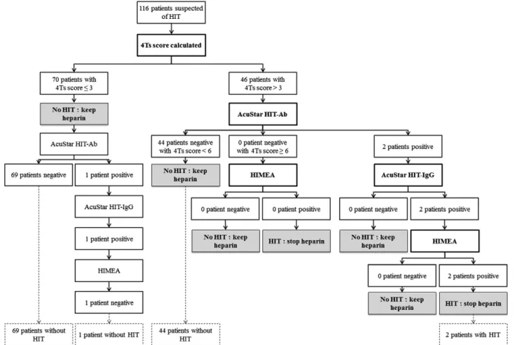
Routine laboratory diagnosis was performed on HIT patients according to our optimized algorithm (Fig. 1) (except for AcuStar HIT-Ab which was performed on each patient). Thisflow chart is based on (1) 4Ts score, (2) AcuStar HIT (PFA-H) (immunological assay), and (3) HIMEA (functional assay).
4Ts score and clinical diagnosis
HIT was suspected because of a rapidly decreasing platelet count oc-curring in hospitalised patients under heparin therapy. Subsequently, the“4Ts score” was calculated (based on four criteria: the severity of the thrombocytopenia and its timing, the occurrence of a thrombosis and the exclusion of other causes of thrombocytopenia). Clinical data were recorded in real time in the hospital medical database. Clinical outcomes were retrospectively and independently confirmed by two investigators (VM and FM), not aware of the results of the laboratory assays. Clinical diagnoses made by these 2 local investigators were 100% concordant among them and with conclusions of the medical database. Several clinical criteria have to be fulfilled for the confirmation of clinical HIT diagnosis. Criteria from the ACCP (American College of Chest Physicians) guidelines were used to make the clinical diagnosis of HIT: (1) Thrombocytopenia, defined as at least a 30% decline in the platelet count, with a platelet count increase after heparin cessation; (2) Timing of platelet count fall after the initiation of heparin occurring between 4 and 14 days, or occurring within 24 to 48 hours (in case of prior heparin exposure within 30 days); and (3) lack of other, predom-inant causes of thrombocytopenia[11–13]. Other causes of thrombocy-topenia analysed in this study were: neoplasia, current pregnancy or
Fig. 1. Flow chart with results. Thresholds for AcuStar HIT-Ab, AcuStar HIT-IgG and HIMEA are, respectively, 9.41 U/mL, 2.89 U/mL and 276 U/mL at 1 IU/mL UFH and a reductionN 80% at 385 IU/mL UFH.
postpartum, autoimmune disease, sepsis, disseminated intravascular co-agulation, intra-aortic balloon pump counterpulsation, multitransfusion, multi-trauma, shock syndrome and drug-induced thrombocytopenia (quinolone,β-lactam, vancomycin, teicoplanin, rifampicin, isoniazid, amphotericin,fluconazole, chemotherapy, anti-GPIIb/IIIa; furosemide and proton pump inhibitors). All these 3 clinical criteria have to be fulfilled for the confirmation of clinical HIT diagnosis. The following infor-mation was taken into consideration: patient’s medical history, types (fractionated vs. unfractionated) and doses of heparin administered, thrombotic complications, alternative diagnoses, therapeutic attitude, clinical and platelet count evolution, co-suspected medications, and phy-sician’s diagnoses[10,13].
Laboratory testing
Blood was collected with a 20 gauge needle via atraumatic antecubital venipuncture into polyethylene terephthalate tubes Venosafe® (Terumo Europe, Leuven, Belgium) containing buffered sodium citrate (109 mM, nine parts of blood to one part sodium citrate solution). Plasmas were tested prospectively in real time.
Immunological assay: AcuStar HIT-IgG (PF4-H) and AcuStar HIT-Ab (PF4-H) The HemosIL® AcuStar HIT-IgG (PF4-H) and HIT-Ab (PF4-H) (In-strumentation Laboratory, Bedford, MA, USA) are two chemilumines-cent two-step immunoassays detecting the presence of anti-PF4-heparin antibodies. Thefirst one is specific for IgG, the second one is polyspecific (IgG, IgA and IgM). These assays have been described by Legnani et al.[7]. The immunological isotype mainly responsible for HIT is IgG[14]. IgM and IgA only play a minor role but we cannot rule out the possibility of these immunoglobulins to contribute to some degree of thrombocytopenia[15]. Consequently, in our algorithm, we decided to perform afirst screening with AcuStar HIT-Ab (PF4-H). AcuStar HIT-IgG (PF4-H) is then applied in case of a positive result. AcuStar HIT-Ab (PF4-H) was performed for each patient regardless the 4Ts score. AcuStar HIT-IgG (PF4-H) was carried out on patients accord-ing to ourflow chart (Fig. 1).
The threshold recommended by the manufacturer is 1.00 U/mL. We used the thresholds determined in our previous retrospective study, i.e. 2.89 U/mL and 9.41 U/mL for AcuStar HIT-IgG (PF4-H) and AcuStar HIT-Ab (PF4-H), respectively.
Functional assay: Heparin-induced multiple electrode aggregometry (HIMEA)
Multiple electrode aggregometry (MEA) is a method for the assess-ment of platelet function in whole blood (Multiplate® analyzer; Dynabyte, Munich, Germany)[16]. MEA is based on the principle of im-pedance aggregometry. Our protocol has already been described[10]. Each HIMEA assay is performed in real-time with the fresh blood of one of the two determined healthy donors. Results are expressed
using the area under the aggregation curve (AUC) and the recording time is 30 minutes. The result is considered to be positive when the platelet aggregation occurred in the presence of low heparin concentra-tion (1 IU/mL UFH) but is partially inhibited (reducconcentra-tionN 80%) by a high concentration of heparin (385 IU/mL UFH). The threshold determined in our retrospective study was 276 AU at 1 IU/mL UFH.
Data analysis
Data analysis was performed using Medcalc software (version 10.4.8) (MedCalc Software bvba, Ostend, Belgium). The sensitivity, the specificity, the negative predictive value (NPV) and the positive predic-tive value (PPV) of AcuStar HIT-Ab (PF4-H) were assessed using clinical diagnosis as reference.
Results
Out of the 116 patients included in this study, two (1.7%) were con-sidered to have developed HIT based on our clinical diagnosis. Thefirst one experienced a pulmonary embolism and the other one died during his hospitalisation following coronary thrombosis. Complete compres-sion ultrasonography and multidetector spiral computed tomography were performed for suspected thrombosis.
Assessment of our optimized diagnostic algorithm (Fig. 1)
Among 116 patients included in this study, 70 patients (60.3 %), 44 patients (37.9%) and 2 patients (1.7%) presented a low (≤3), medium (4–6) and high (7–8) 4Ts score, respectively. No patient with a low PTP was diagnosed as having HIT.
Among 46 patients with a medium-high PTP score, HIT was exclud-ed in 44 patients with AcuStar HIT-Ab (PF4-H) (threshold: 9.41 U/mL) (Fig. 1). The NPV and PPV of AcuStar HIT-Ab (PF4-H) was 100.0% (95% CI: 91.9%-100.0%) and 100.0% (95% CI: 19.3%-100.0%), respectively (Table 1). The 2 patients AcuStar HIT-Ab (PF4-H) positive were also pos-itive on AcuStar HIT-IgG (PF4-H) (threshold: 2.89 U/mL) and on HIMEA (threshold: 276 AU at 1 IU of UFH). These two patients had clinically confirmed HIT. The sensitivity, specificity, PPV and NPV of this combina-tion using our thresholds, for the 116 patients were 100.0 % (95% CI: 16.6%-100.0%), 100.0% (95% CI: 100.0%), 100.0% (95% CI: 96.8%-100.0%) and 100.0% (95% CI: 16.6%-96.8%-100.0%), respectively. (SeeTable 2.) Comparison of performances of AcuStar HIT-Ab (PF4-H) at our threshold (9.41 U/mL) and at manufacturer’s threshold (1.00 U/mL) (Table 1)
AcuStar HIT-Ab (PF4-H) was performed on each patient of the study regardless of the 4Ts score. For all patients (n = 116) at manufacturer’s threshold, there were 11 false positives against 1 false positive at our threshold. The false positive result on AcuStar HIT-Ab (9.96 U/mL)
Table 1
Comparison of HemosIL® AcuStar HIT-IgG (PF4-H), HIT-Ab (PF4-H) and optimized diagnostic algorithm with the clinical diagnosis (n = 116: all patients suspected of HIT with clinical diagnosis available; n = 46: patients with medium-high pre-test probability; AU: arbitrary unit, NPV: negative predictive value, PPV: positive predictive value).
Clinical diagnosis Sensitivity % (95%CI) Specificity % (95%CI) NPV % (95%CI) PPV % (95%CI) Negative Positive
AcuStar HIT-Ab (n = 116) Threshold:N1.00 U/mL Negative 103 0 100.0 (19.3-100.0) 90.4 (83.4-95.1) 100.0 (96.5-100.0) 15.4 (2.4-45.5) Positive 11 2
AcuStar HIT-Ab (n = 116) Threshold:N9.41 U/mL Negative 113 0 100.0 (19.3-100,0) 99.1 (95.2-99.9) 100.0 (96.8-100.0) 66.7 (11.6-94.5) Positive 1 2
AcuStar HIT-Ab (n = 46) Threshold:N1.00 U/mL Negative 40 0 100.0 (19.3-100.0) 90.9 (78.3-97.4) 100.0 (91.1-100.0) 33.3 (5.3-77.3) Positive 4 2
AcuStar HIT-Ab (n = 46) Threshold:N9.41 U/mL Negative 44 0 100.0 (19.3-100.0) 100.0 (91.9-100.0) 100.0 (91.9-100.0) 100.0 (19.3-100.0) Positive 0 2 Optimized diagnostic algorithm (n = 116) Thresholds:N3; N9.41 U/mL; N 2.89 U/mL; N276 AU Negative 114 0 100.0 (16.6-100.0) 100.0 (96.8-100.0) 100.0 (96.8-100.0) 100.0 (16.6-100.0) Positive 0 2
was slightly above our threshold (9.41 U/mL). Specificity was signifi-cantly higher at our threshold, i.e. 99.1% (95% CI: 95.2%-99.9%) than at manufacturer’s threshold, i.e. 90.4% (95% CI: 83.4%-95.1%). Among pa-tients with a medium-high pre-test probability (n = 46), our threshold presented no false positives against the 4 false positive at manufac-turer’s threshold. Using our threshold, the specificity reached 100.0 % (95% CI: 91.9%-100.0%), and it was 90.9% (95% CI: 78.3%-97.4%) with the manufacturer’s threshold.
Discussion
We performed the present study with the aim of assessing prospec-tively our immune HIT diagnostic algorithm with optimized thresholds (Fig. 1). Among 116 inpatients suspected of HIT, every patient was correctly diagnosed using ourflow chart. Specificity was higher at our threshold (99.1% (95% CI: 95.2%-99.9%)) than at manufacturer’s thresh-old (i.e. 90.4% (95% CI: 83.4%-95.1%)).
Applying our diagnostic algorithm, heparin therapy should be continued and no subsequent laboratory assay should be performed in patients with a 4Ts score≤ 3[1]. In this study, no patient with a low probability 4Ts score (≤3) (n = 70) was diagnosed as having HIT. It confirms the effectiveness of the 4Ts score to limit additional biological assays. Cuker et al. also concluded in a systematic review and meta-analysis that 4Ts score of patients with suspected HIT may reduce overtesting and overdiagnosis of suspected HIT patients and that a low probability 4Ts score was a robust tool to exclude HIT[17].
The HemosIL® AcuStar HIT-IgG (PF4-H) and AcuStar HIT-Ab (PF4-H) were proposed as chemiluminescent immunoassays. AcuStar can be run for single sample testing and it presents shorter running-time (30 minutes) than other commercially available immunologic assays (2–3 hours).
In our previous retrospective study, we demonstrated that the use of optimized thresholds markedly improved the specificity of AcuStar HIT-Ab (PF4-H)[10]. A recent study concluded that the diagnostic accu-racy of the anti-PF4/heparin ELISA can be optimized by using a higher cut-off[18]. Using optimized thresholds in our algorithm should reduce overdiagnosis of HIT and the need to perform additional HIMEA. Actual-ly, the manufacturers provided thresholds following a study on healthy subjects and not on a HIT suspected patient’s population.
The HIMEA is a rapid functional assay. It can be performed within 15 minutes and does not require any preparation of patient’s sample. However, the immediate availability of a healthy compatible blood group donor is required.
Our study suffers from some limitations. Firstly, it is a single-centre study, and therefore it does not allow inter-laboratory comparisons. Secondly, because of the intermediate size of our cohort the power of this study is limited. In a recent study, Althaus et al. defined lower cut-offs than in our study. Our cut-offs should be further validated or adjusted using larger sample size in a multicentre study to provide a sufficiently high NPV beside their excellent PPV[19]. However, it should be noted that due to the inter-instrument variation afixed OD threshold of an assay depends of the instrument. Therefore, each laboratory should determine their cut-offs with a cohort of HIT suspected patients. Thirdly, our protocol may differ from other studies since no guideline on standardized HIMEA procedure is available [8,20]. The threshold depends on variables including volume of whole blood of the healthy subject, volume of sample of patients, volume and concentration of heparin, duration of the measurement and the platelet reactivity of the healthy donors. Finally, to ascertain the diagnosis of HIT as recom-mended by the consensus committee of the ISTH working group[21], it is necessary to perform a washed platelet activation assay.14 C-Serotonin Release Assay (SRA) is still considered as one of the reference method but it has not been performed in this study. This assay is indeed not available in most routine hospital laboratories because of its well-known limitations (time-consuming, technically demanding and requires the use of radioactive material).
Ta b le 2 Clini ca l an d lab oratory d ata for th e 2 patient s with a p os itiv e d iagnosis of H IT. Lab o ra to ry as says Clinical data A cu S ta r H IM E A 4 T s Clinical evolution from the H IT sus picio n to th e disch arge from h o spital HI T Ab (U/m L) IgG (U /m L) 1I U /m L (AUC) Thro mb ocy topen ia T imin g o f thro m b o cy to peni a T hr om bo si s o r o ther sequelae O th er ca uses fo r throm bo cyto penia 4 Ts sco re S top h epa rin P lat elet cou n t increase Clin ic al diagnosis Patient 1 1 1 .11 5 .03 4 4 8 P latelet co unt fa ll N 50% an d pla tele t nadir ≥ 20 (2 po ints ) C lea r o ns et at day 7 ,n o p rio r hepa rin ex posure wit h in 3 0 da y s (2 points) Lu ng em bo lis m and deep veno us th ro m b osis (2 points ) Pos sible (1 point) (Tazocin®) 7 + + + Pat ien t 2 4 7 .30 4 6.81 66 4 P lat e let co u n t fa ll N 50% an d pla tele t nadir ≥ 20 (2 po ints ) C lea r o ns et at day 7 ,n o p rio r hepa rin ex posure wit h in 3 0 da y s (2 points) Co ron ary thro m b o si s (2 p oint s) P o ss ib le (1 po int) (Augm e ntin®, N exiam ® ,d ialys is) 7+ N D (d e at h ) +
Conclusion
In this study, we demonstrated that the combination of the 4Ts score, the HemosIL® AcuStar HIT and HIMEA with optimized thresholds may be useful for the rapid and accurate exclusion of the diagnosis of immune HIT among the 116 included patients. A large multicentre prospective study is needed to further validate our algorithm. Authorship Contributions
Contribution: FM, BCH, MGO, JMD conceived the idea and designed the study protocol; VM, NB, JB, JD, SL, BD and FM collected, assembled data and interpreted the data; VM performed statistical analysis; VM and FM wrote the manuscript; and all authors reviewed and approved the manuscript.
Conflict of Interest Disclosures
The authors have no conflict of interest to disclose. Acknowledgments
The authors would like to thank André Bosly, Marc Chatelain and Pierre Gavage for their contribution to this work.
References
[1]Wilke T, Tesch S, Scholz A, Kohlmann T, Greinacher A. The costs of heparin-induced thrombocytopenia: a patient-based cost of illness analysis. J Thromb Haemost 2009;7:766–73.
[2]Chong BH, Isaacs A. Heparin-induced thrombocytopenia: what clinicians need to know. Thromb Haemost 2009;101:279–83.
[3]Lo GK, Sigouin CS, Warkentin TE. What is the potential for overdiagnosis of heparin-induced thrombocytopenia? Am J Hematol 2007;82:1037–43.
[4]Berry C, Tcherniantchouk O, Ley EJ, Salim A, Mirocha J, Martin-Stone S, et al. Overdi-agnosis of heparin-induced thrombocytopenia in surgical ICU patients. J Am Coll Surg 2011;213:10–7.
[5]Elalamy I, Lecrubier C, Horellou MH, Conard J, Samama MM. Heparin-induced thrombocytopenia: laboratory diagnosis and management. Ann Med 2000;32 (Suppl. 1):60–7.
[6]Greinacher A, Ittermann T, Bagemuhl J, Althaus K, Furll B, Selleng S, et al. Heparin-induced thrombocytopenia: towards standardization of platelet factor 4/heparin antigen tests. J Thromb Haemost 2010;8:2025–31.
[7]Legnani C, Cini M, Pili C, Boggian O, Frascaro M, Palareti G. Evaluation of a new automated panel of assays for the detection of anti-PF4/heparin antibodies in pa-tients suspected of having heparin-induced thrombocytopenia. Thromb Haemost 2010;104:402–9.
[8]Elalamy I, Galea V, Hatmi M, Gerotziafas GT. Heparin-induced multiple electrode aggregometry: a potential tool for improvement of heparin-induced thrombocyto-penia diagnosis. J Thromb Haemost 2009;7:1932–4.
[9]Galea V, Khaterchi A, Robert F, Gerotziafas G, Hatmi M, Elalamy I. Heparin-induced multiple electrode aggregometry is a promising and useful functional tool for heparin-induced thrombocytopenia diagnosis: confirmation in a prospective study. Platelets 2013;24:441–7.
[10]Minet V, Bailly N, Douxfils J, Osselaer JC, Laloy J, Chatelain C, et al. Assessment of the performances of AcuStar HIT and the combination with heparin-induced multiple electrode aggregometry: A retrospective study. Thromb Res 2013;132:352–9.
[11]Warkentin TE, Greinacher A, Koster A, Lincoff AM, American College of Chest P. Treatment and prevention of heparin-induced thrombocytopenia: American College of Chest Physicians Evidence-Based Clinical Practice Guidelines (8th Edition). Chest 2008;133:340S–80S.
[12]Linkins LA, Dans AL, Moores LK, Bona R, Davidson BL, Schulman S, et al. Treatment and prevention of heparin-induced thrombocytopenia: Antithrombotic Therapy and Prevention of Thrombosis, 9th ed: American College of Chest Physicians Evidence-Based Clinical Practice Guidelines. Chest 2012;141:e495S–530S.
[13]Tardy B, Presles E, Akrour M, de Maistre E, Lecompte T, Tardy-Poncet B. Experts’ opinion or the serotonin release assay as a gold standard for the diagnosis of heparin-induced thrombocytopenia (HIT)? J Thromb Haemost 2011;9:1667–9.
[14]Greinacher A, Althaus K, Krauel K, Selleng S. Heparin-induced thrombocytopenia. Hamostaseologie 2010;30(17–8):20–8.
[15]Greinacher A, Juhl D, Strobel U, Wessel A, Lubenow N, Selleng K, et al. Heparin-induced thrombocytopenia: a prospective study on the incidence, platelet-activating capacity and clinical significance of antiplatelet factor 4/heparin antibod-ies of the IgG, IgM, and IgA classes. J Thromb Haemost 2007;5:1666–73.
[16]Solomon C, Traintinger S, Ziegler B, Hanke A, Rahe-Meyer N, Voelckel W, et al. Plate-let function following trauma. A multiple electrode aggregometry study. Thromb Haemost 2011;106:322–30.
[17]Cuker A, Gimotty PA, Crowther MA, Warkentin TE. Predictive value of the 4Ts scor-ing system for heparin-induced thrombocytopenia: a systematic review and meta-analysis. Blood 2012;120:4160–7.
[18]Raschke RA, Curry SC, Warkentin TE, Gerkin RD. Improving clinical interpretation of the anti-platelet factor 4/heparin enzyme-linked immunosorbent assay for the diag-nosis of heparin-induced thrombocytopenia through the use of receiver operating characteristic analysis, stratum-specific likelihood ratios, and Bayes theorem. Chest 2013;144(4):1269–75.
[19]Althaus K, Hron G, Strobel U, Abbate R, Rogolino A, Davidson S, et al. Evaluation of automated immunoassays in the diagnosis of heparin induced thrombocytopenia. Thromb Res 2013;131:e85–90.
[20]Morel-Kopp MC, Aboud M, Tan CW, Kulathilake C, Ward C. Whole blood impedance aggregometry detects heparin-induced thrombocytopenia antibodies. Thromb Res 2010;125:e234–9.
[21]Warkentin TE, Greinacher A, Gruel Y, Aster RH, Chong BH, scientific, et al. Laboratory testing for heparin-induced thrombocytopenia: a conceptual framework and impli-cations for diagnosis. J Thromb Haemost 2011;9:2498–500.