ORIGINAL ARTICLES
Left bundle branch block and coronary artery
disease: Accuracy of dipyridamole thallium-201
single-photon emission computed tomography in
patients with exercise anteroseptal
perfusion defects
N. E. Lebtahi, MD, J. C. Stauffer, MD, a n d A. Bischof Delaloye, MD
Background. Reduced septal or anteroseptal uptake of thallium-201 during exercise is frequently observed in patients with left bundle branch block (LBBB) even in the absence of left anterior descending (LAD) coronary artery disease. The purpose of this study was to evaluate prospectively the accuracy of dipyridamole 2°lTl single-photon emission computed tomography (SPECT) in detecting LAD coronary artery disease in patients with LBBB and septal or anteroseptal perfusion defects on exercise 2°lTI SPECT.
• Methods and Results. Twelve consecutive patients (10 men and two women) with complete LBBB and septal or anteroseptal perfusion defects on exercise 2°lTl SPECT underwent dipyridamole 2°lTl SPECT. The delay between dipyridamole and exercise was 2 to 30 days. Coronary angiography was performed during this period in all patients. Six (50 %) of 12 patients with exercise perfusion defects showed normal perfusion after dipyridamole; all had normal coronary angiograms. The remaining six patients also had positive results of dipyridamole studies, two with moderate and four with severe septal or anteroseptal perfusion defects. Coronary angiography showed significant (>50%) LAD coronary artery stenosis in three patients; three patients with severe septal or anteroseptal perfusion defects after dipyridamole had normal coronary angiograms. Neither the evaluation of apical involvement nor the presence of dilated ventricles, decreased left ventricular ejection fraction, or wall motion abnormalities could help to identify (or explain) false-positive results.
Conclusion. This study confirms that dipyridamole is more accurate than exercise in excluding LAD coronary artery disease. However, there are still false-positive results and the severity of the septal or anteroseptal perfusion defect does not add additional information to identify LAD coronary artery disease. Coronary angiography is thus necessary for positive dipyridamole study results to identify coronary artery disease as a major prognostic factor in patients with LBBB. (J Nucl Cardiol 1997;4:266-73.)
Key Words: left bundle branch block • dipyridamole • thallium-201 • single-photon emission computed tomography • coronary artery disease
The high frequency of septal or anteroseptal perfu- sion defects on exercise scintigraphy in patients with left bundle branch block (LBBB) in the absence of left anterior descending (LAD) coronary artery disease has been well documented. 1-~3 Cumulative results (Table 1) From the Divisions of Nuclear Medicine and Cardiology, University
Hospital, Lausanne, Switzerland.
Received for publication March 7, 1996; revision accepted Jan. 2, 1997.
Reprint requests: N. E. Lebtahi, MD, Centre d'Imagerie, CIMED, rue de Locamo 9, 1700 Fribourg, Switzerland.
Copyright © 1997 by American Society of Nuclear Cardiology. 1071-3581/97/$5.00 + 0 43/1/80120
266
indicate that septal or anteroseptal perfusion defects were present in 73% of cases, whereas a significant LAD stenosis was shown by coronary angiography in only 42%. Additional information in Table 1 indicates that the proportion of patients with LBBB and LAD coronary artery disease was only 38%. A proposed mechanism for the perfusion defects in the presence of normal coronary arteries is the occurrence of "relative hypoperfusion ''2,6 caused by abnormal septal motion. Exercise by inducing tachycardia further shortens left ventricular diastole, which could explain the occurrence, as well as the reversibility, of the defects. Thus pharmacologic stress has been suggested to be a more accurate stimulus for the
Journal of Nuclear Cardiology Lebtahi et al. 267
Volume 4, Number 4;266-73 Accuracy of dipyridamole 2°1T1 SPECT
Table 1. C u m u l a t i v e results w i t h e x e r c i s e s c i n t i g r a p h y in patients with LBBB w h o u n d e r w e n t c o r o n a r y a n g i o g r a p h y
No. LAD +
Study No. patients* Tracer No. S or AS + No. TP (total)
Hirzel et al. z 1984 19 Z°lTl 19 4 4
Braat et al. 31 1985 24 2°lTI 7 6 11
Huerta et al. 3 1987 11 ZmTI 8 0 0
Rothbart et al. 4 1987 22 ZmTl 16 5 7
De Puey et al. s 1988 14 ZmTl 12 3 4
Burns et al. 6 1991 16 Z°~T1 12 5 6
Matzer et al. 7 1991 44 Z°lT1 41 24 24
Larcos et al. 8 1991 83 ZmTl 56 26 38
La Canna et al. 9 1992 33 2°1TI 21 0 0
Delonca et al. lo 1992 66 2°1T1 45 16 17
Altehoefer et al. zl 1993 9 99mTc-labeled sestamibi 3 2 5
Knapp et al.ZZ 1993 28 zo 1TI 26 l 5 16
Knapp et al. z2 1993 37 99mTc-labeled sestamibi 34 14 15
Ebersole et al. 11 1993 11 99mTc-labeled sestamibi 8 2 3
O'Keefe et al. 1 z 1993 31 ZO~Tl 20 11 15
Vaduganathan et al. 13 1996 77 Z°lTl 57 29 33
Total 525 385 (73%) 162 (42%) 197 (38%)
S or AS+, Septal or anteroseptal perfusion defect; TP, true positive results (S or AS+ and LAD disease); LAD+, significant LAD coronary artery stenosis.
*Only patients who underwent coronary angiography were considered. detection of coronary artery disease by myocardial thal- lium-201 imaging in patients with LBBB. 6,~2 28 W e studied prospectively the value of dipyridamole in the identification of L A D coronary artery disease in patients with L B B B and septal or anteroseptal perfusion defects on exercise a°IT1 single-photon emission computed to- mography (SPECT).
METHODS
Patients. Twelve consecutive patients (10 men and two women), aged 37 to 77 years (mean age 58 years), with complete LBBB and septal or anteroseptal perfusion defects on exercise 2roT1 SPECT underwent dipyridamole 2°1T1 SPECT within 1 month. All 12 patients underwent coronary angiogra- phy for clinical reasons during this period. Shown in Table 2 are the clinical data of all patients.
Exercise Protocol. Patients underwent a symptom-lim- ited Bruce protocol exercise test on a bicycle ergometer. At peak exercise, a weight-adjusted dose (range 2.5 to 3.5 mCi) of amT1 was administered intravenously and patients were encour- aged to continue exercising for 1 to 2 minutes. SPECT imaging was performed as soon as possible after the end of exercise and again 3 to 4 hours after the 2roT1 injection.
Dipyridamole Protocol. Patients were advised to avoid food and caffeine-containing drinks for the 12 hours preceding the test. All patients had a 4-minute intravenous infusion of 0.56 mg/kg dipyridamole. Cardiac rhythm was monitored
continuously, and 12-lead electrocardiogram, blood pressure, and heart rate were obtained every 2 minutes during the test. Three minutes after the end of dipyridamole infusion, a weight-adjusted dose of 2roT1 (range 2.5 to 3.5 mCi) was administered and SPECT acquisition began 3 minutes later. Redistribution was imaged 3 to 4 hours after 2°1T1 injection.
Z°lTi SPECT. 2°1T1SPECT imaging was performed with a single-head rotating gamma camera (Starcam; General Electric Co., Wilmington, Mass.), equiped with a low-energy, all- purpose collimator interfaced to a dedicated computer. Patients were placed in the supine position and 32 projections were obtained (step by step), 40 seconds each, through a 180-degree orbit beginning in a 45-degree right anterior oblique position and ending in a 45-degree left posterior oblique position. Images were stored in a 64 × 64 byte matrix. Tomographic reconstruction was performed with a filtered back-projection algorithm and a Hann filter. Oblique, orthogonal tomographic slices were used to reconstruct the left ventricle in the short- axis, horizontal long-axis, and vertical long-axis orientations. Anterior and septal walls were considered to represent the LAD coronary artery distribution and perfusion was scored qualita- tively by a consensus of three observers: 0 = severe perfusion defect (which corresponds to >50% of reduced uptake com- pared with normal region), 1 = moderate perfusion defect (>25% and <50% of reduced uptake compared with normal region), and 2 = normal perfusion.
Coronary Angiography. After routine catheterization and premedication with diazepam, images were obtained in 30-degree right anterior oblique views and 60-degree left
268 Lebtahi et al. Journal of Nuclear Cardiology
Accuracy of dipyridamole 2roT1 SPECT July/August 1997
Table 2. Clinical data
Obesity/ Previous Indication
Patient Age (yr)/sex Prior MI Hypertension Diabetes hyperlipidemia CABG or PTCA 2°1TI SPECT
! 61/M No No Yes No Yes Angina (at)
2 50/M No No No No No Angina (at)
3 64/M No Yes Yes No No LBBB only
4 73/M No No No No Yes Angina (ty)
5 43/M No No No No No Angina (ty)
6 77/M No No Yes No No Angina (ty)
7 67/M No Yes No No No Angina (ty)
8 37/M No Yes No Hyperlipidernia No Angina (ty)
9 58/F No No No Obesity No Angina (at)
10 59/M Inferior Yes No No Yes Angina (ty)
11 70/F No Yes No No Yes Angina (at)
12 38/M No Yes No Hyperlipidemia No LBBB only
ML Myocardial infarction; CABG, coronary artery bypass grafting; PTCA, percutaneous transluminal coronary angioplasty; (at), atypical; (ty), typical.
Table 3. Clinical, Z°lTl SPECT, and angiographic findings Resting HR (n) Maximum HR (n)
Development of
typical angina S/AS perfusion
LAD
Patient Exercise Dip Exercise Dip Exercise Dip Exercise Dip > 50%
1 82 54 160 98 No 1 (PR) 2 No 2 88 61 156 95 No 0 (PR) 2 No 3 71 65 106 74 No 1 (RR) 0 (PR) No 4 77 61 112 74 No 0 (F) 0 (PR) No 5 63 60 170 83 No 0 (PR) 2 No 6 102 (AF) 115 151 145 No 0 (F) 2 No 7 70 70 132 91 Yes 0 (F) 0 (F) Yes (75%) 8 60 65 180 95 No 1 (R) 2 No 9 99 75 144 110 No 0 (R) 2 No
10 70 82 132 104 Yes No 0 (PR) 1 (PR) Yes (700/o)
11 62 56 95 78 No 1 (R) 1 (PR) Yes (75%)
12 82 70 186 102 No 0 (PR) 0 (PR) No
Hr, Heart rate; Dip, dipyridamole; S/AS, septal or anteroseptal; LAD >50%, significant LAD coronary artery stenosis; I, moderate peffusion defect; PR, partially reversible; 2, normal peffusion; 0, severe; RR, reverse redistribution; F, fixed perfusion defect; Ai r, atrial fibrillation; R, reversible.
anterior oblique views (cranial and caudal orientations) and lateral views. Significant coronary artery stenosis was de- fined as a 50% or greater reduction in luminal diameter of any major coronary artery, primary branch, or bypass graft. A coronary artery was considered not to be compromised even in the face of significant native vessel disease if a patent bypass graft was inserted distal to the site of the lesion. Quantitative coronary angiographic analysis was performed with the ANCOR (Siemens-Elema, Solma, Swe- den) system. 19
RESULTS
Septal or A n t e r o s e p t a l Perfusion a n d LAD Dis- e a s e . Overall results are shown in Table 3. Six patients with exercise septal or anteroseptal perfusion defects had normal perfusion after dipyridamole (Figure 1); none of them had significant LAD coronary artery stenosis at coronary angiography. In the six patients who had only exercise perfusion defects, four defects were severe and two were moderate. The remaining six patients had
Journal of Nuclear Cardiology Lebtahi et al. 269
Volume 4, Number 4;266-73 Accuracy of dipyridamole 2°1T1 SPECT
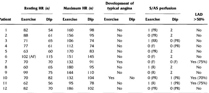
Figure 1. Patient 1. Septal perfusion defect is seen on exercise 2roT1 SPECT images (A). Dipyridamole 2mTl SPECT images of same patient (B) revealed normal perfusion. Coronary angiogram was normal. Rows 1, 3, and 5 fi-om top are initial images, and rows 2, 4, and 6 are redistribution images for both exercise and dipyridmnole. Vertical long-axis images are arranged from septal wall to lateral wall, horizontal long-axis images from anterior wall to inferior wall, and short-axis images from apex to base.
Table 4. Overall results in patients with septal or anteroseptal perfusion defects on both exercise and dipyridamole Z°lTl SPECT (n = 6)
Exercise (n) Dip positive (n)
Mod S/AS Severe S/AS Mod S/AS Severe S/AS Total
defect defect defect defect (n)
Patients 2 4 2 4 6
Patients with LAD >50% 1 2 2 l 3
Patients with normal LAD 1 2 0 3 3
coronary arteries
lMod, moderate; S/AS, septal or anteroseptal; Dip, dipyridamole.
septal or anteroseptal perfusion defects on both exercise and dipyridamole 2°IT1 SPECT. Shown in Table 4 are the comparative results with regard to the severity of the perfusion defect. Three of six patients with positive dipyridamole study results had significant LAD coronary artery stenosis.
Coronary arteriograms were normal in the remaining three patients who had severe septal or anteroseptal perfusion defects after dipyridamole (Figure 2).
Severity of Coronary Artery Disease. Three of 12 patients had coronary artery disease. Two patients (patients 7 and 11) had double-vessel disease; both of
them had multiple scintigraphic perfusion defects with exercise and also with dipyridamole. In addition to LAD coronary artery disease, patients 7 and 11 had, respec- tively, 70% and 100% stenosis of the left circumflex coronary artery (also with the two grafts occluded for patient 11). The last patient had single-vessel disease.
Clinical Data During Exercise and Dipyridam- ole. Table 3 provides detailed clinical data during exer- cise and dipyridamole. Patients with normal dipyridam- ole images had a mean increase of heart rate of 77 -+ 30 beats/min during exercise and 32 + 7 beats/min during dipyridamole infusion. Those with positive dipyridamole
270 Lebtahi et al. Journal of Nuclear Cardiology
Accuracy of dipyridamole 2°iT1 SPECT July]August 1997
Figure 2. Dipyridamole (rows 1, 3, and 5 from top) and
redistribution (rows 2, 4, and 6) 2roT1 SPECT images of patient
12. Severe, partially reversible septal perfusion defect, includ- ing apex, is seen; there is also inferior perfusion defect. Coronary angiogram was normal. Images are arranged as in Figure 1.
images had a mean increase of heart rate of 55 _+ 27 beats/min during exercise and 19 _+ 8 beats/min during dipyridamole infusion.
Analysis of False-Positive Results. Table 5 shows further analysis of false-positive results with regard to presence or absence of apical involvement, dilated left ventricles, anterior or septal wall motion abnormalities, and the mean ejection fraction value. None of these parameters could help to identify or explain false-positive results.
DISCUSSION
Our results showed that six of 12 patients with LBBB and exercise septal or anterior perfusion defects had normal scintigraphic results after dipyridamole. All six patients with normal dipyridamole 2°iT1 SPECT results had normal coronary angiographic results. Only three of 12 patients with exercise septal or anteroseptal perfusion defects had LAD coronary artery disease. On the other hand, three of six patients with a positive dipyridamole study result had LAD coronary artery disease. These data indicate that dipyridamole Z°IT1 SPECT was accurate in nine of 12 patients, whereas exercise was accurate in only three of 12 patients. Nevertheless, there are still false-positive results after dipyridamole with regard to the identification of patients with LBBB and underlying coronary artery disease. Moreover, only one of four patients with severe septal or
anteroseptal perfusion defects after dipyridamole had LAD coronary artery disease, which indicates that the severity of the perfusion defect does not add additional information in identifying LAD coronary artery disease. In patient 7, exercise and dipyridamole 2°1T1 SPECT revealed a fixed anteroseptal perfusion defect with a 75% stenosis of the LAD, without a known previous myocar- dial infarction. However, ventriculography showed sep- tal dyskinesia, which suggests possible transient artery occlusion (non-Q wave myocardial infarction).
In light of the mechanisms proposed to explain perfusion defects in LBBB, 2,6 their occurrence would be expected to be heart rate dependent. This was con- firmed by Vaduganathan et al., 13 who observed a rela- tion between heart rate and false-positive septal defects in the exercise group. Of interest was the much lower frequency of septal defects reported during dobuta- mine than during exercise. 13 They explain these find- ings by the much higher peak heart rate achieved during exercise than with dobutamine. However, the persistence of false-positive results after dipyridamole infusion con- firms that factors other than heart rate contribute to abnormal septal or anteroseptal perfusion in patients with LBBB. In our study, false-positive results after dipyridamole could not be explained by higher heart rate or a larger increase in heart rate compared with patients with normal dipyridamole images (Table 3). The study by Jukema et al. 15 also showed that septal dipyrid- amole perfusion defects were not dependent on heart rate.
In our study neither the severity nor the extent of the perfusion defect could identify patients with underlying LAD coronary artery disease: six of eight patients with severe septal or anterior perfusion defects after exercise and three of four patients with severe defects after dipyridamole had normal coronary angiograms. With regard to the extent of the perfusion defect to the apex on exercise 2roT1 SPECT, we could not confirm the better diagnostic accuracy reported by Matzer et al.7; among five patients who had septal or anterior perfusion defects involving the apex on exercise 2roT1 SPECT, only one had LAD coronary artery disease. Our findings are supported by the results of Larcos et al., 8 who showed that apical extent of the exercise perfusion defect, al- though sensitive, was neither specific nor accurate in the identification of patients with coronary artery disease. Apical involvement was also of no help for dipyridamole in our study; among the three patients with false-positive findings for LAD coronary artery disease, two had perfusion defects involving the apex. The recent study of Vaduganathan et al. 13 reported the results of the largest series to date of patients with LBBB undergoing myo- cardial perfusion scintigraphy with three different mo- dalities of stress. They did not find value in the size of the
Journal of Nuclear Cardiology Lebtahi et al. 271
Volume 4, Number 4;266-73 Accuracy of dipyridamole Z°lT1 SPECT
Table 5.
Analysis of false-positive
resultsFalse positive Other patients
Exercise Dipyridamole Exercise Dipyridamole
(n= 9) (n= 3) (n = 3) (n= 9)
Apical involvement (n) Dilated left ventricle (n) Mean ejection fraction (%) Anterior or septal wall
motion abnormalities
4/9 2/3 1/3 1/9
2/9 0/3 1/3 3/9
54 54 60 57
4/9 1/3 3/3 6/9
perfusion defect or the involvement of the anterior wall, the apex, or another territory in differentiating true from false-positive cases during exercise. They also reported a higher left ventricular ejection fraction in the subgroup of patients who exercised and had false-positive scan responses compared with those with true positive scan results.
Hodge et al. 2° suggested that dilated left ventricles may be responsible for false-positive results in patients with LBBB. In our study, only two of nine patients with false-positive results after exercise and none of three patients with false-positive results after dipyridamole had dilated left ventricles. Therefore we could not confirm this hypothesis.
Our results are in agreement with those of previous studies, 6,12-18 which showed that septal or anteroseptal perfusion defects in patients with LBBB are less frequent with pharmacologic stress, thereby improving the spec- ificity of thallium scintigraphy in those patients. Few studies, however, compared dipyridamole (or adenosine) and exercise in the same patients. In this study, only patients with abnormal septal or anteroseptal perfusion on exercise 2°1T1 SPECT underwent dipyridamole stress, whereas in the other comparative studies 6,~~4 patients without perfusion defects on the exercise study also underwent dipyridamole scintigraphy. Because the limi- tation with exercise is the high number of false-positive results, we chose to evaluate dipyridamole in patients with positive exercise scintigrams. Analysis in our study of clinical data, results of 2°1T1 SPECT in terms of severity and extent of perfusion defect or dilated left ventricle, and ventriculographic and coronary arterio- graphic findings could not explain the false-positive scintigraphic results or help to identify patients with underlying coronary artery disease.
There are several hypotheses to explain the mecha- nism for abnormal perfusion in LBBB. Grines et al. 23 reported that altered electrical activation as a result of LBBB is responsible for global ventricular abnormalities that could lead to abnormal septal motion and abnormal
left ventricular ejection. The occurrence of "relative hypoperfusion" caused by the abnormal septal motion that could cause a prolonged compression of the septal arteries has been proposed by Hirzel et al. 2 Another explanation for this relative hypoperfusion has been proposed by Bums et al. 6 They postulated that in patients with LBBB the septum contracts in early systole with low tension to overcome right ventricular rather than left ventricular outflow resistance as it occurs with normal conduction; therefore the septal wall will receive less coronary flow as a result of coronary autoregulation. Ono et al. 24 used an in vivo animal model to determine whether LBBB itself induces abnormal myocardial per- fusion and ischemia. They demonstrated that during LBBB the mean septal intramyocardial pressure in- creased significantly in the T D phase, which may result in reduced myocardial perfusion in the septum. Neverthe- less, they did not observe indirect signs of nayocardial ischemia such as increased septal glucose uptake or lactate production. In view of these data, perfusion of the septal or anteroseptal territories may be reduced for functional rather than anatomic reasons and the so-called false-positive 2°1T1 study results might well demonstrate relative hypoperfusion.
The occurrence of septal or anteroseptal perfusion defects is obviously not restricted to the use of 2mTl. Results are somewhat conflicting on the occurrence of septal or anterior defects with exercise sestamibi. Alte- hoefer et al. 21 postulated that 99mTc-labeled sestamibi scintigraphy could be more accurate for the detection of coronary artery disease in patients with LBBB because most of these patients (55%) revealed normal septal uptake. However, the findings of Knapp et al. 22 differ because they described a septal defect (reversible or not) on 99mTc-labeled sestamibi scintigraphy in 34 (91.8%) of 37 patients. In both studies the authors speculated that the reversibility of the defect could identify patients with coronary artery disease. Nevertheless, false-positive re- sults with sestamibi according to the criterion of revers- ibility have been described. 25,26 Another study 1~ revealed
272 Lebtahi et al. Journal of Nuclear Cardiology
Accuracy of dipyridamole 2°1T1 SPECT July/August 1997
the conflicting results of 99mTc-labeled sestamibi scintig- raphy in LBBB. In this study, Ebersole et al., 1~ who compared exercise and adenosine stress, pointed out that the specificity in the territory of the LAD coronary artery was only 25% with exercise, whereas that for adenosine was 87.5%. Moreover, in their study, all but one patient who had false-positive results with exercise had a revers- ible defect, which confirms that the criterion of revers- ibility could not help to identify patients with LBBB and underlying coronary artery disease. They also conclude by using 99mTc-labeled sestamibi scintigraphy to the improved specificity of pharmacologic stress compared with exercise.
Limitation of the Method. It can be argued that tomographic analysis in our study was qualitative. How- ever, very good correlations between visual and quanti- tative analysis have been described. The superiority of the quantitative analysis has not been proved clearly, and there is no significant difference (statistically significant) between the two modalities of analysis. 27 In fact, the problem with myocardial scintigraphy in patients with LBBB is the high frequency of perfusion defects in the absence of LAD artery disease, which means that the specificity of thallium scintigraphy in detection of LAD disease is of concern. It is known that quantitative analysis is equivalent to or lower than visual analysis in this setting. 27-3° Moreover, even with quantitative anal- ysis, several studies 4-6,8,12,~3 reported the low specificity of exercise 2°1T1 scintigraphy in detecting LAD coronary artery disease in patients with LBBB. Last, in our study, six of eight and three of four patients with severe perfusion defects (which means clearly positive results) after, respectively, exercise and dipyridamole had nor- mal coronary arteries. Quantitative analysis cannot help increase diagnostic accuracy in these patients.
Conclusion. In view of the cumulative results of exercise scintigraphy and the conflicting results with other alternatives (apical involvement and exercise ses- tamibi), pharmacologic stress by either dipyridamole or adenosine seems the preferred choice to exercise scin- tigraphy in patients with LBBB, by reducing the number of so-called false-positive results. In case of normal perfusion with dipyridamole, coronary artery disease can reasonably be excluded and there is no real indication to perform coronary angiography; therefore the number of coronary angiograms in patients with LBBB may be reduced by use of pharmacologic stress. However, even severe septal or anteroseptal perfusion defects on dipy- ridamole studies may occur in the absence of LAD coronary artery disease. The presence or absence of apical involvement, dilated left ventricles, decreased left ventricular ejection fraction, or wall motion abnormali- ties could not help in the identification of patients with false-positive results. Coronary angiography is thus still
indicated in studies with positive results to identify patients with underlying coronary artery disease, a major prognostic factor in patients with LBBB.
References
1. Mc Gowan LR, Welch TG, Zaret BL, Bryson AL, Martin ND, Flamm MD. Noninvasive myocardial imaging with potassium-43 and rubidium-81 in patients with left bundle branch block. Am J Cardiol 1976;38:422-8.
2. Hirzel HO, Senn M, Nuesch K, Buettner C, Pfeiffer A, Hess OM,
et al. Thallium-201 scintigraphy in complete left bundle branch
block. Am J Cardiol 1984;53:764-9.
3. Huerta EM, Rodriguez Padial L, Castro Beiras JM, Illera JP, Asin
Cardiel E. Thallium-201 scintigraphy in patients having complete left bundle branch block with normal coronary arteries. Int J Car-
diol 1987;16:43-6.
4. Rothbart MR, Beller GA, Watson DD, Nygaard TW, Gibson RS.
Diagnostic accuracy and prognostic significance of quantitative
thallium-201 scintigraphy in patients with left bundle branch block. Am J Noninvas Cardiol 1987;1:197-205.
5. DePuey EJ, Guertler-Krawcynska E, Robbins WL. Thallium-201 SPECT in coronary artery disease patients with left bundle branch block. J Nucl Med 1988;29:1479-85.
6. Bums RJ, Galligan L, Wright LM, Lawand L, Burke R J, Gladstone PJ. Improved specificity of myocardial thallium-201 single-photon
emission computed tomography in patients with left bundle branch
block by dipyridamole. Am J Cardiol 1991;68:504-8.
7. Matzer L, Kiat H, Friedman JD, Van Train K, Maddahi J, Berman DS. A new approach to the assement of tomographic thallium-201
scintigraphy in patients with left bundle branch block. J Am Coil
Cardiol 1991;17:1309-17.
8. Larcos G, Gibbons RJ, Browm ML. Diagnostic accuracy of
exercise thallium-201 single photon emission computed tomogra-
phy in patients with left bundle branch block. Am J Cardiol 1991 ;68:756-60.
9. La Canna G, Giubbini R, Metra M, Arosio G, Cumis A, Cicogna R, et al. Assessment of myocardial peffusion with thallium-201
scintigraphy in exercise-induced left bundle branch block: diag-
nostic value and clinical significance. Eur Heart J 1992;13:942-6. 10. Delonca J, Camenzid E, Meier B, Righetti A. Limits of thallium- 201 exercise scintigraphy to detect coronary disease in patients
with complete and permanent bundle branch block: a review of 134 cases. Am Heart J 1992;123:1201-7.
11. Ebersole DG, Heironimus J, Toney MO, Billingsley J. Comparison
of exercise and adenosine technetium-99m sestamibi myocardial scintigraphy for diagnosis of coronary artery disease in patients with left bundle branch block. Am J Cardiol 1993;71:450-3.
12. O'Keefe JH, Bateman TM, Barnhart CS. Adenosine thallium-201
is superior to exercise thallinm-201 for detecting coronary artery disease in patients with left bundle branch block. J Am Coll
Cardiol 1993;21:1332-8.
13. Vaduganathan P, Zuo-Xiang HE, Raghavan C, Mahmarian JJ, Verani MS. Detection of left anterior descending coronary artery
stenosis in patients with left bundle branch block: exercise, adenosine or dobutamine imaging? J Am Coll Cardiol 1996;28:
543-50.
14. Morals J, Soucy JP, Sestier F, Lamoureux F, Larnoureux J, Danais S. Dipyridamole testing compared to exercise stress for thallium- 201 imaging in patients with left bundle branch block. Can J Cardiol 1990;1:5-8.
15. Jukema JW, Van der Wall EE, Van der Vis-Melsen MJE,
Kruyswijk HH, Bruschke AVG. Dipyridamole thallium-201 scin- tiga'aphy for improved detection of left anterior descending coro-
Journal of Nuclear Cardiology Lebtahi et al. 273 Volume 4, Number 4;266-73 Accuracy of dipyridamole 2°lTl SPECT
nary artery stenosis in patients with left bundle branch block. Eur Heart J 1993;14:53-6.
16. Rockett JF, Wood WC, Moinuddin M, Loveless V, Parrish B. Intravenous dipyridamole thallium-201 SPECT imaging in patients with left bundle branch block. Clin Nucl Med 1990;15:40t-7. 17. Larcos G, Brown MI, Gibbons RJ. Role of dipyridamole thallium-
201 imaging in left bundle branch block. Am J Cardiol 1991;68: 1097-8.
18. Patel R, Bushnell DL, Wagner R, Stumbris R. Frequency of false positive septal defects on adenosineF°~T1 images in patients with left bundle branch block. NucI Med Commun 1995;16:137-9. 19. Pope DL, Parker DL, Gustafson DE, Clayton PD. Dynamic search
algorithms in left ventricular border recognition and analysis of coronary arteries. Comp Cardiol 1984;4:7 i-5.
20. Hodge J, Mattera J, Fetterman R, Williams B, Wackers FJ. False positive TL-201 defects in left bundle branch block: relationship to left ventricular dilatation. J Am Coil Cardiol 1987;9:137A. 21. Altehoefer C, Vom Dahl J, Kleinhans E, Uebis R, Hanrath P, Buell
U. 99mTc-methoxyisobutylisonitrile stress/rest SPECT in patients with constant complete left bundle branch block. Nucl Med Commun 1993; 14:30-5.
22. Knapp WH, Bentrup A, Schmidt U, Ohlmeier H. Myocardial scintigraphy with thallium-201 and technetium-99m-hexakis-me- thoxyisobutilisonitrile in left bundle branch block: a study in patients with and without coronary artery disease. Eur J Nucl Med 1993;20:219-24.
23. Grines CL, Bashore TM, Boudoulas H, Olson S, Shafer P, Wooley CF. Functional abnormalities in isolated left bundle branch block:
the effect of interventricular asynchrony. Circulation 1989;79:845- 53.
24, Ono S, Nohara R, Kambara H, Okuda K, Kawai C. Regional myocardial perfusion and glucose metabolism in experimental left bundle branch block. Circulation 1992;85:1125-31.
25. Campeau R J, Garcia OM, Colon R, Agusala M, Con'ea OA. False positive Tc-99m sestamibi SPECT in a patient with left bundle branch block. Clin Nucl Med 1993;18:40-2.
26. Altehoefer C, Vom Dahl J~ Bull U. Falsch-positiver Befund in der ~)~mTc-MIBI SPECT bei Linksschenkelblock und angiographisch ausgeschlossener KHK. Nucl Med 1994;33:46-8.
27. Iskandrian AS, Verani MS. Nuclear cardiac imaging: principles and applications. Philadelphia: FA Davis, 1996:114-7.
28. Tamaki N, Yoshiham Y, Mukai T, Kodama S, Kadota K, Kambara H, et al. Stress thallium-201 transaxial emission computed tomog- raphy: quantitative versus qualitative analysis for evaluation of coronary artery disease. J Am Coll Cardiol 1984;4:1213-21. 29. DePasquale EE, Nody AC, DePuey EG, Garcia EV, Pilcher G,
Bredlean C, et aT. Quantitative rotational thallimn-201 tomography for identifying and localizing coronary artery disease. Circulation 1988;77:316-27.
30. Mahmarian JJ, Boyce T, Goldberg RK, Cocanougher MK, Roberts R, Verani MS. Quantitative exercise thallium-201 single photon emission computed tomography for the enhanced diagnosis of ischemic heart disease. J Am Coll Cardiol 1990;15:318-29. 31. Brant SH, Bmgada P, Bar FW, Gorgels APM, Wellens HJJ.
Thallium-201 exercise scintigraphy and left bundle branch block. Am J Cardiol 1985;55:224-6.