Received: 22 January 2003 Accepted: 27 May 2003 Published online: 5 August 2003 © Springer-Verlag 2003
S. Bresson-Hadni · D.-A. Vuitton World Health Organisation Collaborating Centre for Prevention and Treatment of Human Echinococcosis and Research Group on Alveolar Echinococcosis, University of Franche-Comté (SERF), Health and Rural Environment Research Unit,
University of Franche-Comté (SERF), Besançon, France
M. Gillet
Liver Transplant Unit, CHUV Lausanne, Lausanne, Switzerland
incurable AE. Methods: Our review summarizes the results of this expe-rience based on a series of 47 Euro-pean patients who received trans-plants between 1985 and 2002, tries to specify the real place of LT for AE, and underlines the measures that could be undertaken in the future to improve the results. Results: Five-year survival was 71%. Five-Five-year survival without recurrence was 58%. Major technical difficulties related either to previous lapar-otomies or to the loco-regional in-volvement were observed. The nine early deaths concerned AE patients with a long past-history of symptom-atic AE (iterative cholangitis, secondary biliary cirrhosis). Five late deaths were directly related to ongo-ing AE, located in the brain in three cases, a very rare AE location that was not investigated before LT in these patients. Conclusions: In general, the pre-LT screening for distant AE metastases appeared insufficient in this series. Heavy im-munosuppressive schemes, absence or delayed re-introduction of BZM after LT have clearly played a role in this unfavourable course. This unique experience indicates that, despite major technical difficulties, LT for incurable AE is feasible and could be discussed in very symptom-atic cases. Before LT, interventional radiology should be preferred to repeated laparotomies. Pre-LT and post-LT BZM treatment is
mandato-Solange Bresson-Hadni Stéphane Koch Jean-Philippe Miguet Michel Gillet Georges-André Mantion Bruno Heyd Dominique-Angèle Vuitton a European group of clinicians
Indications and results of liver transplantation
for
Echinococcus alveolar infection:
an overview
The authors wrote this report in collabora-tion with a European group of clinicians, which included the following participants. France: Jean-Pierre Crumbach, Sabine Kurz, Liver Transplant Unit, CHU Besançon; Anne Minello, Patrick Hillon, CHU Dijon and Liver Transplant Unit, CHU Besançon; Didier Samuel, Henri Bismuth, Centre Hepato-biliaire, Hopital Paul-Brousse, Paris; Maxime Audet, Philippe Wolf, Daniel Jaeck, Strasbourg; Patrick Boissel, Nancy; Filomena Conti, Yves Chapuis, Hopital Cochin, Paris; Jean Chipponi, Clermont-Ferrand; Michel Pouyet, Hôpital de la Croix Rousse, Lyons; Martine Neau-Cransac, Jean-Yves Lacut, Jacques Carles, Bordeaux; Belgium: Jean Van de Stadt, Yves Carlier, Brussels; Switzerland: Philippe Morel, Gilles Mentha, Petro Majno, Geneva; Germany: Markus Golling, Gerd Otto, Heidelberg; Juergen Klempnauer, Karl Oldhafer, Hanover; Ewert Schutte-Frohlinde, Peter Vorwald, Munich; Olaf Guckelberger, Berlin; Carl Allers, Frankfurt; Johannes Scheele, Jena; Tobias Beckurts, Cologne S. Bresson-Hadni (
✉
)Division d’Hepato-Gastroentérologie, Département de Médecine Interne, Hopital Universitaire Cantonal de Genève, Rue Micheli du Crest, Geneva, Switzerland e-mail: solange.bresson-hadni@ufc-chu.univ-fcomte.fr
S. Bresson-Hadni · S. Koch · J.-P. Miguet G.-A. Mantion · B. Heyd · D.-A. Vuitton Liver Transplant Unit,
CHU Besançon, Besançon, France
Abstract Background: Alveolar
echinococcosis (AE) of the liver, caused by the larval stage of the fox tapeworm Echinococcus multilocula-ris, has the characteristics of a slow-growing liver cancer. It is one of the rare parasitic diseases for which a parasitolytic drug is not yet avail-able, and AE is lethal in the absence of appropriate therapeutic manage-ment. Complete surgical resection of the parasite at an early stage of in-fection provides favourable pros-pects for cure, but, due to a long clinical latency, many cases are diagnosed at an advanced stage, so that partial liver resection can be performed in only 35% of patients. Benzimidazole (BZM) treatment is given in inoperable cases but these compounds are only parasitostatic, and lifelong therapy is required. During the past 20 years some cen-tres have considered liver transplan-tation (LT) for the treatment of
Introduction
Alveolar echinococcosis (AE) of the liver is a rare para-sitic disease due to the intra-hepatic development of Echinococcus multilocularis larvae. It behaves like a slow-growing liver cancer [1]. Moreover, extra-hepatic extension of the parasitic disease may be possible, either by loco-regional progression, or by a haematogenous spread [1]. It might seem quite surprising for liver trans-plantation (LT) followed by lifelong immunosuppressive therapy to be considered for an infectious disease that sometimes affects extra-hepatic organs. However, AE is one of the sole parasitic diseases with no parasitolytic medication able to destroy the larvae and cure the pa-tient. Moreover, until recently, due to the long clinical latency, diagnosis of advanced AE was the rule [2]. In these severe cases, not accessible to partial curative sur-gery, invasion of the bile ducts and vessels and necrosis in the central part of the parasitic lesion lead to very se-vere complications, such as cholangitis, liver abscesses, septic shock, portal hypertension and biliary cirrhosis, which can seriously affect not only the quality of life of these generally young patients but also their survival. In such patients, LT may offer the only chance for survival and cure, and this procedure has been proposed in ad-vanced AE cases since 1985 [3, 4, 5]. Concomitantly, striking changes have occurred in the diagnosis and treatment of AE [2, 6], which have contributed to an im-provement in the status of patients affected by this para-sitic disease and made the indication for LT rarer in the recent period.
The aim of this review is to give results of this ulti-mate treatment for AE, based on a series of 47 European patients, and to specify the real place of LT for this dis-ease as well as the particularities of AE patient manage-ment before and after LT.
Liver transplantation in AE: which patients?
What was observed and done?A recent retrospective inquiry allowed us to collect data from 45 AE patients who had been engaged in a LT pro-cedure from 1985 to 1999. The detailed analysis of this series has been published recently [7]. Most of the cases come from French centres, with a total of 37 patients (two-thirds of whom received transplants in Besançon,
Eastern France, located in a highly endemic area for this disease). Interestingly, the large majority of LTs took place between 1985 and 1991 (80% of the cases) and re-cent contacts with these re-centres indicate that there were only two additional LTs for AE between 2000 and 2002. Involved centres are listed in Table 1. Patients included 25 women and 22 men. The mean age at LT was 46.5 years (range 16–67). The mean time interval between di-agnosis and LT was 5 years (range 0–23).
The two main indications for LT were biliary disease, related to parasitic involvement of the hilum (Fig. 1), and tumoral disease invading both lobes of the liver, with or without chronic parasitic Budd-Chiari syndrome. Some patients had two LT indications. Thus, 57% of the cases were referred for LT because of life-threatening cholangitis and/or abscesses, 30% of the cases because of secondary biliary cirrhosis, 11% for a huge bifocal AE involving the hilum (“tumoral” AE), and, in 7% of the cases, because of chronic Budd-Chiari syndrome with in-volvement of the hepatic veins (“vascular” AE).
Thirty four (72%) underwent previous upper abdomi-nal operations: an average of 2.3 operations per patients (range 0–7) were undertaken. Major extra-abdominal surgical procedures needed to control extra-hepatic AE extension were performed in five patients. Details of all these previous operations are given in Table 2. Intervent-ional procedures were performed before LT in 35 cases. ry. A careful evaluation of possible
distant metastases should be done before the decision for LT is made. After LT, the possibility of an ongo-ing AE must be permanently kept in mind. This could be reduced by
lightening the immunosuppressants, carefully following the specific circulating antibodies, and applying a systematic radiological evaluation, not only to the graft but also to the lungs and the brain.
Keywords Alveolar
echinococcosis · Echinococcus multilocularis · Treatment · Liver transplantation · Benzimidazoles
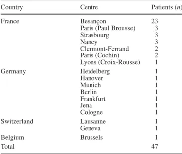
Table 1 European experience of LT for AE: centres involved
(1985–2002)
Country Centre Patients (n)
France Besançon 23
Paris (Paul Brousse) 3
Strasbourg 3 Nancy 3 Clermont-Ferrand 2 Paris (Cochin) 2 Lyons (Croix-Rousse) 1 Germany Heidelberg 1 Hanover 1 Munich 1 Berlin 1 Frankfurt 1 Jena 1 Cologne 1 Switzerland Lausanne 1 Geneva 1 Belgium Brussels 1 Total 47
In particular, there were nine radiological drainages of the bile ducts or of liver abscesses: five external drainag-es, two internal–external drainages and two endoscopic-ally inserted stents.
Fifteen patients were in an extremely poor condition at the time of LT, due to a long past-history (mean dura-tion from diagnosis to LT: 8.5 years) of symptomatic “biliary AE” with iterative cholangitis, and/or secondary biliary cirrhosis leading to ascites or digestive-tract bleeding. Nine of them died either during the operation,
due to haemodynamic disorders, or in the immediate post-operative period from severe infectious complica-tions (bacterial and/or fungal). Most of these early deaths (7/9) occurred during the first 5 years of this experience.
Among patients suffering from “vascular” or “tumor-al AE”, severe symptoms were observed in only h“tumor-alf of the cases: chronic parasitic Budd-Chiari syndrome in one case leading to chronic ascites, iterative life-threatening right atrial and pulmonary embolisms in one case, huge hepatomegaly leading to abdominal pains in two other cases. Moreover, in two patients the diagnosis of AE was obtained by serological screening. Despite mild symp-tomatology, they were referred for LT because of very impressive radiological images that led to the fear of acute Budd-Chiari syndrome occurrence or extra-hepatic progression via the inferior vena cava. Unfortunately, these two patients died from chronic cholangitis (not re-lated to recurrent AE) 3 and 5 years after they had under-gone LT.
What should be done in the future?
Early deaths could certainly be avoided by the selection of patients before the terminal stage of the disease. In the case of “biliary” AE, the situation is quite similar to that of primary sclerosing cholangitis or Caroli’s disease: when acute cholangitis and/or biliary cirrhosis occur, LT must be considered without delay to avoid recurrent epi-sodes of sepsis and prolonged cholestasis that may alter nutritional status and make the peri-operative period more chaotic. When LT is being performed, such pa-tients should certainly receive not only antibiotics but also a systematic anti-fungal peri-operative prophylaxis. In the other situations, represented by chronic parasitic Budd-Chiari syndrome and/or huge pseudo-tumoral AE, the decision to perform LT is far more difficult to make. In fact, such patients, even with very impressive radio-logical imagings, may be asymptomatic or pauci-symp-tomatic, due to the very slow progression of the parasitic larvae. The extensive use of abdominal ultrasonography and serological screening for AE in endemic areas has led to the more frequent discovery of such forms of AE in recent times [2, 8]. The natural history of these “tu-moral” and/or “vascular” AE was unknown. That ex-plains the initial tendency for early indication for LT in such cases, due to the fear of acute Budd-Chiari syn-drome occurrence or extra-hepatic progression via the inferior vena cava [9]. However, to our knowledge, acute parasitic Budd-Chiari syndrome has never been reported in AE. Moreover, a medical option, the application of continuous albendazole therapy, without the periodic in-terruptions of the treatment that were advised in the past and at higher dosage than previously proposed, seems to be able to stabilize such forms of incurable AE. In the Besançon centre we have chosen this policy for five
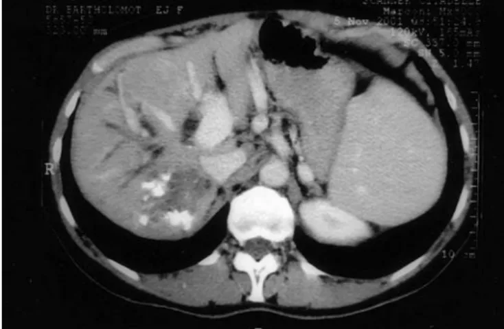
pa-Fig. 1 A case of incurable AE in a 44-year-old woman, revealed
by obstructive jaundice and complicated by acute digestive-tract bleeding, which led the treating surgeons to opt for LT. The ab-dominal angio-CT scan shows a typical AE lesion in the right lobe of the liver (segment VI and VII) extending to the hilum and the hepatic pedicle. There is major dilation of the intra-hepatic biliary ducts. The right portal vein is totally involved in the parasitic tis-sue and the portal trunk is encircled by the disease. This vascular involvement leads to portal hypertension, with huge venous collat-erals and splenomegaly
Table 2 LT for alveolar echinococcosis: details of the surgical
procedures needed before LT
Type of surgery Number of patients (%) Abdominal operations
Bilio-enteric anastomosis 18 (38) Explorative laparotomy 16 (34) Right or left partial hepatectomy 10 (21)
Abscess drainage 6 (13)
Atypical liver resection 5 (11)
Spleno-renal shunt 2 (4)
Extra-abdominal operations
Pulmonary lobectomy 2a(4)
Atrial resection 2b(4)
Explorative thoracotomy 2c(4) a In two patients with AE lung metastases
b In one patient, due to recurrent parasitic clot within the right
atri-um responsible for iterative pulmonary embolisms
c To explore carefully the supra-hepatic inferior vena cava in two
tients with such AE localizations: they remained asymp-tomatic, and the lesions are stable, with a mean follow-up of 5 years (S. Bresson-Hadni, personal communica-tion). If one clinical episode occurs in such cases (asci-tes, bleeding, or septic complication), LT must, of course, be promptly considered.
Liver transplantation in AE:
what surgical difficulties?
What was observed and done?Technical difficulties are summarized in Table 3. They were numerous, related to the regional extension of the parasitic disease and the frequent past-history of previ-ous iterative laparotomies responsible for adherences. One patient died during the hepatectomy phase and three after the liver removal. For the engrafting procedure the supra-hepatic vena cava reconstruction required an anas-tomosis with the right atrial ostium in three cases. In 12 cases, due to the parasitic invasion, the proper or com-mon hepatic artery was either resected or unusable. The arterial reconstruction was done by direct implantation on the coeliac aorta in five cases and by allograft bypass to the infra renal aorta in seven cases. The biliary recon-struction required a choledocho-jejunal anastomosis in 57% of the cases. The overall mean length of the LT procedure was 8.3 h (range 4–20). The mean transfusion requirement was 10.6 units (range 0–88). Ten patients needed early re-operation after LT for the following rea-sons: peri-hepatic clotting (n=4), hepatic artery thrombo-sis (n=2), hepatic artery folding (n=1), diaphragmatic rupture (n=1), primary non-function leading to emergen-cy liver re-transplantation (n=2).
What could be done to prevent or solve these difficulties?
It seems obvious that many patients have been penalized by a prolonged waiting time between diagnosis of incur-able AE and LT. They belong to a past period, before there was access to interventional radiology. At that time
the only means to get over life-threatening infectious ep-isodes depended on iterative palliative surgical mea-sures. This has clearly contributed to making LT very technically difficult. Selection of incurable AE patients before the terminal stage of the disease and use of percu-taneous or endoscopic procedures to temporarily over-come an infectious complication are now clearly recom-mended, whereas multiple laparotomies must be avoid-ed. It should clearly reduce the per-operative difficulties and make the early post-operative period less complicat-ed. It also seems important for one to perform a very precise evaluation of the regional extension of the para-sitic disease to anticipate the per-operative difficulties, the possible additional surgical measures (diaphragmatic resection, peri-aortic cleaning-out, etc.) and to specify what type of vascular anastomosis will be preferable and where to do it. Thus, depending on AE localization, liver arteriography, magnetic resonance imaging (which ap-peared useful for information on hepatic veins, inferior vena cava and diaphragmatic invasion), and inferior vena cava opacification, should be part of the pre-LT evalua-tion.
Liver transplantation in AE:
what medical difficulties?
What was observed and done?In this series, with a median follow-up duration of 6 years, the major long-term medical concern was related to the possible continuation of the parasitic disease after LT. In fact, at the time of surgery, LT was considered as palliative in 15 patients, due to inextirpable residual par-asitic tissue noticed by the surgeon, or due to known AE metastases: seven patients had lung metastases, and one a splenic localization. Moreover, in seven patients ini-tially considered as cured, the morphological follow-up revealed new parasitic foci, and finally, the LT was con-sidered as palliative in 50% of the cases. The “new” AE lesions discovered during follow-up were in the spleen in one case (Fig. 2), in the brain in three cases, and in the lung in three cases. Moreover, a recurrence in the graft itself was observed in six cases: among the patients which such a location, only two were symptomatic, with diffuse re-infection leading to abscesses. In one, the dif-fusion occurred a few weeks after LT, clearly through a haematogenic pathway, from a splenic location of AE that could not be removed during the operation [10]. In the other one, multiple AE lesions developed 5 years post-LT, presumably from residual diaphragmatic AE fo-ci, shortly after a liver re-transplantation for chronic cholangitis, at the time of a heavy immunosuppressive regimen. The four remaining patients were asymptomat-ic: the recurrent liver AE foci were limited in size and discovered by the radiological follow-up.
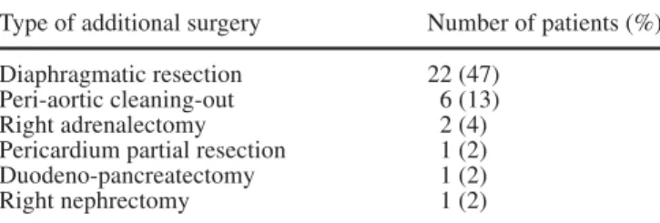
Table 3 Liver transplantation (LT) for alveolar echinococcosis:
additional surgical measures during LT related to regional parasit-ic extension
Type of additional surgery Number of patients (%) Diaphragmatic resection 22 (47)
Peri-aortic cleaning-out 6 (13)
Right adrenalectomy 2 (4)
Pericardium partial resection 1 (2) Duodeno-pancreatectomy 1 (2)
Data concerning the specific serological follow-up after LT were obtained by a study performed on 15 patients in the Besançon centre [11]. An enzyme-linked immunosorbent assay (ELISA) with two parasitic anti-gens, a crude heterologous antigen extracted from E. granulosus (EgAg), and the specific Em2 antigenic fraction was used for the follow-up [12]. It showed that the anti-Em2 ELISA, which is the standard test used in the patient’s follow-up after partial resection for AE [13], did not appear useful in detecting recurrence in this particular setting. On the contrary, the EgAg ELISA, which is useless after a partial resection (most sera, even in cured patients, remained positive long after the opera-tion) was here able to discriminate palliative and cura-tive LT well and diagnose residual AE early. In fact, in one patient with an apparently curative LT, a secondary marked increase of anti-Eg IgG was observed at the end of the first post-operative year, 9 months before the ra-diological discovery of a small recurrent AE focus in segment VIII of the graft. On the contrary, the anti-Em2 IgG remained negative during the whole follow-up peri-od [14].
Five late deaths were directly related to ongoing AE: three from brain AE, at 5, 6.5, and 7.5 years; one from disseminated AE graft recurrence at 5.7 years post-LT, and one from diffuse pulmonary AE at 10 years post-LT.
Three factors have certainly played a role in explain-ing this high rate of continuexplain-ing AE after LT: (a) the lack of precise pre-LT evaluation of the extra-hepatic exten-sion in most cases; (b) the lack of systematic benzimid-azole (BZM) use before and early after LT; (c) a too-heavy immunosuppressive scheme during the first year post-LT.
Lack of precise pre-LT evaluation of AE extra-hepatic extension
The pre-LT screening included a chest X-ray and an abdominal CT scan in all cases, but a thoracic CT scan was performed in only 20 patients (42%) and a brain radiological investigation (CT scan or MRI) in only 12 cases (25%).
Lack of systematic BZM use before and early after LT Only 28 (59%) of the patients received pre-LT BZM therapy, with a mean duration of 4 years. Three patients had been given flubendazole only, seven patients re-ceived mebendazole only, nine patients albendazole only and nine other patients the last two compounds, consecu-tively. Interestingly, one patient of the palliative sub-group who receive pre-LT mebendazole for more than 2 years had a particular evolutive pattern. Despite the per-operative observation of a small retropancreatic AE focus, no recurrent AE lesion was able to be detected at the long-term follow-up, and the patient showed rapid and persistently negative results of the specific serologi-cal tests. After LT, during the first years of this experi-ence, due to the physicians’ fear of interactions between BZM and immunosuppressants, only patients with evi-dence of residual AE received BZM: among the 22 patients with residual AE foci, albendazole (ABZ) was given in 19 cases; the mean time period post-LT before this drug was introduced was 28 months (range 1–115). The mean duration of therapy at the time of this study was 29 months (range 1–80). No serious adverse reac-tions or interacreac-tions with immunosuppressive drugs were observed. Efficacy of this treatment could be evaluated in 15 cases: the parasitostatic treatment led to a favour-able course in ten patients, stabilization in seven cases and regression in three cases. In five cases, the disease progressed despite treatment.
Interestingly, among the five patients who died be-cause of the growth of residual parasitic tissue, four did not receive pre-LT benzimidazoles. Two had adverse reactions to ABZ after LT (one showed digestive intoler-ance and after 1 month refused to take the medication; the other showed increased liver enzymes, which made the follow-up difficult and led to frequent interruption of the therapy). Among the last three patients, two exhibited progression of the residual AE despite long term ABZ therapy, and one received ABZ for only 18 months, when brain AE metastases were discovered, 2 years post-LT.
In the more recent period, “adjuvant” ABZ therapy, begun early after LT in apparently cured patients, was adopted in six cases. One of them is not evaluable, due to too-short a follow-up. The other five patients are alive, with a mean post-LT survival of 5 years (range 1–7) and without any evidence of recurrent AE.
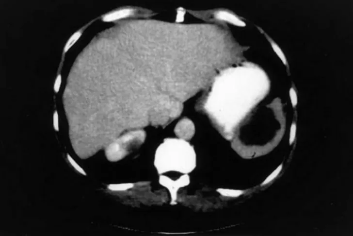
Fig. 2 Appearance of new AE localizations after LT for AE. In
this patient an abdominal CT scan systematically performed 2 years after LT allowed the discovery of a splenic AE metastasis. Further introduction of ABZ with a current duration of 11 years led to stabilization of this lesion
Too heavy an immunosuppressive scheme during the first year post-LT
At the beginning of this experience, there was no specif-ic scheme of immunosuppression for these patients and the majority received a standard immunosuppressive treatment according to the period and the different liver transplant centres’ protocols. Therefore, a triple immu-nosuppressive therapy was used in 56% of the cases, including cyclosporin or tacrolimus, azathioprine or mycophenolate and corticosteroids. The time interval be-fore azathioprine interruption was only available in the Besançon centre. This medication was discontinued at an average of 20 months post-LT (range 2–46). Moreover, four patients from this centre were included in a multi-centric trial using prophylactic anti-CD3 monoclonal an-tibodies for 14 days. Two other patients needed anti-lym-phocyte thymoglobulins. A methylprednisolone bolus (1 or 2 g) was given to 14 patients to treat acute rejection episodes. All six patients having had heavy prophylactic anti-rejection treatment (anti-CD3 or anti-lymphocyte globulins) had a parasitic recurrence. Recurrent AE was observed in only four of the 14 patients who received methylprednisolone bolus.
In 1992, attention was paid to the facilitating role of immunosuppressants on residual AE foci growth [14, 15], and a clear tendency to reduce the immunosuppres-sive regimen as early as possible was adopted by all the centres.
What could be done to prevent or solve these difficulties?
From the results of this retrospective analysis it seems very important, as for liver cancer patients, that an exten-sive morphological pre-LT assessment be performed, including thoracic and brain CT scans in all cases. Our experience suggests that, in the case of brain AE, LT should be ruled out. In the case of pulmonary AE metas-tases, the decision to maintain LT indication depends on the severity of the liver disease, the number, size and lo-cation of the metastases and the tolerance to BZM. In fact, two patients had pre-LT resection of AE lung local-izations with good long-term results. In the other patients a stabilization of the lesions was obtained with long-term BZM therapy. The only patient who died from the pro-gression of pre-existing lung metastases, 10 years after LT, did not receive ABZ because of adverse effects. Therefore, pre-LT BZM should also be recommended, firstly to assess tolerance to the drug, and secondly to try to stabilize the disease while the patient is awaiting LT. In this series, the number of patients having had pre-LT BZM is small. We cannot exclude the hypothesis that re-currence of AE after LT could have been favoured by the absence of pre-LT BZM treatment. Most of the patients
of the palliative sub-group received no BZM or only a short course of it and exhibited a clear progression of the residual AE foci, sometimes associated with occurrence of new localizations. In contrast, the only patient of this sub-group who received MBZ for 2 years before LT has been apparently cured by LT despite the per-operative observation of a small inextirpable retro-pancreatic le-sion. The hypothesis of a parasitolytic effect of MBZ on this small extra-hepatic focus, leading to a fibrous scar, can be made, as recently suggested by other teams [16, 17].
After LT, we advise the early re-introduction of BZM, before the end of the first post-operative month, when the liver tests are normalized. As recommended after a partial surgical resection [18], all patients who undergo transplantation for AE, even if all the parasitic tissue has apparently been removed, should receive BZM for at least 2 years after LT. A regular radiological check-up (abdominal, thoracic and cerebral CT scans once a year) associated with anti-Eg antibodies follow-up, is manda-tory. It seems reasonable that BZM be stopped if all these investigations prove negative at the end of the 2nd year post-LT. Conversely, patients who show evidence of continuing AE should receive BZM indefinitely. We also recommend that the immunosuppressive regimen be kept to a minimum and that the use of lymphocyte anti-bodies be avoided.
Liver transplantation in AE: what outcome?
What is observed, 18 years after the first attempt? Short-term and long-term survivalKaplan–Meier actuarial survival rates were 77%, 71% and 49% at 1, 5 and 10 years, respectively. Survival without recurrence was 58% at 5 years and 45% at 10 years. Given the severity of this disease, this result is quite satisfactory. Some patients, in a very bad condition at the time of LT, are alive and apparently cured, 20 years after the diagnosis of incurable AE and have re-turned to normal activities with a quite satisfactory qual-ity of life. This was quite inconceivable before the LT era, and those patients have clearly benefited from this new therapeutic option. On the other hand, analysis of this series indicates that this very particular indication generates specific problems that absolutely need to be known by teams involved in the care of AE patients.
What could be done to improve the outcome; is there a future for LT in AE?
On the basis on this analysis, one must undertake a very careful management of AE patients with inoperable le-sions, as soon as the diagnosis has been made, keeping
in mind that LT could be part of the therapeutic options taken in the future. However, the major technical diffi-culties during LT, the uncertainty concerning the com-plete cure of the disease, added to the weight of post-LT management, lead us to think that this last therapeutic option should be proposed only to patients with very symptomatic AE. In asymptomatic or pauci-symptomatic cases, a medical option, applying ABZ therapy at higher dosage than previously proposed and using continuous and indefinite administration without the periodic inter-ruptions that were recommended in the past, seems to be worthwhile, as it seems able to stabilize such forms of incurable AE.
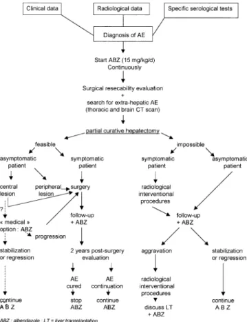
In conclusion, based on this large European experi-ence, LT for incurable AE appears feasible and should be proposed for very symptomatic patients. In the perspec-tive of possible LT, management of septic complications should prioritize interventional radiology and avoid re-peated laparotomies. Pre-LT BZM treatment is mandato-ry to tmandato-ry to stabilize the disease while the patient is awaiting LT and to check the tolerability to these com-pounds. Post-LT BZM, as a continuous administration for at least 2 years, is necessary after LT to minimize the risk of recurrent AE. A meticulous pre-LT evaluation of the extra-hepatic extension of the disease must be per-formed. Due to the high risk of AE recurrence, we rec-ommend that the immunosuppressive regimen be kept to a minimum, anti-Eg IgG be regularly monitored and a systematic careful radiological follow-up be performed. A proposal for current AE management, aiming to place LT among the various options of AE patient manage-ment, is given in Fig. 3.
Fig. 3 Decision-maker tree for the current management of alveolar
echinococcosis of the liver
References
1. Bresson-Hadni S, Vuitton DA (2001) Echinococcoses. Rev Prat
51:2091–2098
2. Bresson-Hadni S, Vuitton DA, Bartholomot B, et al (2000) A twenty-year history of alveolar echinococco-sis: analysis of a series of 117 patients from eastern France. Eur J Gastroen-terol Hepatol 12:327–336
3. Chapuis Y, Houssin D, Brouzes S, et al (1987) Hepatic transplantation in alveolar echinococcosis. 3 attempts. Chirurgie 113:634–640
4. Mboti B, Van de Stadt J, Carlier Y, et al (1996) Long-term disease-free survival after liver transplantation for alveolar echinococcosis. Acta Chir Belg 96:229–232
5. Bresson-Hadni S, Franza A, Miguet JP, et al (1991) Orthotopic liver transplantation for incurable alveolar echinococcosis of the liver: report of 17 cases. Hepatology 13:1061–1070
6. Ammann RW, Ilitsch N, Marincek B, et al (1994) Effect of chemotherapy on the larval mass and the long-term course of alveolar echinococcosis. Hepatology 19:735–742
7. Koch S, Bresson-Hadni S, Miguet JP, et al (2003) Experience of liver transplantation for incurable alveolar echinococcosis: a 45 case European collaborative report. Transplantation 75:856–863
8. Bresson-Hadni S, Laplante JJ, Lenys D, et al (1994) Seroepidemio-logical screening of E. multilocularis infection in 7,884 subjects of a European endemic area of alveolar echinococcosis. Am J Trop Med Hyg 51:837–846
9. Gillet M, Miguet JP, Mantion G, et al (1988) Orthotopic liver transplan-tation in alveolar echinococcosis of the liver: analysis of a series of six pa-tients. Transplant Proc 20:573–576 10. Slim K, Chipponi J, Pezet D, et al
(1996) Récidive d’une échinococcose alvéolaire après transplantation hépatique. Lyon Chir 92:301–303 11. Bresson-Hadni S, Koch S, Beurton I,
et al (1999) Primary disease recurrence after liver transplantation for alveolar echinococcosis: long-term evaluation in 15 patients. Hepatology 30:857–864 12. Gottstein B, Schantz PM, Wilson JF
(1985) Serological screening of Echinococcus multilocularis infections with ELISA. Lancet i:1097–1098
13. Gottstein B, Tschudi K, Eckert J, et al (1989) Em2 Elisa for the follow-up of alveolar echinococcosis after complete surgical resection of liver lesions. Trans R Soc Trop Med Hyg 83:389–393
14. Bresson-Hadni S, Miguet JP, Lenys D, et al (1992) Recurrence of alveolar echinococcosis in the liver graft after liver transplantation. Hepatology 16:279–280
15. Liance M, Bresson-Hadni S, Vuitton DA, et al (1992) Effects of cyclosporin A on the course of murine alveolar echinococcosis and on specific cellular and humoral responses against Echino-coccus multilocularis. Int J Parasitol 22:23–28
16. Ammann RW, Hoffmann AF, Grimm F, et al (1998) Long-term mebendazole therapy may be parasitocidal in alveo-lar echinococcosis. J Hepatol 29:994–998
17. Wang X, Liu Y, Yu D (1996) Continu-ous therapy with albendazole for hepat-ic alveolar echinococcosis associated with obstructive jaundice. Clin J Intern Med 35:261–264
18. WHO Informal Working group on Echinococcosis (1996) Guidelines for treatment of cystic and alveolar echinococcosis in humans. Bull World Health Organ 74:231–242