HAL Id: hal-02135135
https://hal.archives-ouvertes.fr/hal-02135135
Submitted on 21 May 2019
HAL is a multi-disciplinary open access
archive for the deposit and dissemination of
sci-entific research documents, whether they are
pub-lished or not. The documents may come from
teaching and research institutions in France or
abroad, or from public or private research centers.
L’archive ouverte pluridisciplinaire HAL, est
destinée au dépôt et à la diffusion de documents
scientifiques de niveau recherche, publiés ou non,
émanant des établissements d’enseignement et de
recherche français ou étrangers, des laboratoires
publics ou privés.
Biomechanical properties of transosseous bony Bankart
repair in a cadaver model
Philippe Clavert, Florence Aim, Nicolas Bonnevialle, Marine Arboucalot,
Matthieu Ehlinger, Thomas Bauer
To cite this version:
Philippe Clavert, Florence Aim, Nicolas Bonnevialle, Marine Arboucalot, Matthieu Ehlinger, et al..
Biomechanical properties of transosseous bony Bankart repair in a cadaver model. Orthopaedics
and Traumatology - Surgery and Research, Elsevier, 2019, pp.1-4. �10.1016/j.otsr.2018.10.022�.
�hal-02135135�
O
pen
A
rchive
T
oulouse
A
rchive
O
uverte
(OATAO)
OATAO is an open access repository that collects the work of some Toulouse
researchers and makes it freely available over the web where possible.
This is
an author's
version published in:
https://oatao.univ-toulouse.fr/23088
Official URL :
https://doi.org/10.1016/j.otsr.2018.10.022
To cite this version :
Any correspondence concerning this service should be sent to the repository administrator:
tech-oatao@listes-diff.inp-toulouse.fr
Clavert, Philippe and Aim, Florence and Bonnevialle, Nicolas and Arboucalot, Marine and
Ehlinger, Matthieu and Bauer, Thomas Biomechanical properties of transosseous bony
Bankart repair in a cadaver model. (2019) Orthopaedics & Traumatology: Surgery &
Research. 1-4. ISSN 1877-0568
OATAO
Biomechanical
properties of transosseous bony Bankart repair in a
cadaver
model
Philippe
Clavert
a,b,c,∗,
Florence Aim
d,
Nicolas Bonnevialle
e,
Marine Arboucalot
e,
Matthieu
Ehlinger
a,f,
Thomas Bauer
d,
the SOFCOT
ga Équipe 12 matériaux multi-échelles et biomécanique, institut de mécanique des fluides et des solides, laboratoire ICube, GEBOAS, UMR 7357, CNRS UMR 7357, 2–4, rue Boussingault, 67000 Strasbourg, France
b D’anatomie normale, faculté de médecine, fédération de médecine translationnelle, FMTS, 4, rue Kirschleger, 67085 Strasbourg, France c Service de chirurgie du membre supérieur, hôpitaux universitaires de Strasbourg, CCOM, avenue Baumann, 67400 Illkirch, France d Service de chirurgie orthopédique, CHU Ambroise Paré, AP–HP, 9, avenue Charles-de-Gaulle, 92100 Boulogne, France
e Département de chirurgie orthopédique, hôpital Riquet, CHU de Toulouse, place Baylac, 31059 Toulouse, France
f Service de chirurgie orthopédique et de traumatologie, hôpital universitaire de Strasbourg, hôpital de Hautepierre, 1, avenue Molière, 67098 Strasbourg, France
g French Society for Orthopedic and Trauma Surgery, 56, rue Boissonnade, 75014 Paris, France
Keywords: Shoulder Glenoid fracture Osteosynthesis Biomechanics Model
a b s t r a c t
Introduction: We compared two arthroscopic repair techniques to an intact shoulder using a biomechan-ical model of anterior shoulder dislocation with an anterior glenoid rim fracture (Ideberg IA fracture). We hypothesized that transosseous repair is sufficient to effectively stabilize the glenoid fracture. The primary objective was to define the mechanical properties of transosseous repair of an Ideberg IA fracture relative to an intact shoulder (control group). The secondary objective was to determine the contribution of supplemental anteroposterior screw fixation of the bone fragment.
Materials and methods: Fifteen fresh cadaver shoulders were divided into two groups: 5 specimens in the control group and 10 in the fracture fixation group, with sequential performance of transosseous repair followed by transosseous repair + screw fixation. A fracture at the inferior portion involving more than 30% of the glenoid’s surface area was made.
Results: The load to failure was 457 N in the control group, 277 N in the transosseous repair group and 325 N in the transosseous repair + screw fixation group. The stiffness of the constructs was 26.2 N/mm for the control group, 14.6 N/mm for transosseous repair and 24.6 N/mm for transosseous repair + screw fixation. The difference between the two repair techniques was significant for the load to failure (p = 0.02) and stiffness (p = 0.001).
Discussion/Conclusion: This study showed that transosseous repair restores the shoulder’s anatomy but not the mechanical strength of the native glenoid. Adding screw fixation significantly improves the construct.
Level of evidence: IV, basic science study.
1. Introduction
Glenohumeral dislocation most often occurs when the arm is cocked, thus abducted and externally rotated[1–7]. This results in avulsion of anterior capsule/labrum structures on the glenoid rim
[6]; the bony structures may also be damaged. Under certain condi-tions – injury mechanism, patient age, osteoporosis – the energy of
∗ Corresponding author at: faculté de médecine, institut d’anatomie normale, 4, rue Kirschleger, 67085 Strasbourg, France.
E-mail address:philippe.clavert@chru-strasbourg.fr(P. Clavert).
the trauma can cause an intra-articular fracture of the anteroinfe-rior portion of the glenoid fossa, type IA in the Ideberg classification
[8–10]. These fractures are generally treated conservatively. But in
cases with a large fracture fragment and significant displacement (more than 20% of articular surface involved and more than 10 mm displacement[11]), the natural history includes recurrence of the instability and osteoarthritis[9,11,12]. Until recently, surgical fixa-tion was recommended[13–16]. However, the need to detach and then suture the subscapularis tendon contributed to the develop-ment of arthroscopic fixation of this type of fracture with suture anchors. This technique has good clinical and radiological out-comes[17–22]. The advantages of these arthroscopic techniques
are that they provide better reduction of the intra-articular fracture, limit the tissue dissection and reduce the morbidity[23]. However, there are no studies on the biomechanics of these arthroscopic techniques, which are needed to evaluate the risk of secondary displacement and to support postoperative recommendations.
The primary objective of our study was to define the pri-mary mechanical properties of transosseous repair of an Ideberg IA fracture relative to intact shoulders (control condition). The secondary objective was to determine the contribution of supple-mental anteroposterior screw fixation of the bone fragment. We hypothesized that transosseous repair is sufficient to effectively stabilize an anterior glenoid rim fracture (Ideberg IA).
2. Materials and methods
We used an anterior glenohumeral instability model that has been described previously[24].
2.1. Dissection
Fifteen fresh cadaver shoulders from bodies donated to the Anatomy Institute of the Strasbourg Faculty of Medicine were used for the study (9 male, 6 female). The mean age of the subjects was 76 years (range 69–82). Before starting, each shoulder underwent X-rays with the humerus in the frontal plane to calculate the Tingart index used to balance out the groups based on the degree of osteo-porosis[25]. Arthroscopy was performed by the posterior route to eliminate all shoulders with osteoarthritis or damage to anterior capsule and labrum structures. Next, 5 specimens were randomly placed in the control group and 10 in the reconstruction group.
All of the tissues were dissected to leave only 15 cm of the humerus, the glenohumeral joint capsule and the rotator cuff ten-dons. Two traction sutures were placed in the subscapularis tendon (Ethibond No. 6, Ethicon, Somerville, NJ, USA). In the reconstruc-tion group, a fracture of the anteroinferior porreconstruc-tion of the glenoid fossa (between 3 and 6 o’clock) involving nearly 30% of the surface area was created with an osteotome under fluoroscopy control. The continuity of the capsule/periosteum was preserved as was the integrity of the labrum. First, transosseous repair was done by placing interrupted sutures on two or three 3.5-mm diame-ter absorbable PLLA suture anchors (Panalok, Mitek, Norwood, MA) depending on the length of the osteotomy (which varied based on the subject’s size). All the repairs were performed directly, leaving the subscapularis tendon reflected. After the biomechanical testing was completed, the same shoulder was re-used. A new repair was done using the same protocol but with a larger diameter suture anchor, inserted in the same hole as the first anchor, in the cranial and caudal segments of the fracture. A 3.5-mm diameter cannulated screw was added to the suture repair, perpendicular to the fracture line, and in the middle of it, to provide additional bicortical fixation of the fracture fragment.
2.2. Biomechanical testing
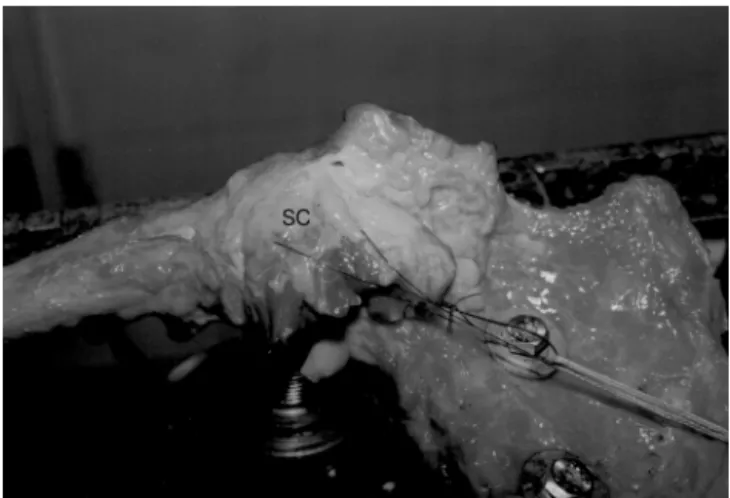
Biomechanical testing was carried out on a materials testing machine (Instron 8500 PLUS, Instron Corp., High Wycombe, Buck-inghamshire, United Kingdom). The shoulders were positioned in a jig that allowed 80◦abduction on the scapular plane and 90◦ exter-nal rotation of the humerus[26]to simulate the arm-cock position. The scapula was secured between two plates and then a K-wire was inserted through the humerus to maintain its position (Fig. 1). A 20 N pre-load was applied to the subscapularis tendon using the two, previously applied traction sutures. A 10 N pre-load was applied to the construct by the actuator of the testing machine on the posterior side of the humeral head. Only at this point was the final position of the humerus set by locking the K-wire.
Fig. 1. Test jig with the arm placed in the cocked position through the
scapulo-humeral joint. Traction sutures are placed in the subscapularis (SC) tendon.
The humeral head was then translated anteriorly at 160 mm/s. The load and displacement were recorded. The 10 specimens that underwent transosseous repair were tested, then the same 10 specimens were re-tested with the screw fixation added to the transosseous repair.
We analyzed the mode of failure, site of failure, load at failure and stiffness, which corresponded to the slope of the linear portion of the load-displacement curve. The specimens were kept moist throughout the experiments.
2.3. Statistical analysis
A one-way ANOVA was used to compare the two reconstruc-tion techniques to the control group. The results of the consecutive repairs on the same shoulder were compared using the non-parametric Wilcoxon test. The alpha level was 5%. The software XLstat 2007 (Addinsoft, Paris, France) was used.
3. Results
3.1. Fragment area
All the fractures involved more than one-third of the inferior articular surface, calculated as described by Sugaya et al.[12]. 3.2. Type of failure
In all specimens in the control group, the failure occurred at the anterior capsule/labrum area (3 to 6 o’clock). There were no fractures of the anterior rim of the glenoid fossa or capsular failure at the humeral neck.
In the transosseous repair group, none of the suture anchor points failed. In every specimen, the failure occurred with breaking of the sutures themselves and displacement of the fragment (Fig. 2). In the transosseous repair + screw fixation group, the capsule failed in all specimens, with fragmentation of the screwed anterior glenoid rim.
3.3. Mechanical properties
The load to failure and stiffness are summarized inTable 1. The mechanical strength (p = 0.001) and stiffness (p = 0.001) were sig-nificantly less in the two reconstruction groups than in the control group. Adding a screw significantly increased the load to failure (p = 0.02) and the stiffness (p = 0.001) relative to the group with transosseous repair only.
Fig. 2. Failure of the sutures with displacement of the bone fragment, which remains
attached to the capsule.
Table 1
Mean values (SD) of the load to failure and stiffness for the control group and two fracture fixation techniques.
Load to failure (N) Stiffness (N/mm)
Control 457 (187.1)a 26.2 (7.5)a
Transosseous repair 277 (48.7)a 14.6 (3.2)a
Transosseous repair + screw fixation 325 (129.4)a 24.6 (8.4)a
aSignificantly different (p-values in the text).
4. Discussion
The results for the control group (load to failure of 457 N) are comparable to published values[24], thus making for a valid com-parison with the reconstruction groups. This study clearly shows that arthroscopic transosseous repair for Ideberg IA glenoid fracture restores the anatomy of the glenoid fossa within the scapula but does not restore the primary mechanical properties of the native glenoid. Adding an anteroposterior screw significantly improves the mechanical stability of the construct, both in terms of the load to failure (p = 0.02) and the stiffness (p = 0.001). Note that in the transosseous repair group, the load at failure and stiffness were much lower than in the other two groups. This can be explained by the fact that the suture anchor + screw construct is very rigid once the screws are inserted, thus the hardware is being tested more than the construct itself. This study was not designed to determine the minimum size of fragment needed to allow a screw to be added to the suture anchor construct.
Since only one-third of the humeral head makes contact with the glenoid’s articular surface, this joint is only partially constrained by its bony relationships. However, lesions in the inferior portion of the glenoid make the glenohumeral joint highly unstable in cases with a displaced intra-articular fracture. Moreover, anterior liga-ment structures also contribute to the joint’s stability[5,27]. Thus it seems necessary to not only reduce the articular fragment, but also to secure the anterior capsule and labrum in their anatomi-cal position in order to control the bone and ligament structures. This is likely the reason why arthroscopy improves the quality and stability of repairs, as it allows associated lesions such as partial or complete rotator cuff tears and/or superior or posterior extension of labrum tears to be repaired (up to 78.3% in the Scheibel study
[28]) and better fixation of ligament structures.
Cameron[21]was the first to propose performing arthroscopic fixation of intra-articular glenoid fractures using a 3.5-mm diam-eter cannulated screw. Later on, it was shown that transosseous fixation stabilizes the fragments well enough for the fracture to
heal in an anatomical position[17–22]. However, all of these stud-ies were done with fragments involving less than 20% of the glenoid surface. Scheibel et al.[27]proposed using an absorbable screw to secure the bone fragment. With a 100% union rate, these authors suggested that along with better stability, adding the screw com-presses the fracture site more and contributes to its healing. The alternative to using a screw is performing a double-row repair[29]. In a cadaver study, it was shown that this fixation technique pro-vides greater stability than suture anchor fixation on the anterior rim of the glenoid (single-row repair performed typically). 4.1. Study limitations
As with any cadaver study, the number of specimens was lim-ited to the minimum needed for use of non-parametric statistical tests. Moreover, this study was done with donors older than the age of patients who typically suffer this type of injury. It can only mea-sure the primary mechanical strength of the construct at T0 and does not take into account remodeling and healing that occurs in vivo. We did not test different types of sutures, in particular newer non-absorbable sutures. Also, the amount of plastic deformation in the joint capsule cannot be determined in vivo, thus cannot be reproduced in a laboratory setting. Lastly, the dislocation mech-anism, by application of an anteroposterior translation load does not always reflect reality and other mechanisms come into play in clinical practice. For this reason, this study did not evaluate the lesions associated with a fracture/dislocation and their impact on the stability of the construct.
5. Conclusions
Arthroscopic transosseous repair of Ideberg IA intra-articular glenoid fractures appears to correctly stabilize the anteroinfe-rior fragment. Adding screw fixation significantly improves the strength of the construct. However, these constructs do not fully restore the primary mechanical properties of the native glenoid. Thus we feel it is necessary to immobilize the patients’ shoulder postoperatively.
Disclosure of interest
PhC: consultant and product development for Wright-Tornier, Associate editor for OTSR.
NB: consultant for Smith & Nephew, Wright-Tornier, Stryker; royalties from SBM.
ME: consultant and product development for DePuy-Synthes, Lépine, Symbios, Newclip, Amplitude®, Associate editor for the SOFCOT instructional lectures.
FA declares that he has no competing interest. TB: consultant for Arthrex®, Zimmer/Biomet®. Funding
None.
Author contributions
All the authors contributed to the study design, validation and writing of the manuscript.
References
[1]Branch T, Lawton R, Lobst C, Hutton W. The role of glenohumeral capsular ligaments in internal and external rotation of the humerus. Am J Sports Med 1995;23:632–7.
[2]Matsen F, Fu F, Hawkins R. The shoulder: a balance of mobility and stability. In: Matsen F, Fu F, Hawkins R, editors. The shoulder: a balance of mobility and
stability. Rosemont: IL: American Academy of Orthopaedic Surgeons; 1993. p. 279–304.
[3]Moseley H, Overgaard B. The anterior capsular mechanism in recurent anterior dislocation of the shoulder. J Bone Joint Surg Br 1962;44–B:913–27.
[4]Ovesen J, Nielsen S. Stability of the shoulder joint: cadaver study of stabilizing structures. Acta Orthop Scand 1985;56:149–51.
[5]Terry G, Hammon D, France P, Norwood L. The stabilizing function of passive shoulder restraints. Am J Sports Med 1991;19:26–34.
[6]Bankart A. The pathology and treatment of recurrent dislocation of the shoulder joint. Br J Surg 1938;26:23–9.
[7]Perry J. Anatomy and biomechanics of the shoulder in throwing, swimming, gymnastics and tennis. Clin Sports Med 1983;2:247–70.
[8]Ideberg R, Grevsten S, Larsson S. Epidemiology of scapular fractures Incidence and classification of 338 fractures. Acta Orthop Scand 1995;66:395–7.
[9]Ideberg R. Unusual glenoid fractures: a report on 92 cases. Acta Orthop Scand 1987;58:191–2.
[10]Ideberg R. Fractures of the scapula involving the glenoid fossa. In: Bateman J, Welsh R, editors. Surgery of the shoulder. Philadelphia: B.C. Decker; 1984. p. 63–6.
[11]De Palma A. Fractures and fracture-dislocations of the shoulder girdle. Philadel-phia: JB Lippincott; 1983.
[12]Sugaya H, Moriishi J, Dohi M, Kon Y, Tsuchiya A. Glenoid rim morphology in recurrent anterior glenohumeral instability. J Bone Joint Surg 2003;85:878–84.
[13]Mayo K, Benirschke S, Mast J. Displaced fractures of the glenoid fossa. Results of open reduction and internal fixation. Clin Orthop Relat Res 1998;347:122–30.
[14]Schandelmaier P, Blauth M, Schneider C, Krettek C. Fractures of the glenoid treated by operation. A 5- to 23-year follow-up of 22 cases. J Bone Joint Surg 2002;84:173–7.
[15]Sinha J, Miller A. Fixation of fractures of the glenoid rim. Injury 1992;23:418–9.
[16]Kavanagh B, Bradway J, Cofield R. Open reduction and internal fixation of displaced intra-articular fractures of the glenoid fossa. J Bone Joint Surg 1993;75:479–84.
[17]Sugaya H, Kon Y, Tsuchiya A. Arthroscopic repair of glenoid fractures using suture anchors. Arthroscopy 2005;21, 635e1-e5.
[18]Bauer T, Abadie O, Hardy P. Arthroscopic treatment of glenoid fractures. Arthroscopy 2006;22, 569e1-6.
[19]Tauber M, Moursy M, Eppel M, Koller H, Resch H. Arthroscopic screw fixa-tion of large anterior glenoid fractures. Knee Surg Sports Traumatol Arthrosc 2008;16:326–32.
[20]Gigante A, Marinelli M, Verdenelli A, Lupetti E, Greco F. Arthroscopy-assisted reduction and percutaneous fixation of a multiple glenoid fracture. Knee Surg Sports Traumatol Arthrosc 2003;11:112–5.
[21]Cameron S. Arthroscopic reduction and internal fixation of an anterior glenoid fracture. Arthroscopy 1998;14:743–6.
[22]Porcellini G, Campi F, Paladini P. Arthroscopic approach to acute bony Bankart lesion. Arthroscopy 2002;18:764–9.
[23]Marsland D, Ahmed H. Arthroscopically assisted fixation of glenoid fractures: a cadaver study to show potential applications of percutaneous screw insertion and anatomic risks. J Shoulder Elbow Surg 2011;20:481–90.
[24]Clavert P, Kempf J, Kahn J. Biomechanics of open Bankart and coracoid abut-ment procedures in a human cadaveric shoulder model. J Shoulder Elbow Surg 2009;18:69–74.
[25]Tingart M, Apreleva M, von Stechow D, Zurakowski D, Warner J. The cortical thickness of the proximal humeral diaphysis predicts bone mineral density of the proximal humerus. J Bone Joint Surg 2003;85:611–7.
[26]Johnston T. The movement of the shoulder joint. Aplea for the use for ‘the plan of the scapula’ as the reference for movement occuring at the humero-scapula joint. Br J Surg 1937;25:252–60.
[27]Warner J, Deng X, Warren R, Torzilli P. Static capsuloligamentous restraints to superior-inferior translation of the glenohumeral joint. Am J Sports Med 1992;20:675–85.
[28]Scheibel M, Hug K, Gerhardt C, Krueger D. Arthroscopic reduction and fixation of large solitary and multifragmented anterior glenoid rim fractures. J Shoulder Elbow Surg 2016;25:781–90.
[29]Millett P, Braun S. The “bony Bankart bridge” procedure: a new arthro-scopic technique for reduction and internal fixation of a bony Bankart lesion. Arthroscopy 2009;25:102–5.