HAL Id: tel-02619041
https://tel.archives-ouvertes.fr/tel-02619041
Submitted on 25 May 2020HAL is a multi-disciplinary open access
archive for the deposit and dissemination of sci-entific research documents, whether they are pub-lished or not. The documents may come from teaching and research institutions in France or abroad, or from public or private research centers.
L’archive ouverte pluridisciplinaire HAL, est destinée au dépôt et à la diffusion de documents scientifiques de niveau recherche, publiés ou non, émanant des établissements d’enseignement et de recherche français ou étrangers, des laboratoires publics ou privés.
Étude multi-approches des voiles bactériens du
Paléoprotérozoïque (2,1 Ga, Gabon) : Biogénicité,
minéralogie et biogéochimie
Jérémie Aubineau
To cite this version:
Jérémie Aubineau. Étude multi-approches des voiles bactériens du Paléoprotérozoïque (2,1 Ga, Gabon) : Biogénicité, minéralogie et biogéochimie. Sciences de la Terre. Université de Poitiers, 2019. Français. �NNT : 2019POIT2278�. �tel-02619041�
THESE
Pour l’obtention du Grade de
DOCTEUR DE L’UNIVERSITE DE POITIERS
Institut de chimie des milieux et matériaux de Poitiers - IC2MP
Faculté des Sciences Fondamentales et Appliquées
Diplôme National - Arrêté du 25 mai 2016
Ecole Doctorale :
Chimie Ecologie Géosciences Agrosciences « Théodore Monod »
Secteur de Recherche : Terre solide : géodynamique des enveloppes supérieures,
paléobiosphère
Présentée par
Jérémie AUBINEAU
************************
Étude multi-approches des voiles bactériens du Paléoprotérozoïque
(2,1 Ga, Gabon) : Biogénicité, minéralogie et biogéochimie
************************
Directeur de Thèse : Abderrazak El Albani
************************
Soutenue le 27 septembre 2019 devant la Commission d’Examen
************************
JURY
Rapporteur Olivier Rouxel Docteur, IFREMER, Brest Rapporteur Bénédicte Ménez Professeur, IPGP, Paris
Directeur de thèse Abderrazak El Albani Professeur, Université de Poitiers Examinateur Laurent Caner Professeur, Université de Poitiers
Examinateur Claude Geffroy Maître de conférence, Université de Poitiers Examinateur Claire Rollion-Bard Ingénieur de recherche CNRS, IPGP, Paris Examinateur Timothy Lyons Professeur distingué, Université de Riverside Examinateur Ernest Chi Fru Professeur associé, Université de Cardiff
i Résumé
La Série sédimentaire du Francevillien âgée de 2,1 milliards d’années du Gabon a fait l’objet de plusieurs investigations notamment à des fins économiques en lien avec les gisements uranifère et manganésifère. Ceci a également promu de nombreuses recherches pour reconstruire les paléoenvironnements et la paléobiodiversité du bassin de Franceville. Les sédiments abritent les plus anciens macrofossiles de tailles et de formes variées, ainsi que les traces laissées par des organismes mobiles, mettant en valeur l’enregistrement grandissant des formes primitives complexes et organisées au Paléoprotérozoïque. Cependant, le style de vie microbien, qui a émergé plus d’un milliard et demi d’années avant la sédimentation du Francevillien, a été peu décrit. Une étude multi-approches et pluridisciplinaire a révélé une grande diversité de structures liées aux voiles (MRS). Ces communautés microbiennes étaient principalement construites par des Cyanobactéries oxyphototrophiques qui ont prospéré dans des environnements peu profonds et dans la zone photique. Etant donné que ces bactéries peuvent avoir produit de grandes quantités d’oxygène très localement, ceci explique à priori la présence rependue de formes de vie avancées à proximité des MRS. Les structures fragiles bactériennes ont ensuite été analysées d’un point de vue minéralogique et géochimique. Les analyses montrent un assemblage minéralogique argileux riche en potassium (K) localisé dans les MRS mais inexistant dans les sédiments encaissants sous-jacents constitués de grès et d’argiles riches en matière organique (black shales). Cela suggère un piégeage des cations K+ par les MRS. Ce K, qui provient de l’eau de mer, a été ensuite relargué dans l’espace
interstitiel pendant la dégradation de la matière organique, permettant ainsi la néoformation argileuse riche en K. Ceci confirme l’enrichissement potassique induit par des microbes. En ce qui concerne la teneur en éléments traces (TE) dans les MRS, aucun enrichissement en lien avec les microorganismes a été observé. La concentration de certains TE dans les MRS est plus élevée que celle du sédiment encaissant, mais des facteurs physiques environnementaux et non biologiques pourraient avoir causé ces enrichissements. Les données du redox local de l’eau de mer pour le sédiment encaissant montrent que le milieu de dépôt se traduit par des fluctuations des conditions redox (oxiques/suboxiques). Les signaux isotopiques du carbone organique et de l’azote de la roche totale sont similaires dans les structures bactériennes et le sédiment encaissant. La composition des isotopes du carbone suggère l’occurrence d'un recyclage secondaire d’un matériel carboné dérivé de la photosynthèse. Par ailleurs, les isotopes de l’azote indiquent une limitation azotée où les fixateurs de l’azote n’ont pas efficacement compensé la perte de ce dernier. La fixation de l’azote dans la colonne d’eau aurait été passagère et potentiellement contrôlée par la structure redox de l’océan, tandis que cette voie métabolique associée aux MRS est vraisemblablement commune au royaume des voiles benthiques à travers l’histoire de la Terre. L’ensemble de
ii
ces résultats soulignent la manifestation fréquente du mode de vie bactérien dans la série du Francevillien et révèlent si les microbes ont laissé des biosignatures spécifiques. La conservation exceptionnelle des MRS en association avec les macrofossiles du Gabon représente un écosystème marin unique à la suite de la première montée significative de la teneur en oxygène dans l’atmosphère terrestre.
iii
Abstract
The 2.1-billion-year old (Ga) Francevillian Series in Gabon has been intensively studied because of economic interests in their uranium and manganese ore deposits. This also promoted numerous scientific investigations to reconstruct the palaeoenvironments and palaeobiodiversity of the Francevillian basin. The sediments host the oldest reported macrofossils of various sizes and shapes, and traces left by motile organisms, highlighting the growing record of early complex forms in the Palaeoproterozoic. However, the microbial lifestyle, which emerged more than a billion and a half years before the Francevillian deposition, has been poorly described. Through a combination of analytical technics, a large diversity of mat-related structures (MRS) has been observed. These microbial communities were mainly built by oxyphototrophic Cyanobacteria that thrived in shallow water environments within the photic zone. Considering that these bacteria may have locally produced higher amount of oxygen than in the oxygen-stressed water column, this likely explained the widespread presence of advanced forms of life in the vicinity of MRS. The delicate bacterial structures were then analyzed for their mineralogical and geochemical compositions. A distinct potassium (K)-rich clay assemblage characterizes the MRS, but not the underlying host sandstone and black shale sediments. It suggests that the MRS trapped K+ from the seawater
and released it into the pore-waters during degradation of organic matter, resulting in K-rich clay neoformation. This confirms the microbially induced K enrichment in the geological rock record. However, the trace element (TE) content in the MRS does not reveal particular microbially mediated enrichments. The concentration of some TE in the MRS is higher relative to that of the host sediments, but physical environmental factors may overwhelm any potential biological signal. The local seawater redox data for the host sediments show that the depositional setting reflects fluctuations in redox conditions (oxic/suboxic). The organic carbon and bulk nitrogen isotopes between the bacterial structures and host sediments are mostly similar. The carbon isotope composition suggests the occurrence of secondary recycling of photosynthetically derived carbonaceous material, while the nitrogen systematic points to a nitrogen limitation by which the N2 fixers did not sufficiently replenish the nitrogen loss. The
nitrogen fixation in the water column would have been transient and likely controlled by the ocean redox structure, whereas this metabolic pathway in the MRS is likely common to the realm of benthic mats over Earth’s history. Combined, these results underline the common occurrence of bacterial lifestyle in the Francevillian Series and reveal whether the microorganisms left typical biosignatures. The exceptional conservation of the MRS in association with the Gabonese macrofossils represents a unique marine ecosystem in the aftermath of the first significant rise of oxygen content in Earth’s atmosphere.
v
REMERCIEMENTS
Avant de commencer, je tiens à exprimer ma gratitude au Ministère de l’Education Nationale, de l’Enseignement et de la Recherche, à la région Nouvelle-Aquitaine, à l’Université de Poitiers, au Centre National de Recherche Scientifique (CNRS), à l’école doctorale Théodore Monod, et enfin le laboratoire IC2MP qui ont contribué directement et indirectement à ce travail de thèse.
Trois années viennent de s’écouler : DEJA ! On se dit « 3 ans, j’ai le temps de tout faire » mais en fait pas vraiment. Je voudrais remercier dans un premier temps Abder qui m’a fait confiance dès le début il y a maintenant 6 ans. En effet, notre collaboration a débuté lorsque je rangeais et classais des échantillons de roches avec d’autres étudiants pendant un simple stage de licence. J’ai donc pu effectuer ce doctorat dont l’aventure humaine et intellectuelle fut particulièrement enrichissante. J’ai également eu la possibilité de visiter d’autres laboratoires à travers de nombreuses collaborations nationales et internationales, toutes riches en expérience. Pour tout cela merci à toi Abder.
Je tiens à remercier mon jury de thèse, Olivier Rouxel, Bénédicte Ménez, Laurent Caner, Claude Geffroy, Claire Rollion-Bard, Ernest Chi Fru et Timothy Lyons, qui m’a fait l’honneur de lire et de juger mes travaux.
Un grand, grand merci à Alain Meunier qui malgré son statut de retraité était là tous les matins (ou presque) pour nous préparer le café. Au-delà de cela, c’est avec Alain que j’ai échangé le plus durant cette thèse et je pense que je lui dois ma gratitude car sans son aide précieuse ce doctorat n’aurait pas la même valeur.
Merci à tous les membres de l’équipe HydrASA et à l’ensemble du service technique pour leur accueil chaleureux et leur soutien humain et analytique durant cette thèse. Un grand merci également aux filles du service administratif de l’IC2MP pour leur aide, leur disponibilité et leur réactivité. Je souhaite également remercier chaleureusement François Leconte, Olabode Bankole et « papa » Claude qui ont toujours su se montrer disponible et généreux à bien des égards durant ces trois longues années.
Je suis très reconnaissant envers mes collègues Andrey Bekker et Ernest Chi Fru qui m’ont accueilli avec plaisir chez eux. Je n’oublierai pas nos longues discussions scientifiques, parfois tardives, mais toutes enrichissantes. Je souhaite exprimer ma gratitude à Kurt Konhauser qui m’a fourni une aide incroyable dans l’ensemble de nos collaborations et que j’ai finalement rencontré dans un cadre idéal pour la recherche : Banff (Canada).
vi
Petit détour par le Nord… Malgré le fait que le soleil ne soit pas tout le temps au rendez-vous, j’ai pu compter sur le soutien indéfectible de mes nombreux collègues et amis lillois. Je tiens à remercier tout particulièrement Kévin Lepot, Philippe Recourt, Jean-Yves Reynaud, Armelle Riboulleau, Alain Trentesaux et Nicolas Tribovillard pour les nombreux conseils et moments partagés depuis mon Master 2. C’est toujours un plaisir de se rendre à Lille pour travailler, échanger et éventuellement boire quelques bières avec Alexis Caillaud, Martin Jovillet Castelot et Fabio Francescangeli.
Une pensée amicale pour mes amis géologues de longue date, Romain Beuzeval, Gaël Cherfallot, Maxime Comby, Nicolat Tricot, Héloïse Verron sans oublié l’inévitable Christophe Ronez avec lequel j’ai partagé de très bons moments, surtout à Los Angeles (une histoire incroyable).
Ce doctorat n’aurait pas eu la même valeur sans eux. Je souhaite remercier mes collègues post-docs, doctorants géologues, paléontologues et de l’association Pictasciences qui m’ont permis de voir sous un autre angle le travail de thèse, surtout autour d’un verre. A ce titre, merci à Marine Bonnet, Grâce Guiyelegou, Thomas Dabat, André Somavilla, Génia Soldatenko, Anastasia Chupryna, Emilie Berlioz, Marion Corbé, Corentin Gibert, Florian Martin, Laurent Pallas et Gislain Thiery. Une pensée également pour l’ensemble des anciens collègues, actuels stagiaires et étudiants extérieurs qui étaient de passage dans nos locaux. Tout de même, mention spéciale à Christopher Sevin et Axelle Walker pour leur caractère atypique et leur sens hors du commun à épater la galerie.
Un grand merci à mes amis lycéens avec qui nous avons su prolonger notre nostalgie. Malgré leur manque d’intérêt pour la recherche fondamentale, j’ai pu compter sur le soutien plus que précieux de Bruno, Catherine, Léa, Jérémy, Mélanie, Jordan et Norine. Pour finir, je tiens à remercier mon amie Véronique, mon frère, mes parents et grands-parents, qui m’ont toujours rassuré dans les périodes difficiles et soutenu, ce qui n’a jamais été aussi important à mes yeux.
vii
TABLE OF CONTENTS
REMERCIEMENTS __________________________________________________ v
TABLE OF CONTENTS ______________________________________________ vii
LIST OF FIGURES ___________________________________________________ x
LIST OF TABLES __________________________________________________ xvii
LIST OF APPENDICES ____________________________________________ xviii
INTRODUCTION (FRANÇAIS) ________________________________________ 1
Objectifs __________________________________________________________ 2
Structure de la thèse ________________________________________________ 3
INTRODUCTION ___________________________________________________ 5
Objectives _________________________________________________________ 6
Thesis structure ____________________________________________________ 7
PART 1. STATE OF THE ART _________________________________________ 9
1.1. Introduction to microbial life _________________________________ 10
1.1.1. Terminology ____________________________________________ 10 1.1.2. Modern microbial mats ____________________________________ 12 a. Formation and organizational pattern _______________________ 12 b. Nature and role of EPS __________________________________ 14 c. Biomineralization _______________________________________ 15 1.1.3. Evidence for microbial activity at the beginning of Earth’s history ___ 16 a. Stromatolites __________________________________________ 17 b. Microbial mat remnants __________________________________ 18 c. Microfossils ___________________________________________ 18 1.1.4. Recognition of biosignatures in Archaean and early Proterozoic
sediments ___________________________________________________ 21 a. Hydrocarbons biomarkers ________________________________ 21 b. Stable isotopes ________________________________________ 22 c. Biominerals ___________________________________________ 26 d. Bioessential (trace) elements ______________________________ 28
1.2. The Palaeoproterozoic rock record ____________________________ 31
1.2.1. Palaeogeographic reconstruction ____________________________ 31 1.2.2. The “Great Oxidation Event” ________________________________ 33 a. General information _____________________________________ 33 b. Sedimentary evidence ___________________________________ 34
viii
c. Geochemical evidence ___________________________________ 36 1.2.3. The “Lomagundi” Event ____________________________________ 37 1.2.4. The Shunga-Francevillian excursion __________________________ 38
1.3. Geological background _____________________________________ 39
1.3.1. General information _______________________________________ 39 1.3.2. Geodynamic ____________________________________________ 40 1.3.3. Lithostratigraphy _________________________________________ 41 a. FA Formation __________________________________________ 42 b. FB Formation __________________________________________ 42 c. FC Formation __________________________________________ 44 d. FD Formation __________________________________________ 44 e. FE Formation __________________________________________ 44 1.3.4. Biomarkers _____________________________________________ 44 1.3.5. Diagenetic history ________________________________________ 45 1.3.6. Radiometric dating _______________________________________ 46 1.3.7. Synthesis of depositional conditions with evidence for microbial activity in the Francevillian basin ________________________________________ 471.4. Studied area and sampling __________________________________ 48
1.5. Methodology _____________________________________________ 50
1.5.1. Sample preparation _______________________________________ 50 1.5.2. Petrographic and mineralogical analyses ______________________ 50 a. Optical microscopy ______________________________________ 50 b. Raman spectroscopy ____________________________________ 50 c. X-ray diffraction (XRD) ___________________________________ 51 d. Electron microscopy _____________________________________ 52 1.5.3. Geochemical analyses ____________________________________ 53 a. Whole-rock elemental analyses ____________________________ 53 b. Organic elemental analyses _______________________________ 54 c. Isotopic geochemistry ___________________________________ 54 d. Iron speciation _________________________________________ 55 e. X-ray Fluorescence (XRF) microscopy ______________________ 56 1.5.4. Descriptive statistic and statistical treatment ___________________ 56
ix
2.1. Introduction ______________________________________________ 58
2.2. A study of Palaeoproterozoic microbial mats from the Francevillian Series
in Gabon ________________________________________________ 59
2.3. The role of Gabonese MRS closely associated with the advanced forms of
life _____________________________________________________ 89
2.4. Highlights of Part 2 ________________________________________ 90
PART 3: MICROBE-MINERAL INTERACTIONS _________________________ 91
3.1. Introduction ______________________________________________ 92
3.2. The link between biological processes and K-rich phyllosilicates _____ 93
3.3. Highlights of Part 3 _______________________________________ 108
PART. 4: GEOCHEMICAL CHARACTERIZATION _______________________ 109
4.1. Introduction _____________________________________________ 110
4.2. A biocontrolled trace element enrichment? _____________________ 111
4.3. Redox conditions in the FB
2Member _________________________ 132
4.4. Highlights of Part 4 _______________________________________ 154
CONCLUSION AND PERSPECTIVES ________________________________ 155
General conclusions _______________________________________________ 155
Future works _____________________________________________________ 157
CONCLUSION ET PERSPECTIVES (FRANÇAIS) _______________________ 159
Conclusions générales _____________________________________________ 159
Travaux futurs ____________________________________________________ 161
REFERENCES ___________________________________________________ 164
APPENDIX ______________________________________________________ 195
Associated works _________________________________________________ 229
1. Sedimentological and stratigraphic approach combined with a
palaeobiological study in the Francevillian basin _________________ 229
2. Spatial organization between the MRS and multicellular organisms in the
studied area _____________________________________________ 261
3. Implications of the rise of arsenic cycling in the Francevillian Series ___ 267
x
LIST OF FIGURES
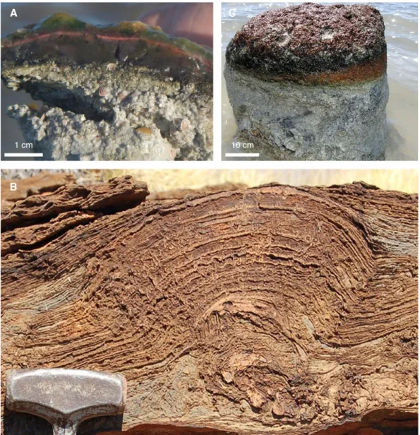
Figure 1: Microbial structures. (A) A modern non-lithified microbial mat from Cayo Coca lagoon, Cuba.
Modified from Pace (2016). (B) A 2.72 Ga-old stromatolites from Tumbiana Formation, Australia. Modified after photo published in Lepot et al. (2009). (C) A modern microbialite from Cayo Coca lagoon, Cuba. Modified from Pace (2016).____________________________________________________ 11
Figure 2: Diel fluctuations of vertical geochemical gradients in microbial mats that are composed of six
major metabolic pathways. Daytime and nighttime metabolic and geochemical reactions lead to distinct microenvironments. Modified from Dupraz et al. (2009). __________________________________ 13
Figure 3: The role of EPS in calcium (and other cations) uptake and liberation. Biologically induced
and/or biologically influenced mineralization may promote carbonate precipitation following three main pathways, including (i) anaerobic microbial respiration, (ii) diagenetic alteration, and (iii) environmentally driven saturation of binding capacity. Modified from Dupraz & Visscher (2005). ________________ 16
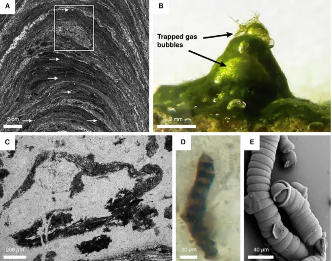
Figure 4: Examples of evidence for the earliest signs of life. (A) Optical photomicrographs of a
stromatolite. Inset shows a fossilized bubble that likely suggests a mat-related gas production. Arrows point to tiny bubble-like features. Modified after photo published in Bosak et al. (2009). (B) Modern analogue of microbial mat producing and trapping O2-rich bubbles. Modified after photo published in
Homann et al. (2015). (C) Rolled-up mat fragment. Modified after photo published in Tice (2009). (D) Controverted bacterial filament. Modified after photo published in Schopf et al. (2002). (E) Synthetic inorganic filaments mimicking biogenic filaments. Modified after photo published in Garcia-Ruiz et al. (2003). _________________________________________________________________________ 19
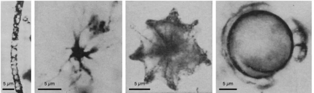
Figure 5: A Gunflint-type assemblage from the earliest discovery of Gunflint Iron Formation.
Filamentous, star-shaped, umbrella-like, and spheroidal morphologies (from left to right) are the most abundant features. Modified after photos published in Barghoorn & Tyler (1965). _______________ 20
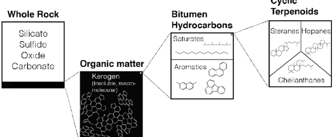
Figure 6: Schematic model of biomarker distribution in organic matter of ancient rocks. Modified from
Waldbauer et al. (2009). ___________________________________________________________ 22
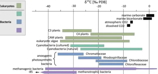
Figure 7: 13C composition of inorganic carbon reservoirs and autotrophs (both bacterial and eukaryotic
domains). Modified from Schirrmeister et al. (2016). _____________________________________ 23
Figure 8: Nitrogen cycling. Black, orange and blue metabolic pathways show the anaerobic N cycle,
suboxic and oxic processes, respectively. Arrow size represents the intensity of fractionation. Adapted from Stüeken et al. (2016). Anammox: anaerobic ammonium oxidation; DNRA: dissimilatory nitrate reduction to ammonium. ___________________________________________________________ 24
Figure 9: Modern and fossilized microorganisms that performed the biologically controlled or biologically
induced mineralization. (A) Complete encapsulation of modern microbial cells by iron-rich minerals. Modified after photo published in Konhauser & Ferris (1996). (B) Precipitation of iron oxides on modern bacterial cell walls. Modified after photo published in Konhauser & Ferris (1996). (C) Intracellular Fe
xi
biomineralization from 1.88 Ga-old cyanobacterial filaments. Modified after photo published in Lepot et al. (2017). _______________________________________________________________________ 27
Figure 10: Synthesis of the evolution of both microbial metabolism and atmospheric oxygen throughout
Earth’s history. The oxidoreductase enzymes are associated with each metabolism. PAL, present atmospheric level; Hd, Hadean; Ph, Phanerozoic. Reproduced from Moore et al. (2017). ________ 29
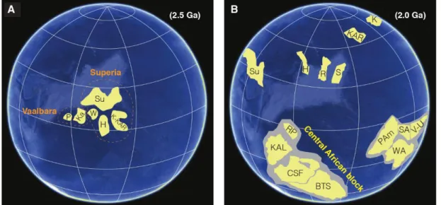
Figure 11: The record of exposed Archaean cratonic blocks. Adapted from Bleeker (2003). ______ 31 Figure 12: Possible palaeogeographic reconstructions. Abbreviations: Su, Superior; P, Pilbara; Ka,
Kaapvaal; W, Wyoming; H, Hearne; K, Kola; KAR, Karelia; R, Rae; S, Slave; RP, Rio de la Plata; KAL, Kalahari; CSF, Congo/São Francisco; BTS, Borborema/Trans-Sahara; Pam, Proto-Amazon; WA, West Africa; SA, Sarmatia; V-U, Volgo-Uralia. (A) Assembly of supercratons during the Neoarchaean-Palaeoproterozoic transition. Adapted from Gumsley et al. (2017) and references therein. (B) Breakup of supercontinents followed by the gathering of cratons (Nuna cycle) during the late Palaeoproterozoic. The most recent plate reconstruction for this time (after D’Agrella-Filho & Cordani, 2017). ________ 32
Figure 13: Schematic representation of redox indicators for the Earth’s oxidation state combined with
the secular carbon isotopic composition and pO2 variations across Archaean-Proterozoic boundary.
Modified from Farquhar et al. (2011) and Bekker & Holland (2012), and references therein. Timelines of arsenic dynamic (after Chi Fru et al., 2019), nitrogen aerobic cycling (after Zerkle et al., 2017; Kipp et al., 2018), and phosphorites (after Papineau, 2010; Soares et al., 2019) are added. Black and red curves show the evolution of carbon isotopes and oxygenation (present atmospheric level; PAL), respectively (after Lyons et al., 2014). ___________________________________________________________ 34
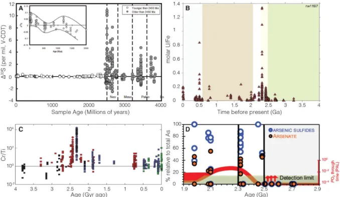
Figure 14: Geochemical evidence for ocean-atmosphere redox evolution following the GOE transition.
(A) Disappearance of S-MIF. Modified from Farquhar et al. (2010). (B) Uranium content in iron-rich rocks. Modified from Partin et al. (2013b). (C) Chromium dynamic in iron formations. Each symbol denotes a type of analysis and each color represents a depositional setting. Cr/Ti ratios have been normalized to the evolving Cr/Ti ratio of upper continental crust (solid line). Modified from Konhauser et al. (2011). (D) Dynamic of arsenic species in shales. Modified from Chi Fru et al. (2019). ________ 37
Figure 15: Simplified geological map of Gabon and cross-section across the four intracratonic
sub-basins. (A) Geological map showing the major lithostructural domains. The orange inset box shows the geological map of Franceville sub-basin in Figure 18. Modified from Thiéblemont et al. (2009). (B) Schematic basin fill architecture of Francevillian basin. Modified from Bouton et al., (2009a). _____ 40
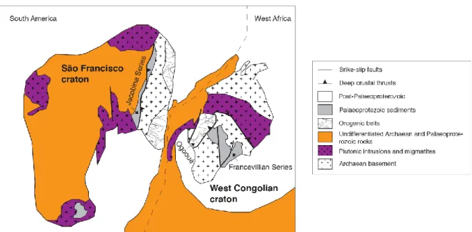
Figure 16: Palaeoreconstruction of West Congolese and São Francisco cratons during the 2.2–2.0
Ga-old Transamazonian–Eburnean orogens. Adapted from Zhao et al. (2002). ___________________ 41
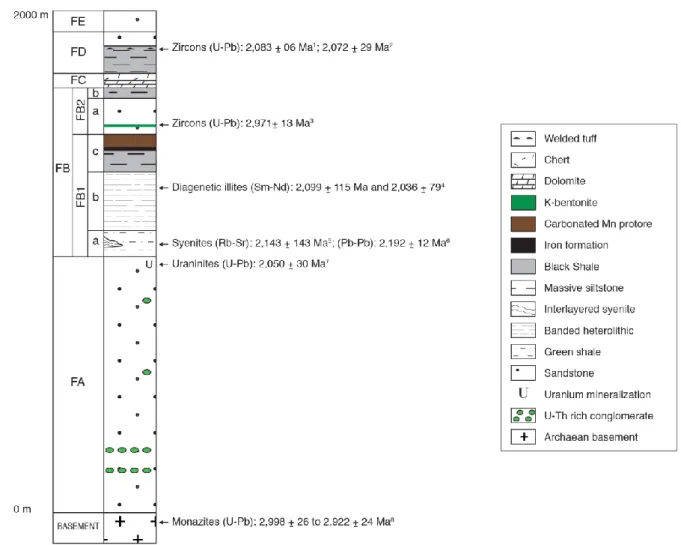
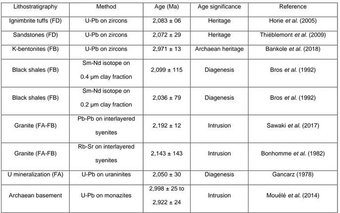
Figure 17: Synthetic lithostratigraphic column of Francevillian Series (1Horie et al., 2005; 2Thiéblemont
et al., 2009; 3Bankole et al., 2018; 4Bros et al., 1992; 5Bonhomme et al., 1982; 6Sawaki et al., 2017; 7Gancarz, 1978; 8Mouélé et al., 2014). ________________________________________________ 43
Figure 18: General geological map of Franceville sub-basin. The Moulendé quarry is shown by the
xii
Figure 19: Synthetic stratigraphic column of the Moulendé quarry in the FB2 Member. __________ 49
Figure 20: Geological map and lithostratigraphic column. (A) Geological map of the Francevillian basin.
The studied quarry is Moulendé (green star). Geological map adopted from Bouton et al. (2009b). (B) Synthetic lithostratigraphy of the Francevillian series. Four sedimentary units rest unconformably on Archean rocks. The red star indicates the detailed lithostratigraphic column observed in the Moulendé quarry (Figure 21). ________________________________________________________________ 62
Figure 21: Detailed lithostratigraphic column. Composite columnar section of the Moulendé quarry in
the FB2 Member showing the vertical distribution of ten representative types of mat-related structures
(MRS) and sedimentary structures (SS). ______________________________________________ 65
Figure 22: Plane view and outcrop pictures of sedimentary facies in the Moulendé quarry. (A)
Representation of the quarry from plane view. Red box indicates the main studied outcrop in B (F8).F = outcrops. (B) Details of the bedding geometry at the transition between massive sandstone beds and thinly laminated black shales. (C) Closer view of B. (D) Cross-section view of decimeter scale hummocky cross-stratifications (HCS), FB2a subunit. (E) Sandstone dyke, FB2a—FB2b transition. Coin diameter: ~2 cm. (F) Cross-section view of convolute structures, FB2b subunit. (G) Bedding plane view of interference ripples, FB2b subunit. (H) Longitudinal view of dark-colored convex laminae associated with cm-scale foreset beds, FB2b subunit. _____________________________________________ 67
Figure 23: Microbial mat structures in the Francevillian B Formation (FB2): Mat-layer structures. (A-C)
“Elephant-skin” textures. (D) Putative macro-tufted microbial mat. (E, F) Clustered domal buildups and flat pyritized microbial structure (red arrow). Macrofossil specimens (white arrows). (G) Isolated domal buildups. (H) Wrinkle marks. ________________________________________________________ 70
Figure 24: Microbial mat structures in the Francevillian B Formation (FB2): Mat-layer structures. (A, B)
Discoidal mats likely representing “fairy ring” structures. (C-E) Disc-shaped mats that display a cauliflower-like pattern. (F) Disc-shaped mat with internal wrinkle structures. (G) Small pyritized circular bodies. (H) Horizontal mat growth pattern. _____________________________________________ 71
Figure 25: Mat-related structures in the Francevillian B Formation (FB2): Mat-protected structures. (A,
B) Parallel wavy wrinkle structures. (C) Cross-cutting wrinkle structures. (D) “Kinneyia” structure. (E)
Linear pattern. Dashed red box indicates the position of the magnification in F. Red arrow shows the location where the Raman spectroscopy was performed. The Raman spectra are visible in Figure 27D. (F) Micrometric spots interpreting as oriented grains. (G) Linear patterns with several parallel ridges. (H) Nodular-like structure. _____________________________________________________________ 75
Figure 26: Optical photomicrographs of mat-related structures. (A) Transmitted thin section of
“elephant-skin” texture. Dashed red box denoting area magnified in F. (B) Transmitted thin section of putative macro-tufted microbial mat. Dashed red boxes denoting areas magnified in G and H. (C) Transmitted thin section of an isolated domal buildup. Dashed red box denoting area magnified in I. (D) Transmitted thin section of parallel wavy wrinkle structures. Dashed red box denoting area magnified in J. (E) Transmitted thin section of a linear pattern. Dashed red boxes denoting areas magnified in K and
xiii
Figure 28E. (F) Tufted microstructures and wavy-crinkly laminae. Dashed red box denoting area magnified in Figure 28A. (G, H) Thickness variation across the mat layer with floating grains embedded by clays (red arrows). Mica (white arrows). Dashed red box denoting area magnified in Figure 28C. (I) Reflected magnified thin section of an entirely pyritized dome. An internal convex lamination is indicated by dashed red lines. (J) Clay laminae do not onlap the rippled siltstone bed but rather well follow its topography. Oriented grains (arrows). (K) High amount of quartz particles (arrows as example) within dark-coloured laminae. Dashed red box denoting area magnified in Figure 28D. _______________ 77
Figure 27: Polished slab of “elephant-skin” texture and Raman spectra of both “elephant-skin” texture
and linear pattern. (A) Polished slab in cross-section perpendicular to bedding plane. Non-homogenous dark layer preserved above a pronounced boundary. Red arrow and white arrow indicate Raman spectra in B and C, respectively. (B) Representative Raman spectra of the microbial mat within bulges. It shows the presence of three carbon peaks (“C”) at ~1202 cm–1 (“D4” disordered peak), 1,336 cm−1 (the “D1”
disordered peak) and 1,603 cm−1 (the “G” graphite peak). (C) Typical Raman spectra of sandstone with quartz (“Q”) peaks. (D) Representative Raman spectra of mat layers of linear pattern indicated in Figure 25E. It shows the presence of three carbon peaks (“C”) at ~1,170 cm−1 (“D4” disordered peak), 1,344
cm−1 (the “D1” disordered peak) and 1,603 cm−1 (the “G” graphite peak). (E) Typical Raman spectra of host sediment of linear pattern, with quartz (Q) peaks and very small intensities of “C” peaks. ____ 79
Figure 28: SEM imaging of mat-related structures. (A) Magnified view of box area in Figure 26F. Upward
clay laminae within tufted microstructures and wavy-crinkly layers. (B). Tufted microstructures and heavy minerals constitute bulges of the “elephant-skin” texture. (C) Magnified view of box area in Figure 26H. Quartz grains, heavy minerals and randomly oriented clays constitute the dark-coloured mat layer. (D) Magnified view of box area in Figure 26K. Detrital particles wrapped by sheet clays. (E) Magnified view of box area in Figure 26E. Clay minerals above and throughout the ridge (arrow). No significant clue of liquefaction nor microbial shrinkage. ____________________________________________ 81
Figure 29: Examples of fossil macroorganisms associated with microbial mats. (A) Pyritized lobate form
just beneath “fairy ring” structures. (B) Disc with radially striated core (arrow) lies on domal buildups. (C,
D) Disc or lobate form and flat pyritized microbial structures on the same strata are closely associated.
(E) Circular discs (arrows) rest on wrinkle marks. (F) Disc and lobate form are close to wrinkle marks on the same level or not. _____________________________________________________________ 87
Figure 30: Geological map and field photographs of the FB2 Member outcrops, Gabon. (A) Geological
map of the Francevillian basin adapted from Bouton et al. (2009b). The studied area is the Moulendé Quarry (green star). (B) Elephant-skin texture on the bedding plane of coarse-grained sandstones. Inset box shows reticulate patterns. (C) Micrometer-thick microbial mat laminae (blue arrow) on the bedding plane of sandstones with rounded pits (white arrows). ____________________________________ 95
Figure 31: Biogenetic fabrics in the mat-related structures. The MRS textures are shown through SEM
images. (A) Tufted microbial fabrics developed above the poorly-sorted quartz sandstone. Yellow and blue arrows point to tufts and quartz grains, respectively. (B) A void-filling titanium oxides that may have filled an oxygen bubble produced within the microbial mat. (C) Nearly-circular void filled with titanium
xiv
oxides at the tip of a cone-like feature (green arrows). Detrital dioctahedral micas (e.g., muscovite) are shown by purple arrows. ___________________________________________________________ 96
Figure 32: K2O/SiO2 ratios from mat-related structures and host sediments. Pyritized MRS are hosted
by black shales and non-pyritized MRS are observed on bedding surface of sandstones. The data for each lithology are represented as box plots with a red square showing the mean, black diamonds corresponding to individual samples, 50% of the data are shown as a box and whiskers extend to 1.5 times the interquartile range. ________________________________________________________ 97
Figure 33: High-resolution sulfur and potassium distribution maps. (A) Pyritized MRS. Red arrows point
to K enrichment between pyrite crystals and in the host sediment of MRS. (B) Black shales. (C) Non-pyritized MRS. Higher X-ray fluorescence (XRF) intensities correspond to higher S and K contents. For easier comparison, a common intensity scale was chosen for the K distribution maps. __________ 98
Figure 34: Mineralogical composition of the <2 µm clay-size fractions. X-ray diffraction profiles of
oriented preparations after air-drying (black lines) and glycolation (red lines) and their transmission-electron images are given. (A) Microbial mat specimen. (B) Large, well-crystallized lath from microbial mat laminae. The inset shows selected area electron diffraction (SAED) pattern. hk0 pattern shows a hexagonal structure typical of phyllosilicates and coherently stacked crystals. (C) Hexagonal habit of illite from a mat sample. (D) Associated sandstone sediment. (E) Lathlike and poorly crystallized particles from the host sandstone. (F) Tiny hexagonal-shaped particles from the host sediment. [Green areas correspond to long-range ordered illite–smectite mixed- layer minerals (R3 I/S MLM); Blue areas represent randomly ordered illite–smectite mixed-layer minerals (R0 I/S MLM); smectite (S); chlorite (Ch); mixed-layer (ML); illite/mica (I/M); anatase (An); quartz (Q); pyrite (Py); barite (Ba); calcite (Ca)]. _______________________________________________________________________________ 99
Figure 35: Geological map of the Paleoproterozoic Franceville sub-basin (A; modified from Bouton et
al. (2009)) and general lithostratigraphic column of the Francevillian Series (B). The studied area (the Moulendé Quarry) is shown with the green star. [1Horie et al. (2005); 2Thiéblemont et al. (2009); 3Bros
et al. (1992); 4Mouélé et al. (2014)]. _________________________________________________ 114
Figure 36: Lithostratigraphic column and representative mat-related structures (MRS) with their
associated petrographic textures from both sandstone and black shale facies. (A) Composite lithostratigraphic section of the studied area in the FB2 Member showing the main microbial levels including pyritized MRS (flat pyritized, domal buildup, and ‘fairy ring’ structures), poorly pyritized MRS (mat-growth and mat-protected structures), and unpyritized MRS (EST: “elephant-skin” texture). (B) Flat pyritized MRS from the FB2b unit. (C) SEM image of pyritized MRS in cross-section perpendicular to the bedding plane. Pyrite grains and clay particles are developed within biofilms. (D) Mat-layer structures (white arrow) on the bedding surface of sandstone from the FB2a unit. (E) SEM image of micrometer-thick poorly pyritized MRS in cross-section perpendicular to the bedding plane. Green and purple arrows show trapped and bound heavy minerals and a gas escape structure, respectively. (F) “Elephant-skin” textures (EST) from the FB2a unit. Inset box shows reticulate pattern. (G) SEM image of tufted microbial fabrics from the EST above the poorly-sorted quartz sandstone. Blue arrow points to tufted microbial
xv
fabrics. Images in B and C, F, and G are modified from Aubineau et al. (2018) and Aubineau et al. (2019), respectively. _____________________________________________________________ 118
Figure 37: Geochemistry of the host rocks, black shales (diamonds) and sandstones (circles). (A)
Chondrite-normalized REE + Y patterns. Chondrite values are from Schmitt et al. (1964) and Evensen
et al. (1978). (B) PAAS-normalized REE + Y patterns. PAAS values are from Taylor & McLennan (1985).
(C) Binary plot of Fe/Ti vs. Al/(Al+Fe+Mn). ____________________________________________ 119
Figure 38: Descriptive statistical treatments. (A) Principal component analysis on 40 individuals
representing the relationship among 3 MRS morphotypes and 2 host sediments (black shales and sandstones) and 26 independent variables (trace element concentrations). (B) Contribution of each variable (%) along PC1 (left) and PC2 (right). The stronger the contribution of TE, the darker the bar is. ______________________________________________________________________________ 120
Figure 39: Stratigraphic profile of enrichment factors (EF) of selected detrital and chalcophile elements
within MRS. EF relative to the average shale (Ti, Zr, Ni concentrations for the average shale are from Taylor & McLennan (2001) and As concentration for the average shale is from Li & Schoonmaker (2003)) were calculated as (X/Al)sample/(X/Al)average shale, where X stands for the element concentration. ____ 121
Figure 40: Relationships between chalcophile elements and sulfur for pyritized MRS, poorly pyritized
MRS, unpyritized “elephant-skin” textures (EST), and Francevillian black shales (from the FB1b and FB1c units and the FD formation). (A) Cross plot of chalcophile element (As, Bi, Co, Cu, Mo, Ni, Pb, and Sb) contents vs. bulk S concentrations for MRS and host black shales. (B) Cross plot of As content vs. bulk S concentration for MRS and host black shales. Inset boxes (A and B) show covariations at low CE and S contents. (C) Cross plot of chalcophile (As, Bi, Co, Cu, Mo, Ni, Pb, and Sb) contents vs. bulk S concentration for MRS and all Francevillian Series black shales (green trend). (D) Cross plot of As content vs. bulk S concentration for MRS and all Francevillian Series black shales (green trend). (E) CE/S ratios for 3 MRS morphotypes and 2 host sediments (black shales and sandstones). CE and S contents for black shales of the Francevillian Series are from Canfield et al. (2013). Data for some samples are below the detection limit; in that case a value of half of the detection limit was used. _ 122
Figure 41: Synchrotron-based XFM showing the distribution of Fe, As, and Ni. (A) Unpyritized EST. (B)
Poorly pyritized MRS. White arrow indicates As-rich pyrite core. (C) Pyritized MRS. The color bars indicate the intensity-scale of the metal X-Ray Fluorescence (XRF) distribution maps in counts/10 ms (black color, below detection limit). Higher XRF intensities correspond to higher metal contents. __ 124
Figure 42: Combined As (green) and Ni (magenta) XRF maps for pyritized MRS. _____________ 125 Figure 43: Conceptual model for the spatial and temporal TE variations in the Francevillian Group FB2
unit MRS. (A) Processes that took place during the living stage of mats. Depositional local- to facies-scale environmental factors controlled the distribution of heavy minerals in the Francevillian MRS. The external mat morphologies provide information on local ecologic conditions. (B) Short-lived first stage of pyrite formation on the earliest phase of diagenesis. The EPS degradation resulted in the release of CE to pore-waters. The amount of CE delivered with EPS decay should be proportional to the organic
xvi
carbon content that is buried. Depending of sulfur and Fe availability, CE were trapped by fine-grained pyrite or diffused back to the water column. (B) Long-lived second stage of pyrite formation as diagenesis progressed. The rest of organic matter was degraded with, thus, larger amounts of H2S and Fe released to pore-waters. High concentrations of biogenic H2S were locally produced, but little CE
were scavenged in coarse-grained pyrite. _____________________________________________ 127
Figure 44: SEM images in backscatter mode of pyrite crystals. (A) Pyritized MRS. (B) Poorly pyritized
MRS. (C) Black shales. ___________________________________________________________ 137
Figure 45: Geochemical data for the MRS and host sediments plotted along the lithostratigraphic profile
of the studied sequence, FB2 Member. Grey and orange vertical lines represent redox distinctions from
Raiswell et al. (2018) and the average PAAS value from Taylor & McLennan (1985), respectively. 139
Figure 46: Data of redox-sensitive metals for the MRS and host sediments plotted along the
lithostratigraphic profile of the studied sequence, FB2 Member. Orange vertical lines correspond to
average PAAS values from Taylor & McLennan (1985). _________________________________ 141
Figure 47: Cross plot of carbon and nitrogen isotopes. __________________________________ 144 Figure 48: Proposed palaeoenvironmental reconstruction during deposition of the Francevillian Series
at the end of the Lomagundi Event (from the FB1c unit to the Lower FC Formation). Ocean redox conditions and biogeochemical cycles are shown (modified from Ossa Ossa et al., 2018). Orange and black dotted arrows indicate the limited expression of upwelling system and microbial processes, respectively. ____________________________________________________________________ 147
xvii
LIST OF TABLES
Table 1: Summary of radiometric ages in the Francevillian basin. ___________________________ 47 Table 2: δ13C values of organic matter in mat-related structures (MRS). ______________________ 83
Table 3: Iron speciation data and whole-rock composition of major and minor elements for all
measurements in this study. _______________________________________________________ 140
xviii
LIST OF APPENDICES
Appendix 1: Additional flat pyritized microbial mats. ____________________________________ 196 Appendix 2: EDS elemental maps of bulges of reticulate patterns in cross-section perpendicular to
bedding plane. BSE and composite elemental maps. Note the wavy-crinkly laminae with a large amount of embedded heavy minerals. ______________________________________________________ 197
Appendix 3: Representative structural formulae of clays from microbial mats studied with EDS. EST,
“elephant-skin” texture; PTMM, putative tufted microbial mat; WS, wrinkle structure; LP, linear pattern. ______________________________________________________________________________ 198
Appendix 4: Organic elemental analyses (carbon and sulfur) on five microbial mats and their host
sediments from the FB2 Formation. EST, “elephant-skin” texture; DB, domal buildup; PWWS, parallel
wavy wrinkle structure. ___________________________________________________________ 200
Appendix 5: Photographs of mat-layer structures found in literature. (A) Analogous ‘elephant-skin’
texture from modern lower supratidal, Bahar Alouane, southern Tunisia. Modified after photo published in Gerdes (2007). (B) Fossil reticulate pattern on bedding plane of siliciclastic beds from the Archaean Tumbiana Formation, Australia. Modified after photo published in Flannery & Walter (2012). (C) A 2.0 billion-years-old tufted microbial mat from Makgabeng Formation, South Africa. Modified after photo published in Simpson et al. (2013). (D) Analogue clustered low mound-like structures with Protichnites trackways on bedding surface of quartz arenites from the Late Cambrian, Elk Mound Group, USA. Modified after photo published in Bottjer & Hagadorn (2007). (E) Modern analogous ‘fairy rings’ on soft muddy sediments from Bretagne salterns, France. Modified after photo published in Grazhdankin & Gerdes (2007). (F) Ancient example of outward-convex, spindle-shaped discoidal structures with concentric rings from the Mesoproterozoic, Sonia Sandstone, India. Modified after photo published in Sarkar et al. (2014). (G) Modern discoidal microbial colony on tidal flat from the Gulf of Cambay, India. Modified after photo published in Banerjee et al. (2014). Lens cap diameter: 6 cm. (H) Analogue discoidal microbial colony on bedding plane of sandstones from the Precambrian Vindhyan Supergroup, India. Modified after photo published in Banerjee et al. (2014). _________________________________ 201
Appendix 6: Photographs of mat-related structures found in literature. (A) Modern example of
submerged wrinkle marks from Redfish Bay, Texas. Modified after photo published in Hagadorn & Bottjer (1997). (B) Patches of wrinkle marks on bedding surface of fine-grained sandstones from the Early Cambrian, Chapel Island Formation, Canada. Modified after photo published in Buatois et al. (2014). (C) Parallel wavy wrinkle structures reproduced in wave tank experiments using microbial aggregates. Modified after photo published in Mariotti et al. (2014). (D) Analogous parallel wavy wrinkle structures on bedding plane of mudstones from the Early Cambrian, Northwest Argentina. Modified after photo published in Buatois & Mángano (2003). (E) Minute “Kinneyia” structures formed with microbial aggregates in wave tank experiments. Modified after photo published in Mariotti et al. (2014). (F) Ancient analogue “Kinneyia” structures on bedding surface of siltstones from the Cambrian, Oeland, Sweden. Modified after photo published in Porada & Bouougri (2007). (G) Linear features from modern tidal flats
xix
of Bhar Alouane, southern Tunisia. Modified after photo published in Porada & Bouougri (2007). (H) Putative linear patterns on bedding surface of fine-grained quartzites from the Neoproterozoic Katanga Supergroup, Zambia. Modified after photo published in Porada & Bouougri (2007). ____________ 202
Appendix 7: Lithostratigraphic columns for the Francevillian basin. (A) Lithostratigraphy of the
Palaeoproterozoic Francevillian Series that comprises four sedimentary formations. (B) Composite stratigraphic section of the FB2 Member in the studied area. It consists of the 15 m-thick coarse-grained
sandstone conformably underlying the 5 m-thick black shale sequence (1Horie et al., 2005; 2Bros et al.,
1992; 3Gancarz, 1978; 4Mouélé et al., 2014; 5Gauthier-Lafaye & Weber, 2003). _______________ 203
Appendix 8: Microbial mat laminae and their host sediments. (A) A µm-thick mat-related structure
preserved as wrinkle on the top of the sandstone bed. (B) Polished slab section of A. (C) Pyritized microbial mat from the black shale facies. (D) SEM imaging of C. Note extensively developed euhedral pyrite, making difficult to separate black shale from pyritized mat structures. (E) SEM image of a thinly laminated mat from the top of the sandstone bed. Note the microtexture of mat laminae. Blue, yellow, and green arrows point to the thin biofilm layers, macrofossil (El Albani et al., 2010, 2014), and heavy minerals, respectively. ____________________________________________________________ 204
Appendix 9: Petrography, SEM, and EDX of bubble-like features in cross-section perpendicular to the
bedding plane. Back-scattered electron (BSE) and composite (Si, Al, Ti, Mg, and Fe) elemental maps show mineral composition of circular structures within the MRS. The cone-like structure mainly consists of Al- and Mg-rich clays. __________________________________________________________ 205
Appendix 10: Whole-rock geochemical data of major elements and carbon. _________________ 206 Appendix 11: Pairwise comparisons between all pairs of groups. __________________________ 208 Appendix 12: Cross plots of selected major elements. (A) SiO2 vs. Al2O3. (B), K2O vs. Al2O3. (C), K2O
vs. Na2O. (D), MgO vs. Al2O3. ______________________________________________________ 209
Appendix 13: XRD patterns of representative randomly oriented powders. (A) Non-pyritized microbial
mats. (B) Sandstones. (C) Pyritized biofilms. (D) Black shales. [Chlorite (Ch); Mixed-layer (ML); Illite/mica (I/M); Quartz (Q); Anatase (An); Florencite (Fl); Di-tri-octahedral chlorite (Ch) (Billault et al., 2002); Gypsum (Gy); Pyrite (Py); Dolomite (Do); Rhodochrosite (Rh)]. ______________________ 210
Appendix 14: XRD patterns of representative oriented <2 µm clay fraction after air-dried treatment
(black lines) and glycolation (red lines). (A) Non-pyritized microbial mats. (B) Sandstones. (C) Pyritized biofilms. The weak signature of short-range ordered I-S MLMs likely indicates contamination from the underlying host sediment. (D) Black shales. [Green areas correspond to long-range ordered (R3) I-S MLMs; Blue areas represent the randomly ordered (R0) I-S MLMs; Chlorite (Ch); Mixed-layer (ML); Illite/mica (I/M); Kaolinite (K); Quartz (Q); Barite (Ba); Calcite (Ca); Gypsum (Gy)]. ____________ 211
Appendix 15: XRD patterns of randomly oriented powders (<2 µm clay fraction) for the MRS and host
sediment are shown at the top and bottom, respectively. The hkl diffraction peaks are typical of illite polytypes with 2M1 and 1Mt polytypes, representing detrital and diagenetic illites, respectively. ___ 212
xx
Appendix 16: Experimental (crosses) and modeled (lines) XRD profiles of selected Ca-saturated MRS
samples. Contribution of different minerals to the profiles is indicated. (A) After air-dried preparation. (B) After ethylene-glycol saturation. ____________________________________________________ 213
Appendix 17: Experimental (crosses) and modeled (lines) XRD profiles of Ca-saturated host sediments.
Contribution of different minerals to the profiles is indicated. (A) After air-dried preparation. (B) After ethylene-glycol saturation. _________________________________________________________ 214
Appendix 18: Structural parameters from the simulation of experimental XRD patterns of Ca-saturated
samples after air-dried treatment and glycolation. ______________________________________ 215
Appendix 19: Transmission-electron images of illite minerals. (A-C) Lath-shaped crystals from MRS.
(D-F) Hexagonal-shaped crystals observed in MRS. (G-H) Lathlike and tiny lath-shaped crystals from host sediments. The inset with Selected Area Electron Diffraction (SAED) pattern shows a hexagonal structure typical of phyllosilicates in (hk0) reflection. ____________________________________ 216
Appendix 20: Whole-rock composition of major and minor elements and enrichment factors (EF) of Ti,
As, Ni, and Zr. __________________________________________________________________ 217
Appendix 21: Bulk-rock data of rare earth elements and calculated Eu anomaly and REE ratios _ 220 Appendix 22: Petrography, SEM, and EDX of a poorly pyritized MRS in cross-section perpendicular to
the bedding plane. Back-scattered electron (BSE) and composite (Al, Si, O, Mg, K, Fe, Ti, and Zr) elemental maps show mineral composition of the poorly pyritized MRS. A number of Ti- and Zr-rich heavy mineral crystals are embedded within these mats. _________________________________ 222
Appendix 23: SEM image of poorly pyritized MRS in secondary electron (left) and BSE (right) modes.
Pyrite-rich layers observed at the bottom of MRS are highly variable in thickness. Inset box shows numerous and tiny sub-euhedral to euhedral pyrite crystals. ______________________________ 223
Appendix 24: Weight of each principal component. _____________________________________ 224 Appendix 25: Stratigraphic profile with enrichment factor (EF) of selected immobile and chalcophile
elements within host sediments. EF is relative to the average shale (Ti, Zr, and Ni concentrations for the average shale are from Taylor & McLennan (2001), and As concentration for the average shale is from Li & Schoonmaker (2003)), and calculated as (X/Al)sample/(X/Al)average shales, where X stands for element
concentration. __________________________________________________________________ 225
Appendix 26: Cross plot of total organic carbon versus total nitrogen for MRS and host sediments.
Dotted lines show the C/N bulk ratios of 5 and 100. The C/N values of modern phytoplanktonic biomass range from 4 to 10 (Gruber & Galloway, 2008; Ader et al., 2016). Remineralization of biomass in the water column and during diagenesis can increase the C/N ratios. __________________________ 226
Appendix 27: Cross plot assessments of data fidelity. (A) Geochemical data for all bulk-rock
measurements from the FB2 Member. (B) Cross plots of K contents with TN and 15Nbulk. _______ 227
Appendix 28: Cross plot of bulk sulfur concentration and TOC for black shale samples from the FB2
1
INTRODUCTION (FRANÇAIS)
Les microbes, connus pour leur grande diversité morphologique, écologique et phylogénique, sont les plus anciennes formes de vie sur Terre et englobent les êtres vivants procaryotiques tels que les archées et les bactéries. Les biofilms et les voiles bactériens font référence à une association fructueuse de microorganismes qui s’est développée au début de l’histoire de la Terre, il y a 3,5 – 3,8 milliards d’années (Ga). Ce mode de vie fournit une large gamme de propriétés émergentes comparé à celui observé des cellules bactériennes isolées. Ces propriétés spécifiques incluent une sorption améliorée pour le piégeage de ressources, une rétention enzymatique, des interactions sociales pour un remodelage dynamique des microorganismes, et des gradients physico-chimiques localisés (Flemming et al., 2016). Les biofilms et les voiles bactériens ont largement été étudiés, cependant la terminologie est souvent confuse sans définition globalement acceptée (Krumbein et al., 2003). Néanmoins, un biofilm peut renvoyer à un groupe de cellules intégré dans une matrice organique dont la structure est monocouche, tandis qu’un voile bactérien définit une distribution multicouche de communautés bactériennes. Ainsi, les voiles bactériens peuvent être perçus comme des biofilms améliorés. Considérant cette terminologie controversée, le terme ‘structures liées aux voiles’ (MRS; Eriksson et al., 2010) a été choisi pour inclure toutes les structures fossilisées induites et/ou préservées par des microorganismes.
Il est largement reconnu que les microbes ont joué un rôle important dans l’évolution du climat et des cycles biogéochimiques de notre planète (Lyons et al., 2014). L’un des plus frappants exemples repose sur le fait que les Cyanobactéries oxyphototrophiques sont capables de produire une grande quantité d’oxygène (O2) à travers la photosynthèse. Bien que le timing de
leur émergence reste débattu, elles sont considérées comme les premiers producteurs d’O2
sur la surface de la Terre. Ainsi, du fait de leur activité, l’ère du Paléoprotérozoïque (2,5–1,6 Ga) fut marquée par la première augmentation significative d’O2 atmosphérique,
communément appelée l’événement de la grande oxygénation (GOE; Holland, 2002), il y a 2,4 Ga. Cet évènement géochimique répandu sur l’ensemble de la surface terrestre a facilité l’apport de nouveaux composés oxydés dans l’océan, ce qui a vraisemblablement boosté la vie microbienne et déclenché une vie eucaryotique (i.e., complexe).
La série du Francevillien au Gabon âgée de ca. 2,1 Ga s’est déposée dans des conditions environnementales principalement oxygénées, en plus d’héberger l’assemblage le plus ancien de fossiles macroscopiques (El Albani et al., 2010, 2014, 2019). Ces macroorganismes coloniaux auraient pu être multicellulaires et fortement organisés du fait de leur grande
2
diversité de taille et morphologie. Quelques preuves d’une activité microbienne ont été soulignées dans les roches du Gabon, incluant des structures ressemblant à des bactéries (Dubois et al., 2015), des filaments de Cyanobactéries (Lekele Baghekema et al., 2017), et des stromatolithes (Bertrand-Sarfati & Potin, 1994), mais il est encore incertain si une vie microbienne prospéra durant le vivant des macroorganismes complexes. De plus, de nombreuses études dans des environnements actuels et anciens ont mis l’accent sur le rôle clé des voiles bactériens dans le développement et la préservation d’animaux primitifs. Les communautés bactériennes peuvent fournir des « oasis à oxygène » localisés et stabiliser la surface du sédiment (e.g., Gingras et al., 2011; Buatois et al., 2014). Dans ce sens, il semble concevable que des microbes auraient pu prospérer en association étroite avec les premiers organismes coloniaux connus pendant le moyen âge de la Terre.
Bien que les MRS ont largement été rapportées de roches sédimentaires archéennes (4,0– 2,5 Ga) et du Mésoprotérozoïque (1,6–1,0 Ga) (Davies et al., 2016), la diversité microbienne du Paléoprotérozoïque est peu connue. Peu de sédiments de ces âges sont non métamorphisés, ce qui explique le manque d’enregistrement des MRS. Néanmoins, les dépôts du Francevillien sont bien préservés avec un effet minimal de la température d’enfouissement et de la diagenèse tardive (Gauthier-Lafaye & Weber, 1989, 2003; Ngombi-Pemba et al., 2014), ce qui a promu d’abondantes recherches sur la sédimentologie, pétrographie, minéralogie et géochimie, principalement publiées dans des thèses de doctorat (Weber, 1968; Azziley Azzibrouck, 1986; Gauthier-Lafaye, 1986; Pombo, 2004; Ossa Ossa, 2010; Ngombi Pemba, 2014; Bankole, 2015; Onanga Mavotchy, 2016; Dubois, 2017; Lekele Baghekema, 2017). Enfin, les minéraux argileux ont fait l'objet d'études approfondies pour élargir la compréhension (i) des conditions de dépôts dans lesquelles les macroorganismes vécurent, il y a 2,1 Ga (Ossa Ossa et al., 2013; Ngombi-Pemba et al., 2014; Bankole et al., 2018) et (ii) du degré des transformations diagénétiques dans les sédiments.
Objectifs
La série du Francevillien a potentiellement abrité une vie microbienne. Tandis que les conditions environnementales (i.e., abondance de nutriments, faible profondeur) ont vraisemblablement favorisé l’émergence d’une vie complexe, il est possible que le style de vie bactérien se soit également épanoui. En effet, des structures ressemblant à des MRS ont été récemment identifiées et collectées sur des grès et des argiles riches en matière organique (black shales). C’est pourquoi, le but de cette thèse est de déterminer leurs morphologies et textures, jusqu’à une échelle micrométrique, à travers une combinaison d’observations et d’une variété de techniques analytiques. Particulièrement, l’étude d’une supposée diversité microbienne dans des roches de 2,1 Ga serait unique. Par conséquent, ce travail vise à :
3
(i) Documenter la vie microbienne et décrire le schéma organisationnel et écologique du biota francevillien ;
(ii) Caractériser la minéralogie des minéraux argileux des MRS et du sédiment encaissant ainsi que de distinguer si les différences sont liées à des processus biologiques ; (iii)Déterminer la distribution des éléments traces (TE) dans les MRS et le sédiment
encaissant pour établir si un enrichissement biocontrôlé est enregistré ;
(iv)Investiguer les compositions isotopiques des MRS et du sédiment encaissant tout en contraignant les conditions locales du redox dans lesquelles les MRS se sont développées.
Structure de la thèse
L’agencement du manuscrit est organisé en quatre parties thématiques distinctes. Les techniques analytiques requises pour chaque investigation sont détaillées dans les sections concernées.
La partie 1 présente l’actuelle connaissance scientifique des voiles bactériens modernes ainsi que les caractères d’identification de telles structures organiques dans l’enregistrement géologique. Par la suite, l’histoire du Paléoprotérozoïque et le contexte géologique du Francevillien sont décrits. L’évolution sédimentaire et stratigraphique de la série du Francevillien avec l’apport des MRS est également mentionnée (Reynaud et al., 2017; associated works §1). Pour finir, l’échantillonnage et la zone d’étude sont succinctement décrits.
La partie 2 met l’accent sur les résultats des analyses macroscopiques et des microtextures des MRS du Francevillien pour confirmer la biogénicité de ces structures. De plus, l’observation d’analogues actuels et fossiles est détaillée pour caractériser l’environnement de dépôt dans lequel les microorganismes prospérèrent. Cette section décrit également l’association entre les MRS et l’assemblage des macrofossiles du biota francevillien (El Albani
et al., 2019; associated works §2). La partie 2 est principalement dédiée à l’article publié dans
le journal Geobiology :
Aubineau J, El Albani A, Chi Fru E, Gingras M, Batonneau Y, Buatois LA, Geffroy C, Labanowski J, Laforest C, Lemée L, Mángano MG, Meunier A, Pierson-Wickmann A-C, Recourt P, Riboulleau A, Trentesaux A, and Konhauser KO (2018) Unusual microbial mat-related structural diversity 2.1 billion years ago and implications for the Francevillian biota. Geobiology 16, 476–497.
La partie 3 présente les analyses géochimiques de la roche totale et in situ des éléments majeurs ainsi que la caractérisation minéralogique des MRS et du sédiment encaissant. Ensuite, l’interaction entre les microbes et les minéraux et le possible impact sur le climat de
4
la Terre primitive sont discutés. Cette partie reproduit l’article publié dans le journal Nature
Communications.
Aubineau J, El Albani A, Bekker A, Somogyi A, Bankole OM, Macchiarelli R, Meunier A, Riboulleau A, Reynaud J-Y, and Konhauser KO (2019) Microbially induced potassium enrichment in Paleoproterozoic shales and implications for reverse weathering on early Earth. Nature Communications 10: 2670.
La partie 4 détaille toutes les données géochimiques acquises pour les MRS et le sédiment encaissant. Tout d’abord, cette partie expose principalement les données géochimiques de la roche totale des éléments traces afin de vérifier si les MRS et le sédiment encaissant peuvent être différenciés. Le rôle des facteurs environnementales et la chimie de la colonne d’eau sur la distribution des TE ont fait l’objet d’une discussion détaillée. De plus, l’investigation du cycle de l’arsenic à travers le GOE (Chi Fru et al., 2019; associated works §3) permet de discuter de l’émergence probable de communautés microbiennes adaptées en réponse à l’arrivée de composés oxydants et toxiques dans la série du Francevillien. Un article soumis dans le journal
Chemical Geology constitue cette première partie. Deuxièmement, cette partie porte
également sur la spéciation de fer pour contraindre les conditions locales du redox dans lesquelles les MRS se développèrent. Les isotopes stables du carbone et de l’azote des MRS et du sédiment encaissant ont été explorés afin d’évaluer les processus géomicrobiologiques impliqués dans l’environnement de dépôt. Cette deuxième section sous la forme d’un article en préparation complète la partie 4.