HAL Id: inserm-00115056
https://www.hal.inserm.fr/inserm-00115056
Submitted on 20 Nov 2006
HAL is a multi-disciplinary open access
archive for the deposit and dissemination of
sci-entific research documents, whether they are
pub-lished or not. The documents may come from
teaching and research institutions in France or
abroad, or from public or private research centers.
L’archive ouverte pluridisciplinaire HAL, est
destinée au dépôt et à la diffusion de documents
scientifiques de niveau recherche, publiés ou non,
émanant des établissements d’enseignement et de
recherche français ou étrangers, des laboratoires
publics ou privés.
prevents nucleo-cytoplasmic shuttling of annexin II.
Jie Liu, Christy Rothermund, Jesus Ayala-Sanmartin, Jamboor Vishwanatha
To cite this version:
Jie Liu, Christy Rothermund, Jesus Ayala-Sanmartin, Jamboor Vishwanatha.
Nuclear annexin
II negatively regulates growth of LNCaP cells and substitution of ser 11 and 25 to glu prevents
nucleo-cytoplasmic shuttling of annexin II.. BMC Biochemistry, BioMed Central, 2003, 4, pp.10.
�10.1186/1471-2091-4-10�. �inserm-00115056�
Open Access
Research article
Nuclear annexin II negatively regulates growth of LNCaP cells and
substitution of ser 11 and 25 to glu prevents nucleo-cytoplasmic
shuttling of annexin II
Jie Liu
1, Christy A Rothermund
1, Jesus Ayala-Sanmartin
2and
Jamboor K Vishwanatha*
1Address: 1Department of Biochemistry and Molecular Biology, University of Nebraska Medical Center, Omaha, NE 68198, USA and 2INSERM U538, Trafic membranaire et signalisation dans les cellules épithéliales, CHU Saint Antoine, 27, rue Chaligny, 75012 Paris, France
Email: Jie Liu - jieliu@unmc.edu; Christy A Rothermund - crothermun@unmc.edu; Jesus Ayala-Sanmartin - joyala@chusa.jussieu.fr; Jamboor K Vishwanatha* - jvishwan@unmc.edu
* Corresponding author
Abstract
Background: Annexin II heavy chain (also called p36, calpactin I) is lost in prostate cancers and in a majority of prostate intraepithelial neoplasia (PIN). Loss of annexin II heavy chain appears to be specific for prostate cancer since overexpression of annexin II is observed in a majority of human cancers, including pancreatic cancer, breast cancer and brain tumors. Annexin II exists as a heterotetramer in complex with a protein ligand p11 (S100A10), and as a monomer. Diverse cellular functions are proposed for the two forms of annexin II. The monomer is involved in DNA synthesis. A leucine-rich nuclear export signal (NES) in the N-terminus of annexin II regulates its nuclear export by the CRM1-mediated nuclear export pathway. Mutation of the NES sequence results in nuclear retention of annexin II.
Results: Annexin II localized in the nucleus is phosphorylated, and the appearance of nuclear phosphorylated annexin II is cell cycle dependent, indicating that phosphorylation may play a role in nuclear entry, retention or export of annexin II. By exogenous expression of annexin II in the annexin II-null LNCaP cells, we show that wild-type annexin II is excluded from the nucleus, whereas the NES mutant annexin II localizes in both the nucleus and cytoplasm. Nuclear retention of annexin II results in reduced cell proliferation and increased doubling time of cells. Expression of annexin II, both wild type and NES mutant, causes morphological changes of the cells. By site-specific substitution of glutamic acid in the place of serines 11 and 25 in the N-terminus, we show that simultaneous phosphorylation of both serines 11 and 25, but not either one alone, prevents nuclear localization of annexin II.
Conclusion: Our data show that nuclear annexin II is phosphorylated in a cell cycle-dependent manner and that substitution of serines 11 and 25 inhibit nuclear entry of annexin II. Aberrant accumulation of nuclear annexin II retards proliferation of LNCaP cells.
Published: 09 September 2003 BMC Biochemistry 2003, 4:10
Received: 09 June 2003 Accepted: 09 September 2003
This article is available from: http://www.biomedcentral.com/1471-2091/4/10
© 2003 Liu et al; licensee BioMed Central Ltd. This is an Open Access article: verbatim copying and redistribution of this article are permitted in all media for any purpose, provided this notice is preserved along with the article's original URL.
Background
Annexins are a family of proteins that have been isolated from a variety of cells and tissues and involved in diverse physiological activities. The amino acid sequence of annexins consists of a variable amino terminal "tail" domain followed by four or eight conserved repeats. The common characteristic of the annexins is that they bind to biological membranes and anionic phospholipids in a Ca2+-dependent manner through their conserved four or
eight repeats [1]. The unique amino terminal tail of each annexin confers its functional and regulatory specificity. Annexin II exists as two forms in the cells, as a heterote-tramer and as a 36 kD monomer. In the heteroteheterote-tramer, 2 molecules of annexin II bind to 2 molecules S100A10. The annexin II tetramer exists in the sub-plasmalemmal cytoskeleton network in different cells types [2,3], and is implicated in a number of membrane-related events, including the Ca2+-dependent regulation of exocytosis in
chromaffin cells and endocytotic pathway [4–6]. It was shown that the binding of p11 to annexin II could increase the affinity of annexin II for Ca2+ and account for
exocytosis in adrenergic cells [7]. As a monomer, annexin II is found in both the cytoplasm and nucleus, but pre-dominantly in the cytoplasm. Given the difference between annexin II and p11 expression levels in different cell types, the heterotetramer and the monomer may have different functions [8,9]. The function of the annexin II monomer in the nucleus was suggested by its purification as part of a primer recognition protein complex that enhances DNA polymerase α activity in vitro [10,11]. The role of annexin II in DNA synthesis and cell proliferation was demonstrated by immunodepletion of annexin II from Xenopus egg extracts which resulted in the loss of DNA replication in these extracts [12], and antisense oli-gonucleotides to annexin II reduced DNA synthesis in HeLa cells and retarded progression of cells through the cell cycle [13,14]. Like many other members of annexin family, annexin II can be phosphorylated in its N-termi-nus. Serine 25 was reported to be phosphorylated by pro-tein kinase C (PKC) both in vivo and in vitro, while serine 11 can only be phosphorylated in vitro [15,16]. In addi-tion to serine 11 and 25, tyrosine 23 is the phosphoryla-tion site of protein tyrosine kinase (PTK) pp60src,
suggesting a function in cell growth [17]. The effects of Tyr or Ser phosphorylation on annexin II function are largely unknown, although previous studies indicate that phos-phorylation affects the lipid binding characteristics of the protein. For example, Tyr phosphorylation of annexin II by pp60src decreases the binding of the protein to
phos-pholipid vesicles at low Ca2+ concentrations [18], whereas
Ser phosphorylation of the protein by protein kinase C inhibits its ability to aggregate phospholipid vesicles [19].
Annexin II expression is lost in prostate cancers and in a majority of prostatic intraepithelial neoplasia (PIN) [20].
Loss of annexin II appears to be specific for prostate cancer because over expression of annexin II is observed in other human cancers, including lung cancer, pancreatic cancer, breast cancer and brain tumors [21–39]. We have previ-ously reported that annexin II is present at high levels in human and hamster pancreatic cancer cells and tissues [38,39], and its expression was localized to invasive areas of the cancer and in metastatic foci.
Recently, a nuclear export signal was identified in the N-terminal of annexin II, which overlaps the p11 binding site and is close to the phosphorylation sites of a 60 kD phosphoprotein encoded by the src oncogene (pp60src)
and protein kinase C [40]. The role of phosphorylation of annexin II is not understood, and because of the proxim-ity of the potential phosphorylation sites to the nuclear export signal, we propose that phosphorylation of these sites may modulate the nucleo-cytoplasmic distribution of annexin II. Our data show that phosphorylated annexin II localizes in the cell nucleus, and that annexin II phosphorylation and the nuclear retention is cell cycle dependent. Annexin II is exported from the nucleus in the annexin II-null LNCaP cells transfected with wild-type annexin II either transiently or stably, and this nuclear export is mediated by CRM1 pathway. Ectopic expression of annexin II results in changes in cell morphology. Accu-mulation of annexin II in the nucleus of NES mutant sig-nificantly reduces cell proliferation. Site directed changes of serines 11 and 25 to glutamic acid prevents nuclear entry of annexin II.
Results
Cell cycle-dependent phosphorylation and nuclear retention of annexin II in cells
The annexin II N-terminus is distinct from the N-termini of other annexins and contains sites for p11 binding and phosphorylation by PKC and pp60src. Thus, modifications
in the N-terminus of annexin II may regulate its cellular function. Recently, a nuclear export signal (NES) was identified in the N-terminus of annexin II [40]. The NES overlaps the p11 binding site and one of the PKC phos-phorylation sites, and it is also in proximity to the PKC and pp60src phosphorylation sites. Phosphorylation
regu-lates nuclear export of other NES-containing proteins [41,42]. Cytosolic and nuclear extracts from unsynchro-nized human K562 cells that express endogenous annexin II were subjected to SDS-PAGE and immunoblot analysis to determine whether annexin II in the cell nucleus is phosphorylated. The results shown in Figure 1 demon-strate that nuclear annexin II has a slower mobility in the SDS-PAGE than the cytosolic annexin II. The slower mobility form is sensitive to calf intestinal alkaline phos-phatase (CIAP) treatment, which results in a change in gel mobility. These data suggest that nuclear annexin II is phosphorylated, and this phosphorylation could regulate
the nuclear retention of annexin II. Additionally, we observed that treatment of cytosolic extract with CIAP resulted in a protein with faster mobility, suggesting annexin II exists in multiple phosphorylation states. Pre-vious reports have described the mono- and diphosphor-ylated state of annexin II [43], and our data are consistent with existence of annexin II in these different phosphor-ylation states.
The activities of many cell cycle regulatory proteins are controlled both by expression levels and phosphorylation status. Annexin II expression is cell cycle regulated [44,45]. We examined if phosphorylation of nuclear annexin II is cell cycle-dependent. K562 cells were
sub-jected to centrifugal elutriation, a method that isolates cells according to their different sizes in different cell cycle phases. The cells in different cell cycle phases were col-lected, and cell cycle distribution in each of the fractions from centrifugal elutriation was confirmed by flow cytom-etry. Nuclear and cytosolic extracts prepared from cells in different cell cycle phases were subjected to SDS-PAGE and western blot analysis. Figure 2 shows the cell cycle dependent phosphorylation and nuclear retention of annexin II. Phosphorylated annexin II is seen in nuclear extracts of G1, G1/S and S/G2 cells, but no phosphorylated annexin II is observed when most of the cells are in S and G2/M phase of the cell cycle. The appearance of phospho-rylated annexin II in the nucleus mimics the annexin II
Annexin II is phosphorylated in nuclear extract of K562 cells Figure 1
Annexin II is phosphorylated in nuclear extract of K562 cells. Human K562 cells were fractionated into nuclear extract (NE) and cytosolic extract (CE). One aliquot of NE and CE was treated with CIAP prior to SDS-PAGE. Untreated CE and NE were subjected to incubation in the phosphatase buffer without CIAP. 20 µg protein from each extract were subjected to SDS-PAGE and immunoblotting with anti-annexin II antisera. Positions of the phosphorylated (upper two) and dephosphor-ylated annexin II are indicated by arrows.
-
+ -
+
AnxII-P
AnxII
Marker NE NE CE CE
30kD
CIAP
RNA levels during the HeLa cell cycle previously reported [44]. Curiously, when the cells were in the S/G2 phase of the cell cycle, we found reappearance of phosphorylated annexin II. Since Figure 1 shows that the anti-annexin II antibody can recognize both faster-mobility cytosolic annexin II and slower-mobility nuclear annexin II, the absence of annexin II in certain phases of cell cycle cannot be explained by the modification of annexin II that blocks the antibody recognition.
Annexin II-null LNCaP cells for the investigation of the functions of annexin II
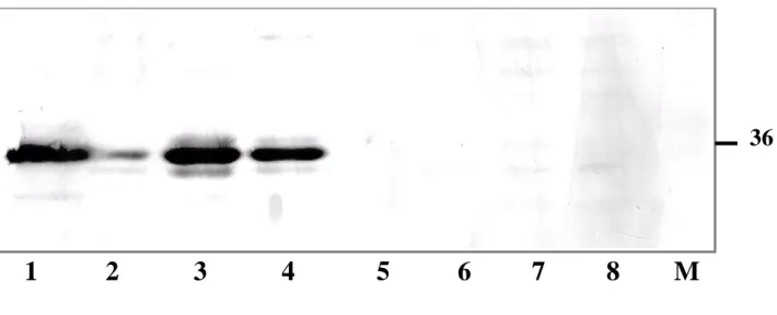
Annexin II is over-expressed in most cancers and cell lines derived from such cancer tissues. However, annexin II
expression is lost in prostate cancers. Hence, we examined a panel of established human prostate cancer cell lines to find an annexin II-null cell line that would serve as a use-ful model for studying the physiological role of annexin II. Figure 3 shows a western blot analysis using whole cell lysates from a panel of prostate cancer cell lines, including both androgen-responsive (LNCaP) and androgen-unre-sponsive (PC-3 and DU-145) cell lines, and cell lines used as positive controls (NIH 3T3 and SkBr-3). High levels of annexin II is expressed in PC-3, DU-145, NIH 3T3 and SkBr-3 cells, but no annexin II is detected in LNCaP cells of different passage numbers tested. LNCaP cells that are androgen-responsive progress to androgen-unresponsive upon continuous passages under normal growth
condi-Phosphorylation of nuclear annexin II and cell cycle distribution Figure 2
Phosphorylation of nuclear annexin II and cell cycle distribution. Human K562 cells were subjected to centrifugal elu-triation followed by flow cytometry to determine the cell cycle distribution of phosphorylated annexin II. Cells enriched in each indicated phase were fractionated to cytosolic extract (CE) and nuclear extract (NE). Twenty µg protein from each sam-ple were subjected to SDS-PAGE followed by immunoblotting with anti-annexin II antibody. Immunoblot of PGK was used as an internal control. The flow cytometric profile of each fraction is presented in the upper panels and the percentage of cells in each cell cycle phase is indicated below the immunoblot under each fraction.
30KD
AnxII-P
PGK
Marker
CE NE CE NE CE NE CE NE CE NE CE NE
Log G1 G1/S S S/G2 G2/M
AnxII
G1 40 92 45 6
3 10
S 46 5 43 93
44 4
G2/M 14 3 11 2 53 86
Log
G2/M
S
G1
G1/S
S/G2
tions [46]. We analyzed if annexin II expression is related to androgen responsiveness. Annexin II expression was not observed in androgen-responsive and androgen-unre-sponsive LNCaP cells, either in the presence or absence of dihydrotestosterone (DHT) in the growth medium (data not shown). Northern blot (Figure 4) analysis was per-formed to determine if the loss of annexin II expression occurs at the transcriptional or at the translational level. Figure 4 shows that annexin II message is expressed at a high level in PC-3 and DU-145 cells. The density of cells growing in monolayer can also influence the expression of genes [47], so the LNCaP cells were seeded in either high or low density and the expression of annexin II gene was observed. No annexin II expression was detected in
LNCaP cells plated at either high or low density with or without treatment with DHT. Our data indicate that annexin II expression is lost at the transcriptional level in LNCaP cells.
Thus, LNCaP cells, which are annexin II-null, serve as use-ful recipient cells to investigate the function of exoge-nously expressed annexin II in the absence of background from endogenous annexin II.
Export of annexin II from the nucleus involves the CRM1 pathway
To confirm whether the annexin II-null LNCaP cells regu-late NES containing proteins by the CRM1 mediated
Immunoblot analysis of annexin II in prostate cancer cells Figure 3
Immunoblot analysis of annexin II in prostate cancer cells. Cell lysates of NIH3T3 (lane 1), SkBr-3 (lane 2), DU-145 (lane 3), PC-3 (lane 4) and LNCaP (passage 181, lane 5; passage 142, lane 6; passage 92, lane 7; passage 34, lane 8) were sub-jected to western blot analysis with mouse anti-human monoclonal anti-annexin II antibody as described in materials and meth-ods. Lane 9 is the protein molecular markers used for SDS-PAGE. The position of annexin II band is indicated on the right side of the panel.
1 2 3 4 5 6
7 8 M
nuclear export pathway, we transfected LNCaP cells with pEGFP-C1 vector, pEGFP-C1-AnxII or pEGFP-C1-AnxII/ L10AL12A, which encode GFP alone, GFP fused to N-ter-minus of wild-type annexin II (GFP-WT), and GFP fused to N-terminus of NES mutant annexin II (GFP-NES), respectively. Confocal microscopy images of transiently transfected LNCaP cells are shown in Figure 5. The pres-ence of GFP was detected by auto-fluorescpres-ence of GFP (green). Immunostaining of annexin II was performed with secondary antibody conjugated to rhodamine (red). Colocalization of GFP and annexin II results in the appearance of yellow fluorescence. Figure 5 shows that both GFP and GFP-fused NES mutant annexin II distrib-ute equally throughout the cell, whereas the GFP fused
wild-type annexin II is observed only in the cytoplasm, indicating that the NES of wild type annexin II is still func-tional. The cellular distribution of wild-type annexin II without GFP is identical to the GFP fused wild-type annexin II (data not shown) indicating no effect of GFP fusion on the cellular distribution of annexin II. Mutation of the conserved leucines (L10 and L12) of the annexin II NES results in retention of annexin II in the nucleus.
We established stable clones of LNCaP cells expressing GFP alone, wild-type annexin II or NES mutant annexin II. In these stable clones, we examined the distribution of annexin II by fractionating the cells and preparing cytosolic and nuclear extracts. These extracts were
sub-Northern blot analysis of annexin II in prostate cancer cells Figure 4
Northern blot analysis of annexin II in prostate cancer cells. Prior to RNA extraction, LNCaP cells of both low level and high level of confluence were treated with 10 nM dihydrotestosterone (DHT) for 2 days. 20 µg RNA were extracted from PC-3 cells (lane 1), DU-145 cells (lane 2) and LNCaP cells treated with (lanes 3, 6) or without (lanes 4, 5) DHT. [32P]-labeled
annexin II cDNA was used as probe (Panel A). The location of full length annexin II cDNA is indicated by an arrow on the left side of the panel. The membrane from panel A was probed with [32P]-labeled GAPDH cDNA for normalization of the annexin
II level (Panel B).
A
B
Annexin II
GAPDH
1 2 3 4 5 6
+ -
-
+
low high low high
PC-3 DU-145 LNCaP
Density
DHT
jected to western blot analysis for annexin II. Figure 6 shows the distribution of GFP, GFP-WT and GFP-NES in LNCaP cells. As expected, there is no annexin II expressed in the LNCaP cell transfected with vector alone, and also there is no endogenous annexin II expressed in LNCaP cells (Figure 6A, lanes 1 and 2). However, in GFP-WT, cytosolic fraction contains a majority of annexin II (Figure 6A, lanes 3 and 4). GFP-NES was found to distribute equally between the cytoplasmic and nuclear extracts (Fig-ure 6, panel A, lanes 5 and 6). In the nuclear extract, the unphosphorylated and phosphorylated annexin II are observed. These results are in agreement with data from K562 cells (Figure 2), as well as previously published lit-erature [48]. We determined that the slower mobility
band in the nuclear extract is phosphorylated by subject-ing the nuclear extract to dephosphorylation with potato acid phosphatase. Figure 6 (panel C) shows that upon treatment with the phosphatase, the slower mobility band is shifted to the position corresponding to annexin II in the cytosolic extracts. These results are consistent with our observations in K562 cells (Figure 1). We performed immunoblot analysis of PGK to ensure that nuclear extracts were devoid of cytosolic contamination (Figure 6, panel B).
Localization of wild type and NES mutant annexin II in transiently transfected LNCaP cells Figure 5
Localization of wild type and NES mutant annexin II in transiently transfected LNCaP cells. Transiently trans-fected LNCaP cells expressing GFP alone, GFP-wild type (GFP-WT) annexin II or GFP-NES mutant (GFP-NES) annexin II were fixed and subjected to immunocytochemistry with anti-annexin II antibody as described in materials and methods. The images were scanned using laser scanning confocal microscope (LSCM). The green color represents auto-fluorescence of GFP, the red color represents the rhodamine staining of annexin II antigen-antibody complex, and the yellow color is the overlay of GFP and annexin II images. Bar: 25 µm.
GFP anti-Annexin II Merge
GFP
GFP-WT
Stable expression of NES-Annexin II retards LNCaP cell proliferation
Because nuclear retention of annexin II is cell cycle dependent, this protein may play a role in regulating cell cycle progression and cell proliferation. We examined the effect of exogenous expression of wild type and NES mutant annexin II on the growth of LNCaP cells. Reex-pression of annexin II results in significant morphological changes to the cells (Figure 7). The vector-alone transfec-tion does not result in any morphological changes. The vector-alone transfection does not result in any morpho-logical changes. However, expression of wild-type annexin II or NES mutant results in the cells taking on a mesenchymal phenotype. To eliminate clonal variation,
which may occur in stably transfected cancer cells, and the possibility that NES mutant itself is affecting proliferation other than nuclear localization, we examined different clones or NES-transfected LNCaP cells. Data from two such clones (NES-1 and NES-4) are shown in Figure 8. The vector transfected cells and wild type annexin II trans-fected cells underwent cell doubling with a doubling time of approximately 34 hours as compared to 30 hours for the untransfected LNCaP cells, whereas the NES-mutant cells had a doubling time of 48 hours. The inhibition of cell proliferation by NES mutant annexin II suggests that abnormal accumulation of annexin II in the nucleus may lead to deregulation or inhibition of DNA replication. We compared the cell cycle distribution of logarithmically
Distribution of GFP and GFP-fused wild type or NES mutant annexin II in LNCaP cells Figure 6
Distribution of GFP and GFP-fused wild type or NES mutant annexin II in LNCaP cells. LNCaP cells expressing GFP, GFP-wild type annexin II (GFP-WT), and GFP-NES mutant annexin II (GFP-NES) were fractionated into cytosolic extract (CE) and nuclear extract (NE). Each extract was subjected to SDS-PAGE and immunoblot with GFP (Panel A) and anti-PGK antibody (panel B). Cytosolic and nuclear extracts from the GFP-NES cells were subjected to potato acid phosphatase (PACP) treatment as described in materials and methods, and an immunoblot analysis was performed for annexin II (Panel C).
GFP GFP-WT GFP-NES
GFP-WT Annexin II
GFP
PGK
CE NE CE NE CE NE
A
B
C
GFP-NES mutant annexin II
PACP -
+ -
+
CE CE NE NE
GFP-NES
growing LNCaP cells and the three transfectants using flow cytometry, and observed no changes in the parental and transfected cells in the distribution of cells in different cell cycle phases (data not shown). This observation indi-cated that nuclear retention of NES-mutant annexin II does not inhibit the growth of LNCaP cells by blocking the cells at any certain phase of the cell cycle. These obser-vations are similar to the finding that the expression of human Cdc6 mutant, HsCdc6E1E2E3, which is exclu-sively nuclear, inhibits initiation of DNA replication [49].
Site-directed mutagenesis of serines 11 and 25 to glutamic acid prevents nuclear entry of annexin II
We examined the role of phosphorylation in the nuclear entry and export of annexin II. Initial experiments with exposure of cells to staurosporine, a protein kinase inhib-itor, in combination with LMB showed no effect of stau-rosporine on nuclear accumulation of annexin II (data not shown), indicating that phosphorylation is not required for annexin II to enter the cell nucleus. To more specifically address the role of phosphorylation in nuclear entry and export of annexin II, we examined the effect of site-directed changes to known phosphoamino acids. Because serines 11 and 25 are known to be phosphor-ylated by protein kinase C [50,51], these residues were mutated to glutamic acid to mimic the negative charge of a phosphate group, or to alanine to mimic an unphospho-rylated state. The mutants were constructed as GFP-fusion proteins and included single mutants (S11A, S11E, S25A, S25E) or in combination (S11AS25A, S11AS25E, S11ES25A, S11ES25E). Transfection of LNCaP cells with these mutants showed no effect on nuclear entry or CRM1-mediated inhibition of nuclear export in all of the mutants except the S11ES25E double mutant. Cells har-boring this double mutation displayed inhibition of annexin II entry to the nucleus (Figure 9), indicating the important regulatory role of the serines at 11 and 25 in the cellular distribution of annexin II. We find annexin II in the nucleus as a phosphoprotein in a cell cycle-dependent manner (Figures 1 and 2), so we conclude that serine 11 and 25 are not the phosphorylation sites involved in nuclear retention of annexin II, and phosphorylation of other residues may be important in nuclear entry or export of annexin II. The contribution of other phos-phoamino acid residues in annexin II to this regulatory function needs further investigation.
Discussion
Phosphorylation of annexin II can regulate its multiple functions in the cell. In the N-terminus of annexin II there are multiple phosphorylation sites suggesting that phos-phorylation of these residues may regulate annexin II physiological functions. When primary cultured chromaffin cells are stimulated by nicotine, annexin II is phosphorylated by protein kinase C at Ser-25 and
phosphorylation at this residue appears to be a prerequi-site for the stimulation of Ca2+-dependent secretion by the
annexin II-p11 complex observed in permeabilized chro-maffin cells [52], indicating that phosphorylation is involved in regulating the exocytotic function of annexin II. In resting chromaffin cells, annexin II is in monomeric form and predominantly exists in cytosol. However, when the cells are stimulated by nicotine, annexin II forms het-erotetramer with p11 and restrict its location to the mem-brane-associated cytoskeleton [53]. The regulatory role of phosphorylation at tyrosine 23 is less well understood. It was shown that Tyr-23 can be phosphorylated by pp60src
[17] and the insulin receptor [54], suggesting its role in growth regulation. Although there is no evidence shown for the function of Tyr-23 phosphorylation in exocytosis, we predict that it may not account for the membrane related processes since Tyr-23 phosphorylation inhibits the formation of annexin II heterotetramer with p11 [18]. Despite the studies mentioned above regarding the role of annexin II phosphorylation on its membrane associated functions, there is no information on the role of phosphorylation on the nuclear function of annexin II. Annexin II was identified to be in a multiprotein complex with DNA polymerase α-primase [11]. We have previ-ously demonstrated that annexin II plays a role in DNA synthesis and cell proliferation. Antisense oligonucle-otides to annexin II reduce DNA synthesis in HeLa cells and retard progression of cells through the cell cycle [13]. Immunodepletion of annexin II from Xenopus egg extract results in loss of DNA replication [12]. Replication extracts made from cells expressing antisense-annexin II in a regulatable vector do not support SV40 replication in
vitro [14]. Annexin II expression is regulated in the mam-malian cell cycle [44], and its levels are enhanced in many cancers [38].
DNA replication is regulated at the level of initiation [55]. The initiation of DNA replication is mediated by a com-mon set of protein factors and some of these factors are activated by phosphorylation catalyzed by cyclin-depend-ent kinase (CDKs) and the Cdc 7 family of protein kinases [55]. Although the specific substrates of phosphorylation have not been identified with certainty, the phosphoryla-tion pattern is definitely cell cycle dependent. Another important mechanism that regulates cell cycle transition is nuclear protein export [56–58]. For example, CDC6 pro-tein, which is essential for initiation of DNA replication, contains a NES involved in exporting phosphorylated forms of CDC6 in a cell cycle-dependent manner [49,59]. Cyclin B is exported from the nucleus and degraded when cells exit mitosis. The N-terminal peptide of annexin II contains both Ser/Thr phosphorylation sites and a Tyr phosphorylation site, and these sites are in proximity to the nuclear export signal (NES). Thus, the activity of annexin II in the nucleus may be regulated not only by its
expression levels but also by phosphorylation and nuclear export. In this study, we have demonstrated that nuclear annexin II is phosphorylated. Interestingly, after CIAP treatment, nuclear annexin II still migrates slower than cytosolic annexin II. This observation suggests that nuclear annexin II may be phosphorylated differently from that in the cytosol, and nuclear and cytosolic annexin II may be regulated through different pathways.
Our data also show that the phosphorylation and nuclear retention of annexin II is cell cycle-dependent. In G1 phase of the cell cycle, annexin II is phosphorylated and accumulates in the nucleus, whereas in S phase annexin II is not detected in the nucleus. These findings are consist-ent with previous reports [44,48] and similar to the obser-vations that the human CDC6 homolog, HsCDC6,
accumulates in the nucleus in G1 phase, but in S phase it is phosphorylated and exported from the nucleus [49]. This similarity indicates that phosphorylation may play a role in the regulation of nuclear entry and export of annexin II. However, another possibility is that annexin II may interact with one or more partner proteins and get transported into the nucleus in a cell-cycle dependent manner, and the phosphorylation may be just a require-ment for its function in the nucleus. Unlike HsCDC6, which contains both NLS and NES, annexin II contains no NLS that can mediate its entry into the nucleus, so it is possible that its nuclear entry is through interaction with a partner protein containing NLS. Our data shows that annexin II NES mutant is found distributed equally between the cytosol and the nucleus. Thus, the nuclear export pathway for annexin II appears to be more active
Morphology of LNCaP cells transfected with wild type annexin II and NES-annexin II Figure 7
Morphology of LNCaP cells transfected with wild type annexin II and NES-annexin II. Untransfected LNCaP and stable clones of vector-alone, GFP-WT and GFP-NES cells were cultured as described in materials and methods. Light micros-copy images of each of the cultures are shown.
GFP-NES
GFP
than the nuclear entry. We have shown that annexin II is exported out of the nucleus by the CRM1 dependent export pathway.
Ectopic expression of NES mutant annexin II results in a drastic reduction in the proliferation of LNCaP cells with an associated increase in cell doubling time. This reduction in cell proliferation may be due to abnormal presence of annexin II during the S phase of the cell cycle where it is usually not present (Figure 2). On the other hand, it is possible that LNCaP cells lack a binding partner for annexin II in the cell nucleus and this results in annexin II becoming a negative regulator of cell prolifera-tion in the prostate cells. LNCaP cells expressing either the
wild-type or the NES mutant change to a more flattened and elongated morphology, indicating that annexin II may cause stabilization of the cytoskeleton. Several lines of evidence suggest that annexin II may be involved in cytoskeleton regulation. First, annexin II has been shown to be associated with the cytoskeleton [60]. Second, it has been suggested that intracellular annexin II heterote-tramer acts as a link between the cytoskeleton and the plasma membrane, although the physiological role of this proposed interaction are unknown [61]. Third, both annexin II monomer and tetramer can bind F-actin in
vitro, and annexin II tetramer can bundle F-actin [62]. It is possible that these annexin II molecules may form com-plexes with p11 underneath plasma membrane and bind
Accumulation of annexin II in the nucleus retards cell proliferation Figure 8
Accumulation of annexin II in the nucleus retards cell proliferation. Parental LNCaP cells and stable clones of vector-alone (GFP), GFP-WT or GFP-NES mutant annexin II were cultured as described in materials and methods for up to 8 days. Two different GFP-NES clones (NES-1 and NES-4) were included. The cell numbers on the indicated days were obtained from a standard curve (data not shown) according to the OD570–690 values. The experiment was done in triplicate and the data were analyzed using GraphPad Prism 3.0. Cell doubling time was determined for each culture, and the values were: LNCaP (30.32 ± 2.1 hours), GFP (33.8 ± 4.6 hours), GFP-WT (34.5 ± 5.1 hours) and GFP-NES (48.1 ± 8.3 hours).
2
4
6
8
0
1000
2000
WT
Vector
NES-1
NES-4
LNCaP
day
Ce
ll
Nu
mbe
r(
X
1
0
3
)
to cytoskeletal molecules and the annexin II heterote-tramer then stabilize cytoskeleton, which results in the morphology change of LNCaP cells. The stabilization of cytoskeleton also accounts for inhibition of cell migra-tion. Our results are also consistent with recent finding showing that re-expression of annexin II inhibits prostate cancer cell migration [63]. Epithelial mesenchymal transi-tion (EMT) has been estimated to occur in as much as 18% of breast tumors in vivo and the EMT generally depicts a more aggressive behavior of the tumor cells [64]. The functional consequence of mesenchymal appearance of annexin II-transfected cells need to be investigated further.
The annexin II NES overlaps the interaction site for p11. Binding of p11 targets annexin II to the cell membrane, and p11 is not localized to the nucleus. Thus, the heterote-tramer formed by annexin II and p11 is exclusively cytosolic and has no role in the nucleus. Previous studies of other proteins containing NES and NLS motifs show that the proteins are shuttling between cytoplasm and nucleus until NES and/or NLS is masked either by phos-phorylation, association with other proteins, or other modification [65,66]. Phosphorylation of annexin II may confer a conformational change resulting in lack of recog-nition by the CRM1-mediated nuclear export pathway. Alternatively, phosphorylated annexin II can interact with additional protein factors in the nucleus, and this interac-tion masks annexin II NES preventing its nuclear export.
Site-directed change of ser11 and ser25 to glutamic acid prevents nuclear entry of annexin II Figure 9
Site-directed change of ser11 and ser25 to glutamic acid prevents nuclear entry of annexin II. GFP-fused annexin II containing Ser to Ala or Ser to Glu either singly or in combination were generated as described in materials and methods. Transient transfection of LNCaP cells was performed followed by treatment with or without LMB. Confocal microscopic observations were made and representative images are shown. Bar: 10 µm.
-LMB
+LMB
The three known phosphorylation sites in the N-terminus of annexin II, Ser11, Ser25, and Tyr23, were identified from studies of the heterotetrameric form of annexin II. The exact phosphorylation sites of the nuclear monomer of annexin II are not known, and it is possible that other potential sites are phosphorylated in the nuclear annexin II. In this study, we have shown that phosphorylation of both Ser11 and Ser 25 prevents annexin II entry to the nucleus. Annexin II phosphorylated in its Ser-11 and 25 cannot form a heterotetramer with p11 [16]. However, diphosphorylated form of annexin II exists as homodimer instead of as monomer [16]. The formation of annexin II homodimer devoid of p11 suggests that the phosphoryla-tion of the heavy chains could induce a head-to-head association [16]. This kind of dimerization has also been observed on a monomeric annexin I in placenta [67] and brain [68]. Thus, this head-to-head dimerization may account for the inhibition of nuclear entry of the diphos-phorylated form of annexin II. From these results, we also conclude that ser11 and ser25 residues are not involved in the regulation of nuclear retention of annexin II.
Conclusions
Data from our studies show that annexin II localized in the cell nucleus is phosphorylated in a cell cycle-depend-ent manner. The cycle-depend-entry of annexin II into the nucleus is prevented by phosphorylation of serines 11 and 25. Stable expression of the NES mutant annexin II results in reduced cell proliferation of LNCaP cells. These results indicate an important role for nuclear annexin II in cell proliferation and the regulation of nucleo-cytoplasmic shuttling of annexin II.
Methods
Cell lines and culture conditions
LNCaP cells were cultured in RPMI 1640 containing 7% fetal bovine serum (FBS) (Gibco-BRL, Rockville, MD), 100 µg/ml penicillin, and 100 µg/ml streptomycin at 37° in a 5% CO2 cell culture incubator. Plasmids that express green fluorescence protein (GFP) alone (pEGFP-C1), GFP-fused wild type annexin II (pEGFP-C1-wild type annexin II), or GFP-fused NES mutant annexin II with lysine 10 and 12 mutated to alanine (pEGFP-C1-AnxII L10A/L12A) were transfected into LNCaP cells using Lipo-fectamine Plus (Gibco-BRL). Stably transfected cell popu-lations were generated by culturing cells in the presence of 0.2 mg/ml G418 and subsequent subcloning. K562 cells were grown in suspension in RPMI 1640 containing 10% FBS, 2 mM L-glutamine, 100 µg/ml penicillin, and 100 µg/ml streptomycin.
Cell fractionation
K562 cell pellets were rinsed twice with Hank's balanced salt solution (HBSS) and once with hypotonic buffer (10 mM Tris-HCl, pH 7.5, 10 mM NaCl and 1 mM MgCl2).
Cells were resuspended and held in hypotonic buffer for 2 hours at 4°C. Cells were disrupted with a Dounce homogenizer, and disruption was monitored by light microscopy. No intact cells were observed by microscopy after homogenization. The homogenate was subjected to centrifugation in a Sorvall SS-34 rotor at 2,000 revs/min for 30 minutes at 4°C. The supernatant was designated as cytoplasmic extract (CE). The nuclear pellet was further rinsed three times with the hypotonic buffer and the supernatant from each wash was added to the cytoplasmic extract. The nuclei were resuspended in 0.4 M potassium phosphate, pH 7.2, 1 mM EDTA, 1 mM dithiothreitol (DTT) and 10% glycerol and extracted for 1 hour at 4°C. Following centrifugation in a Sorvall SS-34 rotor at 10,000 g for 30 minutes at 4°C, the supernatant was collected as nuclear extract (NE). The cytoplasmic and nuclear extracts were dialyzed against 50 mM potassium phosphate, pH 7.2, 1 mM DTT, 1 mM EDTA and 10% glycerol, and stored at minus 80°C until further use. LNCaP cell transfectants grown in 80% confluency were harvested, and the cytosolic extract (CE) and nuclear extract (NE) were obtained using NE-PER Nuclear and Cytoplasmic Extraction Reagents (PIERCE, Rockford, IL).
Electrophoresis and Immunoblot analyses
Protein extracts were prepared from indicated cells. The total amount of protein extracts was quantitated using Bio-Rad BCA protein assay (Pierce). Indicated amount of protein from each extract was separated by 12% SDS-PAGE. After electrophoresis, protein was transferred to polyvinylidene fluoride (PVDF) membranes (Bio-Rad Hercules CA). The membranes were blocked in 1X Tris Buffered Saline with 0.01% Tween 20 (1XTTBS) with 7% powdered milk for one hour at room temperature then probed with monoclonal anti-human annexin II at 1:5000 dilution (BD Transduction Laboratories) and pol-yclonal anti-human PGK at 1:2000 dilution overnight at 4°C. Appropriate secondary antibodies conjugated to horseradish peroxidase (HRP) were used to detect anti-gen-antibody complexes. Membranes were developed using ECL plus (Amersham Pharmacia Biotech, UK) and exposed to films for appropriate amounts of time.
Phosphatase treatment of cytosolic and nuclear extract of LNCaP cells expressing GFP-NES mutant annexin II
LNCaP cells expressing GFP-NES were fractionated into cytosolic and nuclear extracts using NE-PER nuclear and cytosolic extraction reagents (PIERCE, Rockford IL). The extracts were concentrated with Centricon-10 centrifugal concentrator (Millipore Co.), and 10 µg protein was treated with 0.5 units of potato acid phosphatase (Sigma Chemical Co.) for 30 minutes at 37°C in 50 mM PIPES pH 6.0 buffer. The reaction was terminated by adding SDS-PAGE sample buffer and both the untreated and treated extracts were subjected to SDS-PAGE.
Northern blot
Total RNA was extracted using TRIzol standard protocols. A total of 20 µg RNA was used in northern blot experi-ments. Annexin II probe was prepared from the vector pGAF5-1 by the random primer method of labeling with
32P greater than 1 × 10⁸ cpm/µg. Hybridization of the
probe to the membrane was carried out at 42°C overnight in a water bath. The membrane was stripped and re-probed with glyceraldehyde-3-phosphate dehydrogenase (GAPDH) probe and its expression was used as a control to normalize data for annexin II levels.
Immunocytochemistry
LNCaP cells seeded on the glass coverslips were grown to 80% confluency then fixed for 30 minutes in 4% parafor-maldehyde in phosphate buffered saline (PBS) and permeabilized in methanol for 10 minutes. After several rinses with PBS, cells were pretreated with 10% bovine serum albumin (BSA) in PBS for 3 hours at room temper-ature to reduce nonspecific staining. Cells were incubated with monoclonal human annexin II primary anti-body (BD Transduction Laboratories) at 1:5000 dilution in PBS containing 3% BSA for 2 hours at room tempera-ture, washed, and subsequently incubated for 2 hours with biotinylated secondary antibody (Vector Laborato-ries, Inc. Burlingame, CA) diluted 1:500 followed by staining with rhodamine-avidin D (Vector Laboratories Inc. Burlingame, CA) diluted 1:2000 for 30 minutes. The coverslips were washed extensively with PBS and mounted in VECTASHIELD mounting medium for fluo-rescence (Vector Laboratories Inc. Burlingame, CA). Cells were visualized on a Zeiss confocal microscope.
Leptomycin B (LMB) treatment of LNCaP cells
LNCaP cells expressing GFP or GFP-wild type annexin II were grown to 80% confluence on the surface of round glass coverslips placed in 12-well plates. The cells were then treated with 20 nM LMB for 2 hours. The cells were fixed with 4% paraformaldehyde in 1X PBS and observed under confocal microscopy.
Cell proliferation assay
Cell proliferation was measured using 3-(4,5-cimethylthi-azol-2-yl)-2,5-diphenyl tetrazolium bromide (MTT) cell proliferation assay. MTT was dissolved in serum-free and phenol red-free RPMI 1640 at a concentration of 1 mg/ml. A total of 30,000 cells were seeded into 6-well plates. On days 2, 4, 6 and 8, cells were incubated with MTT solution for 3 hours in a CO2 incubator at 37°C. The MTT reagent was removed and placed in 2.0 ml microcentrifuge tubes and centrifuged at 14,000 × g for 5 minutes to pellet and collect any detached cells. The formazan dye was dis-solved by adding 2.0 ml of 99% isopropanol to each sam-ple, and the absorbance was read at 570–690 nm using spectrophotometer. For the standard curve, 20,000 to
140,000 cells were seeded in 6-well plates, and after 24 hours, the cells were treated with MTT and processed as described above. Data were plotted using Prizm 3.0 (GraphPad Software, San Diego, CA).
Construction of GFP-fused site directed mutants of annexin II
GFP fusion proteins with annexin II containing mutations in the Ser11 and Ser25 were constructed by inserting mutant annexin II cDNA into the pEGFP-C1 vector. Ser to Ala and Ser to Glu mutants were generated as previously reported [43]. Annexin II cDNA was generated by PCR with primers containing appropriate restriction sites and inserted into the corresponding restriction sites of the pEGFP-C1 vector. GFP-fusion constructs of single mutants (S11A, S25A, S11E, S25E) and double mutants (S11AS25A, S11AS25E, S11ES25A, S11ES25E) were gen-erated for use in our experiments.
Authors' contributions
JL carried out the phosphorylation experiments, con-structed the GFP-annexin II phosphorylation mutants, conducted the proliferation experiments and drafted the manuscript. CAR performed the transfections. JAS con-structed the annexin II phosphorylation mutants. JKV conceived of the study, and participated in its design and coordination. All authors read and approved the final manuscript.
Acknowledgements
We thank Dr. David A. Eberhard (University of Virginia Health Science Center) for the gift of wild type and NES mutant annexin II vectors and Dr. Minoru Yoshida (University of Tokyo) for the gift of Leptomycin B. We thank the Confocal Microscopy Facility for assistance in our work. This study was supported in part by grants from the Gustavus and Louise Pfeiffer Foundation, Phillip Morris Incorporated and Nebraska Cancer and Smoking Disease Research Program (2003–30).
References
1. Geisow MJ, Walker JH, Boustead C and Taylor W: Annexins--new
family of Ca2+-regulated-phospholipid binding protein. Biosci
Rep 1987, 7:289-298.
2. Gerke V and Weber K: Identity of p36K phosphorylated upon
Rous sarcoma virus transformation with a protein purified from brush borders; calcium-dependent binding to non-erythroid spectrin and F-actin. EMBO J 1984, 3:227-233.
3. Thiel C, Osborn M and Gerke V: The tight association of the
tyrosine kinase substrate annexin II with the submembra-nous cytoskeleton depends on intact p11- and Ca(2+)-bind-ing sites. J Cell Sci 1992, 103 ( Pt 3):733-742.
4. Ali SM, Geisow MJ and Burgoyne RD: A role for calpactin in
cal-cium-dependent exocytosis in adrenal chromaffin cells.
Nature 1989, 340:313-315.
5. Creutz CE: The annexins and exocytosis. Science 1992,
258:924-931.
6. Sarafian T, Pradel LA, Henry JP, Aunis D and Bader MF: The
partic-ipation of annexin II (calpactin I) in calcium-evoked exocyto-sis requires protein kinase C. J Cell Biol 1991, 114:1135-1147.
7. Chasserot-Golaz S, Vitale N, Sagot I, Delouche B, Dirrig S, Pradel LA, Henry JP, Aunis D and Bader MF: Annexin II in exocytosis:
cate-cholamine secretion requires the translocation of p36 to the subplasmalemmal region in chromaffin cells. J Cell Biol 1996, 133:1217-1236.
8. Munz B, Gerke V, Gillitzer R and Werner S: Differential
expres-sion of the calpactin I subunits annexin II and p11 in cultured keratinocytes and during wound repair. J Invest Dermatol 1997, 108:307-312.
9. Zokas L and Glenney-JR Jr: The calpactin light chain is tightly
linked to the cytoskeletal form of calpactin I: studies using monoclonal antibodies to calpactin subunits. J Cell Biol 1987, 105:2111-2121.
10. Jindal HK and Vishwanatha JK: Purification and characterization
of primer recogniton proteins from HeLa cells. Biochemistry
1990, 29:4767-4773.
11. Jindal HK, Chaney WG, Anderson CW, Davis RG and Vishwanatha JK: The protein-tyrosine kinase substrate, calpactin I heavy
chain (p36), is part of the primer recognition protein com-plex that interacts with DNA polymerase a. Journal of Biological
Chemistry 1991, 266:5169-5176.
12. Vishwanatha JK and Kumble S: Involvement of annexin II in DNA
replication: evidence from cell-free extracts of Xenopus eggs. Journal of Cell Science 1993, 105:533-540.
13. Kumble KD, Iversen PL and Vishwanatha JK: The role of primer
recognition proteins in DNA replication: Inhibition of cellu-lar proliferation by antisense oligodeoxyribonucleotides.
Jour-nal of Cell Science 1992, 101:35-41.
14. Chiang Y, Rizzino A, Sibenaller ZA, Wold MS and Vishwanatha JK:
Specific down-regulation of annexin II expression in human cells interferes with cell proliferation. Mol Cell Biochem 1999, 199:139-147.
15. Johnsson N, Nguyen Van P., Soling HD and Weber K: Functionally
distinct serine phosphorylation sites of p36, the cellular sub-strate of retroviral protein kinase; differential inhibition of reassociation with p11. EMBO J 1986, 5:3455-3460.
16. Regnouf F, Sagot I, Delouche B, Devilliers G, Cartaud J, Henry JP and Pradel LA: "In vitro " phosphorylation of annexin 2
heterote-tramer by protein kinase C - Comparative properties of the unphosphorylated and phosphorylated annexin 2 on the aggregation and fusion of chromaffin granule membranes.
Journal of Biological Chemistry 1995, 270:27143-27150.
17. Glenney J.R.,Jr. and Tack BF: Amino-terminal sequence of p36
and associated p10: identification of the site of tyrosine phos-phorylation and homology with S-100. Proc Natl Acad Sci U S A
1985, 82:7884-7888.
18. Powell MA and Glenney JR: Regulation of calpactin I
phospholi-ped binding by calpactin I light-chain binding and phosphor-ylation by p60v-src. Biochemistry Journal 1987, 247:321-328.
19. Johnstone SA, Hubaishy I and Waisman DM: Phosphorylation of
annexin II tetramer by protein kinase C inhibits aggregation of lipid vesicles by the protein. Journal of Biological Chemistry 1992, 267:25976-25981.
20. Chetcuti A, Margan SH, Russell P, Mann S, Millar DS, Clark SJ, Rogers J, Handelsman DJ and Dong Q: Loss of annexin ii heavy and light
chains in prostate cancer and its precursors. Cancer Res 2001, 61:6331-6334.
21. Andronicos NM and Ranson M: The topology of plasminogen
binding and activation on the surface of human breast cancer cells. Br J Cancer 2001, 85:909-916.
22. Brichory FM, Misek DE, Yim AM, Krause MC, Giordano TJ, Beer DG and Hanash SM: An immune response manifested by the
com-mon occurrence of annexins I and II autoantibodies and high circulating levels of IL-6 in lung cancer. Proc Natl Acad Sci U S A
2001, 98:9824-9829.
23. Emoto K, Yamada Y, Sawada H, Fujimoto H, Ueno M, Takayama T, Kamada K, Naito A, Hirao S and Nakajima Y: Annexin II
overex-pression correlates with stromal tenascin-C overexoverex-pression: a prognostic marker in colorectal carcinoma. Cancer 2001, 92:1419-1426.
24. Choi S, Kobayashi M, Wang J, Habelhah H, Okada F, Hamada J, Mori-uchi T, Totsuka Y and Hosokawa M: Activated leukocyte cell
adhesion molecule (ALCAM) and annexin II are involved in the metastatic progression of tumor cells after chemother-apy with Adriamycin. Clin Exp Metastasis 2000, 18:45-50.
25. Kaczan-Bourgois D, Salles JP and Chap H: Expression of annexin II
and associated p11 protein by differentiated choriocarci-noma Jar cells. Am J Obstet Gynecol 1999, 181:1273.
26. Menell JS, Cesarman GM, Jacovina AT, McLaughlin MA, Lev EA and Hajjar KA: Annexin II and bleeding in acute promyelocytic
leukemia. N Engl J Med 1999, 340:994-1004.
27. Balch C and Dedman JR: Annexins II and V inhibit cell
migration. Exp Cell Res 1997, 237:259-263.
28. Blanchard S, Barwise JL, Gerke V, Goodall A, Vaughan PF and Walker JH: Annexins in the human neuroblastoma SH-SY5Y:
dem-onstration of relocation of annexins II and V to membranes in response to elevation of intracellular calcium by mem-brane depolarisation and by the calcium ionophore A23187.
J Neurochem 1996, 67:805-813.
29. Mohiti J, Walker JH and Caswell AM: Studies on annexins in
pri-mary cultures of human osteoblasts and in the human oste-osarcoma cell line MG-63. Biochem Soc Trans 1995, 23:36S.
30. Roseman BJ, Bollen A, Hsu J, Lamborn K and Israel MA: Annexin II
marks astrocytic brain tumors of high histologic grade. Oncol
Res 1994, 6:561-567.
31. Tressler RJ, Updyke TV, Yeatman T and Nicolson GL: Extracellular
annexin II is associated with divalent cation- dependent tumor cell-endothelial cell adhesion of metastatic RAW117 large-cell lymphoma cells. Journal of Cellular Biochemistry 1993, 53:265-276.
32. Ozaki T and Sakiyama S: Molecular cloning of rat calpactin I
heavy-chain cDNA whose expression is induced in v-src-transformed rat culture cell lines. Oncogene 1993, 8:1707-1710.
33. Fox MT, Prentice DA and Hughes JP: Increases in p11 and annexin
II proteins correlate with differentiation in the PC12 pheo-chromocytoma. Biochemical and Biophysical Research Communications 1991, 177:1188-1193.
34. Gress TM, Wallrapp C, Frohme M, Muller Pillasch F., Lacher U, Friess H, Buchler M, Adler G and Hoheisel JD: Identification of genes
with specific expression in pancreatic cancer by cDNA rep-resentational difference analysis. Genes Chromosomes Cancer
1997, 19:97-103.
35. Manda R, Kohno T, Matsuno Y, Takenoshita S, Kuwano H and Yokota J: Identification of genes (SPON2 and C20orf2) differentially
expressed between cancerous and noncancerous lung cells by mRNA differential display. Genomics 1999, 61:5-14.
36. Nygaard SJ, Haugland HK, Kristoffersen EK, Lund Johansen M., Laerum OD and Tysnes OB: Expression of annexin II in glioma
cell lines and in brain tumor biopsies. J Neurooncol 1998, 38:11-18.
37. Cole SPC, Pinkoski MJ, Bhardwaj G and Deeley RG: Elevated
expression of Annexin II (Lipocortin II, p36) in a multidrug resistant small cell lung cancer cell line. British Journal of Cancer
1992, 65:498-502.
38. Kumble KD, Hirota M, Pour PM and Vishwanatha JK: Enhanced
lev-els of annexins in pancreatic carcinoma cells of Syrian ham-sters and their intrapancreatic allografts. Cancer Research 1992, 52:163-167.
39. Vishwanatha JK, Chiang Y, Kumble KD, Hollingsworth MA and Pour PM: Enhanced expression of annexin II in human pancreatic
carcinoma cells and primary pancreatic cancers. Carcinogenesis
1993, 14:2575-2579.
40. Eberhard DA, Karns LR, VandenBerg SR and Creutz CE: Control of
the nuclear-cytoplasmic partitioning of annexin II by a nuclear export signal and by p11 binding. J Cell Sci 2001, 114:3155-3166.
41. Engel K, Kotlyarov A and Gaestel M: Leptomycin B-sensitive
nuclear export of MAPKAP kinase 2 is regulated by phosphorylation. EMBO J 1998, 17:3363-3371.
42. Zhang Y and Xiong Y: A p53 amino-terminal nuclear export
sig-nal inhibited by DNA damage-induced phosphorylation.
Sci-ence 2001, 292:1910-1915.
43. Ayala-Sanmartin J, Gouache P and Henry JP: N-Terminal domain
of annexin 2 regulates Ca(2+)-dependent membrane aggre-gation by the core domain: a site directed mutagenesis study. Biochemistry 2000, 39:15190-15198.
44. Chiang Y, Schneiderman MH and Vishwanatha JK: Annexin II
expression is regulated during mammalian cell cycle. Cancer
Research 1993, 53:6017-6021.
45. Keutzer JC and Hirschhorn RR: The growth-regulated gene 1B6
is identified as the heavy chain of calpactin I. Exp Cell Res 1990, 188:153-159.
46. Rothermund CA, Kondrikov D, Lin MF and Vishwanatha JK:
Regula-tion of Bcl-2 during androgen-unresponsive progression of prostate cancer. Prostate Cancer Prostatic Dis 2002, 5:236-245.
Publish with BioMed Central and every scientist can read your work free of charge
"BioMed Central will be the most significant development for disseminating the results of biomedical researc h in our lifetime."
Sir Paul Nurse, Cancer Research UK Your research papers will be:
available free of charge to the entire biomedical community peer reviewed and published immediately upon acceptance cited in PubMed and archived on PubMed Central yours — you keep the copyright
Submit your manuscript here:
http://www.biomedcentral.com/info/publishing_adv.asp
BioMedcentral
47. Frisa PS and Jacobberger JW: Cell density related gene
expres-sion: SV40 large T antigen levels in immortalized astrocyte lines. BMC Cell Biol 2002, 3:10.
48. Chiang Y, Davis RG and Vishwanatha JK: Altered expression of
annexin II in human B-cell lymphoma cell lines. Biochim Biophys
Acta 1996, 1313:295-301.
49. Saha P, Chen J, Thome KC, Lawlis SJ, Hou ZH, Hendricks M, Parvin JD and Dutta A: Human CDC6/Cdc18 associates with Orc1
and cyclin-cdk and is selectively eliminated from the nucleus at the onset of S phase. Mol Cell Biol 1998, 18:2758-2767.
50. Gould KL, Woodgett JR, Isacke CM and Hunter T: The
protein-tyrosine kinase substrate p36 is also a substrate for protein kinase C in vitro and in vivo. Mol Cell Biol 1986, 6:2738-2744.
51. Jost M and Gerke V: Mapping of a regulatory important site for
protein kinase C phosphorylation in the N-terminal domain of annexin II. Biochim Biophys Acta 1996, 1313:283-289.
52. Sarafian T, Pradel L-A, Henry J-P, Aunis D and Bader M-F: The
par-ticipation of annexin II (calpactin I) in calcium- evoked exo-cytosis requires protein kinase C. Journal of Cell Biology 1991, 114:1135-1147.
53. Sagot I, Regnouf F, Henry JP and Pradel LA: Translocation of
cytosolic annexin 2 to a Triton-insoluble membrane sub-domain upon nicotine stimulation of chromaffin cultured cells. FEBS Lett 1997, 410:229-234.
54. Karasik A, Pepinsky RB, Shoelson SE and Kahn CR: Lipocortins 1
and 2 as substrates for the insulin receptor kinase in rat liver.
J Biol Chem 1988, 263:11862-11867.
55. Kelly TJ and Brown GW: Regulation of chromosome
replication. Annu Rev Biochem 2000, 69:829-880.
56. Dupont S, Sharova N, DeHoratius C, Virbasius CM, Zhu X, Bukrin-skaya AG, Stevenson M and Green MR: A novel nuclear export
activity in HIV-1 matrix protein required for viral replication. Nature 1999, 402:681-685.
57. Kudo N, Khochbin S, Nishi K, Kitano K, Yanagida M, Yoshida M and Horinouchi S: Molecular cloning and cell cycle-dependent
expression of mammalian CRM1, a protein involved in nuclear export of proteins. J Biol Chem 1997, 272:29742-29751.
58. Wen W, Meinkoth JL, Tsien RY and Taylor SS: Identification of a
signal for rapid export of proteins from the nucleus. Cell 1995, 82:463-473.
59. Delmolino LM, Saha P and Dutta A: Multiple mechanisms
regu-late subcellular localization of human CDC6. J Biol Chem 2001, 276:26947-26954.
60. Pol A, Ortega D and Enrich C: Identification of
cytoskeleton-associated proteins in isolated rat liver endosomes. Biochem J
1997, 327 ( Pt 3):741-746.
61. Gerke V and Moss SE: Annexins and membrane dynamics. Bio-chim Biophys Acta 1997, 1357:129-154.
62. Ikebuchi NW and Waisman DM: Calcium-dependent regulation
of actin filament bundling by lipocortin-85. J Biol Chem 1990, 265:3392-3400.
63. Liu JW, Shen JJ, Tanzillo-Swarts A, Bhatia B, Maldonado CM, Person MD, Lau SS and Tang DG: Annexin II expression is reduced or
lost in prostate cancer cells and its re-expression inhibits prostate cancer cell migration. Oncogene 2003, 22:1475-1485.
64. Petersen OW, Nielsen HL, Gudjonsson T, Villadsen R, Rank F, Nie-buhr E, Bissell MJ and Ronnov-Jessen L: Epithelial to
mesenchy-mal transition in human breast cancer can provide a nonmalignant stroma. Am J Pathol 2003, 162:391-402.
65. Komeili A and O'Shea EK: Roles of phosphorylation sites in
reg-ulating activity of the transcription factor Pho4. Science 1999, 284:977-980.
66. Zhu J, Shibasaki F, Price R, Guillemot JC, Yano T, Dotsch V, Wagner G, Ferrara P and McKeon F: Intramolecular masking of nuclear
import signal on NF-AT4 by casein kinase I and MEKK1. Cell
1998, 93:851-861.
67. Pepinsky RB, Sinclair LK, Chow EP and O'Brine-Greco B: A dimeric
form of lipocortin-1 in human placenta. Biochem J 1989, 263:97-103.
68. Pradel LA and Rendon A: Annexin 1 is present in different
molecular forms in rat cerebral cortex. FEBS Letters 1993, 327:41-44.