HAL Id: hal-02268448
https://hal.archives-ouvertes.fr/hal-02268448
Submitted on 20 Aug 2019
HAL is a multi-disciplinary open access
archive for the deposit and dissemination of sci-entific research documents, whether they are pub-lished or not. The documents may come from teaching and research institutions in France or abroad, or from public or private research centers.
L’archive ouverte pluridisciplinaire HAL, est destinée au dépôt et à la diffusion de documents scientifiques de niveau recherche, publiés ou non, émanant des établissements d’enseignement et de recherche français ou étrangers, des laboratoires publics ou privés.
Combinations in Various In Vitro and Ex Vivo HIV-1
Latency Models Identified Bryostatin-1+JQ1 and
Ingenol-B+JQ1 to Potently Reactivate Viral Gene
Expression
Gilles Darcis, Anna Kula, Sophie Bouchat, Koh Fujinaga, Francis Corazza,
Amina Ait Ammar, Nadège Delacourt, Adeline Melard, Kabamba Kabeya,
Caroline Vanhulle, et al.
To cite this version:
Gilles Darcis, Anna Kula, Sophie Bouchat, Koh Fujinaga, Francis Corazza, et al.. An In-Depth Comparison of Latency-Reversing Agent Combinations in Various In Vitro and Ex Vivo HIV-1 La-tency Models Identified Bryostatin-1+JQ1 and Ingenol-B+JQ1 to Potently Reactivate Viral Gene Expression. PLoS Pathogens, Public Library of Science, 2015, 11 (7), pp.e1005063. �10.1371/jour-nal.ppat.1005063�. �hal-02268448�
An In-Depth Comparison of Latency-Reversing
Agent Combinations in Various
In Vitro and Ex
Vivo HIV-1 Latency Models Identified
Bryostatin-1+JQ1 and Ingenol-B+JQ1 to
Potently Reactivate Viral Gene Expression
Gilles Darcis1,2☯, Anna Kula1☯, Sophie Bouchat1, Koh Fujinaga3, Francis Corazza4,Amina Ait-Ammar5, Nadège Delacourt1, Adeline Melard6, Kabamba Kabeya7,
Caroline Vanhulle1, Benoit Van Driessche1, Jean-Stéphane Gatot8, Thomas Cherrier9, Luiz
F. Pianowski10, Lucio Gama11, Christian Schwartz5, Jorge Vila1, Arsène Burny1, Nathan Clumeck7, Michel Moutschen2, Stéphane De Wit7, B. Matija Peterlin3,
Christine Rouzioux6, Olivier Rohr5,12‡, Carine Van Lint1‡*
1 Service of Molecular Virology, Institut de Biologie et de Médecine Moléculaires (IBMM), Université Libre de Bruxelles (ULB), Gosselies, Belgium, 2 Service des Maladies Infectieuses, Université de Liège, Centre Hospitalier Universitaire (CHU) de Liège, Domaine Universitaire du Sart-Tilman, Liège, Belgium,
3 Departments of Medicine, Microbiology, and Immunology, University of California, San Francisco, San Francisco, California, United States of America, 4 Laboratory of Immunology, Brugmann University Hospital, Université Libre de Bruxelles (ULB), Bruxelles, Belgium, 5 Institut Universitaire de Technologie Louis Pasteur de Schiltigheim, University of Strasbourg, Schiltigheim, France, 6 Service de Virologie, Université Paris-Descartes, AP-HP, Hôpital Necker-Enfants Malades, Paris, France, 7 Service des Maladies Infectieuses, CHU St-Pierre, ULB, Bruxelles, Belgium, 8 Service de Génétique, Centre Hospitalier Universitaire (CHU) de Liège, Domaine Universitaire du Sart-Tilman, Liège, Belgium, 9 IGBMC (Institut de Génétique et de Biologie Moléculaire et Cellulaire), Illkirch-Graffenstaden, France, 10 Kyolab, Rua Isaura Ap. Oliviera Barbosa Terini, Sao Paulo, Brazil, 11 Department of Molecular and Comparative Pathobiology, Johns Hopkins University School of Medicine, Baltimore, Maryland, United States of America, 12 Institut de Parasitologie et de Pathologie Tropicale, EA7292, University of Strasbourg, University of Strasbourg, Strasbourg, France
☯ These authors contributed equally to this work. ‡ OR and CVL also contributed equally to this work. *cvlint@ulb.ac.be
Abstract
The persistence of latently infected cells in patients under combinatory antiretroviral therapy (cART) is a major hurdle to HIV-1 eradication. Strategies to purge these reservoirs are needed and activation of viral gene expression in latently infected cells is one promising strategy. Bromodomain and Extraterminal (BET) bromodomain inhibitors (BETi) are com-pounds able to reactivate latent proviruses in a positive transcription elongation factor b (P-TEFb)-dependent manner. In this study, we tested the reactivation potential of protein kinase C (PKC) agonists (prostratin, bryostatin-1 and ingenol-B), which are known to activate NF-κB signaling pathway as well as P-TEFb, used alone or in combination with P-TEFb-releasing agents (HMBA and BETi (JQ1, I-BET, I-BET151)). Using in vitro HIV-1 post-integration latency model cell lines of T-lymphoid and myeloid lineages, we demon-strated that PKC agonists and P-TEFb-releasing agents alone acted as potent latency-reversing agents (LRAs) and that their combinations led to synergistic activation of HIV-1
OPEN ACCESS
Citation: Darcis G, Kula A, Bouchat S, Fujinaga K, Corazza F, Ait-Ammar A, et al. (2015) An In-Depth Comparison of Latency-Reversing Agent Combinations in VariousIn Vitro and Ex Vivo HIV-1 Latency Models Identified Bryostatin-1+JQ1 and Ingenol-B+JQ1 to Potently Reactivate Viral Gene Expression. PLoS Pathog 11(7): e1005063. doi:10.1371/journal.ppat.1005063
Editor: Jonathan Karn, Case Western Reserve University School of Medicine, UNITED STATES
Received: February 27, 2015 Accepted: July 2, 2015
Published: July 30, 2015
Copyright: © 2015 Darcis et al. This is an open access article distributed under the terms of the
Creative Commons Attribution License, which permits unrestricted use, distribution, and reproduction in any medium, provided the original author and source are credited.
Data Availability Statement: All relevant data are within the paper and its Supporting Information files. Funding: We acknowledge grant support from the “Agence Nationale de Recherches sur le SIDA” (ANRS, France), the Belgian Fund for Scientific Research (FRS-FNRS, Belgium), the“Fondation Roi Baudouin”, the NEAT program, the Walloon Region (the Excellence Program“Cibles”) and the “Institut Universitaire de France (IUF)” (to OR). AK is a post-doctoral fellow of "Les Amis des Instituts Pasteur à
expression at the viral mRNA and protein levels. Mechanistically, combined treatments led to higher activations of P-TEFb and NF-κB than the corresponding individual drug treat-ments. Importantly, we observed in ex vivo cultures of CD8+-depleted PBMCs from 35 cART-treated HIV-1+ aviremic patients that the percentage of reactivated cultures following combinatory bryostatin-1+JQ1 treatment was identical to the percentage observed with anti-CD3+anti-CD28 antibodies positive control stimulation. Remarkably, in ex vivo cultures of resting CD4+ T cells isolated from 15 HIV-1+ cART-treated aviremic patients, the combi-nations bryostatin-1+JQ1 and ingenol-B+JQ1 released infectious viruses to levels similar to that obtained with the positive control stimulation. The potent effects of these two combina-tion treatments were already detected 24 hours post-stimulacombina-tion. These results constitute the first demonstration of LRA combinations exhibiting such a potent effect and represent a proof-of-concept for the co-administration of two different types of LRAs as a potential strat-egy to reduce the size of the latent HIV-1 reservoirs.
Author Summary
Persistence of latently infected cells during cART is a major hurdle for HIV-1 eradication. A widely proposed strategy to purge these reservoirs involves the reactivation of latent proviruses. The low levels of active P-TEFb and the cytoplasmic sequestration of NF-κB in resting infected cells largely contribute to maintenance of HIV-1 latency. Therefore, utili-zation of chemical compounds that target both pathways may lead to more potent effects on HIV-1 reactivation than the effect mediated by the individual drug treatments. In this study, we showed that combined treatments of PKC agonists (prostratin, bryostatin-1 and ing-B) with compounds releasing P-TEFb (JQ1, I-BET, I-BET151 and HMBA) exhibited a synergistic increase in viral reactivation from latency. In-depth comparison of combined treatments in various in vitro cellular models of HIV-1 latency as well as in ex vivo primary cell cultures from cART-treated HIV+aviremic patients identified bryostatin-1+JQ1 and ing-B+JQ1 to potently reactivate latent HIV-1. The potent effects of these two combina-tions were detected as early as 24 hours post-treatment. Importantly, bryostatin-1 was used at concentrations below the drug plasma levels achieved by doses used in children with refractory solid tumors. Our mechanistic data established a correlation between potentiated P-TEFb activation and potentiated or synergistic (depending on the HIV-1 latency cellular model used) induction of HIV-1 gene expression observed after the com-bined versus individual drug treatments. In conclusion, our results establish a proof-of-concept for PKC agonists combined with compounds releasing active P-TEFb as a strategy proposed for a cure or a durable remission of HIV infection.
Introduction
Recent advances in cART have greatly improved the quality of life for people with HIV-1 infection. However, cART is not curative and patients must stay on therapy indefinitely. More-over, cART is costly and requires ongoing medical care. Chronic HIV infection, even when suppressed by cART, presents long-term health risks including cancers, cardiovascular diseases or neurocognitive disorders [1,2]. Consequently, achieving either a sterilizing cure (elimination of HIV-1 from the human body) or a remission (a long-term control of HIV in the absence of Bruxelles, asbl". SB is a fellow of the Belgian « Fonds
pour la Recherche dans l’Industrie et l’Agriculture (FRIA) ». BVD is an ANRS post-doctoral fellow. GD and CVL are Aspirant fellow and Research Director of the FRS-FNRS, respectively. The funders had no role in study design, data collection and analysis, decision to publish, or preparation of the manuscript.
Competing Interests: I have read the journal's policy and the authors of this manuscript have the following competing interests: LFP is a share holder of Kyolab laboratories. He also has a contact with Amazonia Fitomedicamentos to develop the ingenol derivatives. This does not alter the authors' adherence to Plos Pathogens policies on sharing data and materials.
cART) remains crucial. Persistence of latently infected cells during cART is a major hurdle for HIV-1 eradication [3]. These latently infected cells contain stably integrated, transcriptionally silent but replication-competent proviruses, thereby representing the state of post-integration latency and some of the HIV-1 latent reservoirs. Although many cells may contribute to the latent reservoirs, including monocytes and monocyte-derived macrophages [4,5] (reviewed in [6,7]), the best characterized one is a small population of long-lived HIV-1-infected resting memory CD4+T cells, maintained throughout patient life by homeostatic proliferation and clonal expansion due to specific HIV integration sites [8,9]. The absence of viral gene expsion in latently infected cells enables viral escape from host immune system. However, the res-ervoirs can be induced by various cellular stimuli and therefore represent one potential source of rebound of viremia after cART interruption [10].
Although the discovery of latent reservoirs diminished hopes for eradication, the French VISCONTI study describes patients characterized not only by an extremely low viral reservoir but also by a skewed distribution of this reservoir, whose viremia remains controlled for several years after treatment interruption [11]. Consequently, a decline of the HIV-1 latent reservoir to a level sufficient to permit an efficient control of the infection by the host immune system would represent an important step in order to achieve long-term virological suppression. A widely proposed strategy to reach this goal involves the reactivation of latent proviruses while maintaining cART in order to prevent new spreading infection. This kind of strategy would allow latently infected cells to die from viral cytopathic effect or host immune response. Multi-ple mechanisms are involved in establishment and maintenance of HIV-1 latency, including (i) epigenetic modifications in the HIV-1 promoter region, (ii) the absence of inducible cellular transcription factors such as NF-κB, (iii) the absence of the HIV-1 transcriptional transactiva-tor (Tat) and Tat-associated factransactiva-tors and (iv) the sequestration of P-TEFb within the 7SK small nuclear ribonucleoprotein (snRNP) repressive complex including the 7SK snRNA, the hexam-ethylene bisacetamide inducible protein 1 (HEXIM1), the 5’methylphosphate-capping enzyme (MePCE) and the La-related protein (LARP7). The increasing understanding of these mecha-nisms allowed the identification of LRAs that can abort the state of proviral quiescence and elicit viral expression.
The protein kinase C (PKC) pathway leading to the activation of NF-κB and AP-1 is one of the most important pathway in HIV-1 reactivation (reviewed in [12–14]). Many PKC agonists were considered for purging the reservoirs of latent HIV-1. The phorbol ester prostratin stimu-lates HIV-1 expression in latently infected T-lymphoid and myeloid cell lines but also in pri-mary cells [15–19] with minimal effects on the immune system [20]. However, the suitability of prostratin for use in humans is still unknown. Bryostatin-1 at low nanomolar concentrations robustly reactivates latent HIV-1 in lymphocytic and monocytic cellular models of post-inte-gration latency and synergizes with histone deacetylase inhibitors (HDACi) to reactivate virus expression in in vitro lymphocytic HIV-1 latency models [21,22]. Of note, bryostatin-1 is a PKC agonist that has been tested in more than 20 clinical trials for cancers and Alzheimer’s dis-ease (reviewed in [23]) and showed minimal toxicity [24]. Recently, derivatives of ingenol ester have been considered for reactivation of HIV-1 expression. In particular, ingenol-B (ingenol-3-hexanoate) referred as to ing-B hereafter, was shown to reactivate HIV-1 in in vitro latently infected T cell lines and in ex vivo CD4+T cells cultures isolated from cART-treated HIV+ aviremic patients [25–27]. Concerning safety, ing-B has been evaluated in vivo in rats and dogs (Aurigon Life Science, Germany, oral communication) and Rhesus macaques (L. Gama, oral communication) by oral dosing and presented low toxic profile. Importantly, these three PKC agonists exhibit anti-viral activity by downregulating the expression of the HIV-1 receptor CD4 and the coreceptors CXCR4 and CCR5 on the host cell surface, which would lead to the blockade of de novo infection [16,22,25].
Although the recruitment of the transcription initiation complex to promoters has long been recognized to play a key role in the regulation of gene expression, the emphasis is now equally placed at subsequent steps of transcription, including elongation. Indeed, the low levels of active P-TEFb in resting CD4+T cells may constitute another major barrier to efficient HIV transcription. Activation of several signaling pathways results in post-translational modifica-tions of P-TEFb subunits and HEXIM-1 [28] which can prevent the sequestration of P-TEFb by the 7SK snRNP complex. Thus, upon cellular activation and when Tat is not produced yet, P-TEFb is released from the 7SK snRNP complex, and associates with the bromodomain-containing protein 4 (BRD4), thereby forming the active P-TEFb complex. P-TEFb is then recruited to the HIV LTR via interactions of the BRD4 bromodomains with acetylated his-tones. Once Tat has been synthesized, on one hand, Tat competes with BRD4 for binding to P-TEFb. On the other hand, Tat is also able to disrupt the inactive P-TEFb complex by displac-ing HEXIM1 and formdisplac-ing a stable complex with P-TEFb. Tat then recruits P-TEFb to the HIV-1 promoter through TAR and increases transcription elongation [14,29]. Tat can also recruit, in addition to P-TEFb, other elongation factors (such as ELL2, AFF4, ENL and AF9), thereby forming the superelongation complex [30]. Thus, the HIV-1 promoter is critically dependent on levels and activity of P-TEFb. Therefore, the sequestration of P-TEFb in the inac-tive 7SK snRNP complex is a key factor contributing to HIV latency.
Chemical compounds named Bromodomain and Extraterminal (BET) bromodomain inhibitors (BETi) block the BET bromodomain interaction with acetylated lysine residues. BET-containing protein BRD4 has been shown to be the most specific target of BETi. BETi inhibit BRD4 interaction with P-TEFb and favor the recruitment of PTEFb by Tat to the HIV-1 promoter [31–33]. Moreover, BETi also act by releasing P-TEFb from the 7SK snRNP com-plex [31]. BETi such as JQ1, I-BET, I-BET151 and MS417 have recently been identified as able to reactivate HIV-1 from latency in vitro in several cellular models of HIV-1 post-integration latency, but also ex vivo in primary cells isolated from cART-treated HIV+aviremic patients [31,32,34–36]. HMBA (Hexamethylene bisacetamide) is another compound that transiently activates the PI3K/Akt pathway leading to the phosphorylation of HEXIM1 and the subsequent release of P-TEFb from its transcriptionally inactive complex with HEXIM1 and 7SK snRNA [37]. Importantly, PKC agonists have recently been described as involved in P-TEFb regulation not only because they can increase the synthesis of CycT1 and CDK9 in resting CD4+T cells [27,38,39], but also because they can release P-TEFb from the 7SK snRNP complex [40].
In the present study, we demonstrated that individual treatments with P-TEFb releasing compounds (JQ1, I-BET, I-BET151 and HMBA) or PKC agonists (prostratin and bryostatin-1) induced HIV-1 expression which was synergistically increased after combined treatments in several in vitro post-integration latency cellular models of T-lymphoid and myeloid lineages. We next compared the reactivation potentials of PKC agonist (including ing-B)+BETi/HMBA co-treatments in ex vivo cultures of CD8+-depleted PBMCs and of resting CD4+T cells isolated from an high number of cART-treated HIV+aviremic patients. We identified for the first time that co-treatments of JQ1 combined with either bryostatin-1 or ing-B induced extracellular genomic HIV-1 RNA levels to a degree comparable to that obtained after anti-CD3+anti-CD28 antibodies stimulation. The measurements of extracellular genomic HIV-1 RNA levels at multiple time points showed that this potent effect of combined treatments was already detectable one day after stimulation. Therefore, these combinations represent promising candi-dates for the shock and kill strategy proposed for HIV cure.
Mechanistically, using four independent assays (immunoprecipitations, biomolecular fluo-rescence complementation assays, reporter gene assays and experiments using various signal-ing pathways inhibitors), we demonstrated that PKC agonists (such as bryostatin-1) and BETi (such as JQ1) caused a more potent activation of P-TEFb when used in combination than
when used individually. Taken together, these mechanistic data established a correlation between the potentiated P-TEFb activation and the potentiated or synergistic (depending on the HIV-1 latency cellular model used) induction of HIV-1 gene expression observed after the combined versus individual LAR treatments. This potentiated release of P-TEFb from the inac-tive 7SK snRNP complex could explain the potentiated or synergistic activation of HIV-1 gene expression induced by PKC agonist+BETi/HMBA combined treatments. Moreover, the syner-gistic activation of HIV-1 obtained after cotreatments was dependent on NF-kB.
Results
Synergistic activation of HIV-1 production in several in vitro latency
models by combined treatments of compounds releasing active P-TEFb
with either prostratin or bryostatin-1
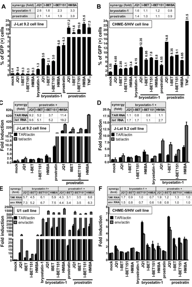
To assess whether compounds releasing active P-TEFb (JQ1, I-BET, I-BET151 and HMBA) synergize with PKC agonists (prostratin and bryostatin-1) in reactivating HIV from latency, we first determined their optimal concentrations in terms of both their HIV-1 reactivation potential and their cellular toxicity (S1 Fig). We measured induction of HIV-1 p24 capsid protein produc-tion by p24-enzyme-linked immunosorbent assay (ELISA) and we estimated the cell viability by tetrazolium salt-based assay which assesses cell metabolic activity in two well-studied HIV-1 latency cellular models, the T-lymphoid J-Lat 9.2 and promonocytic U1 cell lines. Increasing doses of compounds of interest augmented p24 antigen level in the cell supernatants at 24 hours post-treatment in a dose-dependent manner and caused a decrease in metabolic activity of less than 30% (S1 Fig). Based on our reactivation potential and cell viability data we selected two concentrations of BETi/HMBA and three concentrations of PKC agonists for combinations studies (Fig 1). Two compounds synergize when their combination produces higher effect than the sum of effects arising from separate treatments. We observed a synergistic activation of virus production in the J-Lat 9.2 cell line (Fig 1A and 1C). Bryostatin-1 at 5nM and 10nM produced synergistic increases in p24 production with each dose of BETi/HMBA (Fig 1A). Prostratin at 1.25μM and 2.5μM concentrations also synergized with each dose of BETi/HMBA and the fold-synergy was even higher than the one observed for bryostatin-1+BETi/HMBA combinations (compareFig 1C to 1A). We also observed high synergistic activations of virus production in the promonocytic U1 cell line (Fig 1B and 1D). Interestingly, synergy in U1 cells was even higher than the one observed in J-Lat 9.2 cell line. Importantly, combined treatments of 10nM bryostatin-1 or 2.5μM prostratin with 0.5μM BETi and 5mM HMBA were the most potent among the tested combinations and produced in most cases the highest synergies in both cellu-lar models. Of note, the same concentrations also produced synergistic activation of virus pro-duction in another HIV-1 latency model of promyelocytic origin, the OM10.1 cell line (S2 Fig). The OM10.1 cell line constitutes a more physiological model of HIV latency than the U1 mono-cytic cell line, since the latter contains HIV proviruses harboring mutations in tat gene. There-fore, we chose these drug concentrations for the next reactivation studies.
In conclusion, our results demonstrated that PKC agonists such as prostratin and bryosta-tin-1 synergistically increased HIV-1 production when combined with BETi/HMBA in lym-phocytic and myeloid post-integration latency models, representative of the two main cell types infected by HIV-1 in the natural host.
Co-treatments PKC agonist+BETi/HMBA increase HIV-1 expression in a
higher proportion of cells than the drugs alone and synergistically
enhance HIV-1 transcription
We next investigated whether synergistic effects in viral p24 antigen production (Fig 1) follow-ing PKC agonist+BETi/HMBA co-treatments were (i) due to an enhanced HIV-1 expression from those cells whose transcription was already reactivated by the individual drugs or (ii) due to an increase in the number of cells expressing virus. We used the J-Lat 9.2 cell line in which transcriptional activation of the latent provirus can be detected in individual cells by flow cytometry since these cells harbor full-length latent HIV-1 provirus containing gfp gene in place of nef. In the absence of stimulation, the J-Lat 9.2 cells expressed no GFP, indicating the blockade of viral transcription (Fig 2A). Treatments with each compound releasing P-TEFb used individually did not increase the number of GFP-positive cells (Fig 2A). Bryostatin-1 was weaker than prostratin in inducing GFP expression (1.3% compared to 6.3%). However, when we examined the effects of either bryostatin-1 or prostratin combined with BETi/HMBA, we observed similar synergies. The Jurkat CD4+T-cell-based J-Lat clones are the most studied cel-lular models of HIV-1 post-integration latency. However, it is critical to address whether simi-lar effects could be observed in other latency models of other cellusimi-lar origins. For instance, microglial cells are of special importance since they represent the primary host cells for HIV in Fig 1. Compounds releasing active P-TEFb and PKC agonists act synergistically to increase HIV-1 production. J-Lat 9.2 (panels A and C) and U1 (panels B and D) cell lines were mock-treated or treated with two doses of JQ1, I-BET, I-BET151, HMBA alone or in combination with three doses of either bryostatin-1 or prostratin as indicated. At 24 hours post-treatment, viral production was estimated by measuring CA-p24 antigen concentration in culture supernatants. The mock-treated value was arbitrarily set at a value of 1. Means and standard errors of the means from duplicate samples are indicated. One representative experiment from three is represented. For each combinatory treatment, the fold-synergy was calculated by dividing the effect observed after co-treatments by the sum of the effects obtained after the individual treatments.
Fig 2. PKC agonist+BETi/HMBA combined treatments increase HIV-1 expression in a higher proportion of cells than the drug alone and
the brain. Therefore, we took advantage of CHME-5/HIV latently infected microglial cells developed in Jonathan Karn’s laboratory [41]. The CHME-5/HIV cell line containing reporter GFP was treated with prostratin, bryostatin-1, BETi and HMBA alone or in combination. JQ1 was the strongest LRA among the compounds releasing P-TEFb and activated 4.65% of cells (Fig 2B). Conversely, both bryostatin-1 and prostratin activated 4% of cells. Importantly, the proportion of GFP-positive CHME-5/HIV cells was synergistically increased by combined treatments except for the PKC agonist+HMBA and prostratin+I-BET combinations. Combina-tions of PKC agonist+JQ1 led to the highest synergistic increases in the percentage of GFP-positive cells (12% for bryostatin-1+JQ1 and 15% for prostratin+JQ1).
We next examined the effect of combined treatments in the THP89GFP cell line, a cell line of monocytic origin that is chronically infected with full length HIV-1 expressing GFP [42]. Similarly, we observed that combinations of bryostatin-1+JQ1 and prostratin+JQ1 led to the highest synergistic increases in percentage of GFP-positive cells (82.3% and 94.1%, respec-tively) (S3A Fig). We also detected synergistic increases in J-Lat cell lines harboring a latent lentiviral construct expressing GFP and expressing or lacking Tat (A2 and A72, respectively) (S3B and S3C Fig, respectively). Thus, our data, in various latency models of T-lymphocytic (J-Lat 9.2, A2 and A72) and myeloid (CHME-5/HIV, THP89GFP) origins, demonstrated that co-treatments of PKC agonist+BETi/HMBA produced an increase in the number of cells expressing virus. We also compared the mean fluorescence intensities (MFI) of the GFP-posi-tive populations following the PKC agonist+BETi/HMBA treatments (S4A–S4C Fig) and we showed that the amount of GFP produced per cell was also synergistically increased. These data showed that synergy was due to both an increase in the number of cells expressing virus and an enhanced HIV-1 gene expression.
Next, we investigated whether the synergistic effects described above were due to enhanced HIV-1 transcription. To this end, we treated J-Lat 9.2 cells with compounds alone or in combi-nation and we quantified initiated and elongated HIV-1 transcripts by quantitative RT-PCR. Combination prostratin+BETi/HMBA produced synergistic increases in relative amounts of both initiated (TAR probe) and elongated (tat probe) viral transcripts (Fig 2C). Bryostatin-1 was weaker than prostratin in producing synergistic effects (Fig 2D). In addition, synergistic increases in both initiated (TAR probe) and elongated (env probe) viral transcripts were observed in the U1 monocytic cell line (Fig 2E). In CHME-5/HIV microglial cells, synergies were observed for initiated and elongated viral transcripts only for the PKC agonist+JQ1 com-binations (Fig 2F), correlating with the data we obtained by flow cytometry (compareFig 2B and 2F). JQ1 was shown by others to exhibit a good distribution into tissue compartments such as brain and testis which are known HIV-1 sanctuaries [43]. These properties of JQ1 and its ability to reactivate the virus in microglial cells suggest JQ1 as an interesting compound for targeting latent virus in those reservoirs. Importantly, unlike in J-Lat 9.2 cells, in U1 and CHME-5/HIV cells, synergistic effects caused by bryostatin-1 were similar to those caused by gfp gene. The cells were mock-treated, treated with JQ1 (0.5μM), I-BET (0.5μM), I-BET151 (0.5μM), HMBA (5mM), bryostatin-1 (10nM) and prostratin (2.5μM) alone or in combination as indicated. At 24 hours post-treatment, cells were analyzed by flow cytometry to quantify the proportion of cells expressing GFP. Means and standard errors of the means from duplicate samples are indicated. One representative experiment from three is represented. For each combinatory treatment, the fold-synergy was calculated by dividing the effect observed after co-treatments by the sum of the effects obtained from individual treatments. Panels C-F. Measurement of initiated and elongated HIV-1 transcripts following drug treatment. Total RNA was extracted from J-Lat 9.2 (panels C and D), U1 (panel E), CHME-5/HIV (panel F) cells which were mock-treated or treated with BETi, HMBA, bryostatin-1 and prostratin for 24 hours at concentrations described in Fig 2A and 2B. Initiated (primers TAR) or elongated (primers tat or env) transcripts were quantified by quantitative real-time RT-PCR. Values were normalized usingβ-actin gene primers and were presented as fold inductions relative to the values measured in mock-treated cells, which were arbitrarily set at a value of 1. Means and standard errors of the means from duplicate samples are indicated. One representative experiment from two is represented. For each combinatory treatment, the fold-synergy was calculated by dividing the effect observed after co-treatments by the sum of the effects after the individual treatments.
prostratin, indicating that combinations can present cell type-dependent specificity and under-lying the importance of testing LRAs in various cellular models of HIV-1 latency models of dif-ferent cellular origins.
Together, our data demonstrated that co-treatments prostratin/bryostatin-1+ BETi/HMBA resulted in diverse synergies that seemed cell-type specific. Moreover, in all cell lines tested, combined treatments PKC agonists+JQ1 were the most potent combinations and increased viral transcripts levels and percentage of activated cells. Combinations of prostratin/bryostra-tin-1+HMBA led to similar strong synergistic effects, except for treatments in CHME-5/HIV microglial cells.
The combined treatments have a higher potential than the individual
drug treatments in reactivating HIV-1 in CD8
+-depleted PBMCs and in
resting CD4
+T cells from cART-treated HIV
+aviremic patients
In vitro models for HIV-1 latency do not necessarily recapitulate the events occurring during viral latency ex vivo [44]. Above we showed high potential in viral reactivation of treatments combining PKC agonists and P-TEFb-releasing agents in vitro in HIV-1 latency models of lym-phoid and myeloid cell origins. We next determined whether the combined treatments also correlated with HIV-1 recovery in ex vivo cultures of CD8+-depleted PBMCs and resting CD4+ T cells isolated from cART-treated aviremic HIV-1+patients. Firstly, we evaluated cellular via-bility in CD8+-depleted PBMCs cultures purified from blood of 5 uninfected donors following drug treatments (S5 Fig). BETi, HMBA and bryostatin-1 did not alter cellular metabolic activ-ity at any concentration tested. However, we observed a dose-dependent decrease in metabolic activity in prostratin-treated CD8+-depleted PBMCs with a metabolic activity of 25% for the above used prostratin concentration (2.5μM). Therefore, we chose for further experiments a dose of prostratin (0.5μM) presenting a cellular metabolic activity of 60%. In order to evaluate HIV-1 recovery, CD8+-depleted PBMCs purified from blood of 24 cART-treated HIV+ avire-mic patients were mock-treated, treated with anti-CD3+anti-CD28 antibodies as a positive control for global T cell activation or with indicated LARs (Fig 3A,Fig 4andS1 Table). Cell-associated total HIV-1 DNA and extracellular viral RNA were quantified. The number of pro-viruses seeded in each well was estimated from the number of HIV-1 DNA copies/106cells determined by qPCR assuming negligible unintegrated HIV-1 DNA in patients after long-term cART [8]. Measurements of extracellular HIV RNA represents a surrogate marker for virions release from treated cell cultures. Indeed, Mellors’ group has highlighted the necessity to mea-sure viral production and not only unspliced cellular HIV-1 RNA following HIV-1 reactivation since these authors did not observe significant correlation between these two methods after treatment with an other LRA, the HDACi, SAHA [45]. We assumed that extracellular virions would accumulate over the course of several days; therefore, the duration of culture was extended to 6 days to maximize assay sensitivity.
We detected HIV-1 RNA in 2 out of 24 patient’s cultures (8%) in mock-treated conditions. This phenomenon could be explained by the stochastic distribution of HIV+cells in the popu-lation of isolated cells and by the activation of those HIV+cells during the experiment. I-BET led to reactivation of 61% of the patient cell cultures (Fig 4) and caused an increase in recovered viral genomic RNA (mean of 601 HIV-1 RNA copies/ml). JQ1 led to reactivation of 57% of the patient cell cultures (Fig 4) and caused an increase in recovered viral genomic RNA (mean of 461 HIV-1 RNA copies/ml). I-BET151 and HMBA increased the percentage of reactivated cul-tures (50 and 42%, respectively) and the mean levels of extracellular HIV-1 RNA were 478 and 391 HIV RNA copies/ml, respectively (Fig 4). Bryostatin-1 was more potent than prostratin and led to reactivation of 67% of cultures when the latter reactivated 46% of cultures.
Fig 3. PKC agonists and compounds releasing active P-TEFb induce HIV-1 recovery in CD8+-depleted PBMCs and in resting CD4+T cells from
Bryostatin-1 and prostratin produced similar increases in viral RNA levels (mean of 755 HIV-1 RNA copies/ml and of 774 HIV-1 RNA copies/ml, respectively). Combined treatments of prostratin+BETi/HMBA produced increases in the percentage of reactivated cultures as com-pared to individual drug treatments (Fig 4). Among the bryostatin-1 combined treatments, anti-CD3+anti-CD28 antibodies (C+), prostratin (0.5μM), bryostatin-1 (5nM), JQ1 (0.25μM), I-BET (0.25μM), I-BET151 (0.25μM) or HMBA (5mM) alone or in combination as indicated. Six days post-treatment, concentrations of genomic viral RNA were measured in culture supernatants. Statistical comparisons to positive control are indicated if p<0.05 (superiority to positive control). Panel B. Ex vivo cultures of resting CD4+T cells purified from blood of 15 patients were mock-treated, treated with anti-CD3+anti-CD28 antibodies (C+), prostratin (0.5μM), bryostatin-1 (5nM), ing-B (10nM), JQ1 (0.25μM), alone or in combination as indicated. Six days post-treatment, concentrations of genomic viral RNA in culture supernatants were determined. Statistical comparisons to positive control are indicated if p>0.05 (non-inferiority). Panels A and B. Dashed line indicates the 150 HIV-1 RNA copies/ml limit of detection. Panel C. Ex vivo cultures of CD8+-depleted PBMCs purified from blood of 7 HIV+patients were mock-treated, treated with anti-CD3+anti-CD28 antibodies (C+), bryostatin-1 (5nM), JQ1 (0.25μM) or ing-B (10nM) alone or in combination in the presence of cART. One day post-treatment, concentrations of genomic viral RNA in culture supernatants were determined. Statistical comparisons to positive control are indicated if p<0.05 (superiority to positive control). Panel D. Ex vivo cultures of CD8+-depleted PBMCs purified from blood of 11 HIV+patients were mock-treated, treated with anti-CD3+anti-CD28 antibodies (C+), bryostatin-1 (5nM), JQ1 (0.25μM) or ing-B (10nM) alone or in combination in the presence of cART. Three days post-treatment, concentrations of genomic viral RNA in culture supernatants were determined. Statistical comparisons to positive control are indicated if p>0.05 (non-inferiority to positive control). Panel E. Ex vivo cultures of CD8+-depleted PBMCs purified from blood of 11 HIV+patients were mock-treated, treated with anti-CD3+anti-CD28 antibodies (C+), bryostatin-1
(5nM), ing-B (10nM) or JQ1 (0.25μM) alone or in combination in the presence of cART. Six days post-treatment, concentrations of genomic viral RNA in culture supernatants were determined. Statistical comparisons to positive control are not indicated because p<0.05 for all the conditions (superiority of the positive control). Panels C-E. Dashed line indicates the 15 HIV-1 RNA copies/ml limit of detection. Panels A-E. Each symbol represents one cART-treated HIV+patient. The means are represented.
doi:10.1371/journal.ppat.1005063.g003
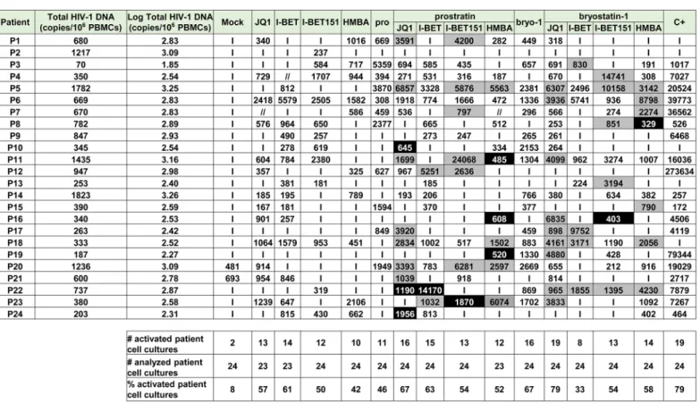
Fig 4. PKC agonists and compounds releasing active P-TEFb induce HIV-1 recovery in CD8+-depleted PBMCs from cART-treated HIV+aviremic
patients. Ex vivo cultures of CD8+-depleted PBMCs purified from blood of 24 patients were mock-treated or treated with anti-CD3+anti-CD28 antibodies,
prostratin (0.5μM), bryostatin-1 (5nM), JQ1 (0.25μM), I-BET (0.25μM), I-BET151 (0.25μM) or HMBA (5mM) alone or in combination as indicated. Six days post-treatment, concentrations of genomic viral RNA in culture supernatants were determined and the values were expressed as HIV-1 RNA copies/ml. Total HIV-1 DNA was expressed as HIV-1 DNA copies/106CD8+-depleted PBMCs. Values representing higher viral production after the combined treatment than
after the single drug treatments are shown in grey. Values representing reactivation of the virus observed exclusively after combined treatments are shown in black.‘I’ indicates below the 150 HIV-1 RNA copies/ml limit of detection. ‘//’ indicates not tested conditions.
only bryostatin-1+JQ1 produced an increase in percentage of activated cultures. From the combined treatments tested, prostratin+JQ1 and bryostatin-1+JQ1 presented the highest per-centages of reactivated cultures (67 and 79%, respectively). Importantly, HIV-1 recovery obtained after bryostatin-1+JQ1 co-treatment (79%) reached the same percentage increase as the one obtained with the positive control (79%). Notably, all combinations of PKC agonist+ compound releasing P-TEFb produced higher mean levels of extracellular HIV-1 RNA than the mean levels obtained with individual drug treatments (Fig 3A). We performed statistical analysis of the reactivation levels observed after individual and combined treatments compared to the level observed after anti-CD3+anti-CD28 stimulation (Fig 3A). Remarkably, the level obtained after the combination bryostatin-1+JQ1 was the only one to exhibit statistical non-inferiority to the positive control level (Fig 3A, p>0.05). This result confirmed the potency we demonstrated inFig 4in terms of frequency of reactivated patient cell cultures.
In addition to the increases in the reactivation frequency and in mean extracellular HIV-1 RNA levels, we demonstrated a higher viral production in several cell cultures following com-bined treatments. For instance, as shown inFig 4, HIV-1 recovery was only observed with the combination but not with the two drugs used separately (Fig 4, see black boxes). Moreover, in several ex vivo cultures, combined treatments allowed higher levels of viral production than the levels observed with the individual treatments (Fig 4, see grey boxes).
Among the cell types present in CD8+-depleted PBMCs, latently infected resting CD4+T cells represent the major reservoir of HIV infection. We therefore evaluated the reactivation potential of individual and combined treatments in this cell type.Fig 3AandFig 4showed that PKC agonist+JQ1 co-treatments led to (i) the highest percentages of reactivated patient cell cultures and (ii) important increases in extracellular viral RNA compared to other PKC agonist +BETi/HMBA co-treatments in CD8+-depleted PBMCs purified from blood of cART-treated HIV+aviremic patients. Therefore, we evaluated in resting CD4+T cells the reactivation poten-tials of JQ1 alone or combined with either prostratin or bryostatin-1 or another PKC agonist, ing-B. Indeed, while we were performing this study, ing-B has been reported to potently reacti-vate latent HIV-1 [25,26] in vitro and we decided to evaluate here its potential ex vivo. We iso-lated HLA-DR-, CD69-, CD25-CD4+T cells from blood of 15 cART-treated HIV+aviremic patients. Ex vivo cell cultures were mock-treated, treated with anti-CD3+anti-CD28 antibodies or with the compounds of interest (Fig 5andS1 Table). Cell-associated total HIV-1 DNA and extracellular viral RNA were quantified. We detected HIV-1 RNA in 3 out of 15 patient’s cul-tures (20%) in mock-treated conditions. Prostratin, bryostatin-1, ing-B, and JQ1 reactivated latent HIV-1 in 60%, 53%, 53%, and 40% of the cultures, respectively (Fig 5). All individual treatments produced increases in mean HIV-1 RNA copies/ml levels that were higher than the 150 copies/ml threshold (Fig 3B). Notably, the combination bryostatin-1+JQ1 and ing-B+JQ1 produced higher increases in mean viral RNA levels relative to the individual treatments. These bryostatin-1+JQ1 and ing-B+JQ1-induced increases reached 1127 and 1547 HIV-1 RNA copies/ml, respectively (Fig 5). Importantly, the very high levels of reactivated virus fol-lowing those treatments were statistically non-inferior (p>0.05) to the level obtained after anti-CD3+anti-CD28 stimulation. In contrast, we did not observe any benefit of combining prostratin with JQ1. All the combined treatments did not increase the frequency of reactivated patient cell cultures relative to the individual treatments, but synergistically increased HIV-1 recovery in most of the cultures (Fig 5, see black and grey boxes). For instance, the ing-B+JQ1 co-treatment led to reactivation in 46% of ex vivo cultures and, remarkably, all these cultures exhibited strong synergistic increases. Of note, we also performed individual and combined treatments using ing-B in CD8+-depleted PBMCs from cART-treated HIV+aviremic patients and we observed strong reactivations of latent HIV-1. This part of our work concerning ing-B
individual treatment of patient cell cultures will be reported in a separate study (L. Gama and colleagues, manuscript in preparation).
Consequently, our results established that PKC agonists acted as powerful LRAs and that their reactivation properties were potentiated when used in combination with P-TEFb-releas-ing agents, especially JQ1. However, it should be noted that we did not observe viral reactiva-tion either after individual, combined or anti-CD3+anti-CD28 treatments in all the patient cell cultures. This observation can be explained by the multifactorial and stochastic nature of viral latency but also by the very low frequency of latently infected cells carrying replication compe-tent proviruses. Indeed, in the experiments described above, we plated 6 million CD8+-depleted PBMCs or 0.5 million resting CD4+ T cells per culture condition. Assuming the scarcity of latently-infected cells, it is possible that some of the experimental wells did not contain any res-ervoir. Moreover, we used a reactivation system without cART in order to increase the sensitiv-ity of our assays since it allowed infection of new cells and multiple viral cycles. Nevertheless, it may have introduced a bias since the compounds used may have sensitized bystander cells to new infections. Based on the results presented inFig 3A and 3B, we thus selected the most potent drug combinations (bryostatin-1+JQ1 and ing-B+JQ1) and tested them in the presence of cART. We plated 12 million of CD8+-depleted PBMCs in order to increase the number of latently infected cells per tested condition. Moreover, we looked at the effect of the drugs not Fig 5. PKC agonists and JQ1 induce HIV-1 recovery in resting CD4+T cells from cART-treated HIV+aviremic patients. Ex vivo cultures of resting CD4+T
cells purified from blood of 15 patients were mock-treated or treated with anti-CD3+anti-CD28 antibodies, JQ1(0.25μM), prostratin (0.5μM), bryostatin-1 (5nM) or ing-B (10nM) alone or in combination as indicated. Six days post-treatment, concentrations of viral RNA in culture supernatants were determined and the values were expressed as HIV-1 RNA copies/ml. Total HIV-1 DNA was expressed as HIV-1 DNA copies/106resting CD4+T cells. Values representing higher viral production after a combinatory treatment than after the corresponding single drug treatments are shown in grey. Values representing reactivation of viral production observed exclusively after combined treatment are shown in black.‘I’ indicates below the 150 HIV-1 RNA copies/ml limit of detection. ‘//’ indicates not tested conditions.
only at day 6 but also at day 1 and day 3 of culture. Of note, continuous bryostatin-1 infusion of 24 hours [46] and 72 hours [47] were previously tested in patients affected by malignancies.
At day 1 (Fig 6andS1 Table), the combination bryostatin-1+JQ1 was the only condition that caused viral reactivation in the cell cultures from 7 patients tested (100% of reactivation). Surprisingly, this combined treatment exceeded the effect observed after anti-CD3+anti-CD28 stimulation (p = 0.0256) (Fig 3C). The combination ing-B+JQ1 reactivated latent HIV-1 in 83% of cultures, a result identical to that observed with ing-B alone and that exceeded the per-centage obtained after stimulation with the positive control (57%) (Fig 6). Importantly, this ing-B+JQ1 combination resulted in a higher mean level of extracellular HIV-1 RNA than the mean level obtained with ing-B alone (mean of 292 HIV-1 RNA copies/ml and of 160 HIV-1 RNA copies/ml, respectively (Fig 3C)).
At day 3 (Fig 6andS1 Table), the combination bryostatin-1+JQ1 reactivated HIV-1 in 10 out of 11 patient cultures (91% of reactivated patient cell cultures). This percentage was thus higher than percentages obtained after treatment with bryostatin-1 alone (64%) or after the positive control stimulation (82%). The conditions ing-B alone and ing-B+JQ1 reactivated HIV-1 in 100% of the ex vivo cultures. Similarly to the results obtained at day 1, ing-B+ JQ1combination resulted in a higher mean level of extracellular HIV-1 RNA than the mean level obtained with ing-B alone (mean of 287 HIV-1 RNA copies/ml and of 184 HIV-1 RNA copies/ml, respectively) (Fig 3D). As indicated inFig 3D, the levels observed after the individ-ual PKC agonist treatments and after the combinations PKC agonists+JQ1 were at day 3 statis-tically non-inferior (p>0.05) to the level obtained after anti-CD3+anti-CD28 stimulation.
At day 6 (Fig 3E,Fig 6andS1 Table), the sensitivity of the RNA quantification allowed us to detect a viral production at very low level even in the mock-treated cultures, probably due to the activation of HIV+cells during the experiment. It is consequently difficult to discuss the percentages of reactivation observed. The combination bryostatin-1+JQ1 produced a higher mean level of extracellular HIV-1 RNA than the mean level obtained with bryostatin-1 alone (mean of 670 HIV-1 RNA copies/ml and of 382 HIV-1 RNA copies/ml, respectively), Fig 6. PKC agonists and JQ1 induce HIV-1 recovery in CD8+-depleted PBMCs from cART-treated HIV+aviremic patients in the presence of cART.
Ex vivo cultures of CD8+-depleted PBMCs purified from blood of 11 patients were mock-treated or treated with anti-CD3+anti-CD28 antibodies, JQ1
(0.25μM), bryostatin-1 (5nM) or ing-B (10nM) alone or in combination as indicated. Concentrations of viral RNA in culture supernatants were determined one day, three days or six days post-treatment. The values were expressed as HIV-1 RNA copies/ml. Total HIV-1 DNA was expressed as HIV-1 DNA copies/106
CD8+-depleted PBMCs. Values representing higher viral production after a combinatory treatment than after the corresponding single drug treatments are
shown in grey. Values representing reactivation of virus production observed exclusively after combined treatments are shown in black.‘I’ indicates below the 15 HIV-1 RNA copies/ml limit of detection,‘//’ indicates not tested conditions.
confirming in the presence of cART the results presented inFig 3Ain the absence of cART. The combination ing-B+JQ1 produced a slightly lower mean level of extracellular HIV-1 RNA than the mean level obtained with ing-B alone. At day 6, the positive control treatment showed a statistical superiority to all the tested conditions. Indeed, unlike the stimulation by the LRAs, the stimulation by the positive control relied on continuous cell stimulation arising from coated anti-CD3 antibodies. This could explain that the level of extracellular genomic viral RNA was increasing in a time-dependent manner. On the contrary, the maximal effect of the bryostatin-1+JQ1 and ing-B+JQ1 combinations appeared earlier (at day 3 and day 1, respec-tively). Importantly, the kinetics of the effect observed with bryostatin-1 corresponded to its clinical use [46,47]. Interestingly, JQ1 did not lead to a significant viral reactivation in the pres-ence of cART (Fig 3C–3E), probably because the reactivation potential of this LRA is weak when used alone and needs to be amplified in order to be detected.
Moreover, we evaluated the cell viability for PKC agonist+JQ1 combinations in CD8+ -depleted PBMCs isolated from blood of uninfected donors and we showed that prostratin +JQ1, bryostatin-1+JQ1 and ing-B+JQ1 combinations presented 82%, 62% and 69% of cellular metabolic activity, respectively (S6A Fig) and caused no cell death (S6B Fig).
Finally, in order to verify that the reactivated viruses were infectious, we performed infec-tions of Jurkat cells with viruses isolated from bryostatin-1+JQ1 treated patient cell cultures and demonstrated the infectivity of the reactivated viruses (S2 Table).
In conclusion, our results established that PKC agonists acted as powerful LRAs and that their reactivation properties were potentiated when used in combination with compounds releasing P-TEFb and thereby increasing transcriptional elongation, especially JQ1. Impor-tantly, HIV-1 reactivation potencies of these combinations were already clearly detected at 24 hours post-treatment.
Synergistic HIV-1 recovery following PKC agonist+BETi/HMBA
combined treatments is dependent on NF-
κB
We assessed the molecular mechanisms underlying the synergistic induction of HIV-1 observed at the viral RNA and protein levels following individual or combined treatments with PKC ago-nists and BETi/HMBA (Figs1–6). Firstly, we evaluated the involvement of two NF-κB binding
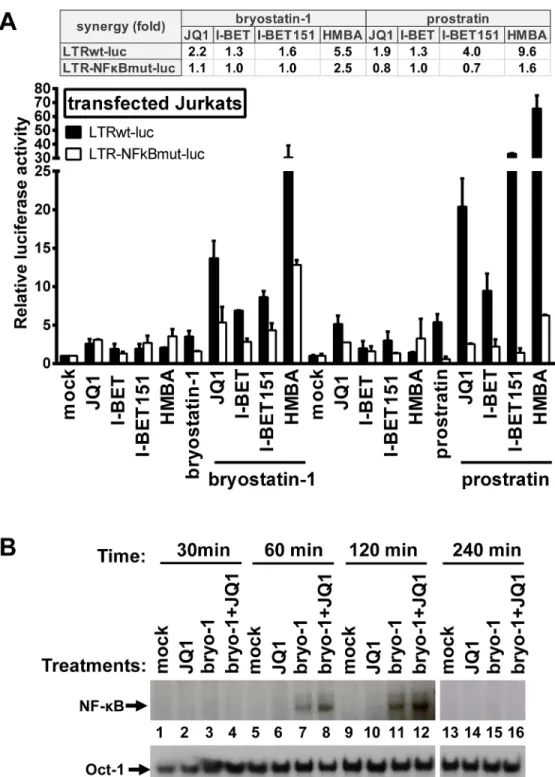
sites located in the enhancer region of the HIV-1 promoter. We transiently transfected Jurkat cells either with episomal vector driving the luciferase expression from wild-type LTR (LTRwt-luc) or with similar reporter vector mutated in two NF-κB binding sites located in HIV LTR (LTR-NFκBmut-luc). Cells were stimulated with JQ1, I-BET, I-BET151, HMBA, prostratin, and bryostatin-1 alone or in combination. Combinations of PKC agonist+BETi/HMBA led to syner-gistic increases in luciferase activity arising from LTRwt-luc transfection (Fig 7A). We observed that lack of the two NF-κB binding sites located in HIV-1 LTR impaired the synergistic increase in luciferase activity following combined treatments. These data indicated the involvement of NF-κB protein binding to the viral promoter in the PKC agonist+BETi/HMBA synergies. To assess the effect of the drugs on the NF-κB binding activity, we next performed electrophoretic mobility shift assays (EMSAs) using an HIV-1 NF-κB probe and nuclear extracts from Jurkat T cells stim-ulated for various periods of time with JQ1, bryostatin-1 or bryostatin-1+JQ1 (Fig 7B). We detected an induction of NF-κB DNA-binding activity in response to a 60 min treatment with bryostatin-1 (Fig 7B, lane 7). JQ1 alone caused no induction of NF-κB binding activity even after
a 240 min treatment (Fig 7B, lanes 2, 6, 10 and 14). When bryostatin-1 was combined with JQ1, an induction of NF-κB binding activity stronger than that obtained with bryostatin-1 alone was observed at 60 and 120 min time points (Fig 7B, compare lane 7 with lane 8 and lane 11 with lane 12). This effect faded away at 240 min time point, confirming transient NF-κB DNA- binding
Fig 7. Synergistic inducibility of HIV-1 LTR promoter by PKC agonist+BETi/HMBA combinatory treatments depends on NF-κB. Panel A. Jurkat cells were transiently transfected with the episomal plasmid containing the luciferase reporter gene driven either by the wild-type HIV LTR promoter (LTRwt-luc) or by the LTR promoter mutated in the two NF-κB binding sites (LTR-NFκBmut-luc). Twenty-four hours later, cells were mock-treated, treated with JQ1 (0.5μM), I-BET (0.5μM), I-BET151 (0.5μM), HMBA (5mM), bryostatin-1 (10nM) and prostratin (2.5μM) alone or in combination. Luciferase activities in cell extracts were measured 24 hours after drug treatments and reported as fold increases over the activity observed in mock-treated conditions (transfection of the reporter plasmid without drug treatment) and arbitrarily set at values of 1. An experiment performed in triplicates representative of two independent experiments is shown. Panel B. Nuclear extracts were prepared from Jurkat cells which were mock-treated, treated with bryostatin-1 (10nM), JQ1 (0.5μM) or with bryostatin-1+JQ1 for different time periods. An oligonucleotide corresponding to the
HIV-activity. Our results demonstrated that JQ1 increased bryostatin-1-induced NF-κB DNA-binding activity. This could explain, at least in part, the synergistic increases in HIV transcription follow-ing combined treatments observed inFig 2.
In conclusion, our data indicated that the synergistic activation of HIV-1 obtained after PKC agonist+BETi/HMBA combinationed cellular treatments was, at least in part, dependent on NF-κB.
Bryostatin-1+JQ1 combination has a higher potential in activating
P-TEFb than the individual drug treatments
To address the molecular mechanisms mediating the synergistic activation of HIV-1 transcrip-tion and productranscrip-tion following the combined bryostatin-1+JQ1 treatment, we investigated the effect on P-TEB release of these LRAs alone or in combination. To this end, Jurkat cells were treated with JQ1, bryostatin-1 or a combination of both compounds for 1 hour and 24 hours. Nuclear extracts were prepared from treated cells and were used to perform immunoprecipita-tion experiments targeting CDK9 and Western blotting addressing the interacimmunoprecipita-tion of CDK9 with either HEXIM1 or CycT1. As shown inFig 8A, for the 1 hour treatment, our results dem-onstrated a transient release of HEXIM1 from the CDK9/CycT1 (P-TEFb) complex after JQ1 treatment. These data are in agreement with a previous report from Peterlin and colleagues [31]. Bryostatin-1 treatment caused a weaker release of HEXIM1 from the P-TEFb complex than the JQ1 treatment (Fig 8A, compare lane 8 with lane 9). Interestingly, the combined treat-ment bryostatin-1+JQ1 led to a much stronger and synergistic HEXIM1 release than the indi-vidual treatments. These results indicated that that combined treatment increased the global availability of the active form of P-TEFb. After 24 hours of treatment (Fig 8B), we observed no P-TEFb release but a potentiated interaction between HEXIM1 and P-TEFb. This 24 hour effect contrasted with the 1 hour short-term effect and indicated reassembly of P-TEFb in the inactive 7SK snRNP complex. These results are consistent with the study of Liu et al. reporting that transient release of P-TEFb results in upregulation of its immediate target gene, HEXIM1, and reincorporation of P-TEFb into the 7SK snRNP [48]. Consistently, we also observed an increased expression of HEXIM1 after the 24 hours treatment compared to the 1 hour treat-ment (Fig 8B, input panel), thereby favoring a negative feedback mechanism of P-TEFb activation.
In order to study the effect of the PKC agonist+BETi/HMBA combined treatments on P-TEFb activation in living cells, we took advantage of the first and very elegant experimental system allowing to monitor quantitatively in vivo the interaction between P-TEFb and its sub-strate, the C-terminal domain (CTD) of RNA polymerase II (RNAPII). This system named V-PAC (visualization of P-TEFb activation by fluorescent complementation) [49] is based on a bimolecular fluorescence complementation (BiFC) assay and uses complementary fragments of fluorescent proteins, the N-terminal region (termed VN) of Venus and the C-terminal region (termed YC) of YFP fluorescent protein. Because activated P-TEFb interacts with and phosphorylates the RNAPII CTD, we employed P-TEFb and the CTD as fusion partners of YC and VN, respectively as described in [49]. Our results demonstrated that the number of YFP-positive cells was higher following the combined bryostatin-1+JQ1 treatment than the numbers obtained after the individual drug treatments (Fig 8C and 8D). Altogether, these data strongly 1 LTR NF-κB sites was used as probe in EMSAs. As control for equal loading, the bottom panel shows comparability of the various nuclear extracts assessed by EMSA with an Oct-1 consensus probe. doi:10.1371/journal.ppat.1005063.g007
indicated that combined LAR treatments led to higher activations of P-TEFb than the corre-sponding individual drug treatments.
To get further insights into the molecular mechanisms underlying the PKC agonist+BETi/ HMBA synergistic gene expression activations induced by the combined treatments, we com-pared the effect of the drugs alone and in combination on P-TEFb-dependent transcription by reporter assays. More specifically, we transiently transfected HeLa cells with a reporter gene construct Hex1(-104)Luc containing the luciferase gene under the control of the HEXIM1 pro-moter which is responsive to active P-TEFb [48]. Transfected cells were mock-treated or treated with either PKC agonists (prostratin, bryostatin-1 and ing-B), either BETi (JQ1, I-BET Fig 8. PKC agonists synergize with BETi in releasing active P-TEFb. Jurkat cells were mock-treated, treated with JQ1 (0.25μM), bryostatin-1 (5nM) alone or in combination for 1 hour (Panel A) or 24 hours (Panel B). Nuclear extracts were prepared from treated cells and subjected to immunoprecipitations (IP) with an anti-CDK9 antibody or the control IgG. The complexes were immunodetected for the presence of CycT1 and HEXIM-1 by Western blotting (right panels). Input controls for CDK9, CycT1 and HEXIM1 are presented (left panels). Levels ofβ-actin were measured to control protein loading. Panels A and B. Histograms represent quantification of band intensities normalized to CDK9 levels in the IP and then normalized to mock-treated condition. Panel C. HeLa cells expressing YC.P-TEFb and VN.CTD were left untreated or were treated as indicated for 1 hour. Venus-positive cells were detected by fluorescence microscopy (upper panels). Bright-field images were also taken (lower panels). Panel D. HeLa cells expressing YC.P-TEFb and VN.CTD were treated as outlined in C and positive cells were counted and averaged from three different areas. Error bars represent differences between counts of Venus-positive cells from the randomly chosen fields under the microscope. Panel E. Hela cells were transfected with the Hex1(-104)Luc reporter plasmid. At 24 hours post-transfection, cells were mock-treated or treated with the different compounds as indicated. Luciferase activities in cell extracts were measured 24 hours after drug treatments and reported as fold increases over the activity observed in mock-treated condition (transfection of the reporter plasmid without drug treatment) and arbitrarily set at a value of 1. An experiment performed in duplicate representative of two independent experiments is shown.
and I-BET151), or HMBA individually and in combination (Fig 8E). Our results demonstrated synergistic increases in luciferase activity following combined treatments. Of note, the PKC agonist+JQ1 combinations were more potent than the other combinations tested.
To further evaluate the signaling pathways leading to HIV-1 synergistic activation in response to PKC agonist+BETi cotreatments, we used chemical inhibitors targeting the NF-κB, P-TEFb and NFAT pathways in both T-lymphoid and monocytic infected cells lines (Fig 9A and 9B, respectively). We showed that BAY 11–7082, an inhibitor of IκBalpha phosphoryla-tion, blocked an average of 50% and of 70% of the viral reactivation mediated by the combined treatment bryostatin-1+JQ1 in J-Lat 9.2 and THP89GFP cells, respectively. We used flavopiri-dol, a selective P-TEFb inhibitor, and we observed that this compound blocked an average of 50% and of 84% of viral activation mediated by the combined treatment bryostatin-1+JQ1 in J-Lat 9.2 and THP89GFP cells, respectively. In contrast, cyclosporin A, a potent NFAT inhibi-tor, had no effect on HIV-1 activation in both cell lines. These data confirmed that NF-κB and P-TEFb, as opposed to NFAT, were involved in induction of HIV-1 in response to bryostatin-1 +JQ1 cotreatment.
Mechanistically, using four independent assays (immunoprecipitations, biomolecular fluo-rescence complementation assays, reporter gene assays and experiments using various signal-ing pathways inhibitors), we demonstrated that PKC agonists (such as bryostatin-1) and BETi (such as JQ1) caused a more potent activation of P-TEFb when used in combination than when used alone. Taken together, these mechanistic data established a correlation between the potentiated P-TEFb activation and the potentiated or synergistic (depending on the HIV-1 latency cellular model used) induction of HIV-1 gene expression observed after the combined versus individual drug treatments. This potentiated release of P-TEFb from the inactive 7SK snRNP complex could explain the potentiated or synergistic activation of HIV-1 gene expres-sion induced by PKC agonist+BETi/HMBA combined treatments.
PKC agonist+JQ1 co-treatments cause differential expression levels of
cell surface activation markers and downregulate the expression of the
CD4 receptor on CD4
+primary cells
Compounds that would be suitable for the therapeutic use in vivo should not lead to non-spe-cific or robust immune activation. Additionally, drug-mediated decrease of CD4 receptor sur-face expression may be an important factor in the blockade of de novo HIV-1 infection. To determine whether individual and combined treatments of PKC agonists with JQ1 led to the cellular activation, we isolated CD8+-depleted PBMCs and resting CD4+T cells from blood of 4 uninfected donors. The activation status of resting CD4+T cells and of CD4+cells analyzed from the mock-treated or treated CD8+-depleted PBMCs population was assessed by flow cytometry analysis of the cell surface activation markers CD69, CD25, HLA-DR and CD38. First, we assessed the activation status of resting CD4+T cells. We observed that JQ1 treatment did not cause any upregulation of T cell activation markers (Fig 10A–10D). Bryostatin-1 and prostratin alone or combined with JQ1 induced the surface expression of CD69 in 10–40% of cells and the surface expression of HLA-DR in 10–20% of cells (Fig 10B and 10C, respectively). These treatments led to low changes in the expression of CD38 and CD25 (Fig 10A and 10D, respectively). Ing-B alone or combined with JQ1 induced CD69 surface expression in 10–15% of cells (Fig 10B). Interestingly, there was no ing-B-induced modulation of other activation markers (Fig 10A, 10C and 10D). In order to extend the data we obtained in resting CD4+T cells, we examined the expression levels of activation markers on the surface of CD4+cells ana-lyzed from the mock-treated or treated CD8+-depleted PBMCs population. Similarly to what we demonstrated in resting CD4+T cells, we observed that JQ1 individual treatment did not
cause any upregulation of activation markers (Fig 10F–10I). We showed that the individual and combined PKC agonists+JQ1 treatments led to differential increases in CD69, CD25, Fig 9. Bryostatin-1+JQ1-mediated HIV-1 reactivation is dependent on NF-κB and P-TEFb and independent from NFAT. The T-lymphoid J-Lat 9.2 (panel A) and monocytic THP89GFP (panel B) cell lines harbor latent HIV-1 provirus containing gfp gene. The cells were pre-treated with the indicated inhibitors for 2 hours and then either mock-treated or treated with the combination bryostatin-1 [10nM]+JQ1 [0.5μM]. At 24 hours post-treatment, cells were analyzed by flow cytometry to quantify the proportion of cells expressing GFP. Means and standard errors of the means from duplicate samples are indicated. One representative experiment from two is represented.
Fig 10. Expression of cell surface activation markers following PKC agonists and JQ1 treatments. Blood from 4 uninfected donors was split and one half was used to purify resting CD4+T cells (panels A-E)
HLA-DR and CD38 surface expression levels. Interestingly, the combined treatments PKC agonists+JQ1 appeared to decrease PKC agonist-driven surface expression increases of CD38, HLA-DR and CD25 (Fig 10F, 10H and 10I, respectively). Since the CD38 marker is also consti-tutively expressed by naive CD4+T cells, we analyzed the frequency of cells expressing simulta-neously CD38 and HLA-DR (S7 Fig). Our data showed that the combined treatments PKC agonist+JQ1 appeared to decrease the PKC agonist-driven surface expression increases of these two activation markers. Next, we assessed the expression of CD4 surface receptor in resting CD4+T cells following treatments with the drugs of interest (Fig 10E). PKC agonists were pre-viously shown to exhibit anti-viral activity by decreasing surface expression of CD4 receptor which would lead to the blockade of de novo infection [16,22,25]. As shown inFig 10E, we demonstrated that that bryostatin-1, prostratin and ing-B strongly reduced the cell surface expression of CD4 receptor. Individual JQ1 treatment had a slight effect on the CD4 surface expression and combined treatments of PKC agonis+JQ1 appeared to slightly augment the PKC agonist-driven CD4 receptor downregulation.
In conclusion, we showed that JQ1 caused no activation of resting CD4+T cells and CD4+ cells analyzed from the treated CD8+-depleted PBMCs population. Combined treatments including JQ1 and PKC agonists caused elevated surface expression levels of CD69 and HLA-DR and no or low upregulation of CD38 and CD25. Notably, the decrease in surface expression levels of activation markers we observed in CD4+cells from PBMCs following com-bined treatments as compared to the PKC agonists individual treatments strengthened the potential of PKC agonist+JQ1 co-treatments for purging strategies because it indicated that the latter combinations decreased the levels of global T-cell activation induced by PKC agonists. Moreover, the combinations PKC agonist+JQ1 led to downregulation of CD4 receptor, which could be beneficial for the blockade of de novo infection.
Discussion
In this report, we performed an in vitro and ex vivo in-depth comparison of the reactivation potentials of two classes of latency-reversing agents, P-TEFb-releasing agents and PKC ago-nists. We demonstrated that combinations of LRAs from both classes led to strong synergistic activation of HIV-1 production in various post-integration latency cell line models of T-lym-phoid and monocytic origins. Most importantly, we identified for the first time two potent combinations of LRAs, bryostatin-1+JQ1 and ing-B+JQ1, which activated latent HIV-1 to a degree comparable to that obtained with the positive control anti-CD3+anti-CD28 antibodies in a high number of ex vivo patient cell cultures and at multiple time points. Mechanistically, we provide a molecular explanation for these synergistic or potentiated viral reactivations by demonstrating a stronger activation of both P-TEFb and NF-κB activities following combined versus individual LRA treatments. We showed by bimolecular fluorescence complementation assays that the potent LRA effects observed resulted from a higher activation of P-TEFb. JQ1 alone failed to potently reactivate HIV-1 from latently-infected T cells. This could be explained and the other half was used to purify CD8+-depleted PBMCs (panels F-I). Cell cultures were mock-treated,
treated with anti-CD3+anti-CD28 antibodies (C+), JQ1 [0.25μM], bryostatin-1 [5nM], prostratin [0.5μM] or ing-B [10nM] alone or in combination for 6 days. Cells were incubated with CD38 (panels A and F), anti-CD69 (panels B and G), anti-HLA-DR (panels C and H), anti-CD25 (panels D and I) or anti-CD4 (panel E) antibodies prior to flow cytometry analysis. The results are presented as percentage of marker expression in the population of CD4+cells (panels A-D and panels F-I) and as median fluorescence intensity (MFI) of CD4+ cells (panel E). Dashed line indicates the percentage of expression obtained in mock-treated cells. The means are represented. Statistical comparisons are indicated. Statistically relevant and not statistically relevant comparisons are indicated by asterisk and“ns”, respectively.
by the fact that the level of P-TEFb is very low in resting T cells due to actions of specific miR-NAs and the cellular factor NF-90 which block translation of CycT1 mRNA [50–53]. However, we demonstrated that JQ1 was a good candidate for combinatory treatments with compounds that increase the amount of P-TEFb.
The combined effects of PKC agonists and BET inhibitors are the result of the induction of P-TEFb production by PKC agonists [27,38,39] and of the release of P-TEFb from the repres-sive 7SK snRNP complex by BETi/HMBA [31,37]. In addition, Peterlin’s group has previously shown that PKC agonists also phosphorylate HEXIM1 at a specific serine residue, leading to release of active P-TEFb from the 7SK snRNP complex and induction of P-TEFb-dependent transcription [40]. In the present study, we confirmed this additional effect of bryostatin-1 and we demonstrated that the combination bryostatin-1+JQ1 caused a higher release of PTEF-b from its inactive 7SK snRNP complex than the drugs alone. We demonstrated a higher activa-tion of P-TEFb following combined treatments of PKC agonist+BETi/HMBA by V-PAC assays. This higher P-TEFb activation can be explained by the dual effect of PKC agonists on P-TEFb (the CycT1 and CDK9 syntheses and the P-TEFb release from the 7SK snRNP com-plex) and by the additional effect of BETi/HMBA on the release of P-TEFb.
Moreover, McNamara and colleagues showed that, during activation of the inflammatory transcriptional program, the inducible recruitment of P-TEFb by NF-κB through PPM1G to promoters of inflammatory-responsive genes disassembles the 7SK snRNP and releases P-TEFb for transcription elongation [54]. Such an interaction could occur at the HIV-1 pro-moter, thereby providing an additional explanation for the synergistic activation of HIV-1 gene expression in response to PKC agonist+P-TEFb-releasing agent combined treatments.
Interestingly, our transient transfection experiments showed that combined treatments induced HIV-1 promoter-driven luciferase activity and that maximal synergistic activations required intact NF-κB binding sites in the enhancer region of the HIV-1 LTR. We showed that JQ1 synergistically increased bryostatin-1-induced NF-κB DNA-binding activity by EMSAs. This observation indicated that JQ1 played a role in controlling NF-κB activity either by target-ing P-TEFb or by still unidentified mechanism(s). Therefore, this increase of NF-κB bindtarget-ing activity following the bryostatin-1+JQ1 treatment could be explained by the stabilization of the NF-κB/PPM1G/P-TEFb complex at the viral promoter. Recently, Zou and colleagues have shown that the BRD4 protein interacts with acetylated NF-κB and that this interaction main-tains NF-κB constitutively active [55]. These authors have also demonstrated that JQ1 treat-ment disrupts this BRD4-acetylated NF-κB protein complex, leading to a decreased NF-κB transcriptional activity [42]. In contrast, the present study demonstrated that combined treat-ments PKC agonists+JQ1 exhibited beneficial effects on NF-κB activity (Fig 7A) and DNA-binding (Fig 7B). Additional studies will be necessary to further elucidate the role of JQ1 in NF-κB regulation.
Of note, the molecular mechanisms mediating the PKC agonists+BETi/HMBA synergy are likely to be highly complex and to implicate phenomena other than the P-TEFb and NF-κB regulations. For example, the following elements could also intervene in the molecular mecha-nisms of synergistic activation after combined PKC agonist+BETi/HMBA treatment: (i) NF-κB interactions with histone acetyltransferase, including p300 and CREB-binding protein (CBP) [56], (ii) direct interaction of P-TEFb with the cellular cofactor CTIP2 (COUP-TF-inter-acting protein 2) previously reported by our laboratory as involved in HIV-1 silencing in microglial cells [57] as well as (iii) the involvement of HMGA-1 (non-histone chromatin pro-tein high mobility group AT-hook 1) in this P-TEFb/CTIP2 regulation [58].
Remarkably, Weinberger’s group has recently observed that conventional transcriptional activators synergize with“noise” modulators that affect gene expression fluctuations without changing mean expression [59]. Our data constitutes a good example of such a regulation since