HAL Id: inserm-02437797
https://www.hal.inserm.fr/inserm-02437797
Submitted on 13 Jan 2020
HAL is a multi-disciplinary open access
archive for the deposit and dissemination of
sci-entific research documents, whether they are
pub-lished or not. The documents may come from
teaching and research institutions in France or
abroad, or from public or private research centers.
L’archive ouverte pluridisciplinaire HAL, est
destinée au dépôt et à la diffusion de documents
scientifiques de niveau recherche, publiés ou non,
émanant des établissements d’enseignement et de
recherche français ou étrangers, des laboratoires
publics ou privés.
heterogeneity and immunosuppression in ovarian cancers
Anne-Marie Givel, Yann Kieffer, Alix Scholer-Dahirel, Philemon Sirven,
Mélissa Cardon, Floriane Pelon, Ilaria Magagna, Géraldine Gentric, Ana
Costa, Claire Bonneau, et al.
To cite this version:
Anne-Marie Givel, Yann Kieffer, Alix Scholer-Dahirel, Philemon Sirven, Mélissa Cardon, et al..
miR200-regulated CXCL12β promotes fibroblast heterogeneity and immunosuppression in ovarian
cancers. Nature Communications, Nature Publishing Group, 2018, 9 (1), pp.1056.
�10.1038/s41467-018-03348-z�. �inserm-02437797�
miR200-regulated CXCL12
β promotes fibroblast
heterogeneity and immunosuppression in ovarian
cancers
Anne-Marie Givel
1,2
, Yann Kieffer
1,2
, Alix Scholer-Dahirel
1,2
, Philemon Sirven
3
, Melissa Cardon
1,2
,
Floriane Pelon
1,2
, Ilaria Magagna
1,2
, Géraldine Gentric
1,2
, Ana Costa
1,2
, Claire Bonneau
1,2
, Virginie Mieulet
1,2
,
Anne Vincent-Salomon
4
& Fatima Mechta-Grigoriou
1,2
High-grade serous ovarian cancers (HGSOC) have been subdivided into molecular subtypes.
The mesenchymal HGSOC subgroup, de
fined by stromal-related gene signatures, is
invari-ably associated with poor patient survival. We demonstrate that stroma exerts a key function
in mesenchymal HGSOC. We highlight stromal heterogeneity in HGSOC by identifying four
subsets of carcinoma-associated
fibroblasts (CAF-S1-4). Mesenchymal HGSOC show high
content in CAF-S1
fibroblasts, which exhibit immunosuppressive functions by increasing
attraction, survival, and differentiation of CD25
+FOXP3
+T lymphocytes. The beta isoform of
the CXCL12 chemokine (CXCL12
β) specifically accumulates in the immunosuppressive
CAF-S1 subset through a miR-141/200a dependent-mechanism. Moreover, CXCL12
β expression in
CAF-S1 cells plays a crucial role in CAF-S1 immunosuppressive activity and is a reliable
prognosis factor in HGSOC, in contrast to CXCL12α. Thus, our data highlight the differential
regulation of the CXCL12α and CXCL12β isoforms in HGSOC, and reveal a
CXCL12β-associated stromal heterogeneity and immunosuppressive environment in mesenchymal
HGSOC.
DOI: 10.1038/s41467-018-03348-z
OPEN
1Institut Curie, Stress and Cancer Laboratory, Equipe labelisée Ligue Nationale Contre le Cancer, PSL Research University, 26, rue d’Ulm, 75005 Paris, France. 2U830, Inserm, 75005 Paris, France.3Institut Curie, Integrative Biology of Human Dendritic Cells and T Cells Laboratory, PSL Research University, U932, Inserm, 26, rue d’Ulm, 75005 Paris, France.4Department of Pathology, Institut Curie Hospital Group, 26, rue d’Ulm, 75248 Paris, France. Correspondence and requests for materials should be addressed to F.M-G. (email:fatima.mechta-grigoriou@curie.fr)
123456789
H
igh-grade serous epithelial ovarian cancers (HGSOC),
commonly treated by the combination of surgery and
chemotherapy, remain one of the deadliest gynecologic
malignancies. Despite an initial response to treatment, many
patients relapse, become resistant, and ultimately die. To date,
treatment strategy mainly relies on clinico-pathologic aspects,
such as histological type, grade and stage without consideration of
molecular phenotypes. HGSOC genomic and transcriptomic
profiles have been helpful for characterizing HGSOC molecular
features and improving patient stratification leading to new
treatment strategies. HGSOC patients carrying BRCA1/2
altera-tions have increased sensitivity to platinum salts and a longer
survival than non-mutated patients, and are now eligible for
anti-PARP therapies
1–5. In addition to genomic characterization,
several groups have defined distinct HGSOC molecular subtypes
based on transcriptomic profiling
6–13. In all studies, one
mole-cular subgroup, referred to as
“Fibrosis” or “Mesenchymal”, has
been systematically identified and is invariably associated with
poor patient survival. Interestingly, one of the
first mechanisms
that differentiates the Fibrosis/Mesenchymal HGSOC from the
other molecular subtypes depends on the miR-200 family of
microRNA
7,13,14. Still, patients suffering from HGSOC of the
Fibrosis/Mesenchymal subtype invariably show poor prognosis
and remain one of the major clinical challenges in ovarian
tumorigenesis.
Transcriptomic
signatures
that
identify
the
“Fibrosis/
Mesenchymal” HGSOC tumors
6–13include several genes
involved in matrix remodeling and stromal components,
sug-gesting a specific role of the stroma in this HGSOC molecular
subtype. Carcinoma-associated
fibroblasts (CAF) are one of the
most abundant components of the tumor microenvironment and
represent attractive targets for therapeutic intervention. Several
studies have demonstrated that the proportion of CAF in ovarian
cancers is associated with poor prognosis
15,16. These cells
con-tribute to tumor initiation, metastasis
17–20, and resistance to
treatment
21. However, CAF identification and molecular
char-acterization remain poorly defined in HGSOC, and nothing is
known about CAF features in the
“Fibrosis/Mesenchymal”
molecular subtype.
Our study highlights new biological properties of the
mesenchymal HGSOC. We describe for the
first time stromal
heterogeneity in HGSOC by identifying four CAF subpopulations
(S1−S4). Moreover, we show that accumulation of the
CAF-S1 subset in mesenchymal HGSOC is associated with an
immu-nosuppressive environment. While the role of the chemokine
(C-X-C motif) ligand 12 (CXCL12) on HGSOC patient survival
remains controversial and the impact of the different CXCL12
isoforms is still largely unknown
22–24, we highlight here that
CXCL12α and CXCL12β isoforms accumulate differentially in the
two subsets of activated
fibroblasts identified (namely CAF-S1
and CAF-S4). Indeed, the CXCL12β isoform specifically
accu-mulates in the CAF-S1 subpopulation, and not in the CAF-S4
subset. This differential accumulation results from a
post-transcriptional mechanism, dependent of miR-200 family
mem-bers, miR-141 and miR-200a. The expression of these two
miRNA leads to the specific downregulation of the CXCL12β
isoform in CAF-S4
fibroblasts and subsequently to its
accumu-lation in CAF-S1 immunosuppressive
fibroblasts. Regulation of
CXCL12 isoforms in CAF-S1 plays a key role in mesenchymal
HGSOC. Indeed, the expression of CXCL12β by CAF-S1
fibro-blasts is essential for T-cell attraction towards CAF-S1-enriched
HGSOC. Once attracted, CAF-S1
fibroblasts enhance the survival,
as well as the content in CD25
+FOXP3
+T lymphocytes. This
latter effect is independent of CXCL12, but mediated through
B7H3, CD73, and IL6 that are highly expressed in CAF-S1 cells.
Thus, our work highlights for the
first-time stromal heterogeneity
in HGSOC and uncover the specific regulation and function of
the CXCL12β isoform in defining stromal and immune features in
mesenchymal HGSOC, one of the most deleterious subtypes of
ovarian cancers.
Results
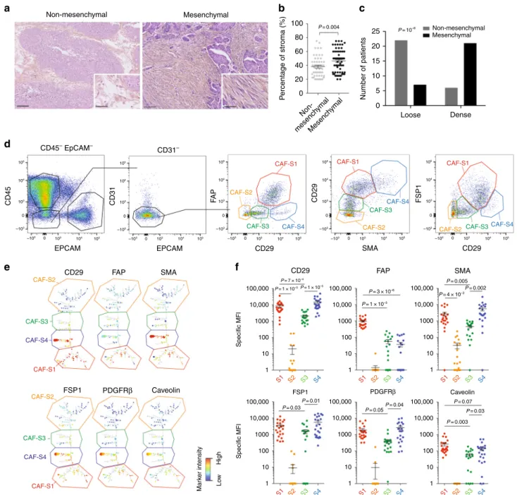
Mesenchymal HGSOC exhibit CAF heterogeneity. Gene
sig-natures defining HGSOC of the mesenchymal subtype are all
composed of stromal genes
6–12. We hypothesized that stroma
could play an important role in the development of mesenchymal
HGSOC. We
first evaluated stromal quantity and cellular density
in HGSOC. We observed that mesenchymal HGSOC exhibited
higher stromal content than non-mesenchymal tumors (Fig.
1
a,
b). Moreover, stroma from mesenchymal HGSOC was compact
and tight with high
fibroblast cellularity (defined as “dense”),
while non-mesenchymal tumors showed scattered and sprinkled
stroma with low cellularity (defined as “loose”) (Fig.
1
a, c). We
next aimed at performing deeper characterization of CAF in
HGSOC. To do so, we performed multicolor
flow-cytometry
(fluorescence-activated cell sorting (FACS)) (Fig.
1
) and
immu-nohistochemistry (IHC) analyses (Fig.
2
) (Table
1
for details on
retrospective cohorts) using concomitantly six different markers,
including FAP (fibroblast activation protein), CD29 (integrin-β1),
SMA (smooth muscle
α-actin), FSP1 (fibroblast-specific protein
1), PDGFRβ (platelet-derived growth factor receptor-β), and
caveolin (see Methods and Supplementary Table
1
for antibody
references). To our knowledge, no study analyzing simultaneously
six
fibroblast markers has ever been done in ovarian cancers.
Among viable cells detected by FACS using fresh human
HGSOC, we identified epithelial, immune, and endothelial cells
using EPCAM, CD45, and CD31 markers, respectively (Fig.
1
d,
left). Fibroblasts were considered as being part of the EPCAM
−
CD45
−CD31
−cells and were further characterized with the
above-mentioned
fibroblast markers (Fig.
1
d, right). Interestingly,
we distinguished four different CAF sub-populations in HGSOC,
according to CD29, FAP, FSP1, and SMA protein levels (Fig.
1
d,
right). These four CAF subsets were named S1 (red),
CAF-S2 (yellow), CAF-S3 (green), and CAF-S4 (blue) (Fig.
1
d, right).
To confirm the existence of the four different CAF subsets in
ovarian tumors, we used an unsupervised algorithm, named
Cytospade
25, an open source platform for network analysis that
organizes cells into hierarchies of related phenotypes. The trees
constructed by applying Cytospade to FACS data enabled us to
validate the presence of four CAF subsets in HGSOC
(Fig.
1
e). Two populations, CAF-S2 and CAF-S3, can be defined
as
“non-activated” CAF based on the lack of expression of SMA,
while both CAF-S1 and CAF-S4 expressed SMA and can be
considered as
“activated” CAF or myofibroblasts (Fig.
1
f).
CAF-S1
fibroblasts expressed high levels of all markers tested, as
opposed to CAF-S2 that were negative for all (Fig.
1
f). CAF-S3
and CAF-S4 were positive for specific but distinct markers
(Fig.
1
f). Indeed, CAF-S4 did not express FAP but showed high
levels of CD29 and SMA proteins, while CAF-S3 exhibited
intermediate to low levels of CD29 and SMA markers, but high
FSP1 protein levels. Of note, caveolin was not detected in CAF-S2
and showed low levels in the other three CAF subsets (Fig.
1
f),
indicating that this
fibroblastic marker was not helpful for
dif-ferentiating CAF subsets in HGSOC and will not be used further
in our study. Thus, CAF subsets can be defined by the following
profiles in HGSOC: CAF-S1: CD29
Med-HiFAP
HiSMA
Med-HiFSP1
Med-HiPDGFRβ
Med-HiCAV1
Low; CAF-S2: CD29
LowFAP-Neg
SMA
Neg-LowFSP1
Neg-LowPDGFRβ
Neg-LowCAV1
Neg;
CAF-S3: CD29
MedFAP
LowSMA
LowFSP1
Med-HiPDGFRβ
MedCAV1-Neg-Low
; CAF-S4: CD29
HiFAP
LowSMA
HiFSP1
HiPDGFRβ
Med-HiThe existence of the four CAF sub-populations was validated
by IHC on serial sections of HGSOC samples (Fig.
2
a), using
five
out of the six
fibroblast markers listed above, except caveolin (see
Supplementary Table
1
for list of antibody references and
Supplementary Fig.
1
a for isotype controls). We evaluated
histological scores (H score) for each marker in the stromal
compartment. We applied a decision tree algorithm (Fig.
2
b) to
determine the global CAF subset enrichment per tumor. In brief,
this decision tree was based on marker intensity thresholds,
first
defined according to FACS data (intensities of each CAF marker
in each CAF subset) from fresh HGSOC samples and next
transposed to IHC values (see Methods, #Development of a
a
d
e
f
b
c
Non-mesenchymal CD45– EpCAM– CD31– 105 105 104 104 103 103 –103 –103 0 105 104 103 –103 0 105 104 103 –103 0 105 104 103 –103 0 105 104 103 –103 0 0 –103 0 103 104 105 105 104 103 –103 0 105 104 103 –103 0 105 104 103 –103 0 CD45 CD31 FA P EPCAM CD29 FSP1 Caveolin Mar k er intensity Lo w High PDGFRβ FAP SMA 100,000 P= 7 × 10⁻⁴ P= 1 × 10⁻⁵P= 1 × 10⁻⁵ P= 3 × 10⁻⁶ P= 4 × 10⁻³ P= 0.005 P= 0.01 P= 0.04 P= 0.07 P= 0.03 P= 0.003 P= 0.05 P= 0.03 P= 0.002 P= 1 × 10⁻⁵ 10,000 1000 Specific MFI 100 10 1 100,000 FSP1 PDGFRβ Caveolin 10,000 1000 Specific MFI 100 10 1 100,000 10,000 1000 100 10 1 100,000 10,000 1000 100 10 1 100,000 10,000 1000 100 10 1 100,000 10,000 1000 100 10 1 S1 S2 S3 S4 S1 S2 S3 S4 S1 S2 S3 S4 S1 S2 S3 S4 S1 S2 S3 S4 S1 S2 S3 S4 EPCAM CD29 CD29 FAP SMA SMA CD29 CD29 FSP1 CAF-S2 CAF-S2 CAF-S2 CAF-S2 CAF-S2 CAF-S1 CAF-S1 CAF-S1 CAF-S1 CAF-S1 CAF-S3 CAF-S3 CAF-S3 CAF-S3 CAF-S3 CAF-S4 CAF-S4 CAF-S4 CAF-S4 CAF-S4 Non-mesenchymal Non-mesench ymal Mesench ymal Mesenchymal Mesenchymal 100 25 20 15 10 5 0 Loose Dense P= 0.004 P= 10–8 80 60 40 20 0 P ercentage of stroma (%) Number of patientsFig. 1 Identification of four CAF subsets in HGSOC. a Representative view of HES staining of non-mesenchymal and mesenchymal HGSOC sections (Institut Curie cohort). Scale bar, 100μm (low magnification) and 40 μm (inset). b Scatter plot showing the percentage of stroma in HGSOC. N = 107. Data are shown as mean ± SEM. P values are from Mann-Whitney test.c Bar plot showing association of mesenchymal HGSOC with stromal features, defined by pathologists as loose (low stromal cellularity) or dense (high stromal cellularity). N= 56. P value is from Fisher’s Exact Test. d Gating strategy to identify CAF subsets in HGSOC by FACS. Results from a representative HGSOC patient are shown. Cells isolated from freshly dissociated human HGSOC were first gated on DAPI−, EPCAM−, CD45−, CD31−cells, for excluding dead cells (DAPI+), epithelial cells (EPCAM+), hematopoietic cells (CD45+), and endothelial cells (CD31+). Selected cells were next examined using sixfibroblast markers. Representative flow cytometry plots show gating strategies based on FAP, CD29, SMA, and FSP1 that allow the identification of four sub-populations of fibroblasts in HGSOC: CAF-S1 are CD29MedFAPHighSMAHigh FSP1High, CAF-S4 are CD29HighFAPLowSMAHighFSP1Med, CAF-S3 are CD29LowFAPLowSMALowFSP1Med/Highand CAF-S2 are CD29LowFAPLowSMALow FSP1Low.e CytoSpade trees annotated with each marker expression in HGSOC analyzed by FACS. Colors show staining intensity for each marker. Size of the nodes is proportional to the number of cells showing similar staining for the markers analyzed.f Scatter plots showing specific mean fluorescent intensity (MFI) detected for each marker in each CAF sub-population. Each dot represents the specific median of fluorescent intensity of the cellular population by patient. N= 22. Data are shown as mean ± SEM. P values are from Student’s t-test
decision tree algorithm for prediction of CAF subset identity).
HGSOC were mainly enriched in activated CAF-S1 and CAF-S4
subsets (Fig.
2
c). Interestingly, mesenchymal HGSOC
accumu-lated more CAF-S1
fibroblasts than non-mesenchymal tumors
(Fig.
2
d). In parallel, we developed a method combining R script
and Fiji plugins to stack IHC staining of serial HGSOC sections.
This method provided maps, where each square corresponded on
average to one cell. Histological scores of the
five CAF markers in
each square defined CAF subset identity at cellular level and
allowed to visualize their geographic repartition within the tumor
1- If (FAP ≥ Q3) thenCAF-S1 2- Else
if(CD29 ≥ Q1 & FSP1≤ median) thenCAF-S4
3- Else
if(CD29 ≥ Q1 & < median) thenCAF-S3 if(CD29 ≥median)
thenCAF-S4 4- Else
if(CD29<Q1 and SMA<Q1) thenCAF-S2 5- ElseCAF-S3 All HGSOC 0 50 100 % Patients
c
Non-mesenchymalMesenchymal 0 50 100 % Patients P= 6.4 × 10⁻⁵d
CAF-S4-enriched tumor CAF-S1-enriched tumora
CAF-S4-enriched tumor CAF-S1-enriched tumor CD29 SMA FAP PDGFRβ FSP1 CD29 SMA FAP PDGFRβ FSP1e
CAF-S1 CAF-S2 CAF-S3 CAF-S4
b
g
CAF-S1 CAF-S2 CAF-S3 CAF-S4 CAF-S1 CAF-S2 CAF-S3 CAF-S4 S1 2 4 6 2 4 6 2 4 6 2 4 6 Distance to epithelium Long Short 0 50 100 % CAF subsets All HGSOC S2 S3 S4 Distance to epithelium S1 S3 S4 Distance to epithelium S1 S2 S3 S4 Distance to epithelium S1 S4 Distance to epithelium Distance to epithelium Long Short CAF-S4-enriched tumors CAF-S1-enriched tumors Epithelial cellsEpithelial cells Epithelial cells
CAF-S1 DAPI CD29 SMA FAP CAF-S4 CAF-S4 CAF-S4 CAF-S4 CAF-S4 CAF-S1 CAF-S1 CAF-S1 CAF-S1 CAF-S4 CAF-S1 CAF-S1 CAF-S1 CAF-S4 CAF-S4 DAPI CD29 SMA FAP CAF-S4 CAF-S1 CAF-S1 CAF-S4 CAF-S4 CAF-S4
f
(Fig.
2
e and Supplementary Fig.
1
b). In addition, we observed on
these maps that there was a significant enrichment of CAF-S1
cells at close proximity of cancer cells (Fig.
2
f), suggesting a
mutual potential benefit between CAF-S1 fibroblasts and cancer
cells, as recently shown in pancreatic cancers
26. Finally, we
confirmed the identity of CAF-S1 and CAFS4 cells by using triple
immunofluorescence staining of CD29, SMA, and FAP markers
on the same HGSOC sections (Fig.
2
g). Thus, IHC data (obtained
from a retrospective cohort of HGSOC patients) confirm FACS
observations described above on fresh HGSOC samples and
demonstrate for the
first time the existence of four distinct CAF
sub-populations in HGSOC, with strong accumulation of the
CAF-S1 subset in mesenchymal HGSOC.
FOXP3
+T lymphocytes accumulate in HGSOC enriched in
CAF-S1. By looking at several HGSOC tumor bed sections, we
observed that the stromal compartment accumulated more
lym-phocytes than the epithelial compartment (see representative
hematoxylin−eosin sections, Fig.
3
a). This led us to hypothesize
that
fibroblasts could play a key role in tumor-infiltrating
lym-phocyte (TIL) recruitment. We thus evaluated T lymlym-phocytes
density by performing CD3, CD8, and FOXP3 IHC staining and
counted the number of TILs per surface of stromal and epithelial
compartments (Fig.
3
b–d; Supplementary Fig.
2
a for isotype
controls). We validated that CD3
+, CD8
+, and FOXP3
+T
lymphocytes were indeed more often detected at the surface of
stroma than of epithelium compartment (Fig.
3
b–d), thereby
highlighting the potential role of CAF on T lymphocytes
infil-tration within tumor bed. We thus sought to investigate the role
of the two most detected
fibroblast subsets in HGSOC, CAF-S1,
and CAF-S4, on TIL recruitment, by comparing CAF-S1- and
CAF-S4-enriched tumors (Fig.
3
e–j). While the density of CD31
+blood vessel was similar in CAF-S1- and CAF-S4-enriched
HGSOC (Supplementary Fig.
2
b, c), the global content in CD3
+lymphocytes was higher in CAF-S1-enriched HGSOC than in
CAF-S4-enriched tumors (Fig.
3
e, f). This effect was driven by the
stroma, as no difference was observed in the epithelium (Fig.
3
f,
right). In contrast to CD3
+T lymphocytes, no significant
dif-ference in the total number of CD8
+cells was observed between
CAF-S1- and CAF-S4-enriched tumors (Fig.
3
g, h), although they
tended to accumulate in CAF-S1-enriched stroma (Fig.
3
h, right).
Interestingly, the most striking difference between CAF-S1- and
CAF-S4-enriched HGSOC was observed with FOXP3
+T
lym-phocytes (Fig.
3
i, j). Indeed, FOXP3
+T cells strongly
accumu-lated in CAF-S1-enriched HGSOC (Fig.
3
j), and this enrichment
was only seen in the stromal compartment (Fig.
3
j). As the total
surface of stroma was larger in CAF-S1-enriched HGSOC than in
CAF-S4-enriched tumors (as shown in Fig.
1
), we normalized the
number of CD3
+and FOXP3
+T lymphocytes per unit surface
area of stroma (Fig.
3
k, l) and observed that the content in CD3
+and FOXP3
+T cells remained significantly higher in the stroma
of CAF-S1-enriched HGSOC, independently of the stromal
content (Fig.
3
k, l). Finally, we took advantage of the CAF map
built on HGSOC (as shown in Fig.
2
e) and calculated the distance
between CAF subsets and CD3+ T lymphocytes (Fig.
3
m). By
comparing the number of CD3
+cells at the surface of each CAF
subset within HGSOC sections, we confirmed that the proportion
of CD3
+T lymphocytes was higher at the surface of CAF-S1-cells
compared to the other CAF subsets (Fig.
3
m). Altogether, these
observations show that HGSOC enriched in CAF-S1
fibroblasts
are highly infiltrated in particular by FOXP3
+T lymphocytes.
CXCL12β expression discriminates CAF-S1 from CAF-S4. In
order to uncover CAF-S1-mediated functions in T-cell
recruit-ment in HGSOC, we compared CAF-S1 and CAF-S4
tran-scriptomic profiles by performing RNA sequencing (RNA-Seq)
on sorted cells from fresh HGSOC using the gating strategy
shown in Fig.
1
(CAF-S1 and CAF-S4 RNAseq data from HGSOC
are available using EBI accession number: EGAS00001002184).
Unsupervised principal component analysis (PCA) (Fig.
4
a) and
hierarchical clustering (HC) (Fig.
4
b) of the 500 most variant
transcripts revealed molecular differences between CAF-S1 and
CAF-S4 subsets in HGSOC. DAVID analysis (
https://david.
ncifcrf.gov
), using GO (Gene ontology) and KEGG (Kyoto
Encyclopedia of Genes and Genomes) databases, of the
differ-entially expressed genes (CAF-S1-specific gene signature provided
in Supplementary Data
1
) showed that CAF-S1 cells were
enri-ched in genes involved in biological adhesion, wound healing
response, extracellular matrix (ECM) protein remodeling, and
skeletal system (Table
2
), while CAF-S4 gene signature
pin-pointed muscle contraction and blood vessel development
(Table
3
). Interestingly, genes expressed in CAF-S1 cells included
many genes enriched in the mesenchymal HGSOC subtype
(Fig.
4
c), thereby confirming that CAF-S1 fibroblasts could be key
components in this HGSOC molecular subtype. The common
genes between CAF-S1 and mesenchymal signatures encoded
ECM components and proteins involved in immune regulation,
such as complement factors (C1S and CFH), cytokines (TNFSF4),
and chemokines (CXCL12β) that could be involved in T-cell
recruitment. We got particularly interested in the detection of the
CXCL12 beta isoform (CXCL12β), as a transcript significantly
upregulated in CAF-S1 subset. Indeed, expression of CXCL12β
was specific of the CAF-S1 fibroblasts and almost undetected in
CAF-S4
fibroblasts. (Fig.
4
d). This differential expression was
specific of CXCL12β. Indeed, CXCL12α was expressed at similar
levels in both CAF-S1 and CAF-S4 subsets (Fig.
4
d, right). In
addition, genes that were either positively- or negatively
corre-lated with expression of the CXCL12β isoform completely
Fig. 2 Mesenchymal HGSOC accumulate mostly the CAF-S1 subset. a Representative views of CD29, SMA, FAP, PDGFRβ, and FSP1 immunostaining of serial sections in CAF-S4- or CAF-S1-enriched HGSOC. Scale bar, 100μm. b Decision tree used to define CAF identity, based on four equal quartiles (Q) and median (Mdn) distribution of each CAF marker intensity. Thresholds (Mdn, Q) and order of decisions werefirst established from FACS data of a prospective cohort of HGSOC patients (N= 22) and next transposed to values of IHC data, using a learning set of tumors containing both non-activated and activated CAF (N= 60). c Bar plot showing percentage of HGSOC according to the predominant CAF subset detected in each tumor. CAF enrichment per tumor is defined by applying the histological scores of all markers on the decision tree described in (b). HGSOC enriched in CAF-S1 (red), CAF-S2 (orange), CAF-S3 (green), or CAF-S4 (blue) are shown as percentage (%). N= 118 HGSOC patients. d Same as in (c) considering mesenchymal (N = 66) versus non-mesenchymal (N= 49) HGSOC. P values are from Fisher’s exact test. e Maps of CAF subsets at cellular level, corresponding to the tumor sections shown in (a). Each square of 225μm2corresponded on average to a single cell. Each CAF subset is represented by a color code and epithelial cells are in grey. The bar plot shows the percentage of HGSOC according to the predominant CAF subset evaluated on CAF maps at cellular level (N= 9). f Representative views of CAF maps, with their corresponding heatmaps showing the distances (shortest in red, farthest in yellow) between cancer cells and CAF subsets. Scatter plots show the distance to epithelial cells according to CAF subsets (distance calculated in a maximum area offive successive tiles in x and y). Data are shown as mean ± SEM (n= 425 cells per image in average). P values are from Student’s t-test. g Representative images showing triple immunofluorescence co-staining of CD29 (red), FAP (green), and SMA (violet) markers in HGSOC enriched in CAF-S1 (arrowheads) or CAF-S4 (arrows). Scale bar, 50μm
recapitulated CAF-S1 and CAF-S4 genetic signatures, respectively
(Fig.
4
e). In contrast, this was not the case for CXCL12α (Fig.
4
f),
thus confirming that only the CXCL12β isoform is discriminant
between CAF-S1 and CAF-S4 cells. As CXCL12 prognostic value
was highly controversial in HGSOC, with variable impacts on
patient survival
22–24, we hypothesized that the isoform-specific
regulation of CXCL12 in CAF subsets could be of particular
interest. We thus analyzed micro-arrays data from three
inde-pendent HGSOC cohorts (Curie, AOCS, and TCGA) (Table
1
)
and showed that high CXCL12β mRNA level was invariably
associated with poor patient survival (Fig.
4
g, for Curie Cohort;
Supplementary Fig.
3
a, for AOCS and TCGA cohorts, and
Sup-plementary Fig.
3
c for analyses by iteration). In contrast,
CXCL12α was not a reliable prognostic factor in HGSOC
(Sup-plementary Fig.
3
b). Importantly, when we considered the
expression of the two detected isoforms together (referred to as
total CXCL12 expression), we observed that the prognostic value
of total CXCL12 followed the one of CXCL12β (Supplementary
Fig.
3
e), arguing for the important role of the CXCL12β isoform
in HGSOC. Consistent with the poor prognosis associated with
high CXCL12β mRNA levels, we observed that CXCL12β
expression was higher in mesenchymal HGSOC compared to
non-mesenchymal HGSOC (Fig.
4
h). This was further validated
using contingency tables based on data from the three cohorts of
patients that showed a strong enrichment of HGSOC with high
CXCL12β mRNA levels among the mesenchymal HGSOC
(Sup-plementary Fig.
3
f). CXCL12α expression was also detected in
mesenchymal HGSOC (Supplementary Fig.
3
g), but the balance
between CXCL12β and CXCL12α expression—assessed by the
ratio of CXCL12β to CXCL12α mRNA levels—was significantly in
favor of the CXCL12β isoform in mesenchymal HGSOC (Fig.
4
i).
Finally, we studied CXCL12/CXCR4 protein expression patterns
in HGSOC by IHC (Fig.
4
j–l). As there is no available antibody
recognizing specifically the CXCL12β isoform, we had to use an
antibody recognizing both CXCL12 isoforms
24,27, for which we
validated the specificity (Supplementary Fig.
4
a−c). As expected,
we confirmed that CXCL12 proteins were mainly detected in the
stroma (Fig.
4
j), and its receptor CXCR4 at the surface of
epi-thelial cells (Fig.
4
k, arrows). CXCR4 was also detected in
endothelial and immune cells (Fig.
4
k, arrowheads), underlying
the role of the CXCL12/CXCR4 axis in HGSOC
microenviron-ment. We observed a huge diversity in CXCL12 histological
scoring in HGSOC (Fig.
4
l). Still, CXCL12 protein significantly
accumulated in mesenchymal HGSOC (Fig.
4
l), thereby
Table 1 Comparative description of the clinical parameters of the Institut Curie, AOCS and TCGA cohorts of HGSOC patients
CURIE AOCS TCGA CURIE
Type of analysis Transcriptomic Transcriptomic Transcriptomic IHC
Number of patients 107 285 484 118
Date of inclusion 1989−2005 1992−2006 1994−2011
Age at diagnostic
Median age (years) 58 59 59 60
Range (years) 31−87 22−80 30−87 35−80 Histotype Serous 82 (76.5%) 264 (92.6%) 484 (100%) 115 (97.4%) Endometrioi ̈d 8 (7.5%) 20 (7%) 2 (1.7%) Mucinous 8 (7.5%) 1 (0.8%) Clear cell 6 (5.5%) Carcinosarcoma 2 (2%) Brenner tumor 1 (1%) Adenocarcinoma 1 (0.4%) Figo substage I 21 (19.6%) 24 (8.4%) 7 (5.9%) II 10 (9.35%) 18 (6.3%) 24 (5%) 9 (7.6%) III 59 (55.14%) 217 (76.1%) 377 (77.9%) 82 (69.5%) IV 17 (15.9%) 22 (7.7%) 78 (16.1%) 13 (11.01%) NA 4 (1.4%) 5 (1%) 7 (5.9%) Grade 1 7 (6.5%) 19 (6.7%) 2 34 (31.5%) 97 (34%) 57 (11.8%) 31 (26.3%) 3 66 (62%) 164 (57.5%) 415 (85.7%) 87 (73.7%) NA 5 (1.8%) 12 (2.5%) Surgery Full 38 (36%) 84 (29.5%) 88 (18.2%) 32 (27.1%) Partial 69 (64%) 164 (57.5%) 339 (70%) 82 (69.5%) NA 37 (13%) 57 (11.8%) 4 (3.4%) Clinical response RC—Complete response 51 (47.7%) 273 (56.4%) 47 (39.8%) RP—Partial response 22 (20.6%) 57 (11.8%) 31 (26.3%) S—Stability 7 (6.5%) 25 (5.2%) 7 (5.9%) P—Progression 11 (10.3%) 36 (7.4%) 3 (2.6%) NA 16 (15%) 93 (19.2%) 30 (25.4%)
Thefirst three columns recapitulate clinical parameters from cohorts used for transcriptomic data analyses. TCGA, AOCS and Curie cohorts have previously been described6–8. The last column concerns
samples used for immunohistochemistry analyses. Tumor samples were obtained from a cohort of consecutive ovarian carcinoma patients, treated at the Institut Curie between 1989 and 2012. All analyzed samples have been collected prior to any chemotherapeutic treatment. Indeed, for each patient, a surgical specimen was taken, before chemotherapy, for pathological analysis and tumor tissue cryopreservation. The median patient’s age was 60 years (with a range of 35–80 years). Ovarian carcinomas were classified according to the World Health Organization histological classification of
gynecological tumors. Pathological analysis identified 115 high-grade serous tumors (97.4%), 2 high-grade endometrioïd tumors (1.7%) and 1 high-grade mucinous tumor (0.8%). Sixteen subjects
(13.5%) were considered as early stage (International Federation of Gynecology and Obstetrics (FIGO) I−II) and 95 subjects (80.5%) were considered as advanced stage (III and IV) of disease. Patients
were treated with a combination of surgery and chemotherapy, the latter including alkylating or alkylating-like agents ± taxane as afirst-line treatment in most cases. All the subjects underwent surgery,
confirming transcriptomic data. In contrast, no difference was
observed for CXCR4 protein levels between mesenchymal and
non-mesenchymal HGSOC (Fig.
4
l, right). Finally, by performing
in situ hybridization using the RNAscope
®technology, we
observed that CXCL12 mRNA was mainly expressed by stromal
cells (Fig.
4
m), observation that was confirmed by using publicly
available data set from primary cell lines (GSE49910 from Gene
Expression Omnibus resources) (Supplementary Fig.
5
). Taken as
a whole, these data indicate that the CXCL12β isoform
accumu-lates more than CXCL12α in mesenchymal HGSOC, consistent
with the accumulation of CAF-S1
fibroblasts in these tumors. All
these features, i.e. mesenchymal molecular subtype, CAF-S1
a
b
e
f
h
j
k
l
m
g
i
c
d
1000CAF-S4-enriched tumors CAF-S1-enriched tumors
CD3 CD8 FO XP3 P= 9.2 × 10⁻¹¹ P= 3 × 10–07 P= 3.2 × 10⁻¹⁰ 1 10 100 1000 1 10 100 1000 1 10 100 10,000 P= 0.004 P= 0.01 P= 0.05 P= 0.14 P= 0.8 (n.s.) P= 0.027 P= 0.046 P= 0.03 P= 0.01 P= 0.04 P= 0.03 P= 0.02 P= 0.07 P= 0.56 (n.s.) P= 5.6 × 10⁻⁶ P= 10–4 P= 9.6 × 10⁻¹⁰ P= 3.0 × 10⁻⁴ P= 1.0 × 10⁻⁴ P= 1.4 × 10⁻⁸ P= 0.85 (n.s.) P= 0.97 (n.s.) P= 1.5 × 10–08 1 Total Total Epithelium Epithelium Epithelium Stroma Stroma Stroma 10 100 1000 100 10 1 1000 10,000 1000 100 10 1 CAF-S1 CAF-S2 CAF-S3 CAF-S4 CD3 Epithelial cells 10,000 1000 100 10 1 1000 100 10 1 80 60 40 20 0 Epith S1 S2 S3 S4 1000 100 10 1 100 10 1 1000 100 10 1 1000 100 10 1 1000 10,000 1 10 100 1000 EpitheliumStroma Epithelium
CAF-S4 CAF-S4 CAF-S1 CAF-S1 Total CAF-S4 CAF-S1
CAF-S4 CAF-S1 CAF-S4 CAF-S1 CAF-S4 CAF-S1 CAF-S4 CAF-S1
CAF-S4 CAF-S1 CAF-S4 CAF-S1 CAF-S4 CAF-S1 CAF-S4 CAF-S1 CAF-S4 CAF-S1 CAF-S4 CAF-S1 Stroma EpitheliumStroma CD3 + cells per mm 2 CD8 + cells per mm 2 CD3 + cells per mm 2 CD8 + cells per mm 2 FO XP3 + cells per mm 2 CD3 + cells per mm 2 of epithelium FO XP3 + cells per mm 2 of stroma FO XP3 + cells per mm 2 of epithelium Nb of CD3
+ cells per section
at surf
ace of
Epithelium or CAF subsets
CD3 + cells per mm 2 of stroma FO XP3 + cells per mm 2
accumulation and CXCL12β expression, are hallmarks of dismal
prognosis for HGSOC patients.
CAF-S1 enhance regulatory T-cell activity at tumor site. As
regulatory T lymphocytes preferentially infiltrate
CAF-S1-enriched stroma, we wondered whether CAF-S1 subset
expres-sing high levels of CXCL12β could favor FOXP3
+T lymphocytes
accumulation by increasing their attraction, enhancing their
survival and/or promoting their activation state, all being
non-exclusive hypotheses. We investigated the impact of CAF-S1
fibroblasts on T-cell attraction by performing in vitro transwell
assays. To do so, we isolated CAF-S1 and CAF-S4
fibroblasts
from ovarian tumors. While CAF-S1
fibroblasts proliferated well
and kept their properties in vitro (Supplementary Fig.
6
a−d),
CAF-S4
fibroblasts quickly died and could not be maintained in
culture, thereby precluding any comparison between CAF-S1 and
CAF-S4
fibroblasts in functional assays. Still, we succeeded in
isolating CAF-S1 primary cells that enabled us to analyze in vitro
the impact of primary CAF-S1
fibroblasts on CD4
+CD25
−and
CD4
+CD25
+T cells isolated from peripheral blood mononuclear
cells (PBMC) of healthy donors (Supplementary Fig.
6
e). We
first
observed that migration of CD4
+CD25
+but not CD4
+CD25
−cells was increased in presence of primary CAF-S1
fibroblasts
(Fig.
5
a), while the survival of the two types of T lymphocytes was
not affected by distant CAF-S1
fibroblasts (Fig.
5
b). Consistent
with CXCL12 function in this process, CXCR4 receptor was
detected at higher levels at the surface of CD4
+CD25
+T cells
than CD4
+CD25
−T lymphocytes (Fig.
5
c, d). Moreover, CXCR4
level at the surface of CD4
+CD25
+T lymphocytes was increased
upon co-culture with CAF-S1
fibroblasts (Fig.
5
c, d, right),
sug-gesting that the CXCL12/CXCR4 axis could be activated in
T-lymphocytes in presence of CAF-S1
fibroblasts and required for
CAF-S1-induced CD4
+CD25
+T-lymphocyte attraction. As
CAF-S1 cells specifically accumulate the CXCL12β isoform, but
also express CXCL12α, we next silenced each of these isoforms in
CAF-S1
fibroblasts (Fig.
5
e) to address their respective function in
CD4
+CD25
+cell attraction. The silencing of CXCL12α decreased
T-cell attraction (Fig.
5
f), confirming previous reports
28,29.
Interestingly, CXCL12β knockdown in CAF-S1 cells, which
mimicked CAF-S4 cells as they express CXCL12α but not
CXCL12β, significantly reduced CD4
+CD25
+T-lymphocyte
attraction (Fig.
5
f). Importantly, silencing both isoforms showed
an additive effect and reduced T-cell attraction that returned to
basal level (i.e. without CAF-S1 (Fig.
5
f)). Thus, co-silencing of
the two isoforms was both necessary and sufficient to completely
abrogate T-lymphocyte attraction by CAF-S1 cells (Fig.
5
f),
indicating that in addition to CXCL12α, the specific expression of
CXCL12β in CAF-S1 is absolutely essential for efficient
T-lymphocyte attraction. While expression of chemokines, such as
CCL17, CCL22, CCL5, CCL28, and CXCL9, well known to be
involved in recruitment of regulatory T lymphocytes
30was not
detected in CAF-S1, CCL2 expression was highly expressed by
CAF-S1 cells (RNAseq data available EGAS00001002184,
Sup-plementary Data
1
for CAF-S1 signature). We thus tested
whe-ther this owhe-ther chemokine could be involved in T-cell attraction
by CAF-S1, besides CXCL12, and found that, in contrast to
CXCL12, CCL2 inhibition in CAF-S1
fibroblasts (Supplementary
Fig.
6
f for silencing efficiency) had no impact on CD4
+CD25
+T-lymphocyte attraction (Fig.
5
g).
To get further insights on the impact of CAF-S1 cells on CD4
+
CD25
+T lymphocytes, we next performed co-culture
experi-ments (Fig.
5
h–m). We observed that the direct contact of
CAF-S1
fibroblasts with CD4
+CD25
+T cells significantly increased
the proportion of CD25
+FOXP3
+T cells among CD4
+T
lymphocytes (Fig.
5
h, i). Moreover, CAF-S1 cells enhanced the
survival of CD25
+FOXP3
+T lymphocytes (Fig.
5
j). Direct
contact of CD25
+FOXP3
+T lymphocytes with CAF-S1 was
required for this effect on T-cell survival, as no impact was
observed in Transwell assays (as shown above Fig.
5
a).
Impor-tantly, the increase in the number of CD25
+FOXP3
+T cells by
CAF-S1 cells was conserved when it was reported to the survival
(Fig.
5
k), suggesting that CAF-S1
fibroblasts increase the global
content of CD25
+FOXP3
+T cells by enhancing their survival
and differentiation. These effects were not affected by the
combined silencing of the two CXCL12 isoforms in CAF-S1
(Fig.
5
h–k), indicating that the two CXCL12 isoforms are
required for attracting CD4
+CD25
+T lymphocytes, but not for
enhancing survival or differentiation into CD25
+FOXP3
+T cells.
Considering the increase of CD25
+FOXP3
+T cells upon
co-culture with CAF-S1
fibroblasts, we next sought to verify if this
effect was associated with an increased T-cell suppressive activity.
To do so, we isolated CD4
+CD25
HighCD127
lowCD45RA
lowT
lymphocytes, strongly enriched in regulatory T cells, and
evaluated their impact on CD4
+effector T cells following
co-culture with CAF-S1
fibroblasts (Fig.
5
l). Interestingly, we found
that the pre-culture of CD25
HighCD127
lowCD45RA
lowT
lym-phocytes with CAF-S1
fibroblasts significantly enhanced their
capacity to inhibit effector T-cell (CD4
+CD25
−) proliferation
rate (Fig.
5
l). Consistent with the increase in CD25
+FOXP3
+T cells, these data suggest that CAF-S1 cells could increase
immunosuppressive activity of regulatory T lymphocytes. Finally,
to get better insights in the mechanisms mediated by CAF-S1 cells
on the differentiation of CD25
HighFOXP3
HighT cells, we took
advantage of the CAF-S1 RNAseq data (EGAS00001002184) and
identified different molecules highly expressed by CAF-S1 cells
that could be involved in this effect. Among them, we observed
that the silencing of CD73/NT5E, B7H3/CD276 and IL6 in
CAF-S1
fibroblasts (Supplementary Fig.
6
f for silencing efficiency)
significantly reduced the proportion of CD25
HighFOXP3
HighT-lymphocyte population (Fig.
5
m, n). Taken as a whole, these data
indicate that, in addition to CXCL12α, the expression of
CXCL12β by CAF-S1 fibroblasts is essential for T-cell attraction
towards CAF-S1-enriched HGSOC. Once in contact, CAF-S1
Fig. 3 CAF-S1-enriched HGSOC accumulate FOXP3+T lymphocytes.a Representative views of HES staining of HGSOC tumor bed sections (Institut Curie cohort) showing lymphocytes accumulation at the surface of the stroma. Scale bar, 100μm (low magnification) and 50 μm (inset). b−d Scatter plots showing the number of CD3+(b), CD8+(c), and FOXP3+(d) lymphocytes per mm2in epithelial and stromal compartments in HGSOC. N= 80 HGSOC (Institut Curie). P values are from Wilcoxon signed-rank test.e, g, i Representative views of CD3+(e), CD8+(g), and FOXP3+(i) immunostaining in HGSOC enriched in CAF-S4 or CAF-S1. Scale bar, 50μm. f, h, i Number of immune cells per mm2in CAF-S1- and CAF-S4-enriched HGSOC, considering either the whole sections referred to as Total or the epithelial and stromal compartments. Positive cells for each staining were counted manually in at least 5−10 fields per tumor at ×20 magnification. The median is indicated. Mann-Whitney statistical test was performed to compare CAF-S1- versus CAF-S4-enriched tumors and Wilcoxon paired test was used to compare epithelial and stromal compartments within tumors. N= 80 HGSOC (Institut Curie). k, l Scatter plots showing the number of CD3+(k) and FOXP3+(j) T lymphocytes per mm2relative to the stromal or epithelial content per tumor, in CAF-S1-or CAF-S4 enriched HGSOC. P values are from Mann-Whitney test.m Representative views of CAF maps, with the corresponding heatmap showing the localization of CD3+T lymphocytes (0.25 mm2). The bar plot shows the number of CD3+T lymphocytes detected at the surface of epithelial cells (Epith) or of each CAF subset cell (n= 936 total cells per image, five images from different HGSOC were analyzed). P values are from Student’s t-test
fibroblasts enhance the survival and the proportion of CD25
+
FOXP3
+T lymphocytes, through at least CD73, B7H3 and IL-6.
CXCL12β RNA is targeted by miR-141/200a in CAF-S4 cells.
As CXCL12β expression in CAF-S1 is essential for T-cell
attrac-tion, we next wondered the mechanism driving its specific
expression in CAF-S1 cells, compared to CAF-S4 cells. We
con-sidered the possibility that CXCL12β could be downregulated in
CAF-S4 cells by miRNA. Indeed, gene signatures differentiating
mesenchymal (high CAF-S1 content) to non-mesenchymal
HGSOC (high CAF-S4 content) were based on miR-141 and
200a family members
7,13. We thus hypothesized that CXCL12β
a
d
h
g
j
l
m
k
i
e
f
b
c
PC2: 18% v a riance Nor maliz ed read counts 20 20 40 0 0 –20 –20 PC1: 46% variance –40 10,000 CXCL12β CXCL12α P= 0.03 P= 0.12 (n.s.) 1000 100 100 15 10 5 0 15 10 5 0 500 400 300 200 100 0 500 400 300 200 100 0 15 1.5 1.0 0.5 0.0 10 5 0 P= 0.01 Curie Curie Curie AOCS TCGA Low CXCL12β High CXCL12β CXCL12 β CXCL12 β / CXCL12 α mRNA le v els P ercent sur viv al CXCL12 CXCL12 H score CXCL12 RNA b y ISH CXCR4 H score CXCR4 50 0 0 50 100 150 Time (month) Non-mesenchymal Non-mesenchymal Mesenchymal Mesenchymal 200 250 Non-mesench ymal Non-mesench ymal Non-mesench ymal Non-mesench ymal Mesench ymal Mesench ymal Mesench ymal Non-mesench ymal Mesench ymal Non-mesench ymal Mesench ymal Mesench ymal 10 1 10,000 1000 100 10 1108 679 535 69 365 144 565 500 190 244 GO: GO: KEGG: Cell adhesion No significant pathway No significant pathway No significant pathway GO: GO: KEGG: KEGG: KEGG: Cell adhesion response to woundingBlood vessel development muscle contraction
Vascular smooth muscle contraction ECM–receptor interaction P= 1.3 × 10–52 P= 10–282 P= 5 × 10–27 P= 10–154 P= 1.4 × 10⁻⁸ P= 10–16 P= 10–16 P= 0.04 P= 0.05 P= 0.2 (n.s.) 431 278 1
CAF-S4 CAF-S1 CAF-S4 CAF-S1
CAF-S1
87
GO: Cell adhesion ECM organization
P= 10–50
KEGG: focal adhesion
50 659 > 1.9 0 < –2 CAF-S4 Mesenchymal signature Genes correlated
with CXCL12β Genes correlatedwith CXCL12α
Genes anti-correlated with CXCL12α Genes anti-correlated with CXCL12β CAF-S1 signature CAF-S1
CAF-S1 signature CAF-S1 signature
CAF-S4 signature CAF-S4 signature
CAF-S4
mRNA could be targeted by miR-141/200a in CAF-S4 cells,
leading to its specific accumulation in CAF-S1 fibroblasts. We
identified two predicted miR-141/200a binding sites in the
3′-UTR of CXCL12β mRNA, which were absent in CXCL12α mRNA
(Fig.
6
a). We
first observed that the CXCL12β 3′-UTR contained
genuine miR-141/200a binding sites (Fig.
6
b). Moreover,
over-expression of miR-141 or miR-200a in CAF-S1
fibroblasts
reduced the total level of endogenous CXCL12β mRNA, but had
no impact on CXCL12α (Fig.
6
c). Furthermore, the CXCL12β
mRNA level was inversely correlated with the 141 and
miR-200a in HGSOC (Fig.
6
d). Consistently, miR-141 and miR-200a
significantly accumulated in non-mesenchymal HGSOC (Fig.
6
e),
which exhibited low levels of CXCL12β mRNA. We and others
have demonstrated that the miR-200 family members are strongly
upregulated by oxidative stress in various cell types, including
fibroblasts
7,13,31–34.
We
thus
wondered
whether
non-mesenchymal HGSOC, accumulating CAF-S4
fibroblasts, could
suffer from oxidative stress. We compared the amount of reduced
(GSH) and oxidized (GSSG) glutathione in non-mesenchymal
and mesenchymal HGSOC (Fig.
6
f). We observed that
non-mesenchymal HGSOC significantly accumulated more GSSG
compared to the mesenchymal ones (Fig.
6
f). Accordingly, while
GSSG and GSH showed similar proportion in mesenchymal
HGSOC, the proportion of GSSG tended to be significantly
higher than GSH in non-mesenchymal HGSOC (Fig.
6
f, right).
Using Gene Set Enrichment Analysis, we detected a strong
enrichment in genes encoding electron transport chain proteins
in CAF-S4 RNAseq data compared to CAF-S1, confirming an
oxidative metabolism in CAF-S4 cells (Fig.
6
g). The small amount
of material obtained from CAF-S1 and CAF-S4 cells sorted by
FACS from HGSOC precluded direct measurement of miR-200
levels in these cells. We thus took advantage that expression of the
miR-141 is strikingly correlated with the transcription of its
immediate upstream gene PTPN6
13, and used PTPN6 as a
read-out of miR-141 expression level in CAF-S1 and CAF-S4. PTPN6
expression was significantly higher in S4 compared to
CAF-S1
fibroblasts (Fig.
6
h), suggesting that miR-141/200c expression
was also upregulated in CAF-S4. Accordingly, three previously
identified targets of miR-141/200a
7,35,36showed a significant
downregulation in CAF-S4
fibroblasts compared to CAF-S1
(Fig.
6
i), similarly to CXCL12β. Taken as a whole, these data
established that the CXCL12β isoform is specifically targeted by
the 200 family members, while CXCL12α is not. The
miR-200 are upregulated in non-mesenchymal HGSOC enriched in
CAF-S4
fibroblasts. Subsequently, the CXCL12β isoform is
spe-cifically downregulated in CAF-S4 fibroblasts, while it
accumu-lates in CAF-S1 cells, further enhancing attraction of CD4
+CD25
+
T lymphocytes (see model Fig.
6
j).
Discussion
Here, we uncover CAF heterogeneity in HGSOC by identifying
four different CAF subpopulations, including two myofibroblast
subsets, referred to as CAF-S1 (CD29
Med-HiFAP
HiSMA
Med-HiFSP1
Med-HiPDGFRβ
Med-HiCAV1
Low) and CAF-S4 (CD29
HiFAP
LowSMA
HiFSP1
HiPDGFRβ
Med-HiCAV1
Neg-Low).
Mesenchymal HGSOC, the molecular subgroup of ovarian
can-cers with poor patient prognosis, exhibit high content of CAF-S1
fibroblasts. CXCL12 is required for CAF-S1-mediated
T-lymphocyte attraction in HGSOC, consistent with previous
observations in pancreatic cancers
37,38. In addition, we highlight
that the CXCL12β isoform, but not CXCL12α, is a key component
for differentiating the CAF-S1 from the other activated CAF-S4
fibroblasts in HGSOC. This specific expression of CXCL12β in
CAF-S1 is essential for T-cell attraction towards
CAF-S1-enriched HGSOC, and thus could be required for CAF-S1
immunosuppressive function. Once in contact, CAF-S1
fibro-blasts enhance the survival, as well as the activation of regulatory
T lymphocytes, independently of CXCL12. Finally, we uncover
the mechanism driving the specific expression of CXCL12β in
CAF-S1. The CXCL12β isoform is regulated by an
miR-141/200a-dependent mechanism that downregulates CXCL12β expression
in CAF-S4
fibroblasts and promotes its specific accumulation in
the CAF-S1 subpopulation (see model, Fig.
6
j).
Based on genetic and transcriptomic analyses, it is now well
established that HGSOC are composed of heterogeneous
mole-cular entities. All studies based on ovarian cancer transcriptomic
profiles
6–13systematically identified a group of HGSOC defined
by a mesenchymal signature that is invariably associated with
poor patient prognosis. This signature contains many
stromal-related genes
6–13, suggesting that the stroma could be an
important feature for the aggressiveness of these tumors.
Inter-estingly, both SMA and PDGFRβ markers, used in our study,
belong to several mesenchymal signatures
6,7. While high
pro-portion of CAF is associated with poor prognosis in HGSOC
patients
17,18,20,39–41, little is known about their identity. Some
studies have analyzed markers, such as SMA, FAP or PDGFRβ,
individually and show a certain degree of heterogeneity in ovarian
cancers
15,42–44. Our current study confirms these observations
but goes beyond by identifying heterogeneous CAF subsets and
characterizing them at histological and molecular levels. To do so,
we combined the use of six different markers, previously tested
individually but never studied concomitantly
45–48. To our
knowledge, the present study is the
first that identifies four
dif-ferent CAF subpopulations in HGSOC, including CAF-S1 and
CAF-S4, which express high levels of polymerized SMA and can
be defined as myofibroblasts. Still, they exhibit different
tran-scriptomic profiles, arguing for distinct functions.
Fig. 4 CXCL12β discriminates CAF-S1 and CAF-S4 cells. a PCA based on the 500 most variant transcripts differentiating CAF-S1 (red) and CAF-S4 (blue). b HC (500 most variant transcripts) using Ward’s method with Euclidean distances. Each column represents a CAF subset and each row a gene. Color saturation shows gene expression deviation from the mean (above in red, below in blue).c Venn diagram showing overlap between mesenchymal signature (defined in ref.7) and CAF-S1 signature (Supplementary Data 1). P value is from hypergeometric test.d Scatter plots of CXCL12α (NM_000609) or CXCL12β (NM_199168) mRNA levels in CAF-S1 and CAF-S4 subsets. e, f Venn diagrams showing overlap between genes correlated or anti-correlated with CXCL12β (e) or CXCL12α (f) and CAF-S1 or CAF-S4 signatures (Supplementary Data1and2). P values are from hypergeometric test.g Kaplan−Meier curves of overall survival according to low- and high-CXCL12β mRNA levels (N = 53 in low-CXCL12β subgroup and N = 54 patients in high-CXCL12β subgroup, Institut Curie). P value is based on log-rank test.h Scatter plots showing CXCL12β mRNA levels in mesenchymal and non-mesenchymal HGSOC from the Institut Curie, AOCS, and TCGA cohorts. Data (log2 of probeset (203666_at) intensity) are shown as mean ± SEM. P values are from Mann-Whitney test.i Scatter plot showing ratio of CXCL12α and CXCL12β expression levels in mesenchymal and non-mesenchymal HGSOC of the Institut Curie cohort. Data are shown as mean ± SEM. P values are from Mann-Whitney test.j, k Representative views of CXCL12 (j) and CXCR4 (k) immunostaining in HGSOC. Scale bar, 50μm (low magnification) and 20 μm (inset). l Scatter plots showing histological scores (H score) of CXCL12 and CXCR4 proteins. H score corresponds to the percentage of positive cells (in CAF and at epithelial cell surface, arrows inj, k) multiplied by the staining intensity. Data are shown as mean ± SEM. P values are from Mann-Whitney test.m Representative view of CXCL12 mRNA detected infibroblasts by in situ hybridization, using RNAscope®Technology on HGSOC tissue section. Scale bar, 20μm (low magnification) and 6 μm (inset)
Table 2 Signi
ficant enriched pathways for genes upregulated in CAF-S1 versus CAF-S4
GO_Biological Process-Term Count % Genes P value FDR
GO:0007155~cell adhesion 79 11.67 NRP2, AEBP1, TLN2, CXCL12, CDSN, SDC3,
CHAD, WISP1, CTGF, COL12A1, ROBO2, COL11A1, BOC, NEGR1, CDH23, SPON1, CYR61, PDPN, CDHR1, SIGLEC11, PCDH7, CERCAM, JUP, CD34, LSAMP, CPXM1, CLDN1, CNTN1, ROR2, VCAN, JAM2, CHL1, CLDN18, PLXNC1, COL3A1, PTK7, NINJ2, ITGB5, SPOCK1, CDH5, DCHS1, ITGBL1, ISLR, IGSF11, ANXA9, FAT4, ITGB8, COMP, COL6A3, SCARB1, COL8A1, COL8A2, THBS2, MLLT4, FLRT2, SVEP1, LRRN2, PPFIBP1, NLGN1, HSPG2, COL16A1, TPBG, COL5A1, COL4A6, EMILIN1, CCL11, LAMA1, OMD, DSG2, PKP2, CDON, FBLN5, DSC3, DSC2, ADAM22, ANTXR1, BMPR1B, NTM, CDH11
1.39E-20 2.44E-17
GO:0001501~skeletal system development
36 5.32 RBP4, AEBP1, PTGS2, FGF9, COL3A1, GLI2,
MMP2, GLI1, CHAD, VDR, CTGF, COMP, COL12A1, COL11A1, PAPSS2, RUNX2, COL10A1, EVC, FBN1, HSPG2, IGF1, ANKH, HOXC10, PTHLH, SMO, CTSK, COL1A2, PDGFRA, GDF10, ROR2, KIAA1217, COL1A1, BMPR1B, IGFBP3, BMP6, CDH11
1.72E-9 3.01E-6
GO:0030198~extracellular matrix organization
19 2.80 LUM, COL3A1, HSPG2, CCDC80, DCN,
SPINK5, COL5A1, COL4A6, EMILIN1, P4HA1, FBLN5, COL1A2, PDGFRA, COL12A1, LOX, COL1A1, COL11A1, COL8A2, CYR61
1.71E-8 2.99E-5
GO:0016337~cell-cell adhesion 29 4.28 CLDN18, NINJ2, CDSN, DCHS1, CDH5,
CHAD, ANXA9, FAT4, CTGF, ROBO2, COL11A1, COL8A2, CDH23, PDPN, CDHR1, NLGN1, PCDH7, CERCAM, JUP, DSG2, CD34, PKP2, CLDN1, DSC3, DSC2, ROR2, BMPR1B, JAM2, CDH11
3.93E-7 6.87E-4
GO:0009611~response to wounding 41 6.05 C7, TLR1, COL3A1, F2RL1, NINJ2, TLR4,
C1S, BDKRB2, GPR68, LMAN1, MDK, CFHR1, HMCN1, SLC1A3, NOD1, CTGF, HMOX1, SERPINE1, CFH, SCARB1, LOX, CFI, PAPSS2, SCG2, NOX4, TNFSF4, PDPN, IGF1, COL5A1, CCL11, SMO, PRKCQ, CD55, SDC1, FBLN5, PDGFRA, VCAN, BMPR1B, GAP43, BMP6, MYH10
3.43E-6 0.006
GO:0030199~collagenfibril organization 9 1.33 P4HA1, LUM, COL3A1, COL1A2, COL12A1,
COL1A1, LOX, COL11A1, COL5A1
4.58E-6 0.008
GO:0001944~vasculature development 25 3.69 NRP2, FGF9, LEPR, COL3A1, FOXO1,
CXCL12, MMP2, CDH5, SHB, ANG, CTGF, HMOX1, PLCD3, SEMA3C, HS6ST1, LOX, SCG2, CYR61, PDPN, MMP19, COL5A1, VEGFC, SMO, COL1A2, COL1A1
7.47E-6 0.01
GO:0001649~osteoblast differentiation 10 1.48 PTHLH, SMO, FGF9, IGF1, COL1A1, GLI2, IGFBP3, RUNX2, GLI1, BMP6
1.11E-5 0.02
GO:0000902~cell morphogenesis 30 4.43 NRP2, SHROOM3, UCHL1, PTK7, GLI2,
EPHB3, CXCL12, EPHB2, DAB2, SLC1A3, UNC5B, ROBO2, ROBO3, CDH23, NOX4, EGR2, PDPN, KIF5C, PRKCI, HGF, NTN1, GAS7, SMO, LAMA1, SEMA6A, VCAN, ANTXR1, BMPR1B, GAP43, MYH10
1.96E-5 0.03
GO:0007411~axon guidance 15 2.21 NRP2, EGR2, KIF5C, EPHB3, GLI2, CXCL12,
NTN1, EPHB2, SEMA6A, UNC5B, ROBO2, ROBO3, BMPR1B, GAP43, MYH10
1.99E-5 0.03
GO:0035295~tube development 22 3.25 RBP4, SHROOM3, PDPN, FGF9, PTK7,
IGF1, GLI2, CXCL12, GLI1, FOXP2, MYCN, WNT2, PTHLH, WNT4, GPC3, CTGF, PDGFRA, TGIF1, ROBO2, HS6ST1, LOX, CYR61
2.80E-5 0.04
GO:0006928~cell motion 35 5.17 NRP2, CTHRC1, IL16, PTGS2, SPOCK1,
GLI2, EPHB3, CXCL12, EPHB2, WNT2, UNC5B, CTGF, ANG, SEMA3C, ROBO2, SCARB1, ROBO3, SCG2, EGR2, KIF5C, IGF1, CERCAM, NTN1, SLC9A10, COL5A1, ELMO1, SMO, SEMA6A, LAMA1, VEGFC, CD34, VCAN, BMPR1B, GAP43, MYH10
5.61E-5 0.09
KEGG_Pathway-Term Count % Genes P value FDR
hsa04512:ECM-receptor interaction 16 0.26 COL3A1, HSPG2, ITGB5, COL5A1,
COL4A6, CHAD, SDC3, LAMA1, SDC1, ITGB8, COMP, COL6A3, COL1A2, COL1A1, COL11A1, THBS2
4.18E-7 4.89E-4
hsa04360:Axon guidance 18 0.30 ABLIM1, PLXNC1, LIMK2, LIMK1, ABLIM3,
LRRC4C, EPHB3, NTN1, CXCL12, EPHB2, SEMA6A, UNC5B, SEMA7A, SRGAP3, SEMA3C, ROBO2, EFNA4, ROBO3
5.84E-6 6.82E-3
Both CAF-S1 and CAF-S4 subsets are the most frequent
sub-populations detected in mesenchymal HGSOC, suggesting they
categorize distinct patients and could exert different functions.
CAF-S1 cells are likely to exert immunosuppressive activities that
could account for the poor survival of mesenchymal HGSOC
patients. We
first observed that the stromal compartment shows a
higher number of T lymphocytes (especially regulatory T cells),
compared to the epithelium, suggesting that CAF are more prone
to attract immune cells than cancer cells. This statement has
previously been described in murine models and in several
human cancers
38,49–52. In that sense, FAP
Poscells have been
shown to promote immunosuppression by triggering an
inflam-matory phenotype, further enhancing the recruitment of
myeloid-derived suppressor cells (MDSC)
50–53. Accordingly, we
demon-strate here that CAF-S1
fibroblasts exhibit immunosuppressive
capacities in vitro acting on CD25
+FOXP3
+T lymphocytes at
three levels by enhancing their attraction, survival, differentiation,
and suppressive activity. CAF-S1 cells have thus functional
similarities to MDSC, well-known to display immunomodulatory
functions
54–57. Human MDSC are commonly described as
Lin-CD11b
+ CD33 + HLADR− cells, with neutrophilic or
mono-cytic phenotype
57. In contrast, none of the markers characterizing
MDSC, such as CD11b
+or CD33
+, are detected in CAF-S1 cells
(EBI study accession number: EGAS00001002184). Furthermore,
we show that the CXCL12 chemokine is a key player involved in
attraction of regulatory T cells in human HGSOC, confirming
previous studies showing the role of CXCL12 in
immunosup-pression
37,38. These effects are independent of
CXCL12-pro-angiogenic function
27,58,59, as we detected them using functional
assays in vitro. Accordingly, CXCL12/CXCR4 blockade reduces
recruitment of regulatory T lymphocytes in immuno-competent
mouse models of cancers
28,29,37,38. In contrast, we did not
observe tumor growth inhibition upon CXCR4 inhibitor
(AMD3100) treatment in immuno-deficient patient-derived
xenograft (PDX) models of ovarian cancer (Supplementary
Fig.
7
), suggesting that immunosuppressive functions mediated
by both CXCL12α and CXCL12β isoforms on T lymphocytes
could be crucial for ovarian tumor growth. In that sense,
Table 3 Signi
ficant enriched pathways for genes upregulated in CAF-S4 versus CAF-S1
GO_Biological Process-Term Count % Genes P value FDR
GO:0001568~blood vessel development
29 7.02 CAV1, PDGFA, PGF, CSPG4, ENPEP, JAG1,
GJA4, PTEN, GJC1, SEMA5A, PTK2, AGT, ANGPT1, ADRA2B, MKL2, FGF1, PLXND1, ANGPT2, COL18A1, FLT1, EPAS1, APOLD1, ITGA4, ARHGAP24, CDH13, LAMA5, PLXDC1, NTRK2, ITGA7
5.45E-12 9.40E-9
GO:0001525~angiogenesis 21 5.08 COL18A1, FLT1, EPAS1, PDGFA, PGF,
CSPG4, APOLD1, JAG1, ENPEP, ARHGAP24, PTEN, SEMA5A, CDH13, PTK2, LAMA5, PLXDC1, ANGPT1, ADRA2B, PLXND1, FGF1, ANGPT2
3.27E-10 5.64E-7
GO:0007010~cytoskeleton organization
35 8.47 DLC1, CAV2, TPPP3, CAV1, PDGFB, CALD1,
ARPC5, PRKG1, DAAM2, DSTN, PTK2, EZR, PACSIN2, MICAL1, CAP1, EHD2, FGD4, ARHGDIB, ACTC1, CAP2, ROCK1, MAP1B, ARHGEF17, MYOZ1, VASP, ARHGAP26, MARK1, ARPC1A, PLCE1, EPB41L1, EPS8, LAMA5, MAP2, MYH11, SYNM
1.10E-9 1.90E-6
GO:0007517~muscle organ development
22 5.32 MEF2C, CAV2, CAV1, MYL4, ACTC1,
TBX2, UTRN, RXRG, CACNB2, CSRP2, PTEN, GJC1, FAM65B, MEF2D, LAMA5, PLN, ITGA7, MYH11, RARB, AGRN, MKL2, SGCA
3.19E-8 5.51E-5
GO:0044057~regulation of system process
25 6.05 CAV1, MYL4, LZTS1, SLC6A1, PPP1R12B,
ADA, SYP, EDNRB, KCNQ4, AGT, GUCY1A3, HRC, FLT1, NTF3, EPAS1, MAP1B, ATP1A2, SSTR2, PLCE1, P2RX1, PLN, NTRK2, HSPB7, NPTN, CACNA1C
3.75E-7 6.47E-4
GO:0042692~muscle cell differentiation
15 3.63 CAV2, ACTC1, NTF3, TBX2, UTRN,
CACNB2, MYOZ1, JAG1, CSRP2, AGT, MYH11, SORT1, RARB, AGRN, MKL2
1.08E-6 0.002
GO:0009190~cyclic nucleotide biosynthetic process
8 1.93 ADCY3, ADCY4, ADCY1, ADCY5, ADCY6,
GUCY1A2, GUCY1A3, GUCY1B3
1.35E-6 0.002
GO:0042310~vasoconstriction 7 1.69 ACTG2, EDNRB, CAV1, ACTC1, P2RX1,
ACTA2, AGT
1.72E-6 0.003
GO:0009187~cyclic nucleotide metabolic process
9 2.17 ADCY3, ADCY4, ADCY1, NUDT4, ADCY5,
NUDT4P1, ADCY6, GUCY1A2, GUCY1A3, GUCY1B3
2.48E-6 0.004
GO:0006936~muscle contraction 16 3.87 MYL4, ACTC1, ACTA2, CALD1, UTRN,
VIPR1, GJC1, EDNRB, ACTG2, SSTR2, AGT, MYH11, MYOM1, CACNA1C, HRC, SGCA
3.69E-6 0.006
GO:0050880~regulation of blood vessel size
10 2.42 CAV2, ACTG2, EDNRB, CAV1, ACTC1,
P2RX1, ACTA2, AGT, GUCY1A3, HBB
3.87E-6 0.007
GO:0014706~striated muscle tissue development
14 3.38 CAV2, ACTC1, CAV1, TBX2, UTRN, RXRG,
CACNB2, PTEN, GJC1, PLN, MYH11, RARB, MKL2, AGRN
5.01E-6 0.008
KEGG_Pathway-Term Count % Genes P value FDR
hsa04270:Vascular smooth muscle contraction
21 5.08 ADCY3, ADCY4, ADCY1, ROCK1, ACTA2,
PPP1R12B, ADCY5, CALD1, ADCY6, MRVI1, PRKG1, KCNMB1, ACTG2, GUCY1A2, PPP1R12A, MYH11, GUCY1A3, GUCY1B3, CACNA1C, MYLK, PLA2G5
4.00E-11 4.55E-8