HAL Id: hal-01057027
https://hal.inria.fr/hal-01057027
Submitted on 21 Aug 2014
HAL is a multi-disciplinary open access
archive for the deposit and dissemination of
sci-entific research documents, whether they are
pub-lished or not. The documents may come from
teaching and research institutions in France or
L’archive ouverte pluridisciplinaire HAL, est
destinée au dépôt et à la diffusion de documents
scientifiques de niveau recherche, publiés ou non,
émanant des établissements d’enseignement et de
recherche français ou étrangers, des laboratoires
Interactive Visualization of Muscle Activity During
Limb Movements: Towards Enhanced Anatomy Learning
Armelle Bauer, Florent Paclet, Violaine Cahouet, Ali Hamadi Dicko, Olivier
Palombi, François Faure, Jocelyne Troccaz
To cite this version:
Armelle Bauer, Florent Paclet, Violaine Cahouet, Ali Hamadi Dicko, Olivier Palombi, et al..
Interac-tive Visualization of Muscle Activity During Limb Movements: Towards Enhanced Anatomy Learning.
VCBM 2014 - Eurographics Workshop on Visual Computing for Biology and Medicine (VCBM 2014),
Sep 2014, Vienne, Austria. pp.191-198, �10.2312/vcbm.20141191�. �hal-01057027�
Eurographics Workshop on Visual Computing for Biology and Medicine (2014) I. Viola, K. Bühler, and T. Ropinski (Editors)
Interactive Visualization of Muscle Activity During Limb
Movements : Towards Enhanced Anatomy Learning
Armelle Bauer1,2,3, Florent Paclet4, Violaine Cahouet4, Ali-Hamadi Dicko1,2,5, Olivier Palombi1,6, François Faure1,2,5, Jocelyne Troccaz3,5
1INRIA,2LJK-CNRS,3TIMC-IMAG,4GIPSA-lab,5Univ. Grenoble Alpes,6LADAF
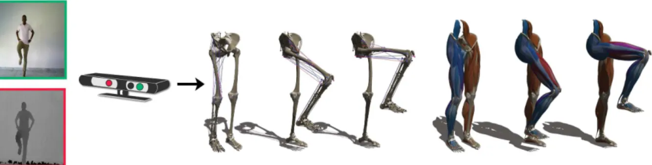
Figure 1: Interactive display of muscle activations during knee flexion. Left: a user moving in front of the Kinect sensor. Middle:
activation displayed on action lines. Right: display on 3D meshes.
Abstract
We propose a framework to investigate a new way to learn musculoskeletal anatomical kinetics using interactive motion capture and visualization. It can be used to facilitate the learning of anatomy by medicine and sports students, and for the general public to discover human anatomy in action. We illustrate our approach using the example of knee flexion and extension by visualizing the knee muscle activation prediction with agonist and an-tagonist co-contraction.
Muscle activation data for specified movements is first measured during a preliminary phase. The user is then tracked in real time, and its motion is analyzed to recognize the motion being performed. This is used to efficiently evaluate muscle activation by interpolating the activation data stored in tables. The visual feedback consists of a user-specific 3D avatar created by deforming a reference model and animated using the tracking. Muscle activation is visualized using colored lines of action or 3D meshes.
This work was made possible by the collaboration of three complementary labs specialized in computer-aided medical intervention, computer graphics and biomechanics.
Keywords : Anatomy Learning, Biomechanical Simulation, Real-Time, Augmented Reality, Embodiment, Motion
Capture and Reconstruction
1. Introduction
Learning functional anatomy and understanding anatomical kinematics is important for medical studies, sport education as well as for education more generally. However, this learn-ing process remains tedious and difficult due to the
complex-ity of the domain which includes a large amount of struc-tured, static and dynamic notions. Understanding the muscu-loskeletal system and its mobility usually requires 3D visuo-spatial abilities [HCD∗14,HCR∗09]. An important difficulty
ob-Bauer et al. / Interactive Visualization of Muscle Activity During Limb Movements : Towards Enhanced Anatomy Learning
jects from books, drawings, photos, or the dissection of ca-davers [GCB∗07]. With the emergence of new technologies
such as 3D modeling and animation, Augmented Reality (AR) systems, etc, new approaches of learning through in-teractions have been proposed [FBE∗13,PR13,KBSC14] to
ease the learning process and the construction of mental rep-resentations of anatomy. Our work is based on two assump-tions :
• Space visualization is fundamental to the understanding and the learning of complex articulated 3D shapes. • Our motor system influences our cognition (Embodiment
theory)
Assuming that the use of one’s own body could make the learning process of musculoskeletal biomechanics more effi-cient led us to sketch the "Living Book of Anatomy" (LBA), an AR system to visualize the relevant anatomical knowl-edge of the trainee body in action. The key idea is to capture a user action (a limb motion for instance) and to visualize its effect on the internal anatomy within an AR paradigm : for instance, the internal structures are superimposed on the image of the trainee. Developing the LBA requires a novel combination of technologies including motion cap-ture, anatomical knowledge modeling, user-to-model map-ping, AR rendering. In this paper, we tackle the challenge of real-time motion capture and muscle activation display using a novel combination of state-of-the-art approaches.
Our biomechanical model is created using an ontology of anatomical structures (MyCorporisFabrica) [PUF∗14,
DGFP12] connected with a complete set of 3D geometrical model of the human body [BUD∗14]. User motion is
cap-tured using a Kinect sensor associated with standard soft-ware and complemented with additional filtering. To avoid intensive, ill-posed inverse dynamics computations, muscle activation is queried in tables based on pose and velocity. Muscles are rendered in real-time as deformed 3D shapes using vertex blending methods, or as colored lines to high-light activation. Our key contribution is the real-time visual-ization of the human body musculoskeletal system kinetics and muscle activity.
The remainder of the paper is organized as follows. In Section2, we briefly survey the related work covering medi-cal learning and real-time applications. In Section3we then describe in detail the proposed pipeline and the input data we are working on. We present and discuss our results in Section 4. We finally conclude and sketch future work in Section5.
2. Related Work
Motion capture of the human body has been a subject of great interest in computer graphics, vision, biomechanics, films and medical applications, and force space reasons we can only scratch the surface of this domain. People gen-erally try to use marker-less motion capture systems
be-cause of the lower price and the simple setup [CSC14]. This approach has also gained popularity in the biomechan-ics community [CSC14,CGMA10,CMC∗06]. Our project
is based on it in order to provide an application everyone could use easily and at a low cost. Some recent papers an-alyze motion capture using Kinect sensors and compare it with the real motion captured with more sophisticated hard-ware [Red13,EPnB∗13].
A lot of studies were carried on about anatomy learn-ing [DRC∗06,PR13,KBSC14,SM08], and 3D models have
become increasingly popular [HCD∗14,HCR∗09,GCB∗07].
Augmented reality (AR), which consists in superimposing data on visible objects, has been increasingly used in the medical domain [DHLD∗02,JBC08,Spe12]. Our project is
inspired by the work of Nassir Navab on the Magic Mirror project [FBE∗13,BKBN12]. We extend it using
biomechan-ical data related to user motion. 3. Materials and Methods
Our method combines data from three sources:
• Muscle activation data for specified movements, mea-sured during a preliminary phase (Sec.3.1)
• Real-time user motion capture and muscle activation eval-uation (Sec.3.2)
• A 3D avatar displayed in the same pose as the user, and highlighting muscle activation (Sec.3.3)
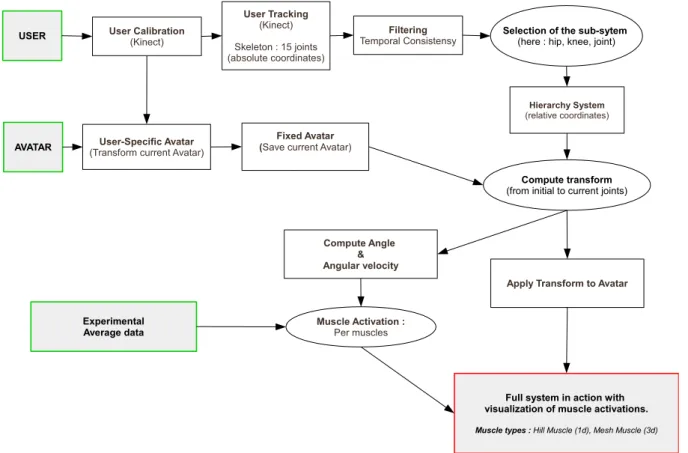
Figure2summarizes the pipeline of the method. In this ex-periment we focus on muscle activation during knee flex-ion/extension for anatomy learning purpose. We used My-CorporisFabrica [BUD∗14] to select the entities (muscles
and bones) involved in the knee flexion and extension. 3.1. Measuring activation
To evaluate musculoskeletal movement, a motion capture system is sufficient. However, muscle and/or tendon ten-sion is difficult to evaluate due to redundancies in the mus-culoskeletal system. Indeed there are more actuators (mus-cles) than degrees of freedom in joints. This leads to an under-determined system of equations, resulting in an infin-ity of possible muscular coordination with different syner-gies and co-contraction patterns to achieve the same task. In this work, motion capture and identification are applied to the flexion-extension of the lower limb, and our goal is to evaluate and display the corresponding muscle activations. To solve the redundancy problem, we use experimental mea-surement data to feed a biomechanical model of the lower limb based on anatomical and functional knowledge, in or-der to compute knee muscle activations including agonist and antagonist co-contraction. The acquisition of the knee flexion muscle activation was made only on the right seg-ment of the human body, since the muscle activation should be the same for each leg.
USER User Calibration (Kinect) Filtering Temporal Consistensy User Tracking (Kinect) Skeleton : 15 joints (absolute coordinates)
AVATAR (Transform current Avatar)User-Specific Avatar
Fixed Avatar (Save current Avatar)
Compute transform
(from initial to current joints)
Selection of the sub-sytem
(here : hip, knee, joint)
Hierarchy System (relative coordinates) Compute Angle & Angular velocity Experimental Average data Muscle Activation : Per muscles
Full system in action with visualization of muscle activations.
Muscle types : Hill Muscle (1d), Mesh Muscle (3d) Apply Transform to Avatar
Figure 2: Pipeline of the application.
Database construction An experimental acquisition is per-formed on a single healthy subject (48yo / 186cm / 77kg) to provide muscle activation patterns corresponding to flex-ion and extensflex-ion. The motflex-ion capture is performed during squat and no-load lower limb flexion-extensions using 12 Vi-con cameras sampled at 250Hz. At the same time, the elec-tromyographic activity of the mono and bi-articular flexor and extensor knee muscles is recorded at 2000 Hz (total 10 muscles) using a Biopac MP150 device.
The subject is standing on a force plate (force plate data not used in this work) and performs right leg flex-ion/extension cycles either in squat or no-load conditions
(see picture3) at three (low, medium and high) velocities. The subject performs 6 flexion/extension cycles in each con-dition. Pairs of silver-silver chloride surface electrodes were used to record the raw activity from the 10 main muscles pro-ducing the knee torque : Vastus Medialis (vast_med), Vastus Lateralis (vast_lat), Rectus Femoris (rect_fem), Tensor Fas-ciae Latae (tfl), Sartorius (sartorius), Gracilis (gracilis), Bi-ceps Femoris (bi_fem_lh), Semi Tendinosus (semiten), Gas-trocnemius (med_gas and lat_gas).
The electrodes are positioned parallel to the muscle fibers
Figure 3: Photos of the experiment.
with an inter-electrode distance of 15mm following care-ful skin preparation to reduce the skin impedance according to the SENIAM (Surface EMG for Noninvasive Assessment of Muscles) recommendations (www.seniam.org). The elec-trodes over the lower-limb muscles are placed centrally over the muscle bellies following palpation of a resisted
isomet-Bauer et al. / Interactive Visualization of Muscle Activity During Limb Movements : Towards Enhanced Anatomy Learning
ric contraction. The ground electrode is placed over the right iliac crest in front of the ilium bone.
Data analysis The Y and Z lateral displacements of each marker are filtered using a fourth-order Butterworth filter with zero phase lag and a cut-off frequency of 4 Hz. Inter-segmental angles are calculated at ankle, knee and hip joints according to the convention shown in Figure4.
Figure 4: Three-link model of the lower limb.
The derivatives of angular position are calculated at each time t using a centred finite difference scheme. Finally, kine-matic values are averaged to give a mean cycle for each con-dition (Figure5).
Figure 5: Hip, knee and ankle angles (rad) evolution during
a mean cycle.
The data is first demeaned then filtered using a Butter-worth filter (4th order, band pass 20-450Hz) with no lag es-sentially to remove movement artefacts. RMS is calculated using a 250ms sliding window with overlapping to get the envelope of the EMG signal. RMS values is normalized us-ing Maximal RMS value (RMSmax) detected across all trials
for each muscle i :
RMSl(t) =
RMSi(t)
RMSmaxi
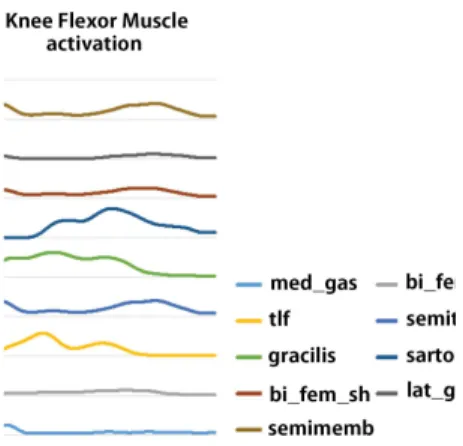
Finally the normalized RMS signal is averaged to pro-vide a mean cycle for each condition, and is filtered using a Butterworth filter (4th order, low pass 4Hz) with no lag to obtain an activation coefficient aifor each knee muscle. The activation of deep muscles (Biceps femoris: short head (bi_fem_short), semimembranosus (semimem), vastus inter-medius (vast_int)), which EMG activities cannot be mea-sured using surface electrodes, is estimated using synergist neighbor muscles. This data can then be used to provide
suit-Figure 6: Activation coefficient of knee flexor muscles
dur-ing a mean cycle.
able information for anatomical structures animation in the LBA.
3.2. Motion capture
We use a Kinect with NiTE and the OpenNI SDK [Ope] to track the user actions. The process starts with a calibra-tion (Sec.3.2.1). We then enter the main application loop. At each time, the user tracking provides us with joint an-gles (Sec.3.2.2), and the motion is analyzed (Sec.3.2.3). 3.2.1. Calibration
In the process of Kinect calibration we find the subject pa-rameters and we transform a reference 3D anatomical avatar to match the user in the learning environment. We use the T-pose (3D avatar and Kinect initial position) to calibrate the user. Figure7illustrates the skeleton used for the initializa-tion.
NiTE provides a user bounding box which we use to find two anthropometric values : the user breadth (width of the bounding box) and the user height [ECA14]. Based on the user and avatar breadth and height we compute the width and height scale factors. To get the third dimension (depth)
Figure 7: NiTE tracking skeleton (15 bones).
we use a ratio between the width and height scale factors. Using these scale factors we find the transformation matrix to deform the reference avatar to fit the user global size, as illustrated in Figure8.
Figure 8: user-specific calibration (user, reference avatar,
user-specific avatar)
3.2.2. Tracking
After the calibration we compute smooth rotation interpola-tions between the frames, while maintaining the distances between the joints. We only take into account the joints needed to control our system : hip joint, knee joint and foot joint.
NiTE re-computes skeleton joints at each frame based on the depth image. Unfortunately, the Kinect sensor offers an imprecise depth image which leads to limits in precision for the skeleton joints. To avoid inconsistencies during the track-ing we save the initial skeleton at initialization time, and we enforce constant distance constraints between the joints dur-ing the trackdur-ing. Before applydur-ing our hierarchical model, we smooth the individual bone trajectories using a 5x5 convolu-tion matrix. The filter is coupled with Dual-quaternion Lin-ear Blending [Ken12,KCZO07] for multiple rotation inter-polations. Each joint is composed of a position (3D vector) and an orientation (quaternion). We separate the joint posi-tions and orientaposi-tions to take only into account the joint ro-tations in the hierarchical joint model. This results in smooth rotation interpolations between frames and avoids jiggle ef-fects.
Figure 9: on the left skeleton tracking without filter, on the
right skeleton tracking with filter
3.2.3. Motion analysis
To avoid complex inverse dynamics computations, the mus-cle activations are retrieved from recorded data, based on joint angles and velocities. To fit our experimental data (Sec.3.1) we have to compute the angle and velocity of the tracked user as well. The current version considers only the hip. To compute the user angle we transform the hip quater-nion to Euler angles and we peek the angle related to the x axis. The angular velocity is computed using finite differ-ences.
Figure 10: experiment and Kinect : hip angle (rad) evolution
during a mean cycle (flexion/extension)
The angular velocity is negative during flexion and posi-tive during extension. Thus based on the angular velocity we know if the user is performing flexion or extension. Then, we use the hip angle to find activation per muscle using a linear interpolation of the experimental data. In the current version, we do not consider the value of the velocity. 3.3. Motion and activation visualization
The mapping from the user to the model/avatar is performed by attaching bones and muscles to the skeleton using Linear
Bauer et al. / Interactive Visualization of Muscle Activity During Limb Movements : Towards Enhanced Anatomy Learning
Blend Skinning. The muscles can be visualized in two ways: 1D muscles (muscle line of action) [The03] and 3D muscles. • 1D (line of action) Hill muscles : to see all muscles be-haviors (even underneath muscles like the "vastus inter-medius"). Using anatomy transfer [AHLG∗13], we adapt
the muscles of the OpenSim [AWLD10,DAA∗07] model
to our system.
• 3D muscle meshes : for more realistic muscles visualiza-tion. Our 3D anatomy avatar provides meshes of lower limb muscles.
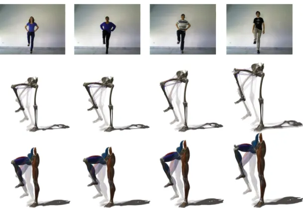
We display muscle activation using a smooth color map from blue (no activation) to red (maximum activation). In theory, muscle activation is a percentage value between 0.0 and 1.0. To better highlight muscle activations, we choose to use the "experimental" maximum instead of "theoretical" maximum to 1.0. For a complete visualization we display all the lower limb entities. Figure12shows the visualization (1D and 3D muscles activation) for 4 of our users. More results are in the video attached.
4. Results and discussion
Our framework is implemented in C++ and runs on a com-modity laptop PC. The real-time capture and activation dis-play run at 25 Hz, with a lag of less than 0.1 s. The visual feedback is currently performed on the screen of the PC, as shown in the accompanying video.
The motion sequences were acquired for 6 young adult men with an average height of 1.81m, an average of weight of 85.3kg, and 4 young adult women with an average height of 1.60m and an average weight of 55.7kg. All of them with-out any type of injury or physical incapacity in their extrem-ities. To get uniform results we work with Kinect sequences made in the same room and with the same lighting condi-tions.
Gender Age Height Weight
F 27 1.59 48 F 23 1.60 50 F 24 1.60 58 F 31 1.63 67 H 30 1.72 74 H 27 1.76 110 H 29 1.82 80 H 26 1.83 72 H 25 1.85 100 H 24 1.91 76
Table 1: Kinect sequence users exact data
Scaling the reference model takes about 4 seconds, then the interactive visualization begins. The user performs flex-ions and extensflex-ions and sees in real-time his/her muscle ac-tivations on the screen.
Figure 11: key positions of our sequence from the Kinect
point of view
We have studied different kinds of flexion-extension movements with various velocities and loading conditions to create a set (library) of averaged lower limb movement references to feed the LBA. This library of muscle activa-tion patterns is used to provide the feedback about the knee muscles for functional anatomy learning. In future work, this experimental process will be greatly improved using addi-tional measurements of muscle activation in maximal vol-untary contraction situation. Using an isokinetic ergome-ter, these activations will be related to knee moment level in various isometric, concentric and eccentric contractions. This knowledge will allow us to more accurately normal-ize EMG signals acquired during flexion-extension move-ments. Finally, the development of a biomechanical model of the lower limb coupled with inverse dynamics, optimiza-tion procedure and EMG informaoptimiza-tion could produce a bet-ter muscle tension estimation in all kind of flexion-extension movement.
The Kinect sensor provides an imprecise depth image which leads to limits in precision for the skeleton joints. This leads to an approximate motion feedback for the user. A bet-ter filbet-tering of the Kinect skeleton [WZC12,MCA07], or a completion of depth sensor data are good response to this problem.
Our user calibration is simplistic while the final goal of our project is to provide a really realistic user-specific avatar. Computing our avatar using real-time anatomy trans-fer [AHLG∗13,GRP10] and anthropometric measurements
[ECSCUQ14] would be a good alternative. An other alter-native would be to combine the Kinect skeleton and silhou-ette [RVHT12,CPLFR13].
5. Conclusion
Our framework is ready to validate the theory of Embod-iment. In this paper we presented a first innovative appli-cation for the visualization of human body kinetics in real-time, by displaying muscle activation. We believe that with accurate anatomically-based models and realistic motion, learning anatomy will be eased.
Figure 12: users : color and depth map, 1D muscles visualization, 3D muscles visualization
short term, we want to improve the personalization of the avatar to reinforce embodiment. We also hope to visualize information on other limbs and their motions. In the longer term, we plan to automatically detect the user motion and deliver knowledge accordingly.
6. Acknowledgments
Many thanks to Estelle Charleroy and Laura Paiardini our Graphist Artists for their great job on the video. To thanks Jonathan Dumon for allowing us to use the MOCA experi-mental platform. To all our kinect subjects.
This work has been partially supported by the LabEx PERSYVAL-Lab (ANR-11-LABX-0025).
References
[AHLG∗13] ALI-HAMADID., LIUT., GILLESB., KAVANL., FAUREF., PALOMBIO., CANIM.-P.: Anatomy transfer. ACM
Transactions on Graphics (proceedings of ACM SIGGRAPH ASIA) 32, 6 (2013).6
[AWLD10] ARNOLDE. M., WARDS. R., LIEBERR. L., DELP
S. L.: A model of the lower limb for analysis of human move-ment. Annals of biomedical engineering 38, 2 (Feb. 2010), 269– 79.6
[BKBN12] BLUM T., KLEEBERGER V., BICHLMEIER C.,
NAVABN.: mirracle: An augmented reality magic mirror system
for anatomy education. 2012 IEEE Virtual Reality (VR) (Mar. 2012), 115–116.2
[BUD∗14] BAUER A., ULLIANA F., DICKO A. H., GILLES B., PALOMBIO., FAUREF.: My Corporis Fabrica : Making Anatomy Easy. In Siggraph 2014 Talks (Vancouver, Canada, May 2014).2
[CGMA10] CORAZZA S., GAMBARETTO E., MÜNDERMANN
L., ANDRIACCHI T. P.: Automatic generation of a subject-specific model for accurate markerless motion capture and biomechanical applications. IEEE transactions on bio-medical
engineering 57, 4 (Apr. 2010), 806–12.2
[CMC∗06] CORAZZA S., MÜNDERMANN L., CHAUDHARI
A. M., DEMATTIOT., COBELLIC., ANDRIACCHI T. P.: A
markerless motion capture system to study musculoskeletal biomechanics: visual hull and simulated annealing approach.
An-nals of biomedical engineering 34, 6 (June 2006), 1019–29.2 [CPLFR13] CHAARAOUI A. A., PADILLA-LOPEZ J. R.,
FLOREZ-REVUELTAF.: Fusion of Skeletal and Silhouette-Based Features for Human Action Recognition with RGB-D Devices.
2013 IEEE International Conference on Computer Vision Work-shops(Dec. 2013), 91–97.6
[CSC14] CESERACCIUE., SAWACHAZ., COBELLIC.: Compar-ison of markerless and marker-based motion capture technolo-gies through simultaneous data collection during gait: proof of concept. PloS one 9, 3 (Jan. 2014), e87640.2
[DAA∗07] DELP S. L., ANDERSON F. C., ARNOLD A. S., LOANP., HABIBA., JOHNC. T., GUENDELMANE., THELEN
D. G.: OpenSim: open-source software to create and analyze dynamic simulations of movement. IEEE transactions on
Bauer et al. / Interactive Visualization of Muscle Activity During Limb Movements : Towards Enhanced Anatomy Learning
[DGFP12] DICKO A. H., GILLES B., FAURE F., PALOMBI
O.: From Generic to Specic Musculoskeletal Simulations us-ing an Ontology-based Modelus-ing Pipeline. In Intelligent
Com-puter Graphics 2012, Plemenos D., Miaoulis G., (Eds.), vol. 441
of Studies in Computational Intelligence. Springer, May 2012, pp. 227–242.2
[DHLD∗02] DAVISL., HAMZA-LUPF. G., DALY J., HAY., FROLICHS., MEYERC., MARTING., NORFLEETJ., LIN K.-C., IMIELINSKAC., ROLLANDJ. P.: Application of augmented reality to visualizing anatomical airways. Proc. SPIE 4711,
Helmet- and Head-Mounted Displays VII 4711 (Aug. 2002),
400–405.2
[DRC∗06] DAMSGAARDM., RASMUSSENJ., CHRISTENSENS.
R. T.R., SURMAE.,DEZEEM.: Analysis of musculoskeletal
systems in the AnyBody Modeling System. Simulation
Mod-elling Practice and Theory 14, 8 (Nov. 2006), 1100–1111.2
[ECA14] ESPITIA-CONTRERASA. SANCHEZ-CAIMAN P. U.-Q. A.: Development of a Kinect-based anthropometric measure-ment application. Virtual Reality (VR), 2014 iEEE (Mar. 2014), 71–72.4
[ECSCUQ14] ESPITIA-CONTRERAS A., SANCHEZ-CAIMAN
P., URIBE-QUEVEDOA.: Development of a Kinect-based an-thropometric measurement application. 2014 IEEE Virtual
Real-ity (VR)(Mar. 2014), 71–72.6
[EPnB∗13] ENCALADAF., PEÑAFIELJ., BECERRAP., IDROVO A., CARREÑOJ., FLORESJ.: Acquisition and analysis of angles between the hip and knee during the sit-up process through the kinect sensor. 2013 IEEE pan american health care exchanges
(PAHCE). Conference, workshops, and exhibits.(2013).2
[FBE∗13] FALLAVOLLITAP., BLUMT., ECKU., SANDORC., WEIDERTS., WASCHKEJ., NAVABN.: Kinect for interactive AR anatomy learning. 2013 IEEE International Symposium on
Mixed and Augmented Reality (ISMAR)(Oct. 2013), 277–278.2 [GCB∗07] GUILLOTA., CHAMPELYS., BATIERC., THIRIET P., COLLETC.: Relationship between spatial abilities, mental rotation and functional anatomy learning. Advances in health
sciences education : theory and practice 12, 4 (Nov. 2007), 491– 507.2
[GRP10] GILLESB., REVERETL., PAID.: Creating and ani-mating subject-specific anatomical models. Computer Graphics
Forum 29, 8 (Dec. 2010), 2340–2351.6
[HCD∗14] HOYEK N., COLLET C., DI RIENZO F., DE ALMEIDAM., GUILLOTA.: Effectiveness of three-dimensional digital animation in teaching human anatomy in an authentic classroom context. Anatomical sciences education 1, 1982 (Mar. 2014), 1–8.1,2
[HCR∗09] HOYEKN., COLLETC., RASTELLOO., FARGIERP., THIRIETP., GUILLOTA.: Enhancement of mental rotation abil-ities and its effect on anatomy learning. Teaching and learning
in medicine 21, 3 (July 2009), 201–6.1,2
[JBC08] JUANC., BEATRICEF., CANOJ.: An Augmented Re-ality System for Learning the Interior of the Human Body. 2008
Eighth IEEE International Conference on Advanced Learning Technologies(2008), 186–188.2
[KBSC14] KAMPHUIS C., BARSOM E., SCHIJVEN M., CHRISTOPH N.: Augmented reality in medical education?
Perspectives on medical education(Jan. 2014).2
[KCZO07] KAVANL., COLLINSS., ZARAJ., O’SULLIVANC.: Skinning with dual quaternions. 39–46.5
[Ken12] KENWRIGHTB.: Dual-Quaternions From Classical
Me-chanics to Computer Graphics and Beyond. 1–11.5
[MCA07] MUNDERMANN L., CORAZZA S., ANDRIACCHI
T. P.: Accurately measuring human movement using articu-lated icp with soft-joint constraints and a repository of articuarticu-lated models. 2013 IEEE Conference on Computer Vision and Pattern
Recognition 0(2007), 1–6.6
[Ope] OpenNi library. URL: http://structure.io/ openni.4
[PR13] PETERSOND., ROBERTSONC.: Utilizing virtual reality in teaching and training: Progress toward virtual surgical simula-tions. In INTED2013 Proceedings (4-5 March, 2013 2013), 7th International Technology, Education and Development Confer-ence, IATED, pp. 3285–3291.2
[PUF∗14] PALOMBIO., ULLIANAF., FAVIERV., LÉONJ.-C., ROUSSETM.-C.: My Corporis Fabrica: an ontology-based tool for reasoning and querying on complex anatomical models.
Jour-nal of biomedical semantics 5(Jan. 2014), 20.2
[Red13] Analysis of movements during the process of the march to ascend stairs by means of sensor. 2013 IEEE pan american
health care exchanges (PAHCE). Conference, workshops, and ex-hibits.(2013).2
[RVHT12] RICHTER M., VARANASI K., HASLER N., THEOBALT C.: Real-Time Reshaping of Humans. 2012 Second International Conference on 3D Imaging, Modeling, Processing, Visualization & Transmission(Oct. 2012), 340–347. 6
[SM08] SLINEYA., MURPHYD.: JDoc: A Serious Game for Medical Learning. First International Conference on Advances
in Computer-Human Interaction(2008), 131–136.2
[Spe12] SPECTF.: First Deployments of Augmented Reality in Operating Rooms. IEEE Computer, Volume 45, Issue 7 (2012), 48–55.2
[The03] THELEN D. G.: Adjustment of Muscle Mechanics Model Parameters to Simulate Dynamic Contractions in Older Adults. Journal of Biomechanical Engineering 125, 1 (2003), 70.6
[WZC12] WEIX., ZHANGP., CHAIJ.: Accurate realtime full-body motion capture using a single depth camera. ACM