Claisen Rearrangement of Graphite Oxide: A
Route to Covalently Functionalized Graphenes
The MIT Faculty has made this article openly available.
Please share
how this access benefits you. Your story matters.
Citation
Collins, William R. et al. “Claisen Rearrangement of Graphite Oxide:
A Route to Covalently Functionalized Graphenes.” Angewandte
Chemie International Edition 50.38 (2011): 8848–8852.
As Published
http://dx.doi.org/10.1002/anie.201101371
Publisher
Wiley Blackwell
Version
Author's final manuscript
Citable link
http://hdl.handle.net/1721.1/74222
Terms of Use
Creative Commons Attribution-Noncommercial-Share Alike 3.0
1
Graphene
DOI: 10.1002/anie.200((will be filled in by the editorial staff))Claisen Rearrangement of Graphite Oxide: A Route to Covalently
Functionalized Graphenes**
William R. Collins, Wiktor Lewandowski, Ezequiel Schmois, Joseph Walish and Timothy M. Swager*
The chemical modification of nano- or micro-molecular surfaces iswidely used for the precise structural and/or electrical manipulation of bulk materials.[1] In particular, atomically thin graphitic
molecules such as fullerene, carbon nanotubes, and graphene display pronounced physicochemical and electronic changes after synthetic derivatization of their surfaces.[2] In many cases these graphitic
substrates can be imbued with a wide range of desirable attributes such as increased solubility in a specific solvent, enhanced mechanical properties, chemosensing, and conductance changes. Because of these attributes, numerous prepared graphitic derivatives have been utilized in a variety of academic and industrial applications.[3]
Unfortunately, few synthetic methods currently exist to covalently functionalize the surface of graphene.[4] This deficiency
is due to both the use of meta-stable colloidal suspensions of reduced graphite oxide (GO) as the starting material for many transformations,[5] as well as the decreased chemical reactivity of
graphene in comparison to the more strained fullerene or carbon nanotubes.[6],[7] Herein we report the covalent functionalization of
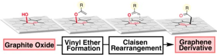
soluble, exfoliated graphite oxide through a Claisen rearrangement (Scheme 1). Specifically, the allylic alcohol functional groups found on the surface of GO,[8] are converted in-situ to vinyl allyl ethers,
and allylically transposed in a sigmatropic-type fashion to form new carbon-carbon bonds. As a direct result of this transformation robust carbonyls are installed on the surface, which can undergo subsequent synthetic manipulations.
The Eschenmoser-Claisen rearrangement variant was chosen as the proof of principle test reaction on GO (Scheme 1, where R = N(CH3)2). In this reaction allylic alcohols are converted to
γ,δ-unsaturated N,N-dimethylamides through the use of the vinyl transfer reagent N,N-dimethylacetamide dimethyl acetal (DMDA). This reaction was chosen specifically for the following reasons: 1) the ability to quantitatively evaluate the efficacy of the transformation through nitrogen incorporation, 2) DMDA’s chemospecificity for alcohol functional groups, 3) only thermal
treatment of DMDA is necessary to affect the vinyl group transfer, and 4) the formation of quaternary carbon-carbon bonds is well established using the Eschenmoser-Claisen variant.[9]
Scheme 1. Allylic oxygen to carbon bond transposition on graphite
oxide (additional substrate oxygenation removed for clarity). For the
Eschenmoser-Claisen rearrangement variant R = N(CH3)2.
We began the investigations into the proposed transformation by mixing graphite oxide with DMDA (~2 equivalents per oxygen on GO, see Supporting Information)[10] in THF at 60 oC for 24 h.[11]
As the reaction progressed the transparent GO-THF solution darkened to an opaque, black suspension. After prolonged standing at room temperature significant sedimentation could be observed. The insoluble product was then subjected to repeated centrifugation, washing cycles and drying. Analysis of the graphitic material (G1) by UV-Vis and FTIR indicated that partial re-conjugation of the π-network had occurred as evidenced by a bathochromic shift of the absorbance maximum from 240 nm in the starting material to 262 nm in the product (see Supporting Information).[12] GO is unstable
with regard to reductive partial deoxygenation and some non-specific reduction was expected. In addition to a decrease in the IR frequencies associated with the C-O bonds in GO (the 1053 cm-1
peak ascribed to surface-bound epoxides is absent in the product)[13]
a new carbonyl stretching–mode at 1635 cm-1 emerges suggesting
amide incorporation (Figure 1a).
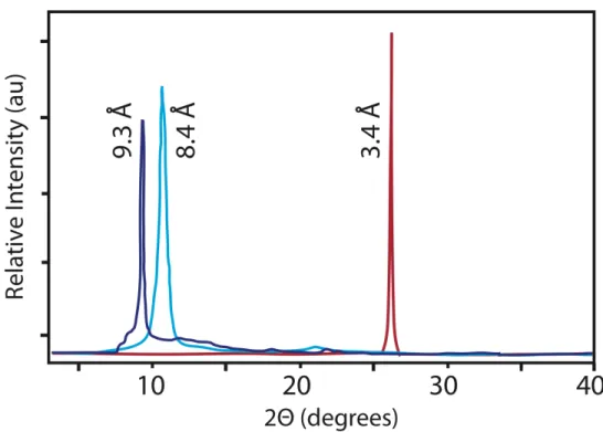
X-ray powder diffraction (XRD) pattern analysis of G1 (Figure 1b) revealed an increase in distance within the intersheet gallery from 0.84 nm in GO to 0.93 nm in the product, which is a qualitative indicator of the installation of additional functionality on the surface of the basal plane of graphene.[14],[15],[16] It is important to
note that simple reductive deoxygenation of GO leads to a decrease in the intersheet spacing.
Figure 1. a) FTIR spectrum of graphite oxide (red) and G1 (blue). b)
XRD patterns of graphite (red), graphite oxide (light blue), and G1 (dark blue).
[∗] Dr. W. R. Collins, W. Lewandowski, E. Schmois, Dr. J.
Walish, Prof. Dr. T. M. Swager
Department of Chemistry and The Institute for Soldier Nanotechnologies
Massachusetts Institute of Technology Cambridge, MA 02139 (USA) Fax: (+1) 617-253-7929 E-mail: tswager@mit.edu
[∗∗] This work was supported by the IC Postdoctoral Research Fellowship Program and the Army Research Office through the Institute for Soldier Nanotechnologies. This project cooperated within the Foundation for Polish Science MPD>Programme co-financed by the EU European Regional Development Fund. We thank Jason Cox for assistance with AFM measurements and Jan Schnorr and Libby Shaw with XPS measurements.
Supporting information for this article is available on the WWW under http://www.angewandte.org or from the author.
2 The quantitative evaluation of the product was accomplished
through both thermogravimetric analysis (TGA) and X-ray photoelectron spectroscopy (XPS). The TGA of G1 showed a large decrease in the surface C-O functionality (epoxides and tertiary alcohols) as evidenced by a reduced mass change at 190-210 oC
(Figure 2, thermograms A and B).[17] Additionally, a new thermal
decomposition is observed between 290-310 oC, which could
potentially be ascribed to amide groups found on the surface of
G1.[18] The relatively high temperature at which this decomposition
occurs suggests that covalent functionalization is occurring in this reaction and is not simply a physical adsorption process.
Figure 2. TGA thermograms of GO (A, green), THF-rearrangement
product G1 (B, dark blue), dioxane-rearrangement product G2 (C, yellow), and diglyme rearrangement product G3 (D, red).
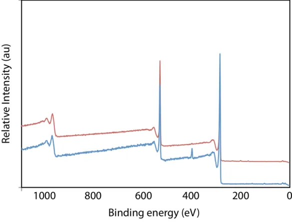
High-resolution XPS analysis of G1 conclusively demonstrated that nitrogen incorporation occurred during the Eshenmoser-Claisen rearrangement (Table 1, entry 1; for spectra see Supporting Information).[19] Through XPS it was determined that
G1 contained 1.4 % nitrogen. A single, amide N(1s) signal was
found at 399.9 eV indicating that all of the starting DMDA material was either consumedin the reactionor removed during the work-up. Importantly, the high resolution N(1s) signal exactly corresponds to that of known XPS spectra of dialkylamide functional groups covalently bound to the surface of activated carbon.[20] The high
resolution C(1s) spectrum (between 284-288 eV) of G1 further confirmed the previous observations that deoxygenation of the C-O functional groups was occurring during the reaction. In effect, the atomic percentage for oxygen content decreased from 27.7% found in the graphite oxide starting material to 19.6% in the product.
With evidence in hand for a successful Claisen rearrangement of graphite oxide, we next attempted to improve the efficacy of the transformation by increasing the reaction temperature. The rearrangement was thus re-run in two higher boiling solvents: dioxane at 100 oC, and bis(2-methoxymethylether)ether [diglyme] at
150 oC. Nitrogen containing solvents were intentionally avoided so
as to not complicate the analysis of the products.[21] In both reactions,
as observed before, the solution rapidly turned black. TGA analysis of the isolated products G2 (from dioxane) and G3 (from diglyme) showed that in both cases a further decrease of the C-O functionality occurred as indicated by an additional drop in the mass loss at 190-210 oC. Importantly, a concomitant increase in the functional
groups that fragment at higher temperatures was also observed in both cases (Figure 2, thermograms C and D).
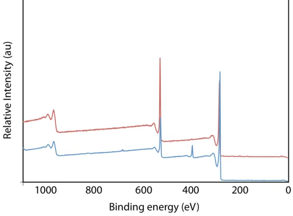
The XPS data for G2 and G3 correlated well with the observed trends in the TGA thermograms. The amide N(1s)
incorporation increased to a total of 3.1% in G3, while the oxygen atomic percent decreased to 11.1% in G3 (Table 1, entries 1-3). When the carbon and oxygen atoms within the appended amide molecule are taken into account (4 carbons, 1 oxygen) this data corresponds to a functional group density of 1 amide per 23 carbons in G3 (1 functional group for 1.25 nm2 of graphitic area).
Due to the fact that the oxygen content decreased in all three transformations, we were interested in whether the deoxygenation of the GO was a sole function of prolonged exposure to high reaction temperatures or whether the DMDA was acting in some capacity as a reducing agent. Therefore, we re-ran the three reactions without the addition of DMDA and evaluated the products by XPS analysis (Table 1, entries 4-6, compare to entries 1-3,7). It can be seen that in all three cases a small to moderate amount of thermal deoxygenation occurs, but that this process alone cannot account for the extensive reduction that is observed.[22] Thus, the DMDA is serving as both
the vinyl transfer reagent in the Claisen reaction and as a reducing agent for GO.
Table 1. List of atomic composition of chemically/thermally modified
graphite oxide by X-ray photoelectron spectroscopy (XPS)[a]
atomic (%) entry temp (solvent/ oC)
C O N
amide
/ C[e] Ratio C/O
1[b] THF / 60 79.0 19.6 1.4 1/52 4.03 2[b] Dioxane / 100 85.5 12.4 2.1 1/37 6.90 3[b] Diglyme / 150 85.8 11.1 3.1 1/23 7.73 4[c] THF / 60 74.0 26.0 -- -- 2.85 5[c] Dioxane / 100 75.5 24.5 -- -- 3.08 6[c] Diglyme / 150 80.1 19.9 -- -- 4.03 7[d] -- 72.3 27.7 -- -- 2.61
[a] Calculated by integration of diagnostic XPS signals. [b] Reaction performed with 2 equiv. DMDA per GO oxygen. [c] Thermal reduction performed without DMDA. [d] Unchanged GO starting material. [e] Ratio = [(C%)/(N%)]-4
Four-point probe measurements on thin films of G3 revealed that the DMDA reduction process sufficiently re-established conjugation within the graphene sheet to produce a moderate level of conductivity of 1.7 S/m (See Supporting Information). Annealing the sample at 250 oC for 24 h (below the temperature at which the
amide groups are thermally eliminated) resulted in a further increase in the conductivity to 538 S/m. As expected, from its increased intersheet spacing as well as covalently bound amide groups disrupting the graphene sheet’s conjugation, G3 displays a lower conductivity than reduced GO (with comparable C/O ratios) produced by hydrazine-, anionic- or thermally-based deoxygenation.[23]
We next determined whether the newly installed amide groups could be chemically converted into alternate functionality. It has been documented that the carboxylate groups on the edges of graphene can electrostatically stabilize colloidal dispersions of exfoliated graphene for short periods of time.[12] It was therefore
postulated that by increasing the carboxylate functional group density through saponification of the surface-bound amide groups, the resulting graphene derivatives could exist as stable colloidal solutions for prolonged periods of time. To this end, we treated G3 under strongly basic conditions (refluxing KOH/H2O/ethanol) to
saponify the amide groups. Aliquots were removed during the reaction timecourse at 12 h increments and the surface charge (zeta potential) of the graphene substrate was monitored (See Supporting Information). From the resulting measurements it was determined
3 that the weakly charged (-19 mV) starting material G3 increasingly
developed surface charge as the reaction progressed, with the zeta potential reaching a maximum of -68 mV at 36 h.
After acidification of the reaction solution the resulting graphene derivative could be precipitated and isolated. Following successive washings, centrifugations, and dryings, the resulting graphene derivative’s (G4) chemical composition could be quantitatively assessed. XPS analysis showed that the atomic weight percent of nitrogen had decreased from 3.1% in the starting material to 0.6% (see Supporting Information for spectrum). This data translates to an amide functional group density drop to 1 group per ca. 80 graphitic carbons. In conjunction with the abovementioned zeta potential measurements, the loss of the amide N(1s) signal suggested that extensive saponification had in fact occurred on the graphene surface.
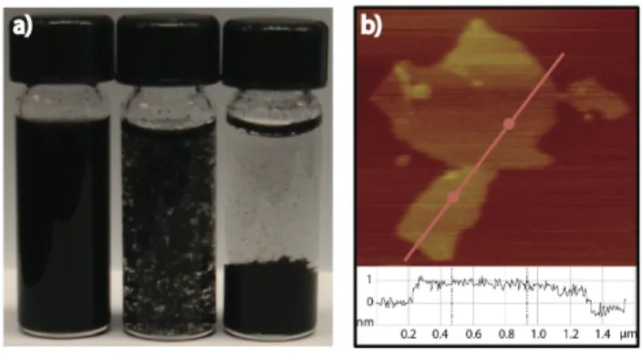
Visual observations of the solubility of G4 provided additional support for the existence of surface bound carboxylate groups. Specifically, the graphene derivative’s solubility profile in aqueous environments proved to be highly dependant upon the pH of the solution (Figure 3a). In aqueous, acidic solutions below pH 5, G4 proved to be largely insoluble in water. Under pH neutral or basic solutions G4 readily formed a homogeneous colloidal dispersion. Relatively high concentrations of up to 5 mg/mL of G4 in neutral pH water have been generated and shown to be stable for over 3 months without sedimentation. Tapping-mode atomic force microscopy (AFM) images of drop-cast solutions of G4 in neutral water show that the graphene sheets in these solutions are largely exfoliated in nature and not composed of higher-order graphitic aggregates (Figure 3b).
Figure 3. a) Solubility profile of G4 in water at pH 7.2, 5.2, and 3.3. b)
Tapping-mode AFM image of G4 with a height profile taken across the red line. The imaged sample was prepared by drop-casting a [0.01 M] solution of G4 in neutral water onto mica followed by evaporative drying of the sample.
A turbidity analysis was undertaken to determine the precise correlation between the pH of the aqueous environment and the solubility profile of G4. This was accomplished by measuring UV-Vis absorbance spectra over a pH ranging from 7.26 to 3.5. Within the pH range of 7.26 to 6 only small changes occur in the UV-Vis absorbance intensity. A much larger drop in the absorbance begins near or at pH 5. This spectral change is matched by the beginning of a noticeable flocculation of the graphene sheets in solution. Below pH 5 the absorbance dramatically decreases due to the aggregation and subsequent sedimentation of the graphene sheets in solution (Figure 4).
Figure 4. UV-Vis absorbance of G4 in an aqueous solution with pH
ranging from 7.26 to 3.5. (Inset: 262 nm UV-Vis maximum absorbance of G4 plotted against the pH range of the aqueous solution.)
Zeta potential measurements performed over the same pH range correlate well with what is observed in the UV-Vis turbidity analysis (see Supporting Information). By lowering the pH below 7.26 the surface charge correspondingly decreases from the initial -68 mV. Between pH 6.3 and 4.6 a significant drop to -15 mV is observed. At pH 3 most of the anionic functionality on the sheet has been protonated as indicated by a near-neutral zeta potential.
Taken together, the turbidity analysis, zeta potential measurements, and visual observations all suggest that the carboxylate functionality is not only present on the surface of G4 but that these groups play an important role in electrostatically stabilizing the graphene sheets in aqueous environments. In particular, when the pH of the aqueous solution is above the pKa of the surface bound carboxylic acid groups the resulting anionic surface charge acts to effectively disperse the sheets. Alternately, when the pH is reduced to pH 5 or below, the carboxylate functional groups are protonated, the electrostatic charge is lost, and the sheets hydrophobically aggregate and begin to sediment.[24],[25]
In summary, this study reports the sigmatropic-type transformation on a graphitic surface. Utilizing the Eschenmoser-Claisen rearrangement on graphite oxide, the allylic alcohol surface functional groups were directly converted to carbon-bound N,N-dimethylamide groups through the use of the reagent DMDA.[25]
These amide groups, when saponified under strongly basic conditions, were converted to the corresponding carboxylates, which have been shown to dramatically increase the solubility of the graphene derivative in aqueous environments without the need of co-solvents or additives.[26] Current efforts are underway to expand
the scope and application of this transformation and to investigate the possibility of additional Claisen rearrangements on graphitic substrates.
Received: ((will be filled in by the editorial staff))
Published online on ((will be filled in by the editorial staff))
Keywords: graphene · Claisen rearrangement · surface
4
[1] a) J. C. Love, L. A. Estroff, J. K. Kriebel, R. G. Nuzzo, G. M.
Whitesides, Chem. Rev. 2005, 105, 1103-1169; b) G. Decher, Science,
1997, 277, 1232-1237.
[2] For fullerenes see: S. Campidelli, A. Mateo-Alonso, M. Prato, in
Fullerenes, Principles and Applications (Eds: F. Langa, J.
Nierengarten) RSC Publishing, Cambridge, 2007, pp. 191-211; For carbon nanotubes see: F. Hauke, A. Hirsch, in Carbon Nanotubes and
Related Structures: Synthesis, Characterization, Functionalization, and Applications (Eds.: D. M. Guldi, N. Martin) WILEY-VCH,
Weinheim, 2010, pp. 135-179; For graphene see: M. J. Allen, V. C. Tung, R. B. Kaner, Chem. Rev. 2010, 110, 132-145.
[3] a) C. Nguyen, T. Yamada, P. Sarrazin, J. Li, J. Li, E. V. Barrera, M. L.
Shofner, E. L. Corral, M. Meyyappan, in Carbon Nanotubes, Science
and Applications (Ed.: M. Meyyappan), CRC Press, Boca Raton, 2005,
pp. 171-352; b) H. G. Chae, J. Liu, S. Kumar, C. A. Dyke, J. Tour, in
Carbon Nanotubes: Properties and Applications (Ed. M. J.
O’Connell) CRC Press, Boca Raton, 2006, pp. 213-295.
[4] For reviews on the chemical functionalization of graphene see: a) K.
P .Loh, Q. Bao, P. K. Ang, J. Yang, J. Mater. Chem. 2010, 20, 2277-2289; b) O. C. Compton, S. T. Nguyen, Small, 2010, 6, 711-723; c) D. W. Boukhvalov, M. I. Katsnelson, J. Phys. Cond. Mater. 2009, 34, 1-12; d) S. Park, R. S. Ruoff, Nat. Nanotech. 2009, 4, 217-224; e) G. Cravotto, P. Cintas, Chem. Eur. J. 2010, 16, 5246-5259; f) C. N. Rao, A. K. Sood, K. S. Subrahmanyam, A. Govindaraj, Angew. Chem. Int.
Ed. 2009, 48, 7752-7777.
[5] For synthetic methods performed on reduced graphite oxide see: a) X.
Zhong, J. Jin, S. Li, Z. Niu, W. Hu, R. Li, J. Ma, Chem. Commun.
2010, 46, 7340-7342; b) T. A. Strom, E. P. Dillon, C. E. Hamilton, A.
R. Barron, Chem. Commun. 2010, 46, 4097-4099; c) V. Georgakilas, A. B. Bourlinos, R. Zboril, T. A. Steriotis, P. Dallas, A. K. Stubos, C. Trapalis, Chem. Commun. 2010, 46, 1766-1768; d) M. Quintana, K. Spyrou, M. Grzelczak, W. R. Browne, P. Rudolf, M. Prato, ACS Nano,
2010, 4, 3527-3533; e) J. R. Lomeda, C. D. Doyle, D. V. Kosynkin, W.
Hwang, J. M. Tour, J. Amer. Chem. Soc. 2008, 130, 16201-16206.
[6] For synthetic methods performed directly on graphene see: a) E.
Bekyarova, M. E. Itkis, P. Ramesh, C. Berger, M. Sprinkle, W. A. de Heer, R. C. Haddon, J. Amer. Chem. Soc. 2009, 131, 1336-1337; b) H. Liu, S. Ryu, Z. Chen, M. L. Steigerwald, C. Nuckolls, L. E. Brus, J.
Amer. Chem. Soc. 2009, 131, 17099-17101; c) L. H. Liu, M. M.
Lerner, M. Yan, Nano Lett. 2010, 10, 3754-3756; d) S. Ryu, M. Y. Han, J. Maultzsch, T. F. Heinz, P. Kim, M. L. Steigerwald, L. E. Brus,
Nano Lett. 2008, 8, 4597-4602; e) D. C. Elias, R. R. Nair, T. M.
Mohiuddin, S. V. Morozov, M. P. Halsall, A. C. Ferrari, D. W. Boukhvalov, M. I. Katsnelson, A. K. Geim, K. S. Novoselov, Science
2009, 323, 610-613; f) S. B. Bon, L. Valentini, R. Verdejo, J. L. Fierro,
L. Peponi, M. Lopez-Manchado, J. M. Kenny, Chem. Mater. 2009, 21, 3433-3438.
[7] a) A. Hirsch, O. Vostrowsky, in Functional Molecular Nanostructures
(Ed.: D. A. Schluter), Springer-Verlag, Berlin, 2005, pp 193-237; b) Z. F. Chen, W. Thiel, A. Hirsch, Chem. Phys. Chem. 2003, 4, 93-97; c) M. A. Hamon, M. E. Itkis, S. Niyogi, T. Alvaraez, C. Kuper, M. Menon, R. C. Haddon, J. Amer. Chem. Soc. 2001, 123, 11292-11293; d) R. C. Haddon, J. Amer. Chem. Soc. 1990, 112, 3385-3389; e) R. C. Haddon, Science, 1993, 261, 1545-1550.
[8] a) D. Dreyer, S. Park, C. Bielawski, R. S. Ruoff, Chem. Soc. Rev.
2010, 39, 228-240; b) W. Gao, L. B. Alemany, L. Ci, P. M. Ajayan,
Nat. Chem. 2009, 1, 403-408; c) D. W. Boukhvalov, M. L. Katsnelson, J. Amer. Chem. Soc. 2008, 130, 10697-10701; d) K. N. Kudin, B.
Ozbas, H. C. Schniepp, R. K. Prud’homme, I. A. Aksay, R. Car, Nano.
Lett. 2008, 8, 36-41.
[9] a) S. N. Gradl, D. Trauner, in The Claisen Rearrangment: Methods
and Applications (Eds.: M. Hiersemann, U. Nubbermeyer),
WILEY-VCH, Weinheim, 2007, pp. 367-394; b) A. M. Martin Castro, Chem.
Rev. 2004, 104, 2939-3002; c) A. E. Wick, D. Felix, K. Steen; A.
Eschenmoser, Helv. Chim. Acta 1964, 47, 2425-2427.
[10] A large excess of DMDA is utilized to compensate for the monolayer of water found within the intersheet gallery of graphite oxide (see: N. V. Medhekar, A. Ramasubramaniam, R. S. Ruoff, V. B. Shenoy, ACS
Nano, 2010, 4, 2300-2306.).
[11] Eschenmoser Claisen rearrangements on small molecules are typically
performed at or above 100 oC. It was believed at the onset of this
study that the weak bond strength of the surface C-O functionality would facilitate this transformation and allow for a lower reaction temperature.
[12] D. Li, M. B. Muller, S. Gilje, R. B. Kaner, G. G. Wallace, Nat.
Nanotech. 2008, 3, 101-105.
[13] a) S. Stankovich, R. Piner, S. T. Nguyen, R. Ruoff, Carbon, 2006, 44, 3342-3347; b) Y. Si, E. T. Samulski, Nano. Lett. 2008, 8, 1679-1682; c) S. Park, K. S. Lee, G. Bozoklu, W. Cai, S. T. Nguyen, R. S. Ruoff,
ACS Nano, 2008, 2, 572-578.
[14] S. Stankovich, D. A. Dikin, O. C. Compton, G. H. Dommett, R. S. Ruoff, S. T. Nguyen, Chem. Mater. 2010, 22, 4153-4157. [15] EFTEM imaging of the basal plane of the amide-bound graphene
derivative G3 correspondingly identified nitrogen incorporation throughout the surface (See Supporting Information).
[16] The Eschenmoser Claisen rearrangement on GO contrasts sharply with known edge-based amide forming reactions on graphene in that this method functionalizes the basal plane.
[17] a) A. Lerf, H. Y. He, M. Forster, J. Klinowski, J. J. Phys. Chem. B,
1998, 102, 4477-4482; b) S. Stankovich, D. A. Dikin, P. D. Piner, K.
A. Kohlhaas, A. Kleinhammes, Y. Jia, Carbon, 2007, 45, 1558-1566. [18] Alkylamides covalently bound to the edges of graphene eliminate at
300 oC. For the TGA see: S. Niyogi, E. Bekyarova, M. E. Itkis, J. L.
McWilliams, M. A. Hamon, R. C. Haddon, J. Amer. Chem. Soc. 2006,
128, 7720-7721).
[19] To remove the possibility that adsorption of hydrolyzed DMDA and not covalent incorporation was occurring, GO was stirred under the three reaction conditions in the presence of dimethylacetamide (hydrolyzed DMDA). After workup, analysis by XPS showed no nitrogen incorporation (see Supporting Information for more details) [20] a) R. J. Jansen, H. Bekkum, Carbon, 1995, 33, 1021-1027; b) R. J.
Jansen, A. Sinnema, H. Bekkum, Carbon, 1995, 33, 550-551 [21] Irreversible incorporation of nitrogen by-products into graphene
through high-temperature NMP decomposition has been shown by: S. Dubin, S. Gilje, K. Wang, V. C. Tung, K. Cha, A. S. Hall, J. Farrar, R. Varshneya, Y. Yang, R. B. Kaner, ACS Nano, 2010, 4, 3845-3852. [22] For a chemical reduction method on graphite oxide that gives
comparable C/O ratios to the diglyme-DMDA procedure see [13b]. [23] a) S. Park, J. An, R. P. Piner, S. Jin, X. Li, A. Velamakanne, R. S.
Ruoff, Nano. Lett. 2009, 9, 1593-1597; b) H. Chen, M. B. Muller, K. J. Gilmore, C. G. Wallace, D. Li, Adv. Mater. 2008, 20, 3557-3561; c) S. Park, J. An, R. D. Piner, I. Jung, D. Yang, A. Velamakanni, S. T. Nguyen, R. S. Ruoff, Chem. Mater. 2008, 20, 6592-6594. [24] a) I. W. Hamley, in Introduction to Soft Matter: Polymers, Colloids,
Amphiphiles and Liquid Crystals, 2nd ed.; Wiley, New York, 2007, p
340; b) J. N. Israelachvili, in Intermolecular and Surface Forces, 2nd
ed.; Academic Press, San Diego, 1992, p. 450.
[25] For an example of water soluble, covalently functionalized graphene see: Y. Si, E. T. Samulski, Nano Lett. 2008, 8, 1679-1682. [26] For examples of aqueous graphene solutions stabilized by additives
see: a) . Li, Y. Bao, Q. Zhang, D. Han, L. Niu, Langmuir, 2010, 26, 12314-12320; b) M. Lotya, Y. Hernandez, P. J. King, R. J. Smith, V. Nicolosi, L. S. Karlsson, F. M. Blighe, S. De, Z. Wang, I. T. McGovern, G. S. Duesberg, J. N. Coleman, J. Amer. Chem. Soc. 2009,
131, 3611-3620; c) Y. Xu, H. Bai, G. Lu, G. Shi, J. Amer. Chem. Soc.
5
Entry for the Table of Contents
Layout 2:
Graphene
William R. Collins, Wiktor Lewandowski, Ezequiel Schmois, Joseph Walish, and Timothy M. Swager __________ Page
– Page
Claisen Rearrangement of Graphite Oxide: A Route to Covalently
Functionalized Graphene A one-pot, oxygen to carbon bond allylic transposition reaction and chemical reduction of graphite oxide has been demonstrated. Utilizing N,N-dimethylacetamide dimethylacetal, the basal plane allylic alcohol functionality of graphite oxide can be converted to N,N-dimethylamide groups through an Eschenmoser-Claisen sigmatropic-type rearrangement. These groups can be subsequently saponified to the carboxylic acid, which, when deprotonated, electrostatically stabilizes the graphene sheets in an aqueous environment.
![Table 1. List of atomic composition of chemically/thermally modified graphite oxide by X-ray photoelectron spectroscopy (XPS) [a]](https://thumb-eu.123doks.com/thumbv2/123doknet/14319593.496811/3.892.456.829.454.627/table-composition-chemically-thermally-modified-graphite-photoelectron-spectroscopy.webp)
![Figure S1. TGA of amide functionalized graphene (G1, blue, [B]) and graphite oxide (green, [A])](https://thumb-eu.123doks.com/thumbv2/123doknet/14319593.496811/11.918.225.720.593.888/figure-tga-amide-functionalized-graphene-graphite-oxide-green.webp)

![Figure S7. TGA of amide functionalized graphene (G2, yellow, [C]) and graphite oxide (green, [A])](https://thumb-eu.123doks.com/thumbv2/123doknet/14319593.496811/14.918.159.772.674.1041/figure-amide-functionalized-graphene-yellow-graphite-oxide-green.webp)