HAL Id: tel-02886136
https://tel.archives-ouvertes.fr/tel-02886136
Submitted on 1 Jul 2020
HAL is a multi-disciplinary open access
archive for the deposit and dissemination of sci-entific research documents, whether they are pub-lished or not. The documents may come from teaching and research institutions in France or abroad, or from public or private research centers.
L’archive ouverte pluridisciplinaire HAL, est destinée au dépôt et à la diffusion de documents scientifiques de niveau recherche, publiés ou non, émanant des établissements d’enseignement et de recherche français ou étrangers, des laboratoires publics ou privés.
The role of integrin αv expressed by VSMCs in vascular
fibrosis
Lei Tian
To cite this version:
Lei Tian. The role of integrin αv expressed by VSMCs in vascular fibrosis. Cellular Biology. Sorbonne Université, 2018. English. �NNT : 2018SORUS103�. �tel-02886136�
1
Acknowledgement
I thank my supervisor Dr. Zhenlin Li. He is a very hard-working scientist, his spirit inspires me to do my best every day. He is an erudite scientist, he always gives me some useful suggestions, He is a responsible scientist, when I meet some difficulties in my PhD project, he always tries to solve my problems. He provides me a platform to do my research and accomplish my PhD career. I thank Pr. Denise Paulin. She is so important in my PhD career. She helps me to open a window to understand the French society. She is so kind to me. Each time, when I am in difficult conditions, I always want to ask her for help. She is so tolerant, sometimes, even though I make some mistakes, she always forgives me and gives me chances to try again. Her personality is an example for me and gives me forces to go ahead in the future. Her tolerances and patience give me a relatively ideal environment to do my research.
I thank Pr. Bertrand Friguet. As a director of our department, he gives me so many supports. He is always so optimistic. His encouragements give me the courage to overcome difficulties. This time, he also agrees to be the president of my PhD defense, it is a great honor for me.
I thank Dr. Dario Coletti. He gives me so many helps. From the experimental designs to the statistic knowledge, from the experimental reagents to the thesis revision. He always tries his best to help me to solve my problems. I learn a lot from him, not only the knowledge of statistic and electronic microscope, but also his personality.
I thank my PhD school. Dr. Catherine Monnot gives me so many supports, her responsibility and supports inspire me to accomplish my PhD thesis better and better.
I thank Dr. Céline Fassot. As a reviewer of my thesis, she helps me revise my manuscript and gives me many useful suggestions. More importantly, she gives me so many positive evaluations.
I thank Pr. Marina Bouche. As a reviewer of my thesis, she gives me careful revision of my manuscript. Your careful corrections and important suggestions help me to improve the quality of my manuscript.
I thank Dr. Isabelle Brunet. As the tutor in my PhD career. About 2 years ago, I first met her, from then on, she gives me some useful suggestions for my PhD project. She builds a bridge between me and PhD school. This time, it is my
2
honor to invite her to be examiner of my PhD jury.
I also would like to express my thanks to Dr. Jean-Sébastien Silvestre. He is the famous scientist in my research field. He gives me some suggestions. His kindness gives me a deep impression.
I also thank Dr. Carine le Goff. I was nervous, when I first met her. She gave me so many patience to express myself. I can feel her friendship and sincerity. I thank Dr. Mathias Mericskay. We have not spent a lot of time together, but he is so kindness to me. Every time I meet him, he always gives me encouragement. In my PhD career, he indeed gives me so many critical suggestions.
I thank Mrs. Jocelyne Blanc. She helps me do the first experiment in this new lab. As a secretary of our lab, she knows all kinds of affaires in our labs. She is so kindness and always gives me some useful information in my experiments. I thank Dr. Jean-François Decaux, Dr. Zhigang Xue, Mrs Jie Gao and Dr. Ara Pariakian. They help me do some experiences and give me some good suggestions. They teach me how to make some kinds of experimental reagents by ourselves. There are so many useful formulas in our labs. They also help me to improve my French. Through their helps, I can understand the French society better.
I thank Rachel Gergondey, Maria Kisara, Ekaterini Kordeil, Gaelle Revet, Janek Hyzewicz, Fanny Canesi, Elodie Bosc, Marie-Paule Hamon. We are in the same big lab, they give me so many helps. they help me get the softwares, they lend me some chemical reagents, they teach me how to use the machine……they create a harmony environment for me. Their kindness give me a deep impression and good memory for my PhD career.
I thank Dr. Emmanuelle Lacaze. She teaches me how to use Atomic Force Microscope. With her help, I have a better understanding of this microscope. I have gotten some meaning results by using this microscope.
I thank my parents. They always give me a lot of unconditional supports. Even though they are in China, When I am sad, I always want to talk with them. In the past several years, they have suffered a lot with me. They are my harbor forever.
3
List of publications
Langlois B, Belozertseva E, Parlakian A, Bourhim M, Gao-Li J, Blanc J, Tian L, Coletti D, Labat C, Ramdame-Cherif Z, Challande P, Regnault V, Lacolley P, Zhenlin L. Vimentin knockout results in increased expression of sub-endothelial basement membrane components and carotid stiffness in mice. Sci Rep. 2017 Sep 14;7(1):11628.
Tian L, Chen K, Cao J, Han Z, Gao L, Wang Y, Fan Y, Wang C. Galectin‑3 induces the
phenotype transformation of human vascular smooth muscle cells via the canonical Wnt signaling. Mol Med Rep. 2017 Jun;15(6):3840-3846.
Tian L, Chen K, Cao J, Han Z, Gao L, Wang Y, Fan Y *, Wang C* Galectin-3 elicited by
oxLDL promotes the phenotype transformation of vascular smooth muscle cells. Mol Med Rep. 2015 Oct;12(4):4995-5002.
Cao J*, Han Z*, Tian L*, Chen K, Fan Y, Ye B, Wang C, Huang Z. Curcumin inhibits EMMPRIN and MMP-9 expression through AMPK-MAPK and PKC signaling in PMA induced macrophages. J Transl Med. 2014 Sep 21;12(1):266.
Cao J, Ye B, Lin L, Tian L, Yang H, Wang C, Huang W, Huang Z. Curcumin Alleviates oxLDL Induced MMP-9 and EMMPRIN Expression through the Inhibition of NF-κB and MAPK Pathways in Macrophages. Front Pharmacol. 2017 Feb 14;8:62.
Han Z, Cao J, Song D, Tian L, Chen K, Wang Y, Gao L, Yin Z, Fan Y, Wang C. Autophagy is involved in the cardioprotection effect of remote limb ischemic postconditioning on myocardial ischemia/reperfusion injury in normal mice, but not diabetic mice. PLoS One. 2014 Jan 23;9(1):e86838.
4
The role of integrin αv expressed by VSMCs in
vascular fibrosis
5
Content
Abstract ... 10
Abbreviations ... 12
Introduction ... 15
Part I Integrins are related to fibrosis ... 15
1.1 Integrins and integrin-related proteins ... 15
1.2 The role of integrins in fibrosis ... 16
Part II VSMCs play an important role in vascular fibrosis ... 18
2.1 Microanatomy of arteries ... 18
2.2 VSMCs is one of the main cell types in the atrial wall ... 19
2.3 Two important phenotypes of SMCs: contractile and synthetic SMCs ... 20
2.4 ECM plays an important role in vascular fibrosis ... 21
Part III Ang II or TGF-β induces vascular fibrosis ... 22
3.1 Ang II is an important factor in cardiovascular fibrosis ... 22
3.1.1 Ang II induces cardiovascular fibrosis... 23
3.1.2 Ang II and its receptors in cardiovascular fibrosis... 23
3.1.3 Downstream of Ang II Receptors: signaling pathways in cardiovascular fibrosis ... 27
3.2 TGF-β1 is a crucial determinant in cardiovascular fibrosis ... 29
3.2.1 TGF-β1 interacts with integrins via LAP and induces cardiovascular fibrosis ... 30
3.2.2 TGF-β1 and its receptors ... 32
3.2.3 TGF-β1 and its signaling pathways in cardiovascular fibrosis ... 32
Part IV Galectin-3: a novel factor involved in fibrosis and cardiovascular diseases ... 37
4.1 Structure and expression of galectin-3 ... 37
4.2 Galectin-3 is related to fibrosis ... 38
4.3 The role of galectin-3 in a variety of cardiovascular diseases ... 39
4.4 Galectin-3 is related to fibrosis in cardiovascular system ... 42
4.4.1 Galectin-3 mediates the fibrosis in several different cell types in the cardiovascular systems. ... 42
4.4.2 Galectin-3 is involvememnt in mechanisms of cardiovascular fibrosis suggests its potential as a therapeutic target ... 44
4.5 The relationship between galectin-3 and integrins ... 45
Part V Vascular stiffness is a multifactor process ... 45
5.1 The importance of vascular stiffness in cardiovascular diseases ... 45
5.2 Factors regulating vascular stiffness ... 46
Aim of the thesis ... 51
Results ... 52
Part I Ang II or TGF-β1 induces the vascular fibrosis via integrin αv ... 52
1.1 Knock-out integrin αv in smooth muscle cell reduces Ang II-induced vascular fibrosis ... 52
1.2 Transcriptomic analysis of αvSMKO and WT mice. ... 57 1.3 Ang II or TGF-β induces the upregulation of fibrosis-related proteins by integrin
6
αv in vitro ... 66
1.4 Ang II or TGF-β induces the activation of ERK and smad-2/3 signaling pathways via integrin αv... 68
Part II Galectin-3 induces the activation of VSMCs via integrin αv/AKT/Wnt/β-catenin signaling pathway ... 71
2.1 Integrin αv interacts with galectin-3 and mediates galectin3-induced synthesis of fibrosis-related proteins in VSMCs ... 71
2.2 Integrin αv mediates galectin-3-induced activation of AKT and Wnt/β-catenin signaling pathways ... 74
2.3 Integrin αv mediates the proliferation and migration induced by galectin-3 in VSMCs ... 77
2.4 Integrin αv does not mediate the endocytosis of galectin-3 ... 78
2.5 Galectin-3 induces activation of Wnt signaling pathway through AKT signaling pathway ... 79
2.6 Galectin-3 induced the proliferation and migration of VSMCs through AKT signaling pathway ... 80
Part III Knock-down integrin αv increases VSMCs stiffness ... 81
3.1 Knock-down integrin αv affects the stiffness of VSMCs. ... 81
3.2 Knock-down integrin αν increases the expression of β-tubulin ... 81
Discussion ... 84
Perspective ... 91
Materials and Methods ... 93
7
List of Figures
Figure 1. Integrins and Integrin-related proteins. ... 16
Figure 2. Some of the multiple functions of integrins in the cardiac myocyte (CM). . 17
Figure 3. Schematic structure, including main cell types and ECM components in small and large arteries. ... 18
Figure 4. VSMCs are one of the most important cell types involved in atherosclerosis. ... 20
Figure 5. Ultrastructural characteristics of contractile and synthetic SMCs. ... 21
Figure 6. Pathogenesis and risk factors of vascular fibrosis in atherosclerosis. ... 22
Figure 7. Role of Ang II role in cardiovascular pathology. ... 25
Figure 8. Model of mechanical activation of latent TGFβ1. ... 31
Figure 9. Functional and structural characteristics of Smad family members. ... 33
Figure 10. Signaling crosstalk in vascular fibrosis. ... 34
Figure 11. Vascular signaling mediating ECM remodeling, fibrosis, and arterial stiffening in aging and hypertension. ... 36
Figure 12. Western blot analysis of different tissues reveals differential expression levels of galectin-3. ... 38
Figure 13. galectin-3 induces fibrosis through TGF-β dependent and independent mechanisms. ... 39
Figure 14. Galectin-3 is upregulated in a variety of cardiovascular diseases ... 42
Figure 15. Galectin-3 affects the functions of several cell types in the cardiovascular systems. ... 44
Figure 16. Large artery stiffness: cross-talk between local and systemic stiffness in large arteries. ... 50
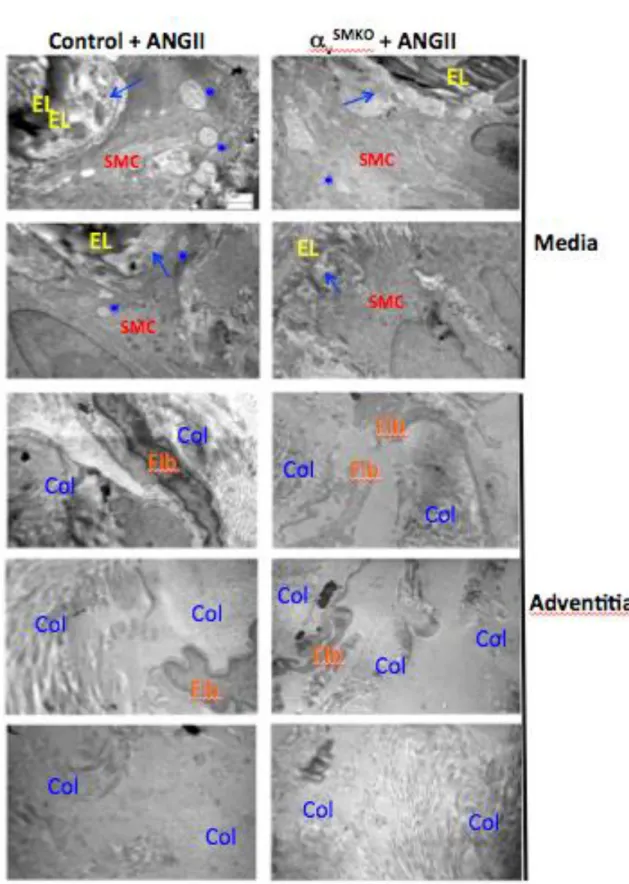
Figure 17. Specific knock-out integrin αv gene in smooth muscle cells of mouse. ... 53
Figure 18. Decrease of fibrosis in Ang II-treated αvSMKO mice. ... 53
Figure 19. Electronic microscopy analysis of mice carotids. ... 56
Figure 20. Decreased TGF-β1 and its receptor in Ang II-treated carotids of αvSMKO mice. ... 57
Figure 21. Western blot analysis of control and integrin αv knock-down (KD) VSMCs at the baseline and under Ang II treatment. ... 67
Figure 22. Western blot analysis of control and integrin αv knock-down (KD) VSMCs at the baseline and TGF-β1 treatment. ... 68
Figure 23. Western blot analysis of phosphorylation of ERK1/2 and smad-2 in control and integrin αv knock-down (KD) VSMCs in response to Ang II treatment. ... 69
Figure 24. Western blot analysis of phosphorylation of ERK1/2 and smad-3 in control and integrin αv knock-down (KD) VSMCs in response to TGF-β1 treatment. ... 70
Figure 25. Galectin-3 interacts with integrin αv. ... 72
Figure 26. Galectin-3 induces expression of ECM proteins via integrin αν. ... 73
Figure 27. Time-dependence of galectin-3-mediated activation of ERK, AKT and Wnt/β-catenin signaling pathways. ... 75
Figure 28. Integrin αv-mediated galectin-3-induced activation of AKT and Wnt/β-catenin signaling pathways. ... 76
8
Figure 29. Integrin αv mediates gal3-induced VSMCs activation. ... 78 Figure 30. Integrin αv does not mediate endocytosis of galectin-3. ... 78 Figure 31. Gal3-induced activation of Wnt signaling pathway through AKT signaling pathway. ... 79 Figure 32. AKT signaling pathway mediates galectin-3 induced proliferation and migration. ... 80 Figure 33. Knock-down integrin αν has little effect on the expression of vinculin and α-tubulin. ... 82 Figure 34. Knock-down of integrin αν could obviously increase β-tubulin. ... 83 Figure 35. Proposed model in which integrin αv mediates galectin-3 induced activation of Wnt/β-catenin signaling in VSMCs. ... 87 Figure 36. Hypothetic schema for the influence of integrin αv on the vascular fibrosis in the Ang II treatment. ... 90 Figure 37. Flowchart of in vitro experiments. ... 98
9
List of Tables
Table 1. Growth factors and cytokines involved in expression of AT1R ... 24
Table 2. Number of genes that have been differently expressed... 58
Table 3. The genes whose expression is increased by knock-out of integrin αv ... 58
Table 4. The genes whose expression is decreased by knock-out of integrin αv... 59
Table 5. The five pathways more implicated in the change of gene expression ... 60
Table 6. Genes involved in fibrosis pathway ... 61
Table 7. Genes involved in TGF-β pathway ... 63
Table 8. The genes involved in the actin cytoskeleton pathway ... 64 Table 9. Summary of Young's modulus of control and integrin αv knock down cells 81
10
Abstract
Arterial stiffness is an independent risk factor for cardiovascular morbidity/mortality. It has been demonstrated that arterial stiffness is linked to arterial fibrosis manifested by increased synthesis of collagen and other extracellular matrix components. Integrins, Transmembrane receptors mediating cell-cell and cell-matrix signaling pathways, are involved in tissue fibrosis. Galectin-3, a novel marker for diagnosis and prognosis of heart failure patients, also plays an important role in fibrosis. However, the molecular mechanisms whereby galectin-3 induces vascular fibrosis are still unclear. We studied the role of integrin αv in Ang II-induced VSMCs arterial fibrosis and stiffness via a SMC specific knock-out of integrin αv mouse model (αv SMKO), induced in adult mice by injection of tamoxifen. We could not find any difference in vascular fibrosis in basal conditions between control and mutant mice. However, decreased arterial fibrosis was observed in αv SMKO mutant mice 28-day after Ang II perfusion. Analysis of RNA from aorta of control and mutant mice by Affymetrix microarrays indicated alteration of TGF-β pathway in Ang II-treated mutant mice. In order to examine the mechanism associated to the decreased fibrosis in VSMCs of αvSMKO mice, we used integrin αv-floxed VSMCs in culture and biochemical methods to analyze the phosphorylation of signaling components and fibrosis-related proteins synthesis following integrin αv inactivation and/or treatment of TGF-β1, Ang-II or galectin-3. Our results indicated that TGF-β1 or Ang-II increased the expression of collagen and fibronectin at the protein level as well as the phosphorylation of ERK and smad2/3 in the control cells, while inactivation of integrin αν partly inhibited the TGF-β1- and Ang-II-induced effects above. Integrin αv was required for Ang II-induced expression of galectin-3 in the VSMCs. Duolink method demonstrated that galectin-3 interacted directly with integrin αv. We also showed that galectin-3 activated AKT, ERK, and Wnt/β-catenin signaling components. The activation of AKT and Wnt/β-catenin signaling pathways, but not ERK signaling pathway, by galectin-3 was inhibited by the knock-down of integrin αv. At cellular level, galectin-3-induced an increase in cell proliferation, migration and synthesis of several fibrosis-related proteins were also significantly inhibited by knock-down of integrin αv. The specific inhibitor of AKT signaling pathway (LY294002) inhibited the activation of downstream Wnt/β-catenin signaling pathway and decreased the response of VSMCs to galectin-3 treatment. Our study indicates a role of integrin αv in the Ang II or TGF-β1 induced arterial fibrogenesis. Galectin-3, interacting with integrin αv, depends on integrin αv/AKT/Wnt/β-catenin signaling pathway to regulate the proliferation, migration and expression of fibrosis-related proteins in VSMCs.
11
Visual abstract
Proposed model in vitro, integrin αv mediates galectin-3 induced activation of Wnt/β-catenin signaling in VSMCs. integrin αv/AKT/β-catenin axis mediates galectin-3 induced proliferation and migration in VSMCs. Galectin-3 interacts with integrin αv directly on the cell surface of VSMCs, inducing the phosphorylation of AKT and, consequently, GSK-3β phosphorylation. Activation of AKT signaling pathway could phosphorylate several targets including GSK-3β and lead to the degradation of GSK-3β. In turn, the inactivation of GSK-3β reduces the β-catenin degradation and increases the expression of active β-catenin. Thus, β-catenin translocate to the nucleus and induces gene expression leading to the proliferation and migration of VSMCs.
Proposed model in vivo. Hypothetic scheme for the influence of integrin αv on the vascular fibrosis in the Ang II treatment. Red arrows indicate the transport of TGF-β between media and adventitia. Possible further interaction with endothelial and circulating/resident leukocytes are not considered
12
Abbreviations
AA arachidonic acid
AFM atomic force microscope ALK activin receptor-like kinase Aldo aldosterone
Ang II angiotensin II AP activator protein
AT1R angiotensin type I receptor AT2R angiotensin type II receptor AF atrial fibrillation
Bcl-2 B-cell lymphoma-2
bFGF basic fibroblast growth factor BM basement membrane
BMP bone morphogenetic protein
CADASIL Cerebral autosomal dominant arteriopathy with subcortical infarcts and
leukoencephalopathy
CM cardiac myocytes Col collagen
CRBP cellular retinol binding protein CTGF connective tissue growth factor DVT deep venous thrombosis
ECM extracellular matrix
ECV extracellular volume fraction EGFR epidermal growth factor receptor
ENPP ectonucleotide pyrophosphate/phosphodiesterase EMMPRIN extracellular matrix metalloproteinase inducer ERK extracellular signal–regulated kinases
FAK focal adhesion kinase FOSL1 fos-like 1
G-CSF granulocyte colony stimulating factor cGMP cyclic guanine 3′,5′-monophosphate GPCR G-protein coupled receptors
GS glycine–serine rich HA hyaluronic acid
HB-EGF heparin-binding epidermal growth factor HCII heparin cofactor II
HCM hypertrophic cardiomyopathy HES hairy and enhancer of split HEY HES-related with YRPW motif HF heart failure
HUVEC Human Umbilical Vein Endothelial Cells IL interleukin
13
ILK integrin linked kinase JAK Janus kinases KD Kawasaki disease
LAP latency associated peptide LDL low density lipoproteins LLC large latent complex
LVEF left ventricular ejection fraction MACE major adverse cardiac events
MACO major adverse cardiovascular outcomes MAPK mitogen-activated protein kinase
MCP modified citrus pectin
MCP-1 monocyte chemoattractant protein
MFG-E8 milk fat globule epidermal growth factor 8 MGP matrix gla-protein
MH1 mad-homology 1 MI myocardial infarction MMP matrix metalloprotease MT membrane type
MTT Thiazolyl Blue Tetrazolium Bromide
NADPH nicotinamide adenine dinucleotide phosphate NO nitric oxide
NECD Notch Extra-Cellular Domain NICD Notch Intra- Cellular Domain
NT-proBNP N-terminal prohormone of brain natriuretic peptide PAF pulmonary adventitial fibroblast
PAH pulmonary arterial hypertension PAI-1 plasminogen Activator Inhibitor-1 PAR-1 protease-activated receptor 1
PASMC pulmonary artery smooth muscle cells Pax paxillin
PDBu phorbol dibutyrate
PDGFR platelet-derived growth factor receptor PG proteoglycans
PIIINP propertied of type III collagen type PKC protein kinase C
PLA2 phospholipase A2 PSC pancreatic stellate cells
PTEN phosphatase and Tenzin Homologue PWV pulse wave velocity
PY poly-proline-tyrosine
Pyk2 proline-rich tyrosine kinase 2
RAAS renin-angiotensin-aldosterone system RAS renin-angiotensin system
14
ROCK rhoassociated coiled-coil forming protein kinase RWT relative wall thickness
α-SMA α-smooth muscle actin SAN sinoatrial node
SHR spontaneously hypertensive rat SLC short latent complex
SMC smooth muscle cell
Smurf smad ubiquitination-related factor SM-MHC smooth muscle-myosin heavy chain SOD superoxide dismutase
STEMI ST-elevation MI
αvSMKO knock out integrin αv in smooth muscle cells of mouse
TGF transforming growth factor
TIMP-1 tissue inhibitor of metalloproteinase-1 Tln talin
TNF-α tumor necrosis factor alpha
TRPM transient receptor potential melastatin WKY normotensive Wistar-Kyoto
VANGL2 transmembrane protein Vang-like 2 Vcl vinculin
VEGF vascular endothelial growth factor
15
Introduction
Part I Integrins are related to fibrosis
1.1 Integrins and integrin-related proteins
Integrins are cell surface receptors which are able to sense mechanical forces such as vascular wall stress, through the binding to ECM components (Chao, et al., 2011). These receptors were named integrin because they had an integral membrane nature and they have a role in maintaining integrity of the cellular ECM–cytoskeletal connection (Tamkun, et al., 1986). In human beings, there are about 18 integrin α subunits and 8 integrin β subunits which combine to make up 24 different integrin combinations. The integrin subunits have a molecular weight of 90–160 kDa and generally consist of a large extracellular domain, a single transmembrane spanning domain, and a short cytoplasmic tail (Nermut, et al., 1988). The cytoplasmic domain of many of the β subunits is highly homologous, while the α subunit sequences vary significantly.
Integrins themselves do not possess enzymatic or actin-binding activity, therefore, various adaptor proteins that bind to the cytoplasmic tails of α and β subunits are required to mediate structural or scaffolding properties, and to produce catalytic activity (i.e. outside-in signalling), or, vice versa, to activate integrins to affect ECM binding (inside-out signalling). Some of these proteins are crucial for integrin function in the fibrotic process, including, ILK, FAK, Pax, Vcl, Tln, Kindlin, PINCH, Parvin, actinin and actin (Figure 1) (Chen, et al., 2016, Israeli-Rosenberg, et al., 2014).
It is through the cytoplasmic tail, mainly made of β subunits, that the integrins bind both cytoskeletal linkers and activate intracellular signalling (Figure 1) (Chen, et al., 2016). The extracellular and cytoplasmic domains of both subunits are required for proper heterodimerization, which, in turn, is needed to form a functional integrin receptor(Campbell, et al., 2011).
16 Figure 1. Integrins and Integrin-related proteins. Integrins are surface receptors spanning the cell membrane; they connect and aggregate a range of adapter and signalling proteins such as ILK, FAK, Pax, Vcl, Tln, Kindlin, PINCH, Parvin, actinin and even actin. This allows both bridging of ECM to the intracellular cytoskeleton, and also allows propagation of signals bidirectionally across the cell membrane (Chen, et al., 2016).
1.2 The role of integrins in fibrosis
Integrins have been implicated in the development of fibrosis (Chen, et al., 2016, Shen, et al., 2017). Upregulation of integrins stimulates cellular proliferation and migration and, more importantly, integrins are activated by their binding to ECM proteins (Jessen, et al., 2017, Murray, et al., 2017), however, diffusible factors are also potent integrin activators. Ang II plays a critical role in cardiac and vascular remodelling and it could also accelerate the pathological process of cardiac fibrosis (Ren, et al., 2017). In the process of driving cardiac fibrosis, Ang II increases the expression of TGF-β1 through the AT1R (Chen, et al., 2016). TGF-β1 has been regarded as a major factor in the development of fibrosis in several organs (Mackinnon, et al., 2012). Integrin αv has indirect effects on mediating some signalling pathways of TGF-β1, meanwhile, it has also been shown to play an important role in the activation of TGF-β1 itself (Campbell, et al., 1997, Chen, et al., 2016).
Integrin signalling, mechanotransduction, and integrin-related proteins in the ECM have been reviewed by Israeli-Rosenberg et.al (Israeli-Rosenberg, et al., 2014). Integrins have a wide variety of functions that are related to cardiac
17
fibrosis, including non-cardiac-specific ones such as adhesion, formation of ECM–cytoskeletal junctions, signalling or viral uptake (Figure 2) (Israeli-Rosenberg, et al., 2014). There are also integrin functions that are not yet well understood and are important in the CM, such as modification of ion channel function, or stem cell growth and engraftment, hypertrophic growth, mechanotransduction, and ischemic protection. ERKs play a pivotal role in mediating downstream signalling upon integrin activation (Israeli-Rosenberg, et al., 2014).
Figure 2. Some of the multiple functions of integrins in the cardiac myocyte (CM). Integrins can have a wide variety of functions including adhesion, formation of extracellular matrix–cytoskeletal junctions, signalling or viral uptake, modification of ion channel function, or stem cell growth and engraftment; hypertrophic growth, mechanotransduction, and ischemic protection (Israeli-Rosenberg, et al., 2014).
Being at the interface between cells and ECM, integrins are also involved in different remodelling processes (Chen, et al., 2016). For example, α4β1, α5β1 as well as αvβ3 integrins can mediate expression and activity of MMPs and their effectors in different cellular systems. In turn, some ECM components are able to regulate expression and activity of several MMPs, through interaction with integrin receptor and modulation of downstream signalling. For example, fibronectin regulates MMPs expression by bounding to α4β1 and α5β1 integrins in rabbit synovial fibroblasts (Huhtala, et al., 1995). The interaction between MMP-2 and integrins also regulate cell migration: for instance, MMP-2 is up-regulated in invasive colorectal tumours; also, shedding of β1 integrin followed by subsequent integrin degradation, leads to decreased adhesion and enhanced cell motility (Kryczka, et al., 2012).
In particular, integrin αv plays an important role in fibrosis. Integrin αv mediates Ang II or TGF-β induced cardiac fibrosis. Selective depletion of αv integrin on
18
PDGFRβ+ cells are protected from Ang II-induced cardiac fibrosis, while integrin αv blockade also reduces TGF-β activation in cardiac PDGFRβ+ cells (Murray, et al., 2017). VANGL2 regulates cell surface integrin αvβ3 expression which influence cell adhesion to fibronectin, laminin, and vitronectin, Tammy et.al also found that integrin αvβ3 was a novel VANGL2 binding partner and was required for increased MMP-2 by VANGL2 (Jessen, et al., 2017). The transcription factor FOSL1-dependent negative regulation of integrin αvβ3 expression in HUVEC is required for angiogenesis, increases cell adhesion, and decreases cell mobility (Evellin, et al., 2013).
Part II VSMCs play an important role in vascular fibrosis
2.1 Microanatomy of arteries
The cells of the three layers of the vascular wall, intima, media and adventitia, lie on or are embedded in their ECM (Figure 3). Endothelial cells are widely spread in the intima. Endothelial cells lie on their basement membrane, including, type IV collagen, laminins, perlecan. There is an internal elastic lamina between the intima and the media in small and large arteries. Between these elastic laminae, VSMC and some ECM components (collagen fibers, structural glycoproteins, PGs) are present. Collagen fibers and fibroblasts are mainly found in the adventitia (Jacob. 2003).
Figure 3. Schematic structure, including main cell types and ECM components in small and large arteries. adapted from (Jacob. 2003).
19
2.2 VSMCs is one of the main cell types in the atrial wall
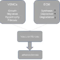
The structure of blood vessels based on lamellar units (an elastic lamella and adjacent VSMCs) varies along the arterial tree (Intengan, et al., 2000). The aorta and proximal branches contain the greatest number of medial elastic layers. The contents of VSMCs decrease from the thoracic aorta to distal arteries (Dinardo, et al., 2014, Greenwald. 2007). VSMCs decrease in arterioles, and only ECs and pericytes are found in the capillaries (Lacolley, et al., 2017).
SMCs are one of the most important cell types in the atrial wall, since they are essential for a good performance of the vasculature. By contraction and relaxation, they alter the luminal diameter, which enables blood vessels to maintain an appropriate blood pressure. However, VSMCs also play an important role in vessel remodeling in physiological and pathophysiological conditions such as pregnancy, exercise, and vascular injury. In these cases, VSMCs synthesize large amounts of ECM components and increase their proliferation and migration (Rensen, et al., 2007). Because of these properties, VSMCs are important not only for short-term regulation of the vessel diameter, but also for long-term adaptation, via structural remodeling by changing cell number and connective tissue composition (Lacolley, et al., 2017, Rensen, et al., 2007). Vascular fibrosis is a risk factors for atherosclerosis, and the latter represents an example of VSMCs role in vascular pathology (Figure 4). The transformation of macrophages into foam cells contributing to fatty streaks in atheroma is a key event during plaque formation (Figure 4). Foam cell and its apoptosis release a variety of cytokines and chemoattractant to induce inflammation in the plaque; on the other hand, apoptotic foam cells, the essential hallmark of vulnerable plaques, constitute the physical center of the plaque that critically impacts plaque progression, destabilization, and rupture (Domschke, et al., 2018, Wu, et al., 2018).
In this pathological process VSMCs are, therefore, one of the major sources of foam cells. VSMCs enter the intima of artery and endocytose LDL; to do so, VSMCs have a high ability of proliferation and migration which could deteriorate the stability of atherosclerosis plaque and induce plaque rupture. Inflammatory cytokines play an important role in the transformation of VSMCs, since inflammation factors promote the migration of VSMCs from the media to the intima of artery (Figure 4). All these changes have also an influence on the vascular stiffness and fibrosis (Part III for further details).
20 Figure 4. VSMCs are one of the most important cell types involved in atherosclerosis. In the pathological process of atherosclerosis, VSMCs are an important sources of foam cells. VSMCs enter the intima of artery and endocytose the Low-Density Lipoproteins (LDL) which accumulate locally. More importantly, these VSMCs have a high ability of proliferation and migration which could deteriorate the stability of atherosclerosis plaque and induce plaque rupture. Inflammation plays an important role in the transformation of VSMCs in atherosclerosis plaque, since it promotes the migration of VSMCs from the media to the intima of artery. http://sphweb.bumc.bu.edu
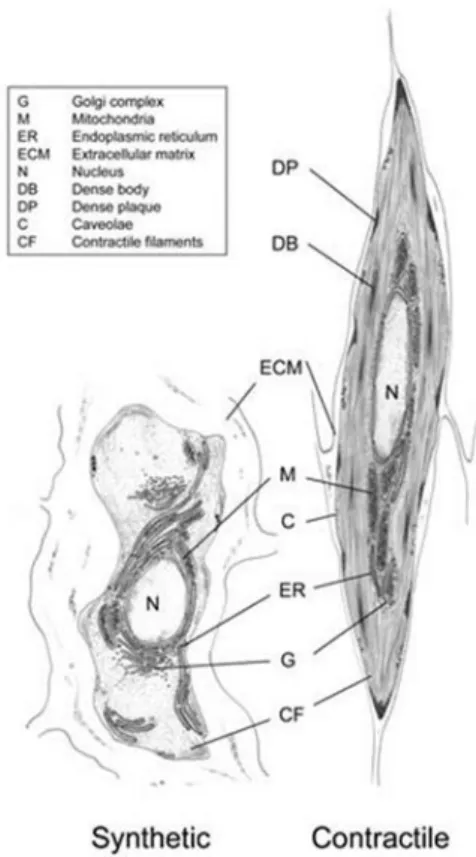
2.3 Two important phenotypes of SMCs: contractile and
synthetic SMCs
Traditionally, there are two populations of SMCs, with a spectrum of intermediate phenotypes: contractile and synthetic SMCs, which are characterized by clearly different morphologies. Contractile SMCs are elongated, spindle-shaped cells (Chamley-Campbell, et al., 1979, Hao, et al., 2003) and have contractile filaments, whereas synthetic SMCs possess a cobblestone morphology and contain a high number of organelles involved in protein synthesis. In addition, synthetic SMCs exhibit higher proliferation and migration activity than contractile SMCs (Hao, et al., 2003). There are a variety of SMC marker proteins which can be used to define SMCs phenotypes (Figure 5): α-SMA, SM-MHC, and smoothening-A/B are usually considered as the contractile SMC phenotype markers. SMemb/non-muscle MHC isoform-B, CRBP-1, h-caldesmon, meta-vinculin can be used to indicate a synthetic
21
phenotype (Glukhova, et al., 1988, Kuro-o, et al., 1991, Neuville, et al., 1997). In fact, these so-called contractile and synthetic phenotypes are just an oversimplification : it is now being recognized that there is a variety of SMC phenotypes, ranging from contractile to synthetic (Hao, et al., 2003, Matsushita, et al., 2007, Rensen, et al., 2007). Actually, recent studies proved that different synthetic and contractile markers could be upregulated at the same time (Hao, et al., 2003, Nangia-Makker, et al., 2000). In some cases, contractile differentiation can be observed in the ‘synthetic’ phenotype and contractile differentiation markers may be expressed along with with matrix synthesis (Carthy, et al., 2012, Rama, et al., 2006, Tian, et al., 2017).
Figure 5. Ultrastructural characteristics of contractile and synthetic SMCs. Contractile SMCs are elongated, spindle shaped cells and have contractile filaments, whereas synthetic SMCs have a cobblestone morphology and contain a high number of organelles involved in protein synthesis (Rensen, et al., 2007).
2.4 ECM plays an important role in vascular fibrosis
ECM plays an important role in vascular fibrosis, since the absolute and relative quantities of collagens and elastin largely affect the biomechanical properties of vessels, particularly of the major arteries and veins (Figure 6) (Arteaga-Solis, et al., 2000, Hartner, et al., 2009, Lan, et al., 2013). Lack of elastin or increased collagen in the vascular wall lead to vascular fibrosis and
22
increased stiffness (Arribas, et al., 2006). The primary sources of the tensile strength of the vessel wall are collagen fibers around fibroblast of the adventitial layer (Arteaga-Solis, et al., 2000). Among the 26 different collagen types, type I and III collagens are the major fibrillar collagens in vessels, representing 60% and 30% of vascular collagens, respectively (Jacob. 2003). However, only mutations in collagen III have been associated so far with vascular diseases. Type III collagen fibrils are relatively more abundant in tissues subjected to periodic stress, such as the vasculature (Jacob. 2003). Type III molecules participate in the tridimensional organization of type I collagen networks (Arteaga-Solis, et al., 2000). In the arterial wall, the structure and function of the ECM are also affected by several other structural glycoproteins, including fibronectin, vitronectin, laminin, entactin/nidogen, tenascin and thrombospondin these glycoproteins have a multidomain structure, potentially mediating interactions between cells and other ECM components (Chothia, et al., 1997, Labat-Robert. 1998).
Figure 6. Pathogenesis and risk factors of vascular fibrosis in atherosclerosis. Vascular fibrosis involves proliferation, migration, hypertrophy and fibrotic features of VSMCs, accumulation of ECM and inhibition of matrix degradation adapted from (Lan, et al., 2013).
Part III Ang II or TGF-β induces vascular fibrosis
23
3.1.1 Ang II induces cardiovascular fibrosis
Arterial stiffness and atherosclerosis-related hypercoagulability increase the risk of stroke. As shown above, VSMCs play a pivotal role in the onset of atherothrombotic diseases. The ability of VSMCs to adapt to environmental cues is related to their high plasticity to reprogram their expression pattern in response to acute stimuli, mainly mediated by ligand-receptor interactions. Ang II has emerged as an important hormone that affects cardiovascular physiology, and long-term exposure to Ang II plays a critical role in cardiac hypertrophy and remodeling (Geisterfer, et al., 1988, Mehta, et al., 2007, Xi, et al., 1999). In the pathogenesis of atherosclerosis, alteration in ECM composition is an important component in the regulation of VSMCs activities, including migration and secretion. Ang II could not only increase the production of collagen, but also regulate fibronectin and TGF-β synthesis by EGFR (Moriguchi, et al., 1999). A series of in vitro and in vivo experiments have shown that Ang II is a pivotal growth factor for VSMCs, causing VSMCs cell proliferation, hypertrophy, and migration (Bumpus, et al., 1991, Dzau. 2001). In Ang II-treated VSMCs, CDK2 activity was suppressed (secondary to failure of p27kip1 repression), leading to G1-phase arrest and cell hypertrophy. In a murine model of atrial fibrillation, Kai et.al found Ang II induced atrial fibrosis depends on the integrins (Friedrichs, et al., 2014).
3.1.2 Ang II and its receptors in cardiovascular fibrosis
Ang I, II, III and their receptors (AT1R, AT2R, Mas and MrgD) have been regarded as the principle components in RAS, an important hormonal cascade regulating blood pressure and heart functions. Ang II, the primary effector molecule of this system, regulates the function of several organs, including heart, kidney and the vasculature. Ang II binds to both AT1R and AT2R, which, in turn have opposite effects. Ang II induces cell proliferation, myocyte hypertrophy, myocardial remodeling, fibrosis, and oxidative stress via AT1R (Mehta and Griendling. 2007). In contrast, stimulation of AT2R produces an anti-fibrotic, anti-proliferative and pro-apoptotic effects (Mehta and Griendling. 2007).
Ang II-mediated cardiovascular responses largely depend on the activation of AT1R (Figure 7) (Lan, et al., 2013). AT1R, which belongs to the seven-membrane superfamily of G protein-coupled receptors, is widely spread in all organs, including liver, adrenals, brain, lung, kidney, heart, and
24
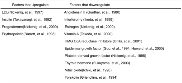
vasculature (Mehta and Griendling. 2007). On the extracellular region of AT1R, the four cysteine residues form disulfide bridges and are essential for Ang II binding (Mehta and Griendling. 2007, Ohyama, et al., 1995). AT1R cytoplasmic tail contains many serine/threonine residues, which are phosphorylated by G protein receptor kinases or GRKs (Mehta and Griendling. 2007). In VSMCs, numerous growth factors and cytokines regulate the expression of AT1R (Table 1).
Table 1. Growth factors and cytokines involved in expression of AT1R Factors that Upregulate Factors that downregulate LDL(Nickenig, et al., 1997) Angiotensin II (Gunther, et al., 1980) Insulin (Takayanagi, et al., 1992) Interferon-γ (Ikeda, et al., 1999) Progesterone(Nickenig, et al., 2000) Estrogen (Nickenig, et al., 2000) Erythropoietin(Barrett, et al., 1998) Vitamin A (Takeda, et al., 2000)
HMG CoA reductase inhibitors (Ichiki, et al., 2001)
Epidermal growth factor (Guo, et al., 1994, Howard, et al., 2000) Platelet-derived growth factor (Nickenig, et al., 1996)
Thyroid hormone (Fukuyama, et al., 2003) Nitric oxide(Ichiki, et al., 1998)
Forskolin (Griendling, et al., 1994) HMG, 3-hydroxy-3-methyl-glutaryl. (Mehta and Griendling. 2007)
Ang II-mediated AT1R activation is one of the main pathogenesis mechanisms of hypertension (Figure 7). Ang II binds to the AT1R activating a series of signaling cascades, including c-Src family kinases, Ca2+-dependent Pyk2, FAK,
JAK, PKC, MAPKs and transactivation of EGFR, PDGFR and insulin receptor (Hunyady, et al., 2006, Suzuki, et al., 2005). The activation of AT1R by Ang II induces cell proliferation, myocyte hypertrophy, myocardial remodeling and fibrosis. Importantly, Ang II increases the expression of TGF-β1 via AT1R, which, in turn, drives fibrosis. Ang II could also induce NADPH oxidase to generate excessive superoxide anion (O2•−), which plays an important role in
25 Figure 7. Role of Ang II role in cardiovascular pathology. The octapeptide Ang II exerts its numerous effects in modulating cardiovascular physiology and pathology by inducing signaling pathways in vascular smooth muscle cells, endothelial cells, and cardiac fibroblasts, and by affecting their interaction with the ECM adapted from (Mehta and Griendling. 2007).
In contrast to the effect of AT1R, the majority of experimental data show that AT2R stimulation produces an anti-fibrotic effect. AT2R also belongs to the seven-membrane superfamily of G protein-coupled receptors. AT2R plays an critical role in fetal development, and is highly expressed in fetal tissue, and its expression decreases after birth (Shanmugam, et al., 1996). In adults, the AT2R has been localized to the heart, kidney, adrenal gland, brain, uterus, pancreas, retina, skin, and both endothelial and VSMCs of the vasculature (Roulston, et al., 2003, Wang, et al., 1999, Wheeler-Schilling, et al., 1999). In the central nervous system, AT2R is expressed in neurons (Steckelings, et al., 2017).
Several cardiovascular pathologies can increase AT2R expression (Jones, et al., 2008). In contrast to AT1R, the activation of AT2R receptors induces vasorelaxation by increasing the production of nitric oxide and cGMP (Jones, et al., 2008). The stimulation of AT2R directly counteracts the dysregulation of sympathetic outflow in neurogenic hypertension. Selective activation of brain AT2R causes moderate decreases in blood pressure in vivo (Steckelings, et al., 2017). AT2R activates the NO/cGMP pathway, and has vasodilatory effects in the vasculature (Savoia, et al., 2006). NO/cGMP activates a cGMP dependent protein kinase causing decreased RhoA activity and AT1R-mediated vasoconstriction (Savoia, et al., 2006, Savoia, et al., 2005).
In the sites of MI and MI repair, ACE, AT1R and AT2R are all expressed at high levels (Jones, et al., 2008, Weber, et al., 1997). There is also a link between upregulation of AT2R expression and fibrosis present in hypertrophied and failing hearts, the increased expression of AT2R was specifically localized to cardiac fibroblasts (Jones, et al., 2008). TGF-β1 and its receptors, and type I
26
and III collagens, are also expressed in AT2R-rich fibroblasts and myofibroblasts (Jones, et al., 2008, Katwa, et al., 1997, Lijnen, et al., 2003). However, the majority of the studies indicate an inhibitory effect of AT2R on cardiac fibrosis. AT2R has also been found in endothelial cells and in VSMCs of vessel (Nora, et al., 1998). In AT2R deficient mice, the antifibrotic effect of AT1R inhibition was not obvious (Wu, et al., 2002, Xu, et al., 2002). Specific over-expression of AT2R in heart decreased the amount of both perivascular and interstitial fibrosis induced by Ang II infusion (Jones, et al., 2008). Recently, it has been shown that AT2R antagonizes the function of AT1R by directly binding to AT1R, but, agonist-induced activation of AT2R could not inhibit AT1R signaling (AbdAlla, et al., 2001).
While the effect of AT1R on collagen synthesis is well established, AT2R seems to have no effect on collagen secretion. Indeed, AT1R blockade, but not AT2R inhibition, suppressed Ang II stimulation of collagen production in cultured rat and porcine cardiac fibroblasts, as well as rat mesenteric VSMCs (Lijnen, et al., 2000, Touyz, et al., 2001, Warnecke, et al., 2001). In VSMCs, AT2R could produce anti-proliferative and pro-apoptotic effects (Mehta and Griendling. 2007). Resveratrol protection against arterial fibrosis is associated with increased expression of AT2R (Kim, et al., 2018). In cultured cardiac fibroblasts, Ang II stimulation induces the decrease in collagenase activity which could be abolished by AT2R blockade but not influenced by AT1R (Brilla, et al., 1994).
AT2R stimulation may also activate lipid-signaling pathways. Ang II increases PLA2 activity and AA release via AT2R (Lokuta, et al., 1994) in rabbit proximal tubule epithelia (Jacobs, et al., 1996) and cultured neurons (Zhu, et al., 1998). Long-term AT2R stimulation by Ang II could also increase synthesis of ceramides, which may then activate stress kinases and caspases involved in the induction of apoptosis (Gallinat, et al., 1999, Lehtonen, et al., 1999)
Ang II could also affect the fibrosis through the cross-talk with other receptors and pathways. TRPM is a group of receptors that could mediate the influx of Ca2+ and Mg2+. Zhong et.al found Ang II could regulate SAN fibrosis through
TRPM7 (Zhong, et al., 2018). TRPM7 mediated both calcium and magnesium homeostasis in the CFs, 2-APB (TRPM7 inhibitor) inhibited Ang II-induced CTGF, SMA expression and CFs proliferation which contribute to fibrosis progress (Yu, et al., 2014).
PAR-1 which is primarily known as the receptor of thrombin also mediates the Ang II-induced vascular and cardiac fibrosis. Knock-out of PAR-1 effectively attenuated the increasing medial wall thickening and perivascular fibrosis which were increased by Ang-II (Antoniak, et al., 2017).
27
TNF has been found to regulate the cardiac structure and function in health and disease (Kleinbongard, et al., 2010, Mann. 2001, Meldrum. 1998). TNF has two distinct TNF receptors, TNFR1 and TNFR2. Duerrschmid et.al found that TNFR1 mediated the Ang-II-induced development of cardiac fibrosis and adverse cardiac remodeling (Duerrschmid, et al., 2013).
C5a, a potent chemotactic and inflammatory mediator, activated monocytes in many remodeling processes. PMX53 is a specific C5aR (receptor of C5a) antagonist that effectively suppressed inflammation and perivascular fibrosis and increased cardiac function in mice treated with Ang II (Zhang, et al., 2014) The transactivation of EGFR, plays a crucial role in the development of atherosclerotic lesion. In VSMCs, Ang II promotes the proliferation and migration of SMCs through the release of HB-EGF and transactivation of EGFR, MMPs also plays an indispensable role in this process (Yang, et al., 2005).
3.1.3 Downstream of Ang II Receptors: signaling pathways in
cardiovascular fibrosis
Ang II activates a series of signaling cascades by binding to its different receptors, which in turn regulate the physiological and pathological effects of Ang II in the cardiovascular system (reviewed by Mehta and Griendling. 2007). Here, we just briefly review some signaling pathways related to fibrosis. Ang II mediates cardiovascular fibrosis primarily via TGF-β-dependent smad2/3 signaling pathway (see the dedicated section in the next chapter). However, many other mediators could also play a role in Ang II-induced activation of TGF-β/smad2/3 signaling pathway (Bhattacharjee, et al., 2016, Chung, et al., 2010). Ang II could also activate smad 2/3 signaling pathway through activin A and its specific downstream component ALK4 (Wang, et al., 2017). Ang II increases ECM production by activating smad signals through the AT1R and ERK/p38MAPK-Smad pathway (Figure 7). Similarly, in diabetes AGE could activate smad2/3 via ERK/p38MAPK-dependent mechanism instead of TGF-β1-pathway (Li, et al., 2004).
MAPKs regulate a plethora of cellular responses, including protein synthesis and metabolism, intracellular transport, cell volume regulation, gene expression, and growth. The signaling cascades including ERK1/2, JNK, and p38MAPK, are implicated in VSMCs differentiation, proliferation, migration, and fibrosis (Srivastava. 2002, Taniyama, et al., 2004). In particular, the MAPK signaling pathway mediates Ang II induced cardiac fibrosis. In atrial fibroblasts,
28
MAPKs are be activated by Ang II, inducing upregulation of TRAF6 and, ultimately, fibroblast proliferation (Gu, et al., 2012). In skeletal muscle cells, Ang II induces the pro-fibrotic factors (TGF-β1 and CTGF) through P38 MAPK activity, instead of ERK1/2 signaling pathway (Morales, et al., 2012, Wong, et al., 2018). Ang II activates P38 MAPK and JNK through TNF-α, and these effects are associated with Ang II-induced hypertension and adverse cardiac remodeling (Sriramula, et al., 2015). P38 MAPK is a downstream target of TGF-β in the pathogenesis of renal fibrosis (Stambe, et al., 2004). Ang II increases the expression of MAPKs/TGFβ1/TRAF6 pathway which is an important signaling pathway in Ang II-induced CTGF expression (Gu, et al., 2017). Ang II activates CaSR, and activates MEK/ERK pathways; interesting, pretreating the cells with CaSR inhibitor (Calhex231) or PD98059 (ERK signaling pathways inhibitor) partially decreased AngII-induced cardiac fibrosis, suggesting the relevance these pathways have in fibrosis (Chi, et al., 2018). G-CSF is a key mediator of neutrophil infiltration and induces fibrosis in infarcted heart; interestingly, it has been shown that Ang II activates ERK1/2 signaling pathway in G-CSF in the heart (Jiang, et al., 2013).
The JAK/STAT signaling pathway plays an important role in Ang II induced expression of granulocyte G-CSF and increases cardiac fibrosis via STAT3 signaling pathway (Jiang, et al., 2013). Accordingly, the proteasome inhibitor bortezomib decreased Ang II-induced hypertrophy by inactivation of AT1R-mediated p38MAPK and STAT3 signaling pathways (Li, et al., 2015). STAT3 is also necessary in Ang II-induced cardiac remodeling, since, Ang II could not increase the mass of myofibrils in STAT3 knock-out mice in contrast to WT mice (Zouein, et al., 2013).
The adaptor molecule CIKS (connection to IKK and SAPK/JNK) played an important role in the IκB kinase/nuclear factor (NF)-κB and JNK/AP-1 pathways. Knock-down CIKS attenuated Ang-II-induced IKK/p65 and JNK/c-Jun phosphorylation, NF-κB and AP-1 activation, and MMP-9 expression (Valente, et al., 2012). ILK, a serine/threonine protein kinase, interacts with β1 and β3 integrin cytoplasmic domains, Ang II stimulated pro-fibrotic process involving crosstalk between ILK and NF-κB activation in cardiac fibroblasts (Thakur, et al., 2014).
Notch proteins share a highly conserved domain architecture (Chillakuri, et al., 2012,Ni, et al., 2018). Notch proteins are a family of transmembrane receptors (300–350 kDa), including an extracellular domain (NECD), an intracellular domain (NICD), and a transmembrane domain. Upon activation the NICD is released and will translocate to the nucleus, based on the presence of an NLS. Binding of Notch ligands trigger the γ-secretase complex to release the NICD into the cytoplasm and the nucleus (Couturier, et al., 2014, Guruharsha, et al., 2012, High, et al., 2008, Ozasa, et al., 2013). In the latter, the NICD regulates
29
transcription of the target genes, HES and HEY (Jarriault, et al., 1995, Maier, et al., 2000, Meester, et al., 2018). Notch signaling pathway play an important role in cell proliferation, differentiation, and apoptosis. Through the AT1R, Ang II increases the expression of NICD, promotes the proliferation and migration of VSMCs, and contributes to the progression of vascular fibrosis (Ozasa, et al., 2013). In mammals, there are four Notch proteins (Notch 1–4). Notch 3 is predominantly expressed in VSMCs of the small arteries (Joutel, et al., 2000, Prakash, et al., 2002), where it plays an important role in their maturation and differentiation (Domenga, et al., 2004, Meester, et al., 2018). Mutations in notch 3 leads to the CADASIL (Joutel, et al., 1996, Meester, et al., 2018). In pathological process of CADASIL, the small cerebral and leptomeningeal arteries show thickening of the arterial wall, which is accompanied by lumen stenosis, destruction of VSMCs, and abundance of ECM proteins (Chabriat, et al., 2009, Meester, et al., 2018, Miao, et al., 2004).
AMPK and some downstream signaling pathways are also related to the Ang II induced fibrosis. Alamandine activates AMPK, induces NO production, and produces the protective effects in cardiomyocytes (Zhang, et al., 2017). Sirtuin 6 increases the expression of pAMPKα-ACE2 signaling and suppress CTGF-FKN pathway, an effect which diminishes Ang II-induced myocardial fibrosis (Jesus, et al., 2018).
NF-κB is a nuclear transcription factor, NF-κB is related to the production of some inflammatory cytokines and its role in inflammation is well established (Zhao, et al., 2018). The NF-kB mediated pathways have been found to be involved in MMPs and EMMPRIN expression in previous study (Cao, et al., 2017, Huang, et al., 2012). Inflammation has also been shown to play a major part in development and progression of atherosclerotic lesion formation (Pant, et al., 2014). In monocytes, macrophages, VSMCs, and endothelial cells, Ang II induces the production of cell adhesion molecules such as VCAM-1, ICAM-1, and E-selectin, and chemokines such as MCP-1, IL-6, IL-8 and IL-18 (Ruiz-Ortega, et al., 2001, Ruiz-Ortega, et al., 2000, Schieffer, et al., 2000) depending on NF-κB.
30
3.2.1 TGF-β1 interacts with integrins via LAP and induces
cardiovascular fibrosis
There are three structurally similar isoforms of TGF-β, including TGF-β1, 2 and 3 in mammals. All three isoforms share the same cell surface receptors and have similar cellular targets. TGF-β1 is the prevalent isoform and is widely spread in mammalian tissues, whereas the other isoforms are expressed in a more limited spectrum of cells and tissues (Biernacka, et al., 2011). Although all three isoforms are expressed in fibrotic tissues, TGF-β1 plays the major role in the development of tissue fibrosis (Ask, et al., 2008).
TGF-β1 associates with LAP, SLC, and LTBP-1, participating in a Large Latent Complex, LLC (Chen, et al., 2016). The covering of TGF-β by LAP prevents it from binding to its receptors and activating related downstream pathway (see below) (Figure 8). Integrin αvβ3, αvβ5, αvβ6 and αvβ8 bind to the integrin-recognition motif (arginine–glycine–aspartic acid) of LAP and mediate the activation of TGF-β (Pozzi, et al., 2011) (see Figure 8). Latent TGFβ1 is converted into its active form by various mechanisms, including integrins, bone morphogenetic protein 1, several MMPs (MMP-2 and MMP-9), plasmin, elastase, thrombin, and cathepsin. Interaction between LAP and TSP-1 also promote latent TGFβ1 activation (Chen, et al., 2016). Integrin αvβ6 interacts directly with TGF-β1-bound LAP of and induces a spatially restricted activation of the TGF-β1 singaling. Anti-β6 integrin blocking antibodies completely inhibit the activation of TGF-β (Annes, et al., 2002). Integrin β6 knock-out mice are protected from bleomycin-induced pulmonary fibrosis (Munger, et al., 1999). TGF-β-dependent and -independent pathways of induction of tubulointerstitial fibrosis were reported in integrin β6- mice (Ma, et al., 2003). The interaction between integrin αvβ8 and an RGD of TGF-β1 results in the MT1-MMP-dependent release of active TGF-β1 which produces autocrine and paracrine effects on cell growth, matrix production and fibrogenesis in lung cancer tumor xenografts (Mu, et al., 2002).
Two main models have been proposed to explain how integrins contribute to the activation of TGF-β. In the first model, integrins simultaneously bind the latent TGF-β1 complex and MMPs. This allows the latent TGF-β1 complex and proteases to come close together and facilitates the enzymatic cleavage and release of active TGF-β1. The second mechanism requires the interaction between the latent complex and ECM, and is independent from proteolysis. Integrin αv can change the conformation of the latent TGF-β1 complex by transmitting cell traction forces (Chen, et al., 2016).
31 Figure 8. Model of mechanical activation of latent TGFβ1. TGFβ1 participate in LLC that consists of TGFβ1 associated with LAP, SLC, and LTBP-1. LAP contains the amino acid sequence motif RGD (Arg-Gly-Asp) which serves as a recognition site for several integrins. Actin / myosin-mediated cell contraction force can be transmitted to an RGD binding site in LAP through the αv integrins and induces a putative conformational change that liberates TGFβ1, activating it (Chen, et al., 2016).
TGF-β induction and activation are consistently observed in experimental models of tissue fibrosis (Biernacka, et al., 2011, Leask and Abraham. 2004, Pohlers, et al., 2009). There are extensive evidences demonstrating upregulation of TGF-β and its key role in the pathogenesis of renal fibrosis in both animal models and humans with kidney diseases (Chen, et al., 2018, Lan. 2011, Meng, et al., 2016). The activated HSC have significant increase in TGF-β expression that acts as an autocrine positive regulator for ECM production. TGF-β signal is associated with the accelerated ECM accumulation (Kisseleva, et al., 2008), TGF-β1 mRNA expression is increased predominantly in alveolar macrophages, but also in pulmonary endothelial cells, mesenchymal cells, fibroblasts, and mesothelial cells in bleomycin-induced pulmonary fibrosis in human patients and experimental animals (Chen, et al., 2016). TGF-β also contributes to fibrogenesis through inflammation in chronic liver disease (Dooley, et al., 2012). TGF-β plays a pivotal role in the development of fibrosis in heart and consistently is involved in a variety of cardiac pathologies (Leask, et al., 2004). In a model of myocardial fibrosis, Ang II increases the expression of TGF-β (Wong, et al., 2018). TGF-β inhibition reduced hepatic (Nakamura, et al., 2000), renal (Fukasawa, et al., 2004) and cardiac fibrosis (Teekakirikul, et al., 2010) highlighting the role of TGF-β in a wide range of fibrotic conditions (Biernacka, et al., 2011). Serum TGF-β1 level is upregulated in AF patients and could be used as an independent predictor of AF recurrence (Zhao, et al., 2014).
32
3.2.2 TGF-β1 and its receptors
There are three types of TGF-β-receptors (TGF-βRI, TGF-βRII and TGF-βRIII). TGF-β signals require a heteromeric complex of type I and type II transmembrane serine/threonine kinase receptors (TGF-βRI and TGF-βRII). TGF-β binds to TGF-βRII, thus recruiting TGF-βRI. The formation of this heteromeric complex of type I and two type II receptors seems to be necessary for signaling (Luo, et al., 1996, Weis-Garcia, et al., 1996, Yamashita, et al., 1994). Phosphorylation of serine and threonine residues in glycine–serine rich (GS)-domain of TGF-βRI could be phosphorylated by TGF-βRII which results in a conformational change of TGF-βRI, Subsequently, phosphorylation of smads transfers the signal into the nucleus (Wieser, et al., 1995). There are two distinct isoforms of the TGF-βRI: the endothelium restricted ALK1 and the widely expressed ALK5. The activation of ALK1 induces smad1, smad5, and smad8 phosphorylation, while ALK5 promotes the phosphorylation of smad2 and smad3 (Lebrin, et al., 2005).
In contrast to the type I and II receptors, our knowledge about the role played by TGF-βRIII in TGF-β biology remains poorly understood. The TGF-βRIII, the most abundant and ubiquitously expressed TGF-β superfamily co-receptor, binds each of the three TGF-β isoforms with high affinity. TGF-βRIII is classically thought to function as a co-receptor, adjusting the TGF-β superfamily ligands to their respective signaling receptors (Cheifetz, et al., 1990). TGF-β has low affinity for TGF-βRII in the absence of TGF-βRIII, and overexpression of TGF-βRIII increases the binding of TGF-β to their receptors and in some cases have been shown to augment TGF-β actions, particularly those of TGF-β2 (Esparza-Lopez, et al., 2001, Lopez-Casillas, et al., 1993, Sankar, et al., 1995).
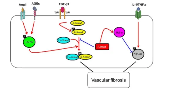
3.2.3 TGF-β1 and its signaling pathways in cardiovascular fibrosis
In general, TGF-β1 is known to signal through smad signaling pathways, which, accordingly, play an important role in fibrosis. The smad family members are well conserved and widely spread in almost all vertebrates (Figure 9). This family has eight members, two TGF-β R-smads (smad2 and 3), three BMP R-smads (smad1, 5 and 8), one Co-smad (smad4) and two I-smads (smad6 and 7). Smad proteins have two globular conserved N-terminal MH1 and C-terminal MH2 domains connected by a linker region. R-smads and Co-smad
33
share two conserved MH1 and MH2 domains (Heldin, et al., 2012, Massague, et al., 2005, Moustakas, et al., 2001, Ross, et al., 2008), whereas I-smads have only a MH2 domain. Both the MH1 and the MH2 domains can interact with cytoplasmic adaptors, transcription factors, co-activators, co-repressors, and chromatin-remodeling factors (Zhang, et al., 2015).
The linker regions between the MH1 and MH2 domains also play an important function in smad regulation. Since this region contains PY motifs and flexible binding sites for Smurf ubiquitin ligases, sites of phosphorylation targeted by various kinases phosphorylation of protein (Moustakas, et al., 2009, Shi, et al., 2003), the R-Smad linker region is involved in pathways including, MAPKs, CDKs (Figure 9) (Zhang, et al., 2015).
Figure 9. Functional and structural characteristics of Smad family members. Smad proteins consist of two conserved globular domains (MH1 and MH2) and a variable linker region R-Smads and Smad4 have a MH1 domain that contains a β-hairpin structure for DNA binding and protein–protein interactions. The R-Smad linker region is involved in pathways for MAPKs and Smurf pathway. The MH2 domain is responsible for Smad oligomerization, transcriptional activation and receptor interaction. Abbreviations: A: acetylation, NES: nuclear export signal, NLS: nuclear localization signal, NPS: nucleopore signal, P: phosphorylation, pA: poly-ADP-ribosylation, S: sumoylation, SAD: Smad activation domain, U: ubiquitination. Adapted from Zhang, et al., 2015.
TGF-β signaling is initiated with serine/threonine kinase receptor oligomerization and R-smad phosphorylation (Derynck, et al., 1998, Moustakas, et al., 2001, Whitman. 1998). Subsequently, a R-smad/Co-smad complex is generated to translocate to the nucleus and regulates downstream gene transcription. I-smads (smad6 and smad7) could block TGF-β signaling pathway by preventing R-smads from interacting with the TGF-β receptor or competing with Co-smad for the generation of R-smad/Co-smad complexes
34
(Figure 10) (Zhang, et al., 2015).
Smad 2/3 signaling pathway has been proved to be related to vascular fibrosis. Some fibrogenic genes have been shown to be the downstream targets of TGF-β/smad3 signaling, including, collagen type 1 α 1, collagen type 1 α 2, collagen type 5 α 2, collagen type 6 α 1, collagen type 6 α 3, CTGF, tissue inhibitor of metalloproteinase-1 (Verrecchia, et al., 2001).
Figure 10. Signaling crosstalk in vascular fibrosis. TGF-β1/TGF-β1R activation phosphorylates R-Smads, Smad2 and 3. Activated R-Smads form a complex with Co-Smad4 which then translocate to the nucleus and regulates gene transcription. The ECM deposition is also mediated through AngII/AGEs—mediated ERK/p38 MAP kinase-Smad crosstalk. Adapted from (Zhang, et al., 2015)
The activation of TGF-β/smads signaling plays a pivotal role in the pathogenesis of cardiac fibrosis and hypertrophy (Flevaris, et al., 2017, Gao, et al., 2017, Jeong, et al., 2015, Xiao, et al., 2018). ALK5 inhibitor inhibited fibrogenesis in a rat model of progressive TGF-β1-induced pulmonary fibrosis (Bonniaud, et al., 2005). Smad3 null mice exhibit attenuated cardiac fibrosis (Bujak, et al., 2007, Dobaczewski, et al., 2010) and are resistant to bleomycin-induced pulmonary fibrosis (Zhao, et al., 2002), dermal fibrosis following irradiation (Flanders, et al., 2002), unilateral ureteral obstruction induced renal interstitial fibrosis (Biernacka, et al., 2011, Sato, et al., 2003). Through binding to the membrane-bound type I and II receptors, TGF-β increases the phosphorylation of smad2 and smad3. Once activated by TGF-β, smad3 and smad4 form a complex, translocate to the nucleus, and recruit some co-activators such as p300 (Xiao, et al., 2018). The smad3/4 complex then binds to the promoter regions of target genes and increases the expression of some fibrosis related protein (Derynck, et al., 2003, Liu, et al., 2017, Wu, et al., 2015). TGF-β1 is not the sole mediator that could activate the
35
smad2/3 signaling pathway. We have mentioned alternative triggers in the Ang II part.
Smad 7, a downstream inhibitory smad in TGF-β signaling (Kavsak, et al., 2000, Rodriguez-Vita, et al., 2005, Wang, et al., 2006), acts in a negative feedback loop. Smad 7 competes with R-smads for receptor binding, thus inhibiting their phosphorylation; in addition, it recruits ubiquitin ligases to the receptors thus promoting their ubiquitination and proteasomal degradation; it also recruits phosphatases to the receptors thus promoting their dephosphorylation and deactivation, and interferes with the binding of smad complexes to DNA (Lorenzen, et al., 2015). For these reasons, Smad 7 stops the progression of cardiac injury via blocking TGF-β/smad3-mediated cardiac fibrosis and NF-kB-driven inflammation (Wei, et al., 2013). Johan M et.al also found that smad 7 is required by Ang II to induce ERK activation as well as AKT signaling pathway and reduced expression of PTEN in the heart (Lorenzen, et al., 2015). In summary, TGF-β signaling activates multiple smad signaling, leading to fibrosis as a main response, in the presence of a finely tuned regulatory feedback.
Recently, there are also some non-smad signaling pathways which are proven to mediate the TGF-β induced vascular fibrosis (Lan, et al., 2013). The JNK/p38, ERK/MAPK, Rho-like GTPase, and PI3/AKT pathways have been found to reinforce, attenuate or modulate downstream cellular responses, accounting for the varying effects of TGF-β (Lan, et al., 2013). ERK, FAK, Src and β-catenin have been proven to be related with integrin signaling (Chen, et al., 2016), further regulating smad-independent TGF-β effects. Worth noting, TGF-β uses non-smad signaling pathways i.e. MAPK pathways, to convey the same fibrogenic signals (Zhang. 2009). MAPK family is a well-known serine/threonine-specific protein kinase which regulate extracellular mitogenic and stress stimuli and regulate cell differentiation, proliferation, survival and apoptosis. TGF-β activates all three known MAPK pathways: ERK, p38 and JNK (Biernacka, et al., 2011). MAPK pathways may further regulate smad proteins or mediate smad-independent TGF-β responses. p38 and JNK usually potentiate TGF-β/smad-induced responses, in contrast, ERK activation either increases or decreases smad signals depending on the cell type (Biernacka, et al., 2011). p38 MAPK mediates TGF-β-induced G0/G1 cell cycle arrest without smad proteins (Seay, et al., 2005). In a mouse model of scleroderma-like fibrosis, activation of a fibrotic gene program was dependent on smad1 and ERK1/2, and not on smad2/3 (Biernacka, et al., 2011, Pannu, et al., 2007). Besides, both in vitro and in vivo findings have suggested that p38 MAPK may play a role in the pathogenesis of renal fibrosis acting downstream of TGF-β (Biernacka, et al., 2011). JNK and ERK could mediate TGF-β1 induced upregulation of fibronectin and α-SMA in pancreatic stellate cells (Xu, et al., 2018). TGF-β1 induced the phenotypic changes in fibroblasts to become