HAL Id: hal-02421115
https://hal.uvsq.fr/hal-02421115
Submitted on 15 Apr 2020
HAL is a multi-disciplinary open access
archive for the deposit and dissemination of
sci-entific research documents, whether they are
pub-lished or not. The documents may come from
teaching and research institutions in France or
abroad, or from public or private research centers.
L’archive ouverte pluridisciplinaire HAL, est
destinée au dépôt et à la diffusion de documents
scientifiques de niveau recherche, publiés ou non,
émanant des établissements d’enseignement et de
recherche français ou étrangers, des laboratoires
publics ou privés.
Exoskeleton Based on User Intention
Jinan Charafeddine, Sylvain Chevallier, Samer Alfayad, Mohamad Khalil,
Didier Pradon
To cite this version:
Jinan Charafeddine, Sylvain Chevallier, Samer Alfayad, Mohamad Khalil, Didier Pradon.
Biokine-matic Control Strategy for Rehabilitation Exoskeleton Based on User Intention. International Journal
of Modeling and Optimization, IJMO, 2019, 9 (6), pp.322-328. �10.7763/IJMO.2019.V9.730�.
�hal-02421115�
Abstract—Rehabilitation exoskeletons require a control
interface for the direct transfer of mechanical power and exchange of information in order to assist the patient in his/her movements. By using co-contraction indexes (CCI), it is possible to accurately characterize human movement and joint stability. But when dealing with human movement disorders, no existing index allows to achieve neuro-motor control with bio-kinematic sensors. Thus, we propose a neuro-motor interactive method for lower-body exoskeleton control. A novel dynamic index called neuro-motor index (NMI) is introduced to estimate the relation between muscular co-contraction derived from electromyography signals (EMG) and joint angles. To estimate the correlation in the state space and enhance the precision of the NMI, we describe an estimation method relying on a two-way analysis of canonical correlation (CCA). A thorough assessment is presented, by conducting two studies on control subjects and on patients with abnormal gait in a medical environment. i) An offline study on control patients showed that NMI captures the complex variation induced by changing walking speed more accurately than CCI, ii) an online study, applied on successive gait cycles of patients with abnormal walk indicates that the existing CCI have a low accuracy related with joint angles while it is significantly higher with NMI.
Index Terms—Exoskeleton for rehabilitation, co-contraction
index, neuro-motor control, bio kinematic.
I. INTRODUCTION
The rehabilitation exoskeleton is a mechatronic device, worn by the neurological patient, designed to increase physical performance and fitted to the shape and function of the human body. This prosthesis is used to provide a high-intensity training for human limbs on user-specific basis, to help the recovery of a neurological disease or disorder [1], [2]. The exoskeleton works mechanically in parallel with the human body [1] and can be controlled in a passive or active way. Typically, a complete exoskeleton, consists of a sensory system that acquires the physiological signals of interest, a processing stage to extract the relevant parameters that can be used to control the exoskeleton accordingly. The main
Manuscript received April 3, 2019; revised June 12, 2019.
Jinan Charafeddine is with Biomedical/Control of Versailles University, LISV 10-12, avenue de l'Europe- 78140 Vélizy, France (e-mail: [email protected]).
Sylvain Chevallier is with Computer Science of Versailles University LISV 10-12, Avenue de l'Europe- 78140 Vélizy, France (e-mail: [email protected]).
Samer Alfayad is with Robotics of Versailles University. LISV 10-12, avenue de l'Europe- 78140 Vélizy, France (e-mail : [email protected]).
Mohamad Khalil is with Biomedical, Lebanese University, Faculty of Engineering, EDST Elmitein street, 1300 Tripoli, Lebanon (e-mail: [email protected]).
Dider Pradon is with Biomechanics of Versailles University, END ICAP.104, Bd Raymond Poincaré 92380 Garches, France (e-mail: [email protected]).
challenge faced by interactive exoskeleton is the direct relationship between the physiological signals and the desired behavior of the exoskeleton. To yield the complete control to the patient, most of the existing approaches are following another approach. The exoskeleton is programed with a predefined behavior that the patient should follow. One of the most difficult cases addressed by researchers in rehabilitation robotics is the training of ground running with exoskeleton, such as HAL [3] of the University of Tsukuba, EXPOS of Sogang University [4]. ReWalk [5], Rex Bionics [6], Indigo [7], Exoatlet [8], Wandercraft [9], among others. There are two known control strategies for these devices (i) impedance or admittance control, which are generally predetermined and do not consider the user's physical condition [10], [11] (ii) electromyogram (EMG)-based control which rely on the detection of muscle activation. But existing approaches are not satisfying in that EMG signals recorded from users with muscular disorder lead to improper operation [10]-[12]. Thus, these two control strategies are not suitable for patients with cerebral palsy (CP) or recovering after stroke, which are two common target populations for rehabilitation exoskeletons. Here, we are interested in an interactive exoskeleton that encompasses a control system driven by the patient. This requires continuous measurements of biological and mechanical signals. The acquired signals are analyzed, interpreted, and used to drive the trajectory of an exoskeleton. This approach relies on physiological signals (electromyography EMG) and kinematic recording (joint angles of walking). The traditional method to quantify the walking movements is based on monitoring the kinematic, EMG and spatio-temporal parameters to assist a patient in his movement [13]. However, this method suffers from several limitations, as it requires off-line measurement, it limits the patient in his own movement, and it does not take into account the variability of the patient's walk [14]. This leads to patient fatigue and pain during rehabilitation session with the exoskeleton [15]. In addition, this method does not allow to distinguish between the deviations and the compensations due to the influence of walking speed [14].
Our approach is based on recording the EMG signals from hamstring and quadriceps muscles due to their bi-articular nature for the knee and the hip [16]. These signals contain information about the intention of the patient [17] and allow to evaluate the muscle co-activation around the joint [18]. It has been suggested that muscle co-activation indicates the achievement of a motor skill [19], [20]. Co-activation is also related to joint stability [20] and is considered as an important factor which contributes to the inefficiency of pathological movement [21]. Most researchers and clinicians rely on EMG measurements to express co-activation [18]. Muscle co-activation has been expressed as a comparison between the
Jinan Charafeddine, Sylvain Chevallier, Samer Alfayad, Mohamad Khalil, and Dider Pradon
Biokinematic Control Strategy for Rehabilitation
Exoskeleton Based on User Intention
measured EMGs for the involved muscles and the reference EMG values [18], [22]-[25]. A different approach was followed by Falconer and Winter [26], Hessee et al. [27] and for another index proposed by Frost et al. [28], Unihan et al. [29]. Those works examine the co-activation around a joint based on the co-contraction index. While it is adequate for offline analysis and diagnosis, these are not suitable for designing a control strategy.
In this paper, we propose to modify those co-contraction indexes for exoskeleton control and we propose a new framework to design a controller based on EMG and kinematics. The proposed approach allows to encompass the patient-specific walking behavior to offer a new interactive control, without any reference to a reference walking cycle. The contributions are the following:
• Introduction of a new index based on the results of the regression in combination with CCI.
• A new methodology of assessment relying on canonical correlation analysis.
• Evaluation of the new index for generalization of walking behavior
• Assessment of the new index for characterizing abnormal walk
The next section of this paper provides the required background, regarding the computation of the co-contraction indexes, the experimental data acquisition and the preprocessing applied on the signal. Section III introduces the neuro-motor index and the adequate tools for its assessment. The experimental results are provided in section IV along with some detailed explanation. Section V concludes this paper.
II. EXPERIMENTAL FRAMEWORK
To incorporate intention information in a control strategy, one could rely on co-contraction indexes. Several indexes are described in the literature; we provide here a unified description of these indexes.
A. Estimating the Co-contraction
A modified version of the method Frost et al. [28] and Unnithan et al. [29] could be written as:
CCI1 (t)= (1)
The method by Hessee et al. [31] is rewritten as:
CCI2 (t)= (2)
The method by Falconer and Winter [30] proposes an index that could be reformulated as
CCI3 (t)= (3)
For the knee study, EMG antago is the envelop of muscles that cause knee flexion movement (quadriceps) and EMG ago is the envelop of muscles that cause knee extension movement of
(hamstring). For the hip study, EMG antago is the envelop of muscles that causes hip flexion movement (hamstring) and EMG ago is the envelop of muscles that cause hip extension movement of (quadriceps). The CCI1 and CCI2, t1 and t2 denote the period of one complete gait cycle. The CCI3: t1 and t2 denote the period where the agonist EMG is less than the antagonist EMG, whereas t2 and t3 denote the period where the antagonist EMG is less than agonist EMG
B. Subjects and Measurements
Patients have been recorded in Gait Laboratory, Raymond Poincaré Hospital, Garches, France. There were twenty subjects; nine healthy adults: four females and five males, aged 50 years ±7; five kids with cerebral palsy: three females and two males, aged 10 years ±2; six adults with stroke: three females and three males, aged 50 years ±7. Each subject was tested for his gait on the ground at different walking velocities, with each subject performing eleven trials for each velocity. The gait analysis was recorded at 100 Hz with a 3D optoelectronic system (Motion Analysis Corporation, Santa Rosa, CA, USA) using eight optoelectronic cameras. Twenty-three markers were placed on the patient’s lower body, following the Helen Hayes model commonly used by the biomechanical community for gait analysis [30]. The relative displacement of each segment was estimated from this coordinate system (flexion / extension, abduction / adduction, internal / external rotation). The marker trajectories were then filtered using a fourth-order zero-lag Butterworth low-pass-filter, with a 6-Hz cut-off frequency [31]. The main kinematic parameters were peak flexion, extension, as appropriate for hips and knees. Spatio-temporal parameters were calculated for both lower limbs, including gait velocity, cadence, step and stride length, step width, and the duration of the single support phase. Kinematic and kinetic parameters were calculated for both lower limbs for each sub-phase of the gait cycle: first double support phase (DS1), the first single phase (SS), second double support phase (DS2) and the swing phase (SWP). The activity of eight muscles were also recorded using a surface EMG system (MA311, Motion Lab Systems, Baton Rouge; band-pass 15-3000 Hz): biceps femoris (Bic-Fem), gastrocnemius lateralis (GasLat), gastrocnemius medialis (Gas-Med), rectus femoris (Rec-Fem), semi-membranous (Semi-Mem), soleus (Soleus), tibialis anterior (Tib-Ant), and vastus lateralis (Vas-Lat) for both legs. The main EMG measurements of quadriceps (Rec-Fem, Vas-Lat) and hamstring (Bic-Fem, Semi-Mem).
C. Processing of Recorded Signals
The EMG raw data were full-wave filtered with a Butterworth 4-band band pass filter and cut-off frequencies at 10 and 400 Hz, rectified and low pass filtered by a Butterworth filter of order 4 at between 4 to 6 Hz according to the cadence of the subject [32] yielding the linear envelopes of each muscle EMG. The EMG of each muscle were then expressed as a percentage of the EMG value during the MVC [33]. The kinetic data were segmented in 1001 values for knee and hip joints to obtain matrices with fixed dimensions, equal to those of recorded EMG matrices and their mean and variance were estimated. The CCIs have been continuously computed with a sliding window with an overlap of 10 points and with a single point increment. ∫𝑡2𝑡1𝐸𝑀𝐺𝑎𝑔𝑜 ∩ 𝐸𝑀𝐺𝑎𝑛𝑡𝑎𝑔𝑜 𝑑𝑡 ∫𝑡2𝑡1𝐸𝑀𝐺𝑎𝑔𝑜 ∪ 𝐸𝑀𝐺𝑎𝑛𝑡𝑎𝑔𝑜 𝑑𝑡 × 100 2 ∫𝑡2𝑡1𝐸𝑀𝐺𝑎𝑔𝑜 ∩ 𝐸𝑀𝐺𝑎𝑛𝑡𝑎𝑔𝑜 𝑑𝑡 ∫𝑡2𝑡1𝐸𝑀𝐺𝑎𝑔𝑜+ 𝐸𝑀𝐺𝑎𝑛𝑡𝑎𝑔𝑜 𝑑𝑡 × 100 2 ∫𝑡1𝐸𝑀𝐺𝑎𝑛𝑡𝑎𝑔𝑜 𝑡2 𝑑𝑡+ ∫ 𝐸𝑀𝐺𝑎𝑛𝑡𝑎𝑔𝑜 𝑑𝑡 𝑡2 𝑡3 ∫𝑡3𝑡1𝐸𝑀𝐺𝑎𝑔𝑜+ 𝐸𝑀𝐺𝑎𝑛𝑡𝑎𝑔𝑜 𝑑𝑡 100 × 323
Fig. 1. Example for one healthy adult subject. Upper graph: normalized EMG of the antagonist and antagonist muscles total muscle activity used to calculate the CCIs. Medial graph: union and intersection for normalized envelop EMGs of the agonist and antagonist muscles used to calculate CCI1.
Lower graph: sum and intersection for normalized envelop EMGs of the agonist and antagonist muscles used to calculate CCI2(t). Vertical lines
indicate the under-phases gait for one leg: DS1, SS, DS2, SWP) CCI values are the estimated values across each phase.
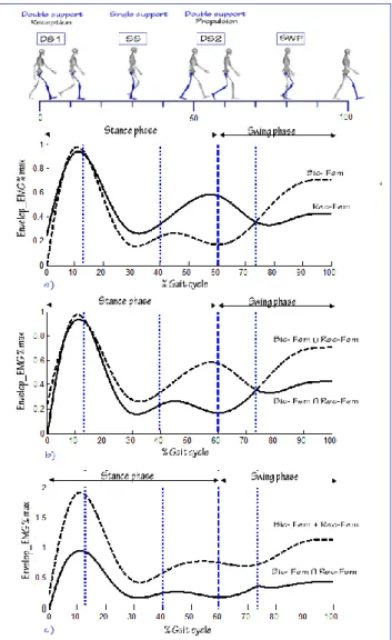
Fig. 2. Example for illustration of the gait angles for a healthy subject and another stroke: upper graph: the mean and variance for hip and knee (flexion/extension) angles of healthy subject; lower graph: the mean and variance for hip and knee (flexion / extension) angles of stroke subject. This graph shows that the gait cycle phases are destroyed in the stroke subject as well the variance between cycles is high.
III. NEURO-MOTOR INDEX AND EVALUATION STRATEGIES
We proposed a neuro-motor index to determine the control function. This index is a sliding evaluation of muscle co-contraction derived from the combination between two co-contraction indexes used by Frost et al. [28], Unnithan et al. [29] and Hessee et al. [27], combined together by a nonlinear regression of the quadriceps and hamstring EMGs. The regression was estimated with a Hermitian polynomial model
He(t).
NMI (t) = { CCI1 (t ) + Rm(t) CCI2 (t ) } (5) where, Rm (t) is a nonlinear regression based on Hermitian
function
Rm (t) = h1 (t) f0 + h2 (t) p0 + h3(t) f1 + h4 (t) p1 The peaks are identified with the following optimization problem:
argmin t | f’(t)|
f (t) = EMGantago (t) ∩ EMGago
emg
AGO(t)h1, h2, h3, h4∈ He (t); p0 and p1 are tangent to f0 and f1.
NMI is an index, derivable from co-activation that indicates achievement of motor skill and that is also related to joint stability. It is considered to be an important factor contributing to the ineffectiveness of human movement. The NMI is a continuous index that could be computed on any acquired EMG and it could help to detect the patient's intention for a given movement. This NMI is based on the detection of the flexion / extension of a joint during a given movement. It does not depend on the definition of a standard cycle (such gait cycle), which is a necessary requisite to work with cerebral palsy patients and patients with stroke. Nonlinear regression ensures that this method can be applied in case of abnormal walking, whether for an adult or for a child. Starting from a minimal calibration to reduce patient fatigue, the NMI allows for direct prediction of patient movement, reliance on residual capacity, and avoidance of pre-recorded control.
A. Validation with Canonical Correlation Analysis
The canonical correlation (CCA) is a multivariable statistical method used to extract the underlying correlation of two sets of data. It finds a pair of linear combinations such that the correlation between the two canonical variables is maximized. CCA extends ordinary correlation for any two sets of variables and is widely used in statistical and information mining [34], [35].
To test the reliability of the NMI, a canonical correlation analysis is applied in the two following studies.
1) First study: Offline assessment of reliability
In order to test the effectiveness of the NMI in the case of speed change, the CCA is applied at the angles, on one hand, and each ICC or/and NMI on the other hand on data extracted for the knee and hip (left and right) walking movement, from healthy subjects, during three velocities with average (slow: 0.59 m/s, normal: 1.18 m/s and fast:1.75) at eleven gait cycles for each velocity, as illustrated in Fig. 3.
Fig. 3. An illustration for usage the correlation by two successive steps. First step: CCA applied at joint angles and in index based in EMG signals. Second step: at the result for CCA in different cases (different velocities).
For three functions Ɵi indicates the angles measured at each gait cycle at different velocities (for 11 gait cycles), IK is the studied indices (CCI1, CCI2, CCI3 and NMI), and Vj symbolizes the three types of velocity (slow, normal and fast) for finding linearly correlated features from Ɵ and I :
Corr ( Ik , Ɵi)
(6)
where is the correlation between two random variables Θ and I with standard deviations and and expected values μƟ and μI to determine the correlation coefficient between result groups. CORR (CCAik)j is the correlation coefficient between the canonical correlation applied between Ɵ1, Ɵ2, ..., Ɵi and I1, I2, …., Ik at V1, V2……, Vj
For three velocities:
CORR(CCAik)j =
(7)
2) Second study: Online control assessment
In order to test the effectiveness of the MNI for several successive input of the data, as illustrated in Fig. 4, the CCA is applied between the angles of the walk on each ICC and the MNI, on the data extracted for the movement of the knee and the hip (left and right) of healthy subjects and patients neurological disorders (CP and stroke). Here, I denotes the index determined from the EMGs for the Hamstring and quadriceps muscle groups, and Θ represents the gait angles (for hip or knee). The linear combinations of I and Θ are I = IT
Wx and Ɵ =Θ T WƟ, respectively. CCA finds the weight vectors,
WI and WƟ, which maximize the correlation between I and Ɵ,
by solving the following optimization problem.
(8)
where ρn are the CCA coefficients obtained with the angle of
reference signals being Ɵ1, Ɵ2, ..., Ɵn. The maximum of ρ with
respect to WI and WƟ is the maximum canonical correlation.
Projections onto WI and WƟ, i.e. I and Ɵ, are called canonical
variants.
Fig. 4. An illustration for usage of CCA applied at I based in EMG signals and joint angles. I, is the multi-channel of index. Ɵref is the reference joint
angles for complete gait cycle.
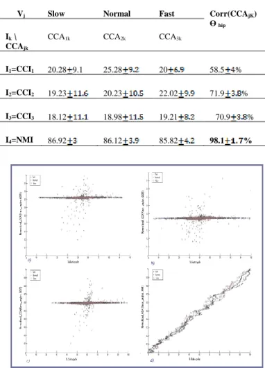
TABLEI:NORMALIZED AVERAGED CCA VALUES APPLIED AT JOINT ANGLES: HIP /INDICES STUDIED, IN THREE VELOCITIES
Vj Slow Normal Fast Corr(CCAjK)
Ɵ hip Ik \
CCAjk
CCA1k CCA2k CCA3k
I1=CCI1 20.28 9.1 25.28 20 58.5 %
I2=CCI2 19.23 20.23 22.02 71.9 %
I3=CCI3 18.12 18.98 19.21 70.9 %
I4=NMI 86.92 86.12 85.82 98.1 %
Fig. 5. Illustration of correlation applied at three groups (Θ, I); Θ represents the knee joint angle during complete gait cycle, and I represents the index used in this study: a) ICC1, b) ICC2, c) ICC3 and d) NMI, after testing the
correlation between his variants (Ɵi, Ik).I1 vs Θ1 test average correlation:
corr1=0.88, and I2 vs Θ2 corr2=0.88, and I3 vs Θ3 test correlation corr3 = 0.87;
type =quasi-linear; corr (CCA)V=0.99.
IV. RESULTS AND DISCUSSION
In the first study on data from healthy subjects, with three velocities (slow, normal, and fast), CCA was applied for knee and hip gait angles (right and left). The correlation points show
𝑐𝑜𝑣 (𝐶𝐶𝐴1,𝐶𝐶𝐴2 ,𝐶𝐶𝐴3) √𝑐𝑜𝑣 (𝐶𝐶𝐴1,𝐶𝐶𝐴1,𝐶𝐶𝐴3).𝑐𝑜𝑣(𝐶𝐶𝐴2,𝐶𝐶𝐴2,𝐶𝐶𝐴3)
(𝑊max 𝐼, 𝑊Ɵ) 𝜌(𝐼,Θ) = 𝐸 (𝐼𝑇Θ) √𝐸(𝐼𝑇 𝐼)𝐸(Θ𝑇 Θ) = 𝐸( 𝑊𝐼 𝑇𝐼 𝐼𝑇 𝑊Ɵ ) √𝐸 (𝑊𝐼𝑇 𝐼 𝐼𝑇𝑊 𝐼 ) 𝐸 (𝑊Ɵ𝑇ΘΘ𝑇 𝑊Ɵ) = 𝜌( 𝐼,Θ) =𝑐𝑜𝑣( Θ,I) 𝜎Ɵ 𝜎𝐼
=
𝐸[( Θ−μƟ)(I−𝜇𝐼)] 𝜎Ɵ 𝜎𝐼𝜌( 𝛩, 𝐼) 𝜎Ɵ 𝜎 𝐼 325
an important variability from one velocity to another, moreover, the percentage of correlation was weak and far from a linear relation. When the CCA was applied at MNI and the gait angles of the same subject, for the three velocities (slow, normal and fast), knee and hip (right and left), the correlation points showed strong similarity for the three velocities. The percentage of correlation is very high; the correlation is almost linear, as shown in Fig. 5. For one healthy subject, during walking, Fig. 5a, b and c, CCA applied to ICCs and the knee angle was low and nonlinear, in addition there was a clear variation from a velocity to another. While, in Fig. 5.d, for CCA applied at NMI and knee angles this correlation had a linear type and was mostly similar at the three different velocities.
Fig. 6. CCA applied at the joint angles and the indexes (CCI1, CCI2, CCI3 and
MNI), for three different velocities during eleven complete gait cycles. Upper graph: CCA estimated for hip (flexion /extension) angles for one healthy subject. Lower graph: CCA estimated for knee (flexion /extension) angles for one healthy subject. Error bars indicate standard deviation.
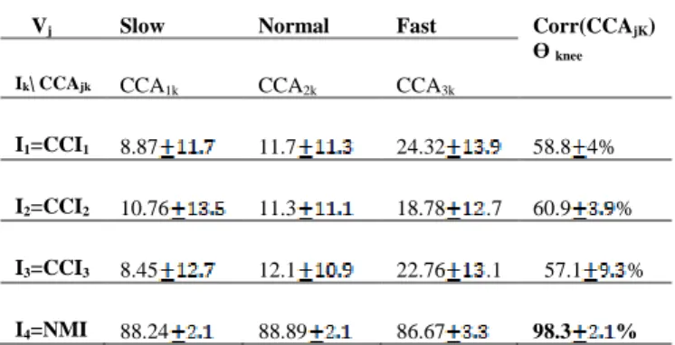
Tables I and II, present the normalized averaged CCA values between joint angles and indices studied (CCI1, CCI2, CCI3 and NMI) during the gait cycle for three different velocities: SV: slow, NV: normal and FV: fast). These tables detail the data presented in histograms of Fig. 6. based on an offline study, eleven gait cycles and three velocities. The CCA applied on one healthy subject indicates that the CCIs have a low relation to angles (average: 18.12 % to 22.02% for hip ;8.45% to 24.32 % for knee; p>0.05) with high variation. While this relation is higher with NMI (average: 85.82 % to 86.92% for hip; 86.67% to 88.89 % for knee) with low variation. The average correlation for CCIs studies are 71.9% for hip; 60.9% for knee, NMI indicates high significance at three velocities (with correlation: average 98.1% for hip; 98.3% for knee; p<0.05).
TABLEII:NORMALIZED AVERAGED CCAVALUES APPLIED AT JOINT
ANGLES:KNEE /INDICES STUDIED, IN THREE VELOCITIES
Vj Slow Normal Fast Corr(CCAjK)
Ɵ knee
Ik\ CCAjk CCA1k CCA2k CCA3k
I1=CCI1 8.87 11.7 24.32 58.8 4%
I2=CCI2 10.76 11.3 18.78 .7 60.9 %
I3=CCI3 8.45 12.1 22.76 .1 57.1 %
I4=NMI 88.24 88.89 86.67 98.3 %
In the second study, a data from healthy subject was used as a reference, to test the ability in determining the control margin angle desired. This was evaluated as much as possible by calculating the difference between joint angles of a reference and others from stroke subject, for the hip and the knee.
Fig. 7. CCA applied at joint angles and the indexes (NMI and CCI using three different methods for eleven complete gait cycles. Upper graph: CCA estimated for hip flexion /extension angles for one healthy subject. Lower graph: CCA estimated for knee flexion /extension angles for one healthy subject and one patient with stroke. Error bars indicate standard deviation. TABLEIII:NORMALIZED AVERAGED CCAVALUES APPLIED AT JOINT ANGLES
(KNEE AND HIP)/INDICES STUDIED, FOR STROKE SUBJECT AND HEALTHY DATA.
Healthy subject (ref) Stroke subject (p)
CCA(Ɵ,IK) Hip Knee CCA(Ɵ,IK) Hip Knee
I1=CCI1 21.25 .6 12.5 .9 I1=CCI1 21.25 .6 10.91 8.7
I2=CCI2 22.12 13.82 10.1 I2=CCI2 22.12 11.91 .1
I3=CCI3 18.81 .6 14.11 .1 I3=CCI3 18.81 .6 11.67 .2
Table III compares successive eleven gait cycles for healthy and stroke subjects (Fig. 7). Based on an online study, the CCA applied on successive gait cycles indicates that the CCIs have a low association to joint angles (i) healthy subject (average: 18.81% to 21.25% for hip; 12.5% to 14.11% for knee); (ii) stroke subject (average: 17.17% to 21.91% for hip; 10.91% to 11.91% for knee) with high variation. However, this relation is significantly higher (p<0.05) with NMI (i) for healthy subject (average 92.65% for hip; 91.85% for knee), than (ii) for stroke subject (average 91.45% for hip; 90.15% for knee ). The normalized averaged CCA values between joint angles and indices, studied (CCI1, CCI2, CCI3 and NMI) during 11 successive gait cycles for stroke subject with comparison with another healthy data, show no access to a control signal from ICCs. The detection of the control margin angle ∆Ɵ wasn’t accomplished in a control function, it was only
achieved in the case of correlation with NMI canonical correlation.
V. CONCLUSION
The effectiveness of using the new neuro-motor index (NMI) for bio kinematic-based control strategy for user-lead rehabilitation exoskeleton was investigated. This index was derived from a combination of two co-contraction indices and the nonlinear regression from a Hermitian function. The results of the studies reported here indicate that the NMI contributed to find a relation between contraction and joint angles. The CCA method is used for extracting angle information from the multi EMG signals. The offline analysis showed that only the coefficient of correlation INM-joint angles at different velocities were important when compared to the correlation CCI- joint angles. With an online test, which introduces successively bio-kinematic stroke data, results in a precise angle margin corrective function.
This approach will have an impact on the development of control for online rehabilitation and for a good coordination patient - exoskeleton in cases of neurological disorders resulting in muscle spasticity such as CP and stroke. This index based on the joint muscular co-contraction, yielding a very good stability. As it estimated from EMG signals, it offers a better detection of the patient intention. The study of the flexion/extension without complying with the gait cycle phases makes it an effective indicator of the different movements around the joints, and not just walking movement. Future work will focus on real-time testing this index on an exoskeleton setting.
REFERENCES
[1] J. L. Pons, Wearable Robots, Chichester, UK: John Wiley & Sons, Ltd, 2008.
[2] K. H. Low, “Robot-assisted gait rehabilitation: From exoskeletons to gait systems,” in Proc. 2011 Defense Science Research Conference and Expo, 2011.
[3] Y. Sankai, “HAL: Hybrid assistive limb based on cybernics,” Springer Tracts in Advanced Robotics, 2010.
[4] K. Kong and D. Jeon, “Design and control of an exoskeleton for the elderly and patients,” IEEE/ASME Trans. Mechatronics, 2006. [5] A. Esquenazi, M. Talaty, A. Packel, and M. Saulino, “The Rewalk
powered exoskeleton to restore ambulatory function to individuals with thoracic-level motor-complete spinal cord injury,” Am. J. Phys. Med. Rehabil., 2012.
[6] B. N. et al., “Results of the first interim analysis of the RAPPER II trial in patients with spinal cord injury: Ambulation and functional exercise
programs in the REX powered walking aid,” J. Neuroeng. Rehabil., 2017.
[7] C. Tefertiller et al., “Initial outcomes from a multicenter study utilizing the indego powered exoskeleton in spinal cord injury,” Top. Spinal Cord Inj. Rehabil., 2017.
[8] S. V. Kotov, V. Y. Lijdvoy, A. B. Sekirin, K. A. Petrushanskaya, and E. V. Pismennaya, “The efficacy of the exoskeleton ExoAtlet to restore walking in patients with multiple sclerosis,” Zhurnal Nevrol. i psikhiatrii im. S.S. Korsakova, 2018.
[9] T. Gurriet et al., “Towards restoring locomotion for paraplegics: Realizing dynamically stable walking on exoskeletons,” 2018. [10] T. Yan, M. Cempini, C. M. Oddo, and N. Vitiello, “Review of assistive
strategies in powered lower-limb orthoses and exoskeletons,” Rob. Auton. Syst., 2015.
[11] G. Chen, C. K. Chan, Z. Guo, and H. Yu, “A review of lower extremity assistive robotic exoskeletons in rehabilitation therapy,” Crit. Rev. Biomed. Eng., 2014.
[12] Applications of EMG in Clinical and Sports Medicine, 2012.
[13] J. Perry and J. Burnfield, “Gait cycle,” in Gait Analysis. Normal and Pathological Function, 2010.
[14] S. R. Simon, “Quantification of human motion: Gait analysis - Benefits and limitations to its application to clinical problems,” J. Biomech., 2004.
[15] W.-H. Yeo et al., “Lower extremity exoskeleton: Review and challenges surrounding the technology and its role in rehabilitation of lower limbs,” Aust. J. Basic Appl. Sci., 2013.
[16] L. Snyder-Mackler, Z. Ladin, A. A. Schepsis, and J. C. Young, “Electrical stimulation of the thigh muscles after reconstruction of the anterior cruciate ligament, effects of electrically elicited contraction of the quadriceps femoris and hamstring muscles on gait and on strength of the thigh muscles,” J. Bone Jt. Surg. - Ser. A, 1991.
[17] T. Lenzi, S. M. M. De Rossi, N. Vitiello, and M. C. Carrozza, “Intention-based EMG control for powered exoskeletons,” IEEE Trans. Biomed. Eng., 2012.
[18] E. Kellis, “Quantification of quadriceps and hamstring antagonist activity,” Sport. Med., 1998.
[19] P. Le, T. M. Best, S. N. Khan, E. Mendel, and W. S. Marras, “A review of methods to assess coactivation in the spine,” Journal of Electromyography and Kinesiology. 2017.
[20] J. V Basmajian, “Motor learning and control: A working hypothesis,” Arch.Phys.Med.Rehabil., 1977.
[21] D. A. Winter, Biomechanics and Motor Control of Human Movement: Fourth Edition. 2009.
[22] J. T. Viitasalo and C. Bosco, “Electromechanical behaviour of human muscles in vertical jumps,” Eur. J. Appl. Physiol. Occup. Physiol., 1982. [23] L. M. Knutson, G. L. Soderberg, B. T. Ballantyne, and W. R. Clarke, “A study of various normalization procedures for within day electromyographic data,” J. Electromyogr. Kinesiol., 1994.
[24] M. Solomonow et al., “The synergistic action of the anterior cruciate ligament and thigh muscles in maintaining joint stability,” Am. J. Sports Med., 1987.
[25] J. T. Viitasalo, A. Salo, and J. Lahtinen, “Neuromuscular functioning of athletes and non-athletes in the drop jump,” Eur. J. Appl. Physiol. Occup. Physiol., 1998.
[26] K. Falconer and D. Winter, “Quantitative assessment of co-contraction at the ankle joint in walking.,” Electromyogr. Clin. Neurophysiol., vol. 25, no. (2-3), pp. 135–149, 1985.
[27] S. Hesse, B. Brandl-Hesse, U. Seidel, B. Doll, and M. Gregoric, “Lower limb muscle activity in ambulatory children with cerebral palsy before and after the treatment with Botulinum toxin A.,” Restor. Neurol. Neurosci., 2000.
[28] G. Frost, J. Dowling, K. Dyson, and O. Bar-Or, “Cocontraction in three age groups of children during treadmill locomotion,” J. Electromyogr. Kinesiol., 1997.
[29] V. B. Unnithan, J. J. Dowling, G. Frost, B. Volpe Ayub, and O. Bar-Or, “Cocontraction and phasic activity during GAIT in children with cerebral palsy.,” Electromyogr. Clin. Neurophysiol., 1996.
[30] M. E. W. O. M. P. Kadaba, H. K. Ramakrishnan, “Measurement of lower extremity kinematics during level walking,” Class. Pap. Orthop., 1990. [31] D. A. Winter, H. G. Sidwall, and D. A. Hobson, “Measurement and
reduction of noise in kinematics of locomotion,” J. Biomech., 1974. [32] R. Shiavi, C. Frigo, and A. Pedotti, “Electromyographic signals during
gait: Criteria for envelope filtering and number of strides,” Med. Biol. Eng. Comput., 1998.
[33] E. Kellis, F. Arabatzi, and C. Papadopoulos, “Muscle co-activation around the knee in drop jumping using the co-contraction index,” J. Electromyogr. Kinesiol., 2003.
[34] R. B. Lund, H. von Storch, and F. W. Zwiers, “Statistical analysis in climate research,” J. Am. Stat. Assoc., 2000.
[35] O. Friman, J. Cedefamn, P. Lundberg, M. Borga, and H. Knutsson, “Detection of neural activity in functional MRI using canonical correlation analysis,” Magn. Reson. Med., 2001.
Jinan Charafeddine received two master diplomas. His first master 2, is a
research diploma in biomedical engineering (electro mechanical-systems) from Doctoral School of Sciences and Technology at Lebanese University (Beirut -Lebanon) in 2012, the second master 2, is a professional diploma in Instrumentation and Industrial data processing, at Faculty of Sciences at Lebanese University (Tripoli -Lebanon) in 2013. Ph.D. Student at Versailles University (UVSQ) since September 2016. The PhD thesis is in control of exoskeleton of rehabilitation by EMG signals. this thesis is collaboration between END-ICAP laboratory (Garches-France), LISV of UVSQ (Velizy- France) and EDST at Faculty of Engineering Lebanese University (Tripoli- Lebanon)
Sylvain Chevallier graduated from Université
Paris-Sud 11 in Cognitive Science (2005) and obtained his PhD in computer science / cognitive science at LIMSI-CNRS (Orsay, 2009). After a postdoc in neurocybernetic team at ETIS (Cergy), he joined INRIA Saclay and Telecom ParisTech as a post-doctoral fellow. He is associated professor in University of Versailles an at LISV since 2011. He is in charge of the coordination of the workgroup Handicap for University Paris-Saclay.
Samer Alfayad received his master diploma in
sciences and technology from Ecole Nationale Supérieure d’Arts et Métiers (ENSAM-Paris) in 2005. His Ph.D was received in robotic development from Versailles University (UVSQ) in 2009. He was awarded of the best Ph.D. thesis in robotics for year 2010 by the French CNRS. Also, he was awarded of the best Ph.D. thesis in robotics for the 20 years’ anniversary of UVSQ. From 2010 to 2011 he was a post-doc researcher at Technische Universitat Munich (TUM-Germany) with a scholarship from the Alexander Von Humboldt foundation. In 2011, he has been appointed as an associated professor in Humanoid robotic design at Versailles University. From 2012, he holds an industrial excellence chair about hydraulic domestication at UVSQ. He has been investigator in several French National projects. He is the leader of the Humanoid research group at LISV laboratory. He is working currently on the two anthropomorphic biped robots called HYDRODï and ROMEO2 at the LISV. He is cocoordinator of Institute for
Control and Decision (iCode Paris-Saclay).
Mohamad Khalil received the DEA in biomedical engineering from the University of Technology of Compiegne (UTC) in France in 1996. He received his Ph.D from the University of Technology of Troyes in France in 1999. He received his HDR (Habilitation a diriger des recherches) from UTC in 2006. He is the chair of the EMBS chapter in Lebanon, chair of ICABME international Conference. His current interests are the signal and image processing problems: detection, classification, analysis, representation and modeling of non-stationary signals, with application to biomedical signals and images. He is currently professor, teacher and researcher at Lebanese University, faculty of engineering.
Didier Pradon received his master diploma in
adapted physical activities from University of Quebec at Trois-Rivieres - Canada in 1999. and a DEA in biomechanics from Claude Bernard University (UCBL) Lyon- France in 2000. His Ph.D was received in biomechanics from Claude Bernard University (UCBL) Lyon- France in 2004. From 10/2004 to 10/2005 he was a post-doc researcher at Movement Analysis Laboratory, Raymond Poincaré University Hospital, CIC-IT 805, AP-HP, Garches - France (Garches Foundation Contracts). He was head of the Movement Analysis Laboratory, EA 4497 GRCTH, CIC-IT 805, Raymond Poincaré University Hospital, APHP in 2005, Head of the Movement Analysis Laboratory, EA 4497 GRCTH, CIC-IT 805, CHU Raymond Poincaré, APHP in 2010, Project leader of the creation of a new sector at the engineering school of ISTY (Institute of Sciences and Techniques of Yvelines) of the University Versailles Saint-Quentin. Creation of the Biomedical Engineering program: Assistance and supplement systems for sensory and cognitive motor disabilities. in 2011, Technological Coordinator of CIC-IT 805 in 2012, from 2011 to 2014 Head of the clinical committee within the "SHIFT" project of the Garches Foundation and the Safran Foundation: Development of two manual wheelchair platforms. Hospital Engineer at UFR of Health Sciences Simone Veil – France since 2005.