Citrobacter rodentium Induced Liver Changes in C57BL/6 Mice:
Animal Model of Acute Inflammatory Stress and Injury
by
Arkadiusz R. Raczynski B.A. Biology, Biochemistry
Carleton College, 2002
MASSACHUSETTS INSTITUTE
L
F
T
y
JUL 1 4 2011
L
IBnprA" R I
ES'--SUBMITTED TO THE DEPARTMENT OF BIOLOGICAL ENGINEERING IN PARTIAL FULLFILLMENT OF THE REQUIREMENTS FOR THE DEGREE OF
DOCTOR OF PHILOSOPHY IN MOLECULAR AND SYSTEMS TOXICOLOGY AND PHARMACOLOGY
AT THE
MASSACHUSETTS INSTITUTE OF TECHNOLOGY JUNE 2011
@ 2011 Massachusetts Institute of Technology. All rights reserved
Signature of Author
Biological Engineering April 25th, 2011
Certified by
Professor Steven R. Tannenbaum Professor of Biological Engineering Thesis Advisor
This doctoral thesis has been examined by a committee of the Department of Biological Engineering as follows:
r
Certified by
U
Professor James G. Fox, MITDepartment of Comparative Medicine
Certified by
Dr. Eric R. Fedyk Director of Immunobiology and Toxicology Millennium Pharmaceuticals: A Takada Company
Citrobacter rodentium Induced Liver Changes in C57BL/6 Mice: Animal
Model of Acute Inflammatory Stress and Injury
By
Arkadiusz R. Raczynski
Submitted to the Department of Biological Engineering on April 25th, 2011 In Partial Fulfillment of the Requirements
For the Degree of Doctor of Philosophy in Molecular and Systems Toxicology and Pharmacology
Abstract
The activation of inflammatory responses, while critical for host defense, contributes to hepatic injury in numerous acute and chronic liver disease states as well as drug-induced liver injury (DILI). The interactions that mediate susceptibility to liver injury and disease, however, are still poorly understood, underscored by the complexity of immune interactions and the diverse cellular composition and functions of the liver.
Using Citrobacter rodentium, a well characterized rodent-specific enteric pathogen as a
source of extrahepatic inflammatory stress; host liver responses, metabolic dysregulation, and susceptibility to injury in C57BL/6 mice were investigated. For the first time, we show altered liver pathology during the early course of C. rodentium infection, characterized by periportal necrosis indicative of thrombic ischemic injury, correlating with distinct circulating and tissue specific cytokine/chemokine profiles.
Using Acetaminophen (APAP), a widely used analgesic and well-characterized hepatotoxin, we evaluated liver responses in isolation and in the context of host inflammation to gain insight into the role of live bacterial infection in altering liver metabolism and susceptibility to DILI. We combined systemic and tissue-specific cytokine/chemokine levels, clinical serum chemistries, and histopathological assessments of hepatic and enteric inflammation and necrosis to measure molecular-level responses to treatment and their physiological effect. Using principal components analysis (PCA), clustering, partial least squares regression (PLSR), and a combination mutual-information-correlation network, enabled detection and visualization of both linear and nonlinear dependencies between molecules and physiological states across tissues and timepoints.
C. rodentium-induced inflammatory stress was finally investigated for its
potential in altering drug pharmacokinetics (PK) of substrates varying in their metabolic biotransformation and clearance mechanisms. Infection resulted in increased systemic oral exposure (AUC) of clinically relevant xenobiotics such as verapamil, propranolol, and digoxin. Functionally, these changes were not found dependent on CYP-mediated biotransformation of parent compounds; rather, they appear driven more by proposed gut barrier compromise.
In conclusion, gastrointestinal infection with C. rodentium alters systemic and hepatocytes specific responses, not previously appreciated from this enteric pathogen,
Acknowledgments
I would like to dedicate my PhD to the late Dr. David Schauer, without his mentorship and support this work would not have been possible. His vision and incredible wisdom has been an inspiration throughout my tenure as a graduate student and this will surely carry on throughout my career. I am also in great debt to my parents Marek and Irena who made tremendous sacrifices in allowing me to pursue advanced studies. The support of Jeanette Raczynski, Derin Tugal, and other family and close friends has also been amazing throughout this process - their love and patience were invaluable.
My deepest thanks go to Steven Tannenbaum who was instrumental in not only his guidance, but also his efforts to continue my support after David's passing. Furthermore, he was extremely flexible in allowing my relocation to Cleveland towards the later part of the thesis write-up - for this I am extremely grateful and most appreciative.
Furthermore, I would like to extend my thanks to the remaining members of the committee: Dr. James Fox, Dr. Linda Griffith, and Dr. Eric Fedyk. Their suggestions and insight were extremely valuable towards a deeper understanding of the work and critical in guiding experiments and conclusions.
Thanks of course go to the Department of Biological Engineering and all of its members. I would like to particularly thank Marcia Weir, Amy Francis, Marcia Ross, JoAnn Sorrento, Roni Dudley-Cowans, and Dalia Fares and all other administrators for their assistance at MIT, and Doug Lauffenburger for all his efforts towards making research in this department so enjoyable and rewarding.
During my time at MIT I was also fortunate to work and collaborate with numerous scientists (and friends) that not only listened during experimental struggles, but whose advice was so valuable to me. These include Megan McBee, Diana Borenshtein, Adrienne Li, Alex Sheh, David Weingeist, Laure-anne Ventorous, Luke Robinson, Mark Bathe, Robbie Barbero, Jaime Rivera, Jeremy Rock, and Justin Pritchard. Many thanks to Emily Miraldi who was instrumental in helping with computational approaches with the APAP-related work and Charles Knutson for his efforts and collaboration in the MGH project. I would like to also acknowledge the support of Biogen Idec, specifically Tonika Bohnert and Lawrence Gan for their efforts and intellectual contributions towards the pharmacokinetics project.
Also, the Department of Comparative Medicine (DCM) has been so incredibly supportive in many aspect of this thesis. From Gladys Valeriano in orchestrating animals space and time to Melissa Mobley and Amanda Potter for their technical
expertise, and Sureshkumar Muthupalani for his pathological assessments and research advice.
Prior to MIT, there is a tremendous amount of guidance that was vital in cultivating a desire to continue a scientific career. At Millennium Pharmaceuticals, Vilmos Csizmadia was instrumental in motivating me as a scientist. It was truly a privilege to have you as a mentor and I know it will undoubtedly continue for years to come. Passing on his depth of knowledge and expertise helped tremendously in my scientific endeavors at MIT. I would also like to thank Vito Sasseville, Scott Barros, Cindy Xia, Mingxiang Liao, and Vic Kadambi for their insightful discussions and guidance as a young scientist. And of course Peter Smith for giving me a chance to intern at MLNM fresh out of college - an invaluable research experience that has opened so many opportunities.
I would like to acknowledge the support of the ISN grant 6915549, and support from the Toxicology Training Grant (ES-070220) and Nitric Oxide Project Program Grant (P01 CA026731).
Table of Contents
ABSTRACT ... ... ... 3
ACKNOW LEDGM ENTS... ... ... ... 4
TABLE OF CONTENTS... .... ... 6
LIST OF FIGURES...9
LIST OF TABLES... .... ... 14 CHAPTER 1: INTRODUCTION ... 1-16
STRESS RESPONSE AND INFLAMMATION ... ... 1-16 INNATE IMMUNE SYSTEM IN STRESS AND INFLAMMATION...1-16
LIVER STRUCTURE AND FUNCTION...1-18
INFLAMMATION AND LIVER DISEASES ... 1-18 DRUG-INDUCED LIVER INJURY (DILI)...1-19 ACETAMINOPHEN-INDUCED LIVER INJURY ... 1-20 ANIMAL M ODELS OF LIVER INJURY...1-21
CITROBACTER RODENTIUM ... 1-22
CONCLUDING REMARKS: ... 1-23 FIGURES AND TABLES...1-25
CHAPTER 2: ENTERIC INFECTION WITH CITROBACTER RODENTIUM INDUCES HEPATIC INFLAMMATION AND COAGULATIVE LIVER NECROSIS PRIOR TO PEAK INFECTION
AND COLONIC DISEASE... 2-26
2.1 INTRODUCTION ... 2-27
2.2 RESULTS...2-28
2.3 DISCUSSION ... 2-32
2.4 SELECTED METHODS...2-35
2.5 FIGURES AND TABLES ... 2-39
CHAPTER 3: SYSTEMS ANALYSIS OF ACETAMINOPHEN-INDUCED LIVER INJURY
UNDER CONDITIONS OF CITROBACTER RODENTIUM-INDUCED INFLAMMATORY
STRESS... 3-57
3.1 INTRODUCTION ... 3-58 3.2 RESULTS...3-60
3.3 DISCUSSION ... 3-67 3.4 SELECTED METHODS...3-75
3.5 FIGURES AND TABLES...3-81
CHAPTER 4: EFFECT OF SYSTEMIC INFLAMMATION FROM GASTROINTESTINAL
INFECTION WITH CITROBACTER RODENTIUM ON ORAL DRUG EXPOSURE ... 4-98
4.1 INTRODUCTION ... 4-99 4.2 RESULTS... 4-100 4.3 DISCUSSION ...--... 4-103 4.4 SELECTED METHODS ... 4-106 4.5 FIGURES AND TABLES... 4-108
APPENDIX A: GENERAL METHODS... A-117
MEDIA AND BACTERIAL STRAINS...A-117 QUANTITATIVE REAL-TIME PCR ... A-117
APPENDIX B: EFFECT OF CITROBACTER RODENTIUM ON EARLY SERUM AND LIVER RESPONSES... B-118
B.1 INTRODUCTION ... B 119
B .2 R ESU LTS...B -1 2 0
B.3 DISCUSSION ... B-121
B.4 SELECTED METHODS ... B-123
B.5 FIGURES AND TABLES...B-124
APPENDIX C: BIOMARKERS OF INFLAMMATORY BOWEL DISEASE ... C-129
C.1 INTRODUCTION ... C-130 C .2 R ESU LTS... C -1 3 1 C .3 D ISCU SSIO N ... C -1 3 3 C.4 SELECTED METHODS ... C-134
C.5 FIGURES AND TABLES... C-136
APPENDIX D: SERUM AND KIDNEY CYTOKINE ANALYSIS OF STX-EXPRESSING
CITROBACTER RODENTIUM ... D-142 D.1 INTRODUCTION... D-143 D .2 R ESU LT S ... D -14 4 D .3 D ISCU SSIO N ... D -14 5 D.4 SELECTED METHODS...D-146 D.5 SELECTED FIGURES ... D-147 APPENDIX E: ABBREVIATIONS...E-149 APPENDIX F: REFERENCES... F-150
LIST OF FIGURES
Figure 1-1. Acetaminophen Metabolism and Elimination...1-25
Figure 2-1. Infection kinetics and body weight changes in C57BL/6 mice infected w ith C. rodentium ... 2-39 Figure 2-2. C. rodentium-induced colonic effects in C57BL/6 mice. ... 2-40 Figure 2-3. C. rodentium-induced necrosis and histological liver changes in C57BL/6
m ic e ... 2 -4 1 Figure 2-4. Activated caspase 3 and KI-67 in C. rodentium-induced liver lesions 2-42 Figure 2-5. C. rodentium significantly increases Ki-67+ labeling index in livers.... 2-43 Figure 2-6. Signal transduction changes in liver due to C. rodentium infection...2-44 Figure 2-7. STAT3 activation in livers of C rodentium inoculated C57BL/6 mice...
... 2 -4 5 Figure 2-8. Serum specific PLS-DA analysis of C. rodentium infected C57BL/6 mice
a t 3 D P I ... 2 -4 6 Figure 2-9. Serum specific OPLS analysis of C. rodentium infected animals at 3 DPI..
... ... 2 -4 7 Figure 2-10. Liver specific PLS-DA analysis of C. rodentium infected C57BL/6 mice
a t 3 D P I ... 2 -4 8 Figure 2-11. Liver specific OPLS analysis of C. rodentium infected animals at 3 DPI..
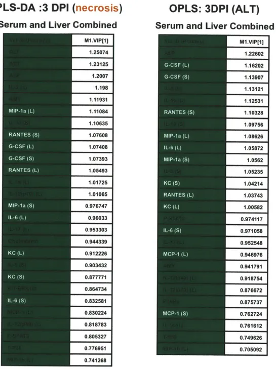
... 2 -4 9 Figure 2-12. Most influential variables in serum and liver for PLS-DA (necrosis
versus no necrosis) and OPLS (ALT) models ranked by Variable in Projection (V IP ) v a lu e s. ... 2 -5 1
Figure 3-2. C. rodentium and APAP-induced liver lesions evaluated by H&E at 3 DPI ... 3 -8 2 Figure 3-3. PLS-DA analysis at 3 DPI:... 3-83 Figure 3-4. C. rodentium and APAP-induced liver H&E and inflammation at 7 DPI..
... 3 -8 4 Figure 3-5. PLS-DA analysis at 7DPI ... 3-85 Figure 3-6. OPLS results for prediction of ALT levels for all APAP treated mice. 3-86 Figure 3-7. OPLS model for prediction of ALT levels for Citro (3DPI) treated mice...
... 3 -8 7 Figure 3-8. Cytokine changes at 3 and 7 DPI with high ALT correlation for Citro or
A P A P a lo n e ... 3 -8 8 Figure 3-9. Correlations and Mutual information of targets for liver pathologies....
... 3 -8 9 Figure 3-10. Mutual information and correlation maps for liver inflammation and
n e cro sis...3 -9 0
Figure 4-1. Effects of C. rodentium infection of Acetaminophen (5mg/kg) exposure. ... 4 -1 0 8 Figure 4-2. Effects of C. rodentium infection on Fexofenadine (5mg/kg) exposure...
... 4 -1 0 8 Figure 4-3. Effect of C. rodentium infection on Propranolol and Digoxin exposure...
... 4 -1 0 9 Figure 4-4. Effects of C. rodentium infection on Verapamil (5mg/kg) exposure...
... 4 -1 1 0 Figure 4-5. Verapamil metabolites monitored after 5mg/kg PO dose ... 4-111 Figure 4-6. Method description and LC/MS/MS results of verapamil metabolite
le v e ls ... 4 -1 1 1 Figure 4-7. Effect of C. rodentium on metabolites of verapamil (5mg/kg)... 4-112
SUPPLEMENTAL FIGURES
Supplemental Figure 2-1. Serum chemistry changes in C57BL/6 mice inoculated w ith C. rod en tiu m ... 2 -5 2 Supplemental Figure 2-2. Systemic cytokine and chemokine changes induced by C.
rodentium infection in C57BL/6 mice... 2-53 Supplemental Figure 2-3. Liver cytokine and chemokine changes induced by C.
rodentium infection in C57BL/6 mice... 2-54 Supplemental Figure 2-4. PLS-DA analysis of 3 and 7 DPI animals...2-55 Supplemental Figure 3-1. Affinity propagation clustering of all measurements3-92 Supplemental Figure 3-2. Animals and measurements clustered by affinity
propagation using Z-score values...3-93 Supplemental Figure 3-3. Mutual information and correlation network at 3 DPI...
... 3 -9 4 Supplemental Figure 3-4. Mutual information and correlation network at 7 DPI...
... 3 -9 5 Supplemental Figure 3-5. Sub-network diagram highlighting interactions for liver
IL -1
p
... 3 -9 6APPENDIX FIGURES
Figure B-1. Signal transduction in liver lysates of C. rodentium infected animals at 1 2 a n d 2 4 H P I ... B -1 2 4 Figure B-2. qRT-PCR for acute phase and liver targets due to C. rodentium infection.
... B -1 2 5 Figure 3. C. rodentium induced systemic cytokines changes at early timepoints..
B-126
Figure C-2. Observed versus predicted disease score: Ulcerative colitis active and inactive patients (STU DY 1& 2) ... C-137 Figure C-3. OPLS for disease score: Crohn's using active and inactive patients
(ST U D Y 1 & 2 )... C -1 3 8 Figure C-4. Observed versus predicted disease score: Crohn's active and inactive
patients (ST U D Y 1& 2)... C-139 Figure C-5. Mann-Whitney of ESR and CRP values for UC active and inactive
p a tie n ts... C -1 4 0 Figure C-6. Potential biomarkers of disease activity obtained from OPLS analysis...
... C -1 4 0 Figure C-7. Total scores for UC and CD patients based on VIP scores obtained from
O P L S ... C -1 4 1 Figure D-1. Serum cytokine and chemokine changes in C57BL/6 mice exposed to
intimin-deficient and STX-expressing C. rodentium...D-147 Figure D-2. Quantitative RT-PCR for expression of kidney cytokines and
chemokines transcript in C57BL/6 mice inoculated with STX-expressing C.
LIST OF TABLES
Table 1-1. Target modulation and susceptibility to APAP-induced toxicity in exp erim ental m odels...1-25 Table 2-1. PLS-DA and OPLS component contributions to discrimination (R2 Y) and
variance (Q 2) of necrosis at 3 DPI...2-50 Table 3-1. Data compendium including animal numbers and measurements
c o lle cte d ... 3 -9 1 Table 4-1. Summary of compound exposure under C. rodentium-induced
in flam m atory stress... 4 -113 Table 4-2. LC/MS/MS Results of propranolol metabolites with and without C.
Chapter 1: Introduction
Stress Response and Inflammation
Acute stress primes immune functions by stimulating cell trafficking from lymphoid organs into systemic circulation and sites of infection. In humans, exposure to even short term stress, like public speaking, results in increased numbers of certain populations of circulating lymphocytes and briefly increase natural-killer-cell activity [1]. Innate immune cells mediate early cytokine release (IL-1, TNF-a, IL-6) promoting the acute phase response, which is critical to appropriately deal with infection and trauma. As an essential response to harmful stimuli such as chemical injury and microbial pathogens, however, inflammatory activation must be tightly coupled with regulatory mechanisms that bring about its own resolution [2]. A failure to properly regulate inflammatory processes can result in sustained collateral damage to healthy cells and tissues (cytokine storms, septic shock), as demonstrated by numerous knockout (KO) models as well as disease states with both genetic and environmental components. Indeed, persistent and aberrant inflammation plays a key role in many, if not most, serious human diseases including atherosclerosis, autoimmune disorders, inflammatory bowel disease, neurodegenerative disease, obesity/diabetes, chronic infection, and cancer [3, 4]. Recently, research groups have begun to address the role of various stressors on liver responses (apoptosis/necrosis) and metabolic function. Such stressors include endotoxin-induced inflammation (lipopolysaccharide (LPS)) [5, 6], pathogenic bacterial infection [7, 8], caging stress [9], temperature [10], electric foot shock [11], and various other 'generic' stress conditions. Infection with pathogenic bacteria is one stress that can lead to inflammation, resulting in the stimulation and recruitment of immune regulators and release of various cytokines and chemokines at local and systemic levels with concomitant release of bacterial antigens that can activate innate and adaptive immune responses.
Innate Immune System in Stress and Inflammation
Pattern recognition receptors (PRRs), and their recognition of highly conserved pathogen-associated molecular patterns (PAMPs), as well as endogenous stress signals termed damage-associated molecular pattern molecules (DAMPs), allow the innate immune system to discriminate a variety of potential pathogens and injuries, furthermore, to coordinate appropriate responses [12]. PRRs include toll-like receptors (TLRs), NOD-like receptors (NLRs), C-type lectin receptors (CLRs), and several others, which contribute to immune activation in response to diverse stimuli, including infection and tissue injury. PRR expression can include the cell
surface, cytoplasm, and endosomal compartments, with levels and expression of receptors varying by cell type. Interestingly, despite a variety of TLR ligands, signaling converges to activation of nuclear factor-kappa B (NF-KB), mitogen-activated protein kinases (MAPKs), extracellular signal-regulated kinase (ERK), p38, and c-Jun N-terminal kinase (JNK). Adapter molecules are also critical in mediating effector function, and of the TLR adaptor proteins identified (MyD88, Mal, TRIF, TRAM, and SARM), MyD88 is utilized by most TLRs (and some NLRs) to regulate cytokine and major histocompatibility (MHC) molecule production through NF-KB [13]. Repressors of MyD88 signaling have also been discovered, specifically IRAK-M, expressed only in monocytes and macrophages, has been implicated in LPS tolerance in sepsis, and can suppress innate immunity mechanistically due to dampened Nf-KB activation [14, 15].
Recently, NLR proteins (NALPs, IPAF) have been reported to form caspase-1-activating complexes called inflammasomes, the process of which has recently been reviewed in depth [16]. Inflammasomes appear to play a critical role in leading to the activation of pro IL-1 and IL-18, key early mediators in inflammation and cell responses. It has also been proposed that NALP inflammasomes may also contribute to antimicrobial defense by inducing the apoptosis of infected cells [17]. In macrophages, IL-1p production requires double stimulation with TLR ligands, which induce gene transcription, and NLR agonists (such as ATP or muramyl dipeptide (MDP)) that activate the inflammasome and subsequently activate pro-IL-1j. Interestingly, monocytes can release active IL-1 upon stimulation with TLR ligands alone [18]. Conceptually, it has been proposed that TLR signaling in the absence of NLRs may result in a 'yellow' alert, indicating for example gut barrier compromise, and in conjunction with activation of inflammasome NLRs, may trigger a 'red alert', indicating exposure to more virulent pathogens [19]. This signal processing is complicated further by an emerging number of molecules known as DAMPs, a group naturally expressed in the cytosol or nucleus, and currently includes S100 proteins, heat-shock proteins (HSPs) and HMGB1, and when released extracellularly, induce signals to the host of tissue damage [20, 21]. HMGB1 is of particular interest, as its release can be active, secreted by macrophages and monocytes (as well as NK cells [22]) in response to exogenous and endogenous inflammatory stimuli (LPS, TNF-a, IL-1, and IFN-y), or passive during necrosis, in both cases mediates innate and adaptive inflammatory responses to infection and injury. Recently, a reactive oxygen species (ROS), hydrogen peroxide (H202), was
shown to induce both active (at low dose) and passive (high dose) HMGB1 release from macrophages and monocytes, a mechanism abrogated by JNK and MEK inhibition [23]. While there is clear diversity in PRRs, PAMPs, and DAMPs, downstream signaling through conserved signaling nodes indicates that the network state might be resolved by interrogating a select number of downstream
Liver Structure and Function
Functionally complex, the liver plays critical roles in metabolism, lipid and protein synthesis, as well as detoxifying functions. The liver is unique in its anatomical proximity affording it continuous blood flow through the sinusoids from the gastrointestinal tract, and diverse in cellular composition; host to metabolically active hepatocytes, non-parenchymal cells (biliary, Kupffer, sinusoidal endothelial, and stellate cells) and various innate and adaptive immune cell populations (neutrophils (PMN), monocytes, dendritic cells (DCs), and lymphocytes (T, B, NK, and NKT cells) that can enter via hepatic arteries or the portal vein [24]. Within this heterogeneous tissue, cytokines and chemokines, small molecular-weight messengers that act in autocrine, paracrine, and endocrine fashion, are critical in mediating effector functions of resident and distal immune cells, as well as intracellular hepatic signaling responses that modulate liver homeostasis [25]. While indispensable in its metabolic function, the liver is also a key "immunological" organ, underscored by its constant perfusion of blood from the gastrointestinal tract which is highly enriched with potential antigens, providing an early line of defense against microbes and toxins crossing the intestinal barrier [26]. The immune cells mentioned above have the potential in initiate both innate and adaptive immune responses, as in the case of infection in response to high levels of systemic LPS or bacterial superantigens, while balancing immunological tolerance in the case of low levels of LPS and innocuous antigens.
Inflammation and Liver Diseases
The activation of the inflammatory response, while critical for pathogen defense, contributes to hepatic injury in numerous cases of liver diseases including alcoholic liver disease (ALD), non-alcoholic fatty liver disease (NAFLD), ischemia/reperfusion (I/R) injury, autoimmune hepatitis and viral hepatitis. Generally it is accepted that acute injury is driven primarily by a Th1 cytokine response, among these tumor necrosis factor-a (TNF-a), in combination with additional pathophysiological factors, seems to play a central role in liver damage by induction of hepatocyte apoptosis, although oncotic necrosis, typically a consequence of acute metabolic perturbation characterized by ATP depletion and mitochondrial dysfunction, can also predominate [27]. Indeed, pure apoptosis or necrosis might just represent extremes in the spectrum of so called "necrapoptotic" responses, and that the more typical response of liver cells to injury might be a mixture of events associated with apoptosis, necrotic cell death, and inflammation. Recently, necrotic mechanisms have been described, indicating a process more regulated than originally thought [28]. It has also become evident that hepatocyte responses to TNF-a are context-dependent, promoting pleitropic cell responses such as apoptosis, proliferation, or survival [29]. Such seemingly disparate outcomes demonstrate dependence on autocrine loops, and sustained intracellular signaling (ex Nf-xB, JNK activation).
Decision processes of cell fate in tissues are also likely intricately linked to the presence and coordinated signaling of PAMPS, DAMPS, and local and systemic cytokine levels. HMGB1, for example engages its receptor RAGE, and results in cooperation with TLR2/4 in inducing inflammatory responses. The role of TLR4 dependent signaling in resident cell types has gained considerable attention, and while it has been generally accepted that Kupffer cells, a TLR4 expressing cell, are critical mediators of liver inflammation, recent findings demonstrate that hepatic stellate cells also express TLR4, and furthermore, are the predominant target through which TLR4 ligands promote fibrosis [30].
Drug-induced Liver Injury (DILI)
Drug induced liver injury (DILI) is a major pharmaceutical and clinical concern that can result in last stage discontinuation of drug development, limit the use and distribution of effective therapeutics, and even lead to market withdrawal [31]. DILI is the leading cause of acute liver failure (ALF) in the United States, accounting for about half of all cases [32]. Drugs that cause severe DILI in humans do not show clear hepatotoxicity in currently used animal models, generally lack dose-related toxicity, and typically demonstrate low rates of severe injury in humans (1 in 5,000 to 10,000 or less). In the US alone, DILI accounts for more than 50% of cases of ALF, with -39% due to acetaminophen and -13% resulting from idiosyncratic drug reactions (IDRs) [32]. Such IDRs could be a reflection on host factors and individual susceptibility, as the majority of patients do not demonstrate adverse drug reactions to these agents[33]. Whether they are the result of genetic or acquired differences has not yet been established, and to date no genetic, metabolic, or other
characteristic has been found to predict severe DILI in an individual.
While inflammation is intricately associated with numerous liver diseases, its importance is also appreciated in DILI: proposed mechanisms include the Hapten and Danger hypotheses as a model for allergic reactions observed with some drugs (ex penicillin) [34]. Others hypothesize that variable host factors such as endotoxin leakage from the gut, for example during pathogenic infection or gut barrier compromise (alcohol ingestion), or underlying inflammatory conditions (ex hepatitis, arthritis) can promote increased basal levels of local or systemic inflammatory cytokine, chemokines, or other soluble factors that predispose the liver to injury[6, 35, 36]. The role of bacteria and cytokines on the regulation of hepatic metabolic enzyme activity, such as cytochrome P450s and more recently drug transporters have been demonstrated and recently well reviewed [37]. These changes are believed to be regulated by altering nuclear receptor activities (PXR, CAR, FXR, RXR, LXR) which drive expression of phase I, II, and III metabolic enzymes. While critical for biotransformation and clearance, the changes in
Acetaminophen-Induced Liver Injury
Acetaminophen (APAP) is the worlds most commonly used analgesic and antipyretic, responsible for approximately a third of cases related to DILI [38]. Under normal liver conditions, -90% of APAP is metabolized to non-toxic conjugated metabolites APAP-glucuronide and APAP-sulfate and -10% of APAP metabolism occurs via cytochrome P450 oxidation (CYP 2E1, 3A4, and to a lesser extent 1A2) to form N-acetyl-p-benzoquinoemine (NAPQI), a highly reactive electrophilic metabolic intermediate [39]. Detoxification of NAPQI normally proceeds by conjugation with intracellular glutathione, either spontaneously or catalyzed by glutathione-s-transferase (GST), the majority of which is excreted into bile via multidrug resistance-associated protein 2 (Mrp2) [40]. When doses exceed glutathione stores, free NAPQI can covalently modify thiol (SH) groups on cellular proteins (summarized in Figure 1-1). While long thought that covalent protein adducts are the cause of liver injury [41], it has been recently suggested that adduct formation is a necessary initiating factor in cell death but not sufficient, requiring amplification through intracellular signaling [42]. In this regard there is a central role for sustained JNK activation [43], a serine/threonine kinase important in responding to environmental stresses, growth factors, and cytokines [44]. Important intracellular activators of JNK are ROS such as H202 generated from
mitochondria and other sources [45, 46]. Indeed APAP-induced ROS from mitochondria is a necessary requirement for APAP induced toxicity, demonstrated by upstream knockout of ASK1, JNK knockout, and INK specific inhibitors [42, 47]. Altering the host's ability to properly balance both protoxicant and hepatoprotective processes has also been shown to alter susceptibility to APAP-induced injury. Germline KO for IL-13 [48], IL-10 [49], IL-6 [50], cyclooxygenase-2 (COX-2) [51], C-C
chemokines receptor 2 (CCR2: receptor for MCP-1), and Nrf2 all demonstrate increased susceptibility to APAP-induced liver injury, while CX-chemokine receptor 2 (CXCR2: receptor for KC and MIP-2) KO is protective [52]. Furthermore, the importance of innate immune cells in mediating injury as demonstrated by Kupffer cell depletion using liposomal chlorondate treatment, which confers protection at early time points after APAP treatment [53], but can lead to more severe injury at later time points [54]. While mechanistically not completely understood, depletion dramatically reduced transcript levels of IL-10 and IL-6, corroborating their importance demonstrated in KO studies. Functionally, IL-10 limits inducible nitric oxide synthase (iNOS) expression [49, 55] and peroxynitrite-induced liver injury after APAP overdose, while IL-6 promotes regeneration. Others have found that APAP toxicity results in liver infiltration of a macrophage (IM) population derived from circulating monocytes, distinct from resident Kupffer cells. The IMs exhibit a phenotype consistent with that of alternatively activated macrophages, with ability to phagocytize apoptotic cells and induce apoptosis of neutrophils. Furthermore, in the absence of the IMs, the resolution of hepatic damage following APAP-induced hepatotoxicity was delayed in CCR2 KO mice at 48 and 72 hours [56], however, necrosis and circulating ALT were comparable at earlier time points (10 and 24 hrs post dose). These animals demonstrated reductions in infiltrating macrophages.
These findings implicate IMs in the processes of tissue repair, including dampening inflammation and promoting angiogenesis.
The role of PRRs has also been examined, and TLR4 is involved in APAP-induced hepatotoxicty, as C3H/HeN (TLR4+/+) mice demonstrated more severe histological lesions, significantly higher ALT, and higher liver and plasma TNF-a levels than C3H/HeJ (TLR4-/-) [57]. Interestingly, endotoxin pretreatment of male C3H/OuJ mice 24hr prior to APAP treatment (400mg/kg) afforded protection but not in male C3H/HeJ, furthermore, protection was mimicked with pretreatment with IL-la alone in both strains [58]. LPS binding protein (LPB) KO mice also show protection to APAP-induced liver injury, and a recently used LPB inhibitory peptide demonstrated protection against APAP-induced hepatotoxicity in C57BL/6 mice [59, 60]. LPB is known to assist binding of LPS to receptors CD14 [61] and TLR4/MD2, promoting inflammatory cytokine production. Furthermore, while the effect of LPS delivery 24 prior to APAP has been shown protective, potentially via suppression of CYPs associated with APAP bioactivation, a distinctly different local and systemic cytokine profile than may be experienced with APAP delivery 2 hrs-post LPS [62], where LPS pretreatment moved the dose response curve to the left, sensitizing animals to injury. Collectively, this indicates a complex role for LPS signaling and predisposition to injury may be context or time dependent.
More recently, C57BL/6 mice deficient in TLR9, a key receptor for bacterial DNA, as well as modified mammalian DNA, demonstrated protection to APAP-induced hepatotoxicity [63]. Mammalian DNA in cells undergoing apoptosis can be modified by caspase-activated DNase-mediated (CAD-mediated) cleavage [64], as well as methylation [65], and oxidative damage [66], modifications shown to increase its ability to activate TLR9 [67]. Downstream targets of TLR9, including components of the inflammasome (Nalp3, apoptosis-associated speck-like protein containing a CARD (ASC), and caspase-1) when knocked out, decreased susceptibility to APAP-induced liver injury and mortality to a normally toxic dose. Mechanistically, it was demonstrated that activation of TLR9 and subsequent caspase-1 activation via inflammasome components, resulted in processing of pro-IL-1 and pro-IL-18 to their respective active forms. APAP appeared to have a 2-signal requirement (TLR9 and Nalp3 inflammasome) mediating toxicity, analogous to the 'red' alert hypothesis of innate immune activation. Although not mentioned by the authors, JNK activation is downstream of TLR4, TLR9, and IL-1R [43], a key mediator of APAP toxicity downstream of metabolic activation.
Animal Models of Liver Injury
demonstrate that drugs such as Ranitidine and Trovafloxacin, which result in idiosyncratic injury in humans, synergize with a subtoxic iv dose of LPS resulting in hepatotoxicity in rats and mice, while innocuous when treated with either drug or LPS alone [68, 69]. Mechanistically, subsequent studies demonstrated critical roles for TNF-ca [70, 71], neutrophils [72], and the coagulation pathway [73], in mediating the toxic response. Although, it has been noted that levels of LPS in this model are not physiologically relevant to what would be observed in clinical patients [33], and the profile of toxicity in these studies is similar to high dose LPS treatment alone. While the use of LPS has helped greatly in the understanding of cytochrome P450
regulation under inflammatory conditions, it may not accurately predict enzyme regulation and cell responses under live bacterial infections, which results in complex pathogen host interactions, and release of diverse PAMPs other than LPS. Moreover, a bolus injection of LPS is unlikely to be physiologically relevant.
Citrobacter rodentium
Citrobacter rodentium is an enteric bacterial pathogen that results in varying
degrees of intestinal inflammation, acute colitis, hyperplasia, and edema in numerous strains of inbred and outbred laboratory mice [74, 75]. Sharing numerous virulence factors with the human infecting enteropathogenic Escherichia
coli (EPEC) and enterohemorrhagic E. coli (EHEC), as well demonstrating superior
pathogenicity to the former in mice, have made C. rodentium infection a highly utilized and robust model for studying the pathogenesis of attaching and effacing (A/E) lesion forming pathogens. In experimentally infected mice, large numbers of bacteria colonize the distal colon and are observed adhering to the epithelial surface. Although highly dependent on mouse strain, peak colonization and hyperplasia can be seen as early as 4 days post inoculation (DPI), with maximal colonic thickening observed between 10 and 12 dpi. In adult C57BL/6, disease recovery and full clearance of C. rodentium occurs by 4 weeks post infection (WPI) and full resolution of colonic lesions by 6 wpi [74, 76].
Recent work by Richardson et al demonstrates that C. rodentium infection alters mRNA levels of phase I (cytochrome P450s) and phase II (UGTs) metabolic enzymes in liver and kidney, as well as hepatic cytokine levels [7, 8]; a time course of regulation that followed colonic inflammation and bacterial colonization, peaking at 7-10 dpi, and returning to normal by 15-24 dpi. Interestingly, when the role of toll-like-receptor 4 (TLR4) was examined in the regulation of hepatic and renal mRNA changes using C3H/HeJ (TLR4-/-) and C3H/HeN (TLR4+/+) mice, the majority of transcriptional changes were TLR4 independent. Subsequent study comparing IV versus oral infection with C. rodentium resulted in both quantitatively and qualitatively different responses in regulation of P450 enzymes [77]. What remains unclear, is the role of potential cross-talk from other TLR receptors, as well as NLRs on regulating hepatic transcriptional changes in this model, furthermore, if cytokine and chemokine secretion due to pathogenic bacterial detection in the gut and subsequent transport to liver via portal blood flow is primarily driving these
changes, and not bacterial antigens leaking from the gut. Furthermore, whether there is a consequential change on drug exposure and clearance under infection conditions.
Concluding Remarks:
While inflammation plays a critical role in liver homeostasis and response to xenobiotics, clear biomarkers that predict host susceptibility factors for hepatotoxicity are currently lacking. Its diverse cellular constituents as well as its many functions critical for host survival underscore the complexity of the liver as a tissue and early responder to pathogenic infection. This includes a population of innate and adaptive immune regulators under constant flux, balancing proper responses to deal with host stress and insult, while also mediating its resolution and eventual homeostasis. Local responses in the liver to intestinal stimuli are a substantial source of circulating cytokines [77], attributable to portal blood (potentially containing bacterial components, cytokines/chemokines etc) draining from the intestines for liver filtration prior to entering systemic circulation. A balance between the gut and liver axis may be critical in mediating host-pathogen equilibrium, a failure of which can result in detrimental consequences in both intestinal, liver, and systemic health [4, 78]. It is possible that these cytokine responses are general to the presence of bacterial products, but may also indicate a liver response to distal infection in the gut. A combination of cytokines, PRRs, DAMPs, and intracellular stress may all synergize to induce such hepatic injury, however, depending on the time course of expression may also contribute to resistance and resolution of injury via immune mechanisms. Particularly interesting is the potential for molecular mimicry of host components (mammalian DNA) as well as active and passive release of HMGB1 that have cross-talk with PRRs, resulting in altered kinetics of signaling, potentially critical in understanding and predicting toxic disposition or resolution of injury. While in vitro attempts to understand these processes have been immensely helpful, to truly understand the complexity may require systems-wide interrogation in vivo using a reproducible physiological model of pathogenic inflammation using multiplex capabilities and computational tools.
Here we have utilized C. rodentium infection, a model of EPEC and EHEC infection
and IBD in humans, to interrogate both systemic and liver-specific effects of gastrointestinal infection in C57BL/6 mice. Using a physiologically relevant source of systemic inflammation and acute stress, we measured liver responses at various pathological states of infection, pathogen-host interactions under conditions of drug-induced-liver injury, and systemic effects on drug clearance across a diverse class of xenobiotics that vary in their biotransformation and clearance mechanisms. Furthermore, using a computational approaches such as PCA, PLSR, affinity
My first aim (reviewed in Chapter 2) focused on taking a systems-level approach in characterizing both systemic and liver-specific changes that occur over the course of
C rodentium infection kinetics using multiplex assays and multivariate
computational modeling approaches. This resulted in the novel discovery and characterization of an infection-induced liver necrosis in the absence of clinically relevant colonic injury, paralleled by local and circulating cytokine and chemokine changes indicative of a host inflammatory component to the injury. In subsequent work, we demonstrated that C rodentium induces systemic and local changes in liver signaling as early as 12 hours post inoculation (Appendix Chapter B). The second aim, and focus of Chapter 3, involved superimposing drug treatment on gastrointestinal infection in order to evaluate the potential effect of local and systemic cytokine changes induced by C rodentium to synergize or protect animals from drug-induced injury. Using APAP treatment at a mildly hepatotoxic dose in the presence and absence of C rodentium-induced inflammatory stress, a compendium of measurements were generated including systemic and local cytokine levels (colon and liver), serum chemistries, as well as pathological assessment of inflammation and necrosis for both liver and colonic sections. We not only confirmed our initial findings of C rodentium induced liver lesions during early course of infections, but determined through computational approaches such as mutual information and partial least squares (PLS), signatures characteristics of both infection-induced and drug-induced liver injury. In the final aim (Chapter 4), we evaluated the effect of gastrointestinal infection on altering oral bioavailability of clinically relevant xenobiotics. C rodentium resulted in increased systemic exposure (AUC) to commonly used pharmaceutical agents, such as verapamil, propranolol with paralleled increases in their metabolites. Mechanistically, it appears that C. rodentium acts to increase intestinal absorption, perhaps through alteration of the mucosal barrier, as a principle mode of PK alteration. In summary, these findings are important in helping to build our understanding of the dynamic interplay between inflammation and systemic/tissue-level homeostasis during enteric infection. Future work focusing on neutralizing antibodies or conditional KO's may help to confirm and expand on the importance of numerous targets implicated.
Figures and Tables
Phase i conjugation Major
L-C u-cree
+
I
-sI Glu/Su-APAPKidneyAcetaminophen
P450 minor (P450 2E1I, 3A4 > I1A2)
I
lumoe )Mimle NAPOI GSH-APAP Bile Mrp2Figure 1-1. Acetaminophen Metabolism and Elimination.
Acetaminophen biotransformation proceeds by phase II glucuronidation or sulphation with subsequent elimination via renal excretion. A minor pathway (10%) involves oxidation via cytochrome p450s (2E1, 3A4 > 1A2) to a reactive quinone imine (NAPQI), followed by detoxification by glutathione stores and biliary elimination via Mrp2. Under conditions of glutathione depletion or overdose, NAPQI reacts with hepatic proteins and nucleic acids resulting in subsequent toxicity. Figure obtained from Steven R. Tannenbaum and amended to include details of P450 activation and elimination pathways.
Table 1-1. Target modulation and susceptibility to APAP-induced toxicity in experimental models. (Modified and updated table from Kaplowitz 2004 [7)
5. * * ' S ' GST-Pi-CYP 2E1+-- IFN-y-/-NALP3 -- TLR9-/-CXCR2+ (KC and MIP-2 receptor) NK/NKT depletion [80] [81] [82] [63] [63] [52] [85] IL-6-/-
IL-10+--IL-4/IL-10 (highly sensitive)
IL-13-/-CCR2 (MCP-1 receptor) COX-2-/-JNK-- (and inhibition)
Ii
I.;'
II 'I
OH [50] [49] [55] [48] [83, 84] [51] [43] ...Chapter 2: Enteric Infection with Citrobacter rodentium
Induces Hepatic Inflammation and Coagulative Liver
Necrosis Prior to Peak Infection and Colonic Disease
Arek Raczynskil, Katherine Schlieper', Sureshkumar Muthupalani2, Steven
Tannenbaum', David Schauer1,2
'Department of Biological Engineering, 2Division of Comparative Medicine,
Massachusetts Institute of Technology, Cambridge, MA, USA
An abridged version of this chapter is to be submitted to Infection and Immunity
2.1 Introduction
Liver responses under acute and chronic forms of inflammation have gained considerable interest, particularly due to the role of inflammation in non-alcoholic steatohepatitis (NASH) [87, 88] and drug induced liver injury (DILI). While genetic manipulation and pharmacological inhibition has helped greatly in our understanding of hepatic homeostasis under inflammatory stress conditions, there still exist few animal models that can properly predict these pathologies in the human case. Furthermore, understanding the host response to environmental pathogens is critical in order to study how exposure to chemicals amplifies, synthesizes with, or mitigates environmentally induced disease.
Citrobacter rodentium (C. rodentium) is an enteric bacterial pathogen that causes
varying degrees of intestinal inflammation, acute colitis, hyperplasia, and edema in numerous strains of mice [74-76]. As a murine homolog of enteropathogenic
Escherichia coli (E. coli) and enterohemorarrhagic E. coli (EPEC and EHEC
respectively), C. rodentium has become a highly utilized animal model of these human infections, providing a reproducible, robust, and physiologically relevant model of inflammation. More recently, is has demonstrated organ-specific effects distal to the main site of attachment and disease with alterations of phase I
(cytochrome P450s) and phase II (UGTs) metabolic enzymes in liver and kidney, as well as increases in hepatic cytokine transcript [7, 8]; a time course of regulation that follows colonic inflammation and bacterial colonization, peaking at 7-10 days post inoculation (DPI), and returning to normal by 15-24 DPI. Such changes in local and systemic cytokines could lead to metabolic dysregulation or alter susceptibility to injury and disease. Furthermore, altered phase I and II metabolic enzymes, which catalyze the biosynthesis and catabolism of endogenous substrates and metabolic clearance of pharmaceuticals, could be of clinical impact for patients presenting inflammatory bowel syndromes (IBS) or pathogenic gastrointestinal infections. Here we examined the host response to C. rodentium at various stages in the course of enteric infection, focusing particularly on systemic and liver-specific cytokine protein profiles. For the first time, we show distinct liver pathology associated with enteric infection with C. rodentium in C57BL/6 mice, characterized by periportal necrotic lesions indicative of thrombic ischemic injury during the early course of pathogenic infection. Histologic liver changes in inflammation and injury.correlate with serum elevation of liver transaminases, systemic and liver resident cytokines, as well as signal transduction changes and liver inflammation prior to peak bacterial colonization and colonic disease. C. rodentium infection in C57BL/6 mice appears to be a useful new model to study acute liver injury and inflammatory stress under conditions of gastrointestinal infection analogous to pathogenic E.coli infection in
2.2 Results
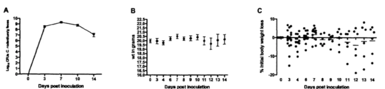
Infection kinetics and body weight changes
As previously reported, fecal shedding of C. rodentium in C57BL/6 mice reached a maximum of -109 CFU/g feces [89], with detectable levels 3 DPI, peaking 7 DPI, with clearance beginning as early as 10 DPI (Figure 2-1A). Although dependent on mouse strain, bacterial load in the colon peaks at 5-14 DPI with approximately 109 colony-forming units [74]. Body weight changes were not significant over the course of infection, although variable with some animals losing close to 20% of their initial body weight (Figure 2-1B & C), in line with previous findings that C.
rodentium infection in adult C5713L/6 mice results in self-limiting disease with
minimal incidence of morbidity and mortality [74, 76, 90].
C. rodentium-induced histological colonic changes
Infection and disease was confirmed by colonic histological changes assessed by H&E with sections scored by a veterinary pathologist blinded to study treatment groups (Figure 2-2). The integrity of epithelial and goblet cells appear intact in control animals but as early as 3 DPI, epithelial defects, colonic foci of inflammation, edema, and hyperplasia were noted (Figure 2-2A and B) with statistically significant increases in inflammation, edema, and epithelial defects by 7 DPI (p <
0.05, Krustkal Wallis with Dunn's post test), appearing maximal by 14 DPI (Figure 2-2C and D). Statistically significant changes in crypt atrophy and hyperplasia were only observed at 14 DPI (Figure 2-2D) (p < 0.05 and p < 0.01 respectively). Although highly dependent on mouse strain, peak colonization and hyperplasia can be seen as early as 4 DPI, with maximal colonic thickening observed between 10 and 12 DPI. In adult C57BL/6, disease peaks approximately 2 weeks post inoculation (WPI) with recovery and full clearance of C. rodentium occurs by 4 WPI and resolution of colonic lesions by 6 WPI [74, 76, 91].
C. rodentium-induced histological liver changes
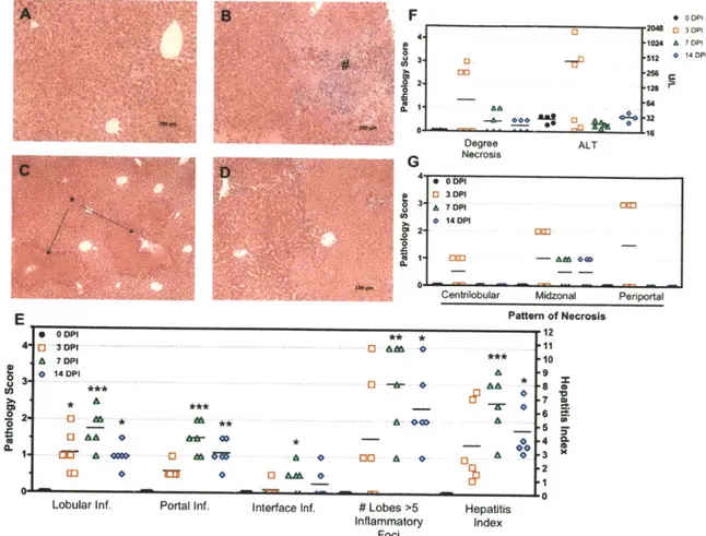
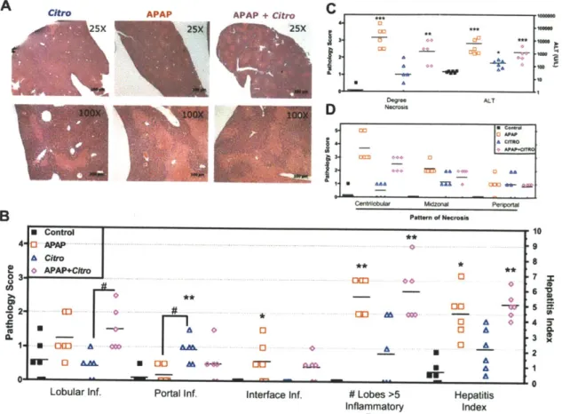
While the effect of C. rodentium infection has been well characterized with respect to colonic changes, we decided to examine in greater detail the effect of pathogenic enteric infection on liver homeostasis. Liver sections were formalin fixed, paraffin embedded, and stained by H&E for pathological assessment (Figure 2-3). C.
rodentium induced statistically significant histological changes as early as 3 DPI
(lobular inflammation) in liver sections and most significant at 7 DPI (portal inflammation, lobular inflammation, interface inflammation, # lobes with >5 inflammatory foci, and hepatitis index), with moderate improvement by 14 DPI (Figure 2-3E). Control livers had no observable histological abnormalities, however, at 3 DPI, the appearance of foci of inflammation indicated a pro-inflammatory state with multifocal vascular coagulative necrosis observed in 3/6 mice (Figure 2-3A-D). Necrotic lesions presented primarily with a periportal pattern of distribution (Figure 2-3G) and indicative of thrombic ischemic injury with the presence of portal venular fibrin thrombi. Necrotic regions contained
hepatocytes with eosinophillic cytoplasms, appearance of pyknotic or absent nuclei, and loss of normal cellular structure. To confirm and measure the extent of necrosis, serum was processed for alanine amino transferase (ALT), and aspartate amino transferase (AST), two accepted markers of hepatic injury. Hepatic necrosis score correlated with increased ALT at 0 vs 3 DPI = (28.60 ± 2.358 U L-1 N=5 versus 415.2 ± 250.4 U L-1 N=6). Mice presenting lesions had ALT levels 56, 16, and 13-fold
higher than average control values (ALT = 1603, 464, and 359 U L-1 respectively), while non-lesions bearing animals at 3 DPI were comparable to controls (Figure 2-3 F). To our knowledge, this is the first reported liver lesion of this kind reported as a result of C rodentium infection and have been repeated and confirmed in subsequent studies (Chapter 3).
Further characterization of necrotic livers by immunohistological staining for activated caspase 3 and Ki-67 revealed labeling index in periportal areas of injury for both markers indicating heterogeneous cell death and active proliferation (Figure 2-4). Livers harboring lesions at 3 DPI contained a higher incidence of positively stained cells for Ki-67, as compared to controls and non-lesions bearing mice at 3 DPI, indicating a proliferative state in these livers with a comparable increase in labeling index at 7 and 14 DPI Figure 2-5A and B). Animals at 7 and 14 DPI did not harbor obvious necrotic lesions, however, the presence of large hepatocytes and areas of necro-granulomatous inflammation indicate prior lesions in these animals may have resolved via inflammatory and proliferative mechanisms.
Serum cytokine and chemistry changes due to C rodentium infection
C rodentium has been shown to induce numerous immune regulators at both
systemic and local levels, and generally associated with a mucosal TH1/Th17-mediated response in C57BL/6 mice ([92, 93]). Recently, serum and colon matched cytokines were analyzed in C rodentium infected C57BL/6 mice at peak colonic disease (14 DPI), demonstrating correlations of colon and systemic levels associated with disease severity [94]. Here we decided to measure both serum specific changes in cytokines/chemokines (23-PLEX Luminex bead-based ELISA) as well as serum chemistries in multiplex at early, peak, and resolving timepoints of bacterial clearance (Supplementary Figures 2-1 and 2-2). We determined numerous circulating cytokines and chemokines to be significantly induced at 3, 7, and 14 DPI. Findings at 3 DPI are particularly interesting as they correlate with our histological findings in the liver and precede peak colonic bacterial colonization and disease. Circulating cytokines were significantly elevated for IL-2, G-CSF, GM-CSF, MCP-1, MIP-1b, and RANTES at 3 DPI as a group (p < 0.05, by one way ANOVA with Tukey's multiple comparison test). Elevations in IL-10 and KC were noted but missed significance. For the majority of targets measured, mice at 7 DPI appeared to have the highest circulating cytokines and chemokines, correlating with peak bacterial
Serum cholesterol levels also increased significantly at 3 DPI and continued to rise until 14 DPI. It has been shown that C. rodentium can alter the transcriptional levels of certain phase I (cytochrome P450s) and phase II metabolic enzymes in both liver and kidney, key in metabolism of endogenous and pharmaceutical substrates [7, 8]. Cholesterols clearance from circulations is partially mediated by oxidation via CYP 7A1, and we have shown decreases in CYP7a1 in liver infected with C. rodentium (Appendix Chapter B). LPS has also been shown to induce expression of ATP-binding cassette transporter Al (ABCA1), a transporter known to promote apolipoprotein-dependent cholesterol efflux from cells, with cholesterol participating in the removal of an immunostimulatory bacterial lipid, lipopolysaccharide (LPS)[95]. Others have also shown that acute phase proteins such as serum amyloid A are loaded into HDL vesicles for transport, indicating a mechanisms for cholesterol in host defense that have previously not been appreciated. Cholesterol homeostasis is complex, and while interesting in this model, would require considerable follow-up to understand the mechanism for its increased serum levels in this infection model. Other serum chemistries and electrolytes, including albumin and glucose, were normal and did not vary significantly across times monitored (Supplementary Figure 2-1).
Liver cytokine and signaling changes due to C. rodentium infection
To determine if systemic cytokine and chemokine profiles correlated with local levels in the liver, we analyzed lysates using the 23-plex cytokine/chemokine panels and discovered numerous targets increased in serum demonstrated similar up-regulation in livers at 3 DPI (Supplemental Figure 2-3). Liver levels of IL-1 (L), G-CSF (L), KC (L), MCP-1 (L), MIP-la (L), and RANTES (L) showed upregulation at 3DPI. Cytokines are known signaling molecules in signal transduction pathways, so we also measured signal transduction in liver lysates using multiplex phospho-kits monitoring both total and phosphorylated forms of
JNK,
AKT, ERK1/2, p38, IKBa, and STAT3 (phosphorylated only). STAT3 showed increases in livers harboring necrotic lesions as confirmed by western analysis, with lower but sustained activation at 7 and 14 DPI (Figure 2-7). Akt activation as assessed by the ratio of phosphorylated/total Akt was statistically significantly upregulated at 14 DPI, although levels were comparably elevated at 3 and 7 DPI but missed significance (Figure 2-7). Phosphorylation of IKBa, which results in proteasome-mediated degradation and subsequent activation of Nf-KB, was increased in animals bearing necrotic lesions, indicating potential activation of pro-inflammatory and tissue repair mechanisms associated with this transcription factor.Multivatiate analysis uncovers serum and tissue cytokines and chemokines discriminate necrosis
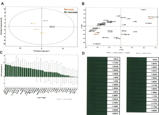
Analyzing data based on averages in representative groups can cause an investigator to miss important correlations on an individual animal basis. Based on this, we decided to take a multivariate computational approach in determining features that covary best in our data compendium leveraging its multiplex nature. PLS-DA (Partial Least Squares Projection to Latent Structures - Discriminant
Analysis) and OPLS (Orthogonormalized Partial Least Squares Regression) were used to determine variables with the highest discriminatory power for lesion bearing animals at 3DPI, furthermore, to determine features that correlate best with serum ALT, a known biomarkers of liver necrosis. We developed non-invasive models (serum targets only), tissue level model (liver targets only), and a combined model using both types of features. The serum PLS-DA model resulted in good separation of lesion bearing vs non-lesion bearing animals (Figure 2-8A), with dummy variables (Y) based on this classification covarying with serum targets that make up the principal components plane (Figure 2-8B). Their relative importance as discriminators was assessed by their variables in projection (VIPs) score for the principle component 1, where values >1 are have positive influence in discriminating between classes and VIP <1 are less influential (Figure 2-8C and D). Similarly using OPLS regression, which reduces dimensions on the basis of their covariance with a specified dependent variable (Y, serum ALT) (principal component 1 - predictive), while ignoring targets orthogonal to this vector (principal component 2 - orthogonal) also resulted in clear separation of animals based on this classifier (Figure 2-9). PLS-DA serum models uncover ALT, AST, immune modulators (IL-6, IL-10), monocytes chemokines/activators (MIP-la, MCP-1), neutrophil chemokines/activators (G-CSF, KC), and t-cell activation (RANTES) as enriched in lesion bearing animals (Figure 2-8 C-D).
This method was repeated for tissue markers (Liver PLS-DA Figures 2-10, and OPLS Figure 2-11) and resulted in a large overlap with the serum specific targets; immune modulators (IL-1a (L), IL-6 (L), IL-12p40 (L)), monocytes chemokines/activators (MIP-1a (L), MIP-1b (L), MCP-1 (L)), neutrophil chemokines/activators (G-CSF (L), KC (L)), and t-cell activation (RANTES (L)) as enriched in lesion bearing animals resulting in enrichment of liver PLS-DA and OPLS models generated using serum targets, liver targets, and combined targets are summarized (Table 2-1). Overall, both serum and tissue models were highly effective at discriminating both necrosis state prediction of necrosis severity. The tissue models were slightly better independently than the serum models, and the combine (serum + tissue) gave the highest R2Y (cumulative) and Q2 (cumulative) with the least number of components. The less non-invasive method of serum cytokine detection makes this a more attractive method even with a modest lost in model prediction.
2.3 Discussion
In the present study we have characterized the systemic and liver effects of C.
rodentium infection in female C57BL/6 mice at early, peak, and resolving timepoints
of bacterial clearance. We demonstrated systemic targets (cytokines/chemokines and serum chemistry markers) that differentiated animals by PLS-DA. Systemic elevations in ALT, AST, with an upregulation of immune modulators (IL-6, IL-10), monocytes chemokines/activators (MIP-la, MCP-1), neutrophil chemokines/activators (G-CSF, KC), and t-cell activation (RANTES) at 3PDI correlated with coagulative liver necrosis, lesions predominantly periportal in nature and associated with fibrin thrombi. Mice harboring lesions also demonstrated induction of STAT3 and IKBa phosphorylation, coupled with liver specific protein elevations of IL-1p (L), IL-6 (L) G-CSF (L), KC (L), IL-12p40 (L), MCP-1 (L), MIP-1a (L), and RANTES (L).
C. rodentium-induced histological liver changes
Pathological assessment of livers discovered that 50% of mice at 3 DPI harbored periportal necrotic lesions that have not been observed and/or reported to our knowledge in this murine model of enteric infection and colonic hyperplasia. This may due in part to the fact that 3 DPI is early in disease pathogenesis coupled by our interest in investigating an organ distal to the primary site of disease pathology and bacterial colonization. Bacterial translocation due to inflammation or overgrowth of commensal bacteria has been shown to occur, with translocation of luminal bacteria to other organs [96, 97], in some cases resulting in sepsis and the subsequent death. Furthermore, C. rodentium has demonstrated translocation to MLNs during infection course [98]. We did not plate liver tissues to determine C. rodentium liver load but follow-up experiments staining liver sections using a C. rodentium specific antibody could confirm this. C. rodentium has been associated with disruption of tight junctions and barrier function in intestinal epithelial cells in vitro and in vivo ([99, 100]). As liver lesions were predominantly periportal in their pattern of distribution, and indicative of thrombic ischemic injury with portal vein thrombi present, it is possible that bacterial components, or C. rodentium (live or dead) at the early stages of bacterial colonization, translocated from the gut lumen via portal blood to the liver inducing a systemic and local inflammatory response. LPS, for example, through interaction with LPS-binding protein has been demonstrated to induce fibrin clots in tissues. Others have proposed that the liver may act to trap LPS, along with other bacterial components as a protective mechanism to prevent systemic spread of bacteria. As a regenerative organ, minimal necrosis via coagulative mechanisms could me manageable. The presence of necro-granulomatous inflammation at later timepoints indicates perhaps of residual effects of clearance mechanisms of prior injury in these animals. The lack of circulating ALT indicates that active necrosis was likely not occurring.