The bright side of sound: perspectives on the
biomedical application of sonoluminescence
Roberto Canaparo,
*
aFederica Foglietta,
bFrancesca Giuntini,
cAndrea Francovich
dand Loredana Serpe
aLight is a physical phenomenon that is very important to human life, and has been investigated in its nature, behaviour and properties throughout human history although the most impressive improvements in the use of light in human activities, and of course in medicine, began just two centuries ago. However, despite the enormous progress in diagnosis, therapy and surgery to assess health and treat diseases, the delivery of light sourcesin vivo remains a challenge. In this regard, several strategies have been developed to overcome this drawback, the most interesting of which is the involvement of ultrasound. In this review, the authors examine how ultrasound may improve light deliveryin vivo with a special emphasis on one of the most intriguing ultrasound-mediated phenomena called sonoluminescence, which is the conversion of mechanical ultrasound energy into light.
Introduction
Light is linked in a wide variety of ways with biological phenomena such as vision, biological clocks and photosyn-thesis. These phenomena are so important that, without light, our world would be different, probably without life or at least
inhospitable for all higher organisms that are around today. Due to the huge impact of light in human evolution, humans have tried, for centuries, to find out all the secrets about this phenomenon and how to take advantage from it. Due to this effort, nowadays, light influences the way we live in ways we could never have imagined just a few decades ago, in the ways we communicate, in the tools we use to explore the frontiers of science and, of course, in the practice of medicine. In this regard, in the 19th century the use of light was introduced in medicine, leading to a rapid increase in the knowledge of its physical nature and basic interactions with matter.
Currently, light and its related optical methodologies have achieved an extensive impact on current medicine, with various laser and optical instruments used in the clinical
aDepartment of Drug Science and Technology, University of Torino, Via Pietro Giuria
13, 10125 Torino, Italy. E-mail: roberto.canaparo@unito.it
bDepartment of Molecular Biotechnology and Health Sciences, University of Torino,
Via Nizza 52, 10126 Torino, Italy
cSchool of Pharmacy and Biomolecular Sciences, Liverpool John Moores University,
Liverpool L3 2AJ, UK
dInstitut de Physiologie, Université de Fribourg, Chemin du Musee 5, 1770 Fribourg,
Switzerland
http://doc.rero.ch
Published in "Photochemical & Photobiological Sciences 19(9): 1114–1121, 2020"
which should be cited to refer to this work.
setting for diagnosis and therapy to evaluate health and cure diseases. Recent improvements, thanks to innovative medical lasers and new optical technologies, have revealed opportu-nities for further progress in photomedicine.1–3 However, for all photomedicine approaches, the delivery of light sources in vivo remains a challenge. Recently, to overcome this signifi-cant drawback, a new idea has been suggested, mainly in the field of photodynamic therapy (PDT) and optogenetics, where ultrasound (US) seems to play an interesting role in triggering common photosensitizers4and in controlling cellular activity.5 In this review, the authors discuss how US can improve specific areas of photomedicine, with a special emphasis on one of the most intriguing US-mediated phenomena called sonoluminescence (SL).
Application of light in medicine
In medicine, the use of light originates from its evolutionary function in biology, indeed the therapeutic use of light takes advantage of the biological effects on tissues derived from the specific wavelength absorption of a diversity of light-responsive molecules. In the human body, these light-sensitive molecules can be endogenous or exogenous, therefore, administered as drugs or inserted by gene-editing methodologies. To date, the main light applications can be subdivided into three main groups: (i) optical imaging, (ii) light-activated therapy and (iii) laser surgery.3
Optical methodologies are extensively utilized in diagnostic medicine, ranging from laboratory measurement and diagnos-tic imaging, to intra-surgery imaging and therapy monitoring. Thanks to the high spatial resolutions of light, optical imaging allows real-time visualization of cells and tissues by reasonably inexpensive and portable devices. Many different technologies for macro- and microscopic imaging have been implemented as a standard of clinical practice, and many others are under development for translation in the clinical setting. Surgical techniques such as laparoscopic surgery, which are based on optical guidance, have decreased the haemorrhage risk and lowered the time of patient recovery. Increased surgical results have also been achieved by using intra-surgery optical imaging with tissue contrast that can exceed what the human eye can distinguish.3
In 1960, T. Maiman provided the first evidence of the ruby laser application, paving the way to the great history of laser medical uses.6Pioneering studies employed photocoagulation for treating retina diseases, skin injuries and cardiovascular lesions. Nowadays, medical lasers are used in routine practice, as well as in ophthalmic surgery, dermatological treatment and tissue removal in internal body organs by using fibre-optics.7
One of the most intriguing properties of light is its capa-bility of influencing photo-responsive cells, proteins and mole-cules that can be effectively exploited in therapeutic appli-cations. Light-mediated treatments, namely phototherapies, rely on the use of an appropriate light source. These
treat-ments, such as PDT, can be very useful for diseases in which unhealthy cells can be destroyed by light-induced oxidative stress. PDT is a clinically proven approach exerted in a variety of fields such as oncology, ophthalmology, dermatology and dentistry.8 Recent innovations in nanomedicine have enabled the development of multimodal nanocarriers with numerous light-activated functions, including conversion of near infrared to visible light, photodynamic and photothermal therapies, drug delivery and imaging.9
Optogenetics represents a new frontier in light-activated therapies, enabling unprecedented control over neural activity and cellular signalling.10 Indeed, optogenetic methodologies have opened up novel avenues into disease connections, leading to immediate clinical results. For instance, in mouse models, the optical section of brain connections has disclosed mechanisms for brain stimulation useful for treatment of Parkinson’s disease.11Thanks to its neuromodulation capabili-ties, optogenetics has attracted interest for treating chronic pain,12depression13and laryngeal paralysis.14
However, to effectively exert the various biological effects induced by light–tissue interactions for clinical applications, light must be carried to the desired tissues with specificity and proper energy, therefore the development of new lasers deliver-ing desired output characteristics will expand both clinical and at-home settings, as well as the development of new opto-electronic devices such as polymer LEDs.3 Unfortunately, all these strategies still suffer from one of the main drawbacks about light delivery, namely the poor penetration of light due to intrinsic absorption and scattering within tissues. In this regard, several attempts have been made to overcome this drawback, with the most promising methods involving implan-table optoelectronic devices.15Innovative instrument concepts and design involve implantable light emitting diodes,16drug delivery controlled by optofluidics17 and optogenetic appli-cations through miniaturized wireless optoelectronic instruments.18,19 A new approach involves working with bio-material photonic devices.20 Biomaterial-based optical wave-guides can be exploited for long-lasting delivery of light, and are required to be displaced if produced with biodegradable materials, such as absorbable sutures made of polyglycolic acid or silk.21 Finally, taking advantage of the intrinsic pro-perties of intracellular lasers to act on specific sites such as inflamed tissues, developing cell-based lasers can represent an innovative way to bring light with selectivity and the possibility to perform– at the same time – a strong multiplex imaging set on narrowband coherent emission.22Recently some research-ers have suggested US as a new tool to overcome certain draw-backs and improve the light delivery, mainly in the field of PDT and optogenetics.5,23–27
Light from sound
Sound is a wave that transports mechanical energy through the local vibration of particles in an elastic medium with no net transport of the particles.28This definition is, of course,
very different from the definition of light because light is described as an electromagnetic wave with the same theore-tical principles that govern all forms of electromagnetic radi-ation. Nevertheless, despite great differences between these two waves such as mechanical versus electromagnetic, a well-known phenomenon called mechanoluminescence (ML, Fig. 1) is able to transform mechanical energy, as well as mincing, smashing, distorting, splitting, shrinking or using US pulses, into light.29Thus, ML can have various applications such as magnetic and electric field detection, light source, dynamic pressure outlining or stress sensing.30Terasaki et al. showed the exploitability of ML as a light source following in vivo exposure to US radiation or for photocatalysis.31,32 Terasaki and colleagues synthesized an ML nanoparticle with a size of 10 nm, small enough to be used as an ubiquitous light source in tissues and cells.31For this purpose, they inves-tigated the ML induced by ultrasonic waves, detecting ML that depends on US irradiated power, then introducing the ultra-sonic wave as a suitable candidate for mechanical stimulation to achieve ML.32However, these are just preliminary results to demonstrate the ability of US-induced ML as a light source, and many issues should be addressed before ML can be uti-lised as a ubiquitous light source in tissues, for example, the toxicity of the materials in living organisms.
ML is not the only mechanism through which mechanical energy can be transformed into light, in nature, a light emis-sion by marine living organisms is very usual, triggered by the mechanical stimulation from water agitation that elicits cell deformations able to initiate action potentials into vacuole membranes, called bioluminescence (BL). The BL molecular mechanism is based on a classic two-component process com-posed of the luciferase enzyme catalysing the BL reaction, and the luciferin molecule acting as a light-releasing system during the reaction (Fig. 1).33Kheirolomoom and colleagues encapsu-latedD-luciferin, in long-circulating liposomes, showing that,
after tumour exposure to US in Met1-luc tumour-bearing mice, an immediate emission of light was detected, enhancing in vivo bioluminescence imaging.34
Another mechanism for light emission is through a chemi-cal reaction, which is chemi-called chemiluminescence (CL, Fig. 1).35 In general, in direct CL, two essential mechanisms are respon-sible for the chemiluminescent reaction, commonly a sub-strate and an oxidant reagent, along with cofactors and often a
catalyst, which react, generating a product or intermediate. In addition, a fraction of the product or intermediate will be gen-erated in an electronically excited state, being able to emit photons after releasing to its ground state. On the other hand, in indirect CL, an energy transfer process from an excited molecule to a fluorophore is pivotal, which, once activated, is able to emit photons when relaxing to its ground state. This mechanism is relevant for molecules that cannot be engaged in direct CL reactions but that are able to transfer their excess energy to a fluorophore.36 The referred reactions can be exploited in a great diversity of practical uses of CL, and recently Le et al. have investigated the combination of CL and US for dual imaging.37 In this investigation, the relevant improvement of using CL with US has been demonstrated by using tissue mimicking materials and ex vivo models.38,39 McMurray and colleagues suggested that the sonochemilumi-nescence intensity could be related to the concentration of HO•.40Recently, this mechanism has been confirmed by the fact that US irradiation of water can lead to acoustic cavitation, a process capable of generating free radicals such as H•and HO•, which are able to produce reactive oxygen species (ROS) therefore, increasing CL.41
Sonoluminescence
Acoustic cavitation, which is the mechanism that underlies sonochemiluminescence, refers to the mechanical interplay between acoustic waves and gas-filled microbubbles present in the exposed liquid.42Acoustic cavitation can take place either as a stable or transient mode. During a significant number of acoustic cycles, in stable cavitation, microbubbles oscillate close to an equilibrium radius without collapsing. Conversely, in transient cavitation, microbubbles grow by rectified diffusion reaching a resonance size, and then collapse. A variety of physical effects can, therefore, be generated when cavitation microbubbles oscillate or collapse, such as shear forces, shock waves and micro-jets. Moreover, the collapse of gas-filled microbubbles during transient acoustic cavitation is nearly adiabatic and produces a temperature in the thousand-degree range in the microbubbles for a very short time period.43Extremely reactive radicals are then generated thanks to these exceptional temperature conditions. For instance, if the cavitation medium is water, OH•and H•radicals are pro-duced by the water homolysis. This generation of radicals has been exploited to perform, for example, organic pollutant degradation and synthesis of polymers or nanomaterials.44 Furthermore, acoustic cavitation has also been found to be valuable in medical diagnosis and therapy.45,46In this regard, in biomedicine, recently the acoustic cavitation use has greatly improved thanks to its unique theranostic features and feasi-bility. The cavitation energy derived from the microbubble generation, growth, and collapse, can produce reversible pora-tion of cell membranes and vessel walls, namely sonoporapora-tion, which can be used for delivery to the target site of bioactive elements such as drugs, genes, peptides or proteins.47,48
However, when cavitation microbubbles oscillate and col-lapse in liquids, another intriguing physical phenomenon
Fig. 1 Schematic illustration of the most known phenomena respon-sible for physical or chemical-mediated light emission.
occurs called sonoluminescence (Fig. 2), and nowadays it is well known that acoustic cavitation is also followed by light emission.49In simple terms, SL refers to the transformation of US mechanical energy into light pulses of about 35–350 pico-seconds and composed of 3 × 10⁴–3 × 10⁵ photons.50 This process is always inefficient, generally transforming only 0.0001 of acoustic energy into photons, however it is remark-able, as the occurring energy density of rising photons over-comes the US driving energy by a 1012fold.51
In 1934, SL was accidentally observed at the University of Cologne by H. Frenzel and H. Schultes studying acoustic radar. The two scientists noticed that many of the microbubbles were flashing in the water. Created by highly powerful US fields, the flashing lights were chaotic and unpredictable. Later, this phenomenon was defined as multi-bubble sonoluminescence. After 50 years, F. Gatian and L. A. Crum began researching SL and were able to achieve single-bubble sonoluminescence (SBSL) for the first time. Effectively trapping a single bubble in a flask, Gatian and Crum energized the micro-bubble, creating the first recorded SBSL.52
The discovery of SBSL initiated a powerful research activity mainly by physicists and obtained consideration from the broader scientific community just after it was suggested as a result of quantum radiation or when it was proposed to be able to produce nuclear fusion. Noteworthy, the representation of SL as a“hot spot”, namely the thermal model, is the univer-sally accepted one, and the model related to the generation of nuclear fusion has been widely rejected.52Therefore, the main question is: could SL have a potential for applications in the biomedical field?.
The first evidence of a biomedical application of SL comes from a Russian study where the authors proposed detecting SL in blood plasma in the differential diagnosis of tuberculosis, cancer and sarcoidosis of the lungs.53 Unfortunately, the manuscript was written in Russian therefore not easily municating the real achievements to the wider scientific
com-munity. In 1996, other Russian scientists published a short manuscript in English, in which 8000 patients with cancer, tuberculosis and some other diseases were enrolled to investi-gate the possible applications of SL in diagnostics. The results show that the SL-method is a promising technique for early diagnostics of cancer, tuberculosis and low-immunity status. Due to the small samples of blood required and the speed of the analysis, this approach could be prospective for mass people examination.54 Recently, A. Casacchia and colleagues developed an experimental apparatus for producing SBSL and subsequent measurement of radial oscillations using optical scattering techniques, allowing the effective characterization of both the biological fluid content and viscoelasticity on the spectra of SBSL light emissions, opening new perspectives of SL in diagnostics.55
Photodynamic therapy
Suggesting a possible practical application of SL in the bio-medical field, we can pose the question“in which therapeutic applications could SL be feasible”, and PDT is the most likely answer. PDT makes use of harmless chemical compounds ( photosensitizers) such as porphyrins, chlorophylls, and other dyes that are employed to cause precise bioeffects on cancer cells following light irradiation. However, this technique may have limited effectiveness in thicker tumours since it pos-sesses a poor penetration depth.56Therefore, to overcome this drawback, a strategy to activate the photosensitizer in deeper tissues is to deliver laser light to the target site through fibre-optics. Thus, nowadays laparoscopic intraoperative incisions and endoscopic procedures are needed to reach the target area and activate the photosensitizer by laser.
The photosensitizer absorption of a proper energy photon causes the molecule to achieve a singlet state excitation. The photosensitizer can then go through intersystem crossing to a triplet state of lower energy. On the assumption that the triplet state has an increased sufficient energy, its decay back to the ground state may have two effects. Firstly, a type I reaction takes place by transfer of an electron to molecular oxygen, leading to the formation of a superoxide radical, thereby initi-ating a cascade of radical reactions, which damage biomacro-molecules and kill cells in the close proximity. A reaction of type II takes place through transfer of energy to oxygen, which is promoted from a ground triplet state to a cytotoxic excited singlet state. Singlet oxygen and radicals cumulatively defined as ROS can cause apoptotic and necrotic cell death.57
Almost all of the porphyrin-derived photosensitizers are excited in the blue region of the visible spectrum, and SL emis-sion is also intense in the same region of the electromagnetic spectrum, raising the question whether SL is able to produce a photon amount sufficient to excite porphyrin-derived photo-sensitizers. Since US is able to reach deep tissues in living organisms, the answer to this question may help to overcome the main drawback in PDT, since the in-depth delivery of PDT relies on relatively invasive approaches.
The first experiments about the possibility of SL employed as an inner source of light to activate other molecules in
solu-Fig. 2 Ultrasound-mediated SL relies on the occurrence of transient cavitation during US exposure of a liquid milieu leading to the formation of gas-filled collapsing microbubbles. Thanks to rectified diffusion, gas and vapour are transported into the microbubbles until they reach a critical size, and then they collapse. The microbubble contents are then compressed rapidly, resulting in extreme local conditions and a range of secondary effects that drive processes such as photon emis-sion, dubbed SL.
tion were performed by M. Ashokkumar and F. Grieser in 1998.58 These authors investigated the excitation of pyranine in water by SL and the ensuing emission, which can be referred to as sonophotoluminescence, drawing the con-clusions that photosensitizing chemical compounds inside the human body can be excited via sonophotoluminescence pro-duced through US as an outer source with respect to the human body. The same authors also investigated other different sensitizers, namely fluorescein, eosin and pyrene where SL, produced in air-saturated non-aqueous and aqueous solutions, was able to directly excite these species, leading to fluorescence emission.59Recently, Beguin and colleagues have published a work where therapeutic US was able to produce SL when phospholipid-coated microbubbles were added in aqueous solution. This investigation provides a mechanistic explanation about the anticancer approach, called sonody-namic therapy (SDT), where light-responsive chemical com-pounds were able to be excited through US-mediated cavitation.60
Sonodynamic therapy
The first evidence about SDT came from a study by Yumita et al. in 1989, where various hematoporphyrin derivatives employed in PDT also caused a relevant cell damage after being exposed to US.61Since then, it has been shown that SDT can treat solid tumours,62–65leukaemia66and atherosclerosis67 and, moreover, remove proliferative scars and kill pathogenic microorganisms.68,69
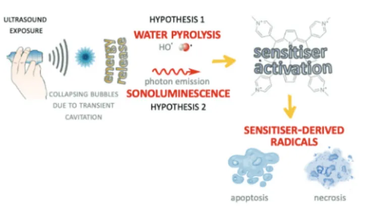
Since SDT was developed on the basis of PDT, some suggest that SDT shares a similar therapeutic mechanism where the ultrasonic wave triggers the sonosensitizer inside the target tissue, with production of ROS that kill the target cells.70Even though there is consensus in ROS production as the main cell killing mechanism, less is known about the mechanism underlying ROS production. At present, two hypotheses have been suggested to explain the mechanism of ROS production following exposure of the sonosensitizer to US wave energy, namely the pyrolysis hypothesis and the SL hypothesis (Fig. 3).
Both hypotheses are based on the acoustic cavitation phenom-enon; according to the first hypothesis, sonosensitisation arises from the sonosensitizer activation nearby or inside the hot collapsing cavitation microbubbles leading to formation of sensitizer-derived radicals via direct pyrolysis or indirect reac-tions with HO• and H• radicals derived from water pyrolysis. These are mainly carbon-centred radicals and are also able to interact with oxygen-generating alkoxyl and peroxyl radicals. In contrast to H•and HO•radicals, also generated by pyrolysis in the cavitation microbubbles, the alkoxyl and peroxyl radical reactivity with organic components is lower, and they therefore have an increased probability of reaching pivotal cellular sites killing target cells. This hypothesis is the most supported by researchers involved in this field even though some evidence has revealed, as mentioned before, that SL seems to play a role, as also suggested from the studies by Giuntini et al. and Dezhkunov et al.24–71 In particular, Giuntini and colleagues pointed out how the activation of a metal-porphyrin complex by US exposure, namely sonodynamic activation, is more likely due to SL-induced photoactivation rather than to pyrolysis, the radical generation occurring through the homolytic bond rupture of water molecules.
Conclusions
In this review we have discussed the possibility of overcoming the poor penetration of light in tissues and the invasiveness of diverse optical strategies, changing the sound into light to improve, mainly, the photodynamic and optogenetics approaches in vivo. We have therefore discussed about SL and how this intriguing phenomenon deserves more attention. However, every kind of new advancement and achievement, mainly in the scientific field, has its own limitations which make rigorous challenges. In this regard acoustic cavitation, considered one of the primary mechanisms underpinning STD and SL, represents the most important challenge in this field. Therefore, additional mechanistic studies have to be con-ducted to develop efficient methods to easily handle acoustic cavitation with the final aim to directly measure and monitor its effects. These improvements in acoustic cavitation compre-hension will also open new perspectives about the SL, evolving this phenomenon, from a physics laboratory curiosity to a reliable, convincing and solid technique to deliver light for in vivo applications.
Con
flicts of interest
There are no conflicts to declare.
Acknowledgements
The authors gratefully acknowledge funding from the Ministry of Education, University and Research (FFABR 2017) and the University of Torino (Ricerca Locale 2019), Italy.
Fig. 3 Schematic illustration of the two main hypotheses concerning the underpinning mechanism of sonodynamic activity. The sensitizer activation leading to radical-mediated cell death can be driven by water pyrolysis or SL induced by the energy release of the collapsing microbubbles.
References
1 S. Mallidi, S. Anbil, A. L. Bulin, G. Obaid, M. Ichikawa and T. Hasan, Beyond the Barriers of Light Penetration: Strategies, Perspectives and Possibilities for Photodynamic Therapy, Theranostics, 2016, 6, 2458–2487.
2 M. M. Lerch, M. J. Hansen, G. M. van Dam, W. Szymanski and B. L. Feringa, Emerging Targets in Photopharmacology, Angew. Chem., Int. Ed., 2016, 55, 10978–10999.
3 S. H. Yun and S. J. J. Kwok, Light in diagnosis, therapy and surgery, Nat. Biomed. Eng., 2017, 1, 0008.
4 F. Giuntini, L. Bourré, A. J. MacRobert, M. Wilson and I. M. Eggleston, Improved peptide prodrugs of 5-ALA for PDT: rationalization of cellular accumulation and protopor-phyrin IX production by direct determination of cellular prodrug uptake and prodrug metabolization, J. Med. Chem., 2009, 52, 4026–4037.
5 W. J. Tyler, Y. Tufail, M. Finsterwald, M. L. Tauchmann, E. J. Olson and C. Majestic, Remote Excitation of Neuronal Circuits Using Low-Intensity, Low-Frequency Ultrasound, PLoS One, 2008, 3, e3511.
6 P. Franck, P. W. Henderson and K. O. Rothaus, Basics of Lasers, Clin. Plast. Surg., 2016, 43, 505–513.
7 Q. Peng, A. Juzeniene, J. Chen, L. O. Svaasand, T. Warloe, K.-E. Giercksky and J. Moan, Lasers in medicine, Rep. Prog. Phys., 2008, 71, 056701.
8 I. Yoon, J. Z. Li and Y. K. Shim, Advance in Photosensitizers and Light Delivery for Photodynamic Therapy, Clin. Endosc., 2013, 46, 7.
9 K. A. Carter, S. Shao, M. I. Hoopes, D. Luo, B. Ahsan, V. M. Grigoryants, W. Song, H. Huang, G. Zhang, R. K. Pandey, J. Geng, B. A. Pfeifer, C. P. Scholes, J. Ortega, M. Karttunen and J. F. Lovell, Porphyrin–phospholipid lipo-somes permeabilized by near-infrared light, Nat. Commun., 2014, 5, 3546.
10 E. S. Boyden, F. Zhang, E. Bamberg, G. Nagel and K. Deisseroth, Millisecond-timescale, genetically targeted optical control of neural activity, Nat. Neurosci., 2005, 8, 1263–1268.
11 V. Gradinaru, M. Mogri, K. R. Thompson, J. M. Henderson and K. Deisseroth, Optical deconstruction of parkinsonian neural circuitry, Science, 2009, 324, 354–359.
12 S. M. Iyer, K. L. Montgomery, C. Towne, S. Y. Lee, C. Ramakrishnan, K. Deisseroth and S. L. Delp, Virally mediated optogenetic excitation and inhibition of pain in freely moving nontransgenic mice, Nat. Biotechnol., 2014, 32, 274–278.
13 S. Ramirez, X. Liu, C. J. MacDonald, A. Moffa, J. Zhou, R. L. Redondo and S. Tonegawa, Activating positive memory engrams suppresses depression-like behaviour, Nature, 2015, 522, 335–339.
14 T. Bruegmann, T. van Bremen, C. C. Vogt, T. Send, B. K. Fleischmann and P. Sasse, Optogenetic control of contractile function in skeletal muscle, Nat. Commun., 2015, 6, 7153.
15 T. Kim, J. G. McCall, Y. H. Jung, X. Huang, E. R. Siuda, Y. Li, J. Song, Y. M. Song, H. A. Pao, R.-H. Kim, C. Lu, S. D. Lee, I.-S. Song, G. Shin, R. Al-Hasani, S. Kim, M. P. Tan, Y. Huang, F. G. Omenetto, J. A. Rogers and M. R. Bruchas, Injectable, cellular-scale optoelectronics with applications for wireless optogenetics, Science, 2013, 340, 211–216.
16 R. H. Kim, D. H. Kim, J. Xiao, B. H. Kim, S. I. Park, B. Panilaitis, R. Ghaffari, J. Yao, M. Li, Z. Liu, V. Malyarchuk, D. G. Kim, A.-P. Le, R. G. Nuzzo, D. L. Kaplan, F. G. Omenetto, Y. Huang, Z. Kang and J. A. Rogers, Waterproof AlInGaP optoelectronics on stretchable substrates with applications in biomedicine and robotics, Nat. Mater., 2010, 9, 929–937.
17 J. W. Jeong, J. G. McCall, G. Shin, Y. Zhang, R. Al-Hasani, M. Kim, S. Li, J. Y. Sim, K. I. Jang, Y. Shi, D. Y. Hong, Y. Liu, G. P. Schmitz, L. Xia, Z. He, P. Gamble, W. Z. Ray, Y. Huang, M. R. Bruchas and J. A. Rogers, Wireless Optofluidic Systems for Programmable In Vivo Pharmacology and Optogenetics, Cell, 2015, 162, 662– 674.
18 K. L. Montgomery, A. J. Yeh, J. S. Ho, V. Tsao, S. Mohan Iyer, L. Grosenick, E. A. Ferenczi, Y. Tanabe, K. Deisseroth, S. L. Delp and A. S. Y. Poon, Wirelessly powered, fully internal optogenetics for brain, spinal and peripheral cir-cuits in mice, Nat. Methods, 2015, 12, 969–974.
19 S. I. Park, D. S. Brenner, G. Shin, C. D. Morgan, B. A. Copits, H. U. Chung, M. Y. Pullen, K. N. Noh, S. Davidson, S. J. Oh, J. Yoon, K.-I. Jang, V. K. Samineni, M. Norman, J. G. Grajales-Reyes, S. K. Vogt, S. S. Sundaram, K. M. Wilson, J. S. Ha, R. Xu, T. Pan, T.-I. Kim, Y. Huang, M. C. Montana, J. P. Golden, M. R. Bruchas, R. W. Gereau and J. A. Rogers, Soft, stretch-able, fully implantable miniaturized optoelectronic systems for wireless optogenetics, Nat. Biotechnol., 2015, 33, 1280– 1286.
20 M. Choi, M. Humar, S. Kim and S.-H. Yun, Step-Index Optical Fiber Made of Biocompatible Hydrogels, Adv. Mater., 2015, 27, 4081–4086.
21 S. Nizamoglu, M. C. Gather, M. Humar, M. Choi, S. Kim, K. S. Kim, S. K. Hahn, G. Scarcelli, M. Randolph, R. W. Redmond and S. H. Yun, Bioabsorbable polymer optical waveguides for deep-tissue photomedicine, Nat. Commun., 2016, 7, 10374.
22 M. Humar and S. H. Yun, Intracellular microlasers, Nat. Photonics, 2015, 9, 572–576.
23 X. Wu, X. Zhu, P. Chong, J. Liu, L. N. Andre, K. S. Ong, K. Brinson, A. I. Mahdi, J. Li, L. E. Fenno, H. Wang and G. Hong, Sono-optogenetics facilitated by a circulation-deli-vered rechargeable light source for minimally invasive optogenetics, Proc. Natl. Acad. Sci. U. S. A., 2019, 116, 26332–26342.
24 F. Giuntini, F. Foglietta, A. M. Marucco, A. Troia, N. V. Dezhkunov, A. Pozzoli, G. Durando, I. Fenoglio, L. Serpe and R. Canaparo, Insight into ultrasound-mediated reactive oxygen species generation by various
metal-porphyrin complexes, Free Radicals Biol. Med., 2018, 121, 190–201.
25 S. Wang, T. Kugelman, A. Buch, M. Herman, Y. Han, M. E. Karakatsani, S. A. Hussaini, K. Duff and E. E. Konofagou, Non-invasive, Focused Ultrasound-Facilitated Gene Delivery for Optogenetics, Sci. Rep., 2017, 7, 39955.
26 J. Kubanek, J. Shi, J. Marsh, D. Chen, C. Deng and J. Cui, Ultrasound modulates ion channel currents, Sci. Rep., 2016, 6, 24170.
27 M. E. Moore, J. M. Loft, W. C. Clegern and J. P. Wisor, Manipulating neuronal activity in the mouse brain with ultrasound: A comparison with optogenetic activation of the cerebral cortex, Neurosci. Lett., 2015, 604, 183–187. 28 T. G. Leighton, What is ultrasound?, Prog. Biophys. Mol.
Biol., 2007, 93, 3–83.
29 Y. Xie and Z. Li, Triboluminescence: Recalling Interest and New Aspects, Chem, 2018, 4, 943–971.
30 A. Feng and P. F. Smet, A Review of Mechanoluminescence in Inorganic Solids: Compounds, Mechanisms, Models and Applications, Materials, 2018, 11, 484.
31 N. Terasaki and C.-N. Xu, Performance of single mechano-luminescent particle as ubiquitous light source, J. Colloid Interface Sci., 2014, 427, 62–66.
32 N. Terasaki, H. Yamada and C.-N. Xu, Ultrasonic wave induced mechanoluminescence and its application for photocatalysis as ubiquitous light source, Catal. Today, 2013, 201, 203–208.
33 A. Fleiss and K. S. Sarkisyan, A brief review of biolumines-cent systems (2019), Curr. Genet., 2019, 65, 877– 882.
34 A. Kheirolomoom, D. E. Kruse, S. Qin, K. E. Watson, C.-Y. Lai, L. J. T. Young, R. D. Cardiff and K. W. Ferrara, Enhanced in vivo bioluminescence imaging using liposo-mal luciferin delivery system, J. Controlled Release, 2010, 141, 128–136.
35 C. Dodeigne, Chemiluminescence as diagnostic tool. A review, Talanta, 2000, 51, 415–439.
36 T. H. Fereja, A. Hymete and T. Gunasekaran, A Recent Review on Chemiluminescence Reaction, Principle and Application on Pharmaceutical Analysis, ISRN Spectrosc., 2013, 2013, 1–12.
37 D. Le, D. Dhamecha, A. Gonsalves and J. U. Menon, Ultrasound-Enhanced Chemiluminescence for Bioimaging, Front. Bioeng. Biotechnol., 2020, 8, 25.
38 N. T. Huynh, B. R. Hayes-Gill, F. Zhang and S. P. Morgan, Ultrasound modulated imaging of luminescence generated within a scattering medium, J. Biomed. Opt., 2013, 18, 020505.
39 M. Kobayashi, N. Kikuchi and A. Sato, Ultrasound-enhanced chemiluminescence tomography in biological tissue, Ultrason. Sonochem., 2016, 31, 1–6.
40 H. N. McMurray and B. P. Wilson, Mechanistic and Spatial Study of Ultrasonically Induced Luminol Chemiluminescence, J. Phys. Chem. A, 1999, 103, 3955– 3962.
41 Y. He, D. Xing, G. Yan and K. Ueda, FCLA chemilumines-cence from sonodynamic action in vitro and in vivo, Cancer Lett., 2002, 182, 141–145.
42 M. Ashokkumar, The characterization of acoustic cavitation bubbles - an overview, Ultrason. Sonochem., 2011, 18, 864– 872.
43 M. Ashokkumar, J. Lee, S. Kentish and F. Grieser, Bubbles in an acoustic field: an overview, Ultrason. Sonochem., 2007, 14, 470–475.
44 K. S. Suslick, Ultrasound. Its Chemical, Physical, and Biological Effects, Science, 1989, 243, 1499–1499.
45 C. C. Coussios and R. A. Roy, Applications of Acoustics and Cavitation to Noninvasive Therapy and Drug Delivery, Annu. Rev. Fluid Mech., 2008, 40, 395–420.
46 S. Umemura, S. Yoshizawa and R. Takagi, Cavitation in therapeutic ultrasound: To be avoided or to be utilized, J. Acoust. Soc. Am., 2016, 140, 3246–3246.
47 M. S. Aw, L. Paniwnyk and D. Losic, The progressive role of acoustic cavitation for non-invasive therapies, contrast imaging and blood-tumor permeability enhancement, Expert Opin. Drug Delivery, 2016, 13, 1383–1396.
48 T. Y. Wang, K. Wilson, S. Machtaler and J. Willmann, Ultrasound and Microbubble Guided Drug Delivery: Mechanistic Understanding and Clinical Implications, Curr. Pharm. Biotechnol., 2014, 14, 743–752.
49 K. S. Suslick and D. J. Flannigan, Inside a collapsing bubble: sonoluminescence and the conditions during cavitation, Annu. Rev. Phys. Chem., 2008, 59, 659–683. 50 K. R. Weninger, P. G. Evans and S. J. Putterman, Time
cor-related single photon Mie scattering from a sonolumines-cing bubble, Phys. Rev. E: Stat. Phys., Plasmas, Fluids, Relat. Interdiscip. Top., 2000, 61, R1020–R1023.
51 P. Axford, L. Lawton, P. Robertson and P. A. Campbell, Multi-bubble sonoluminescence: laboratory curiosity, or real world application? Proc. SPIE, 2008, 7030, 703012. 52 L. A. Crum, Resource Paper: Sonoluminescence, J. Acoust.
Soc. Am., 2015, 138, 2181–2205.
53 V. Z. Zhadnov, R. F. Mishanov and V. V. Chernov, Sonoluminiscence of blood plasma in the differential diag-nosis of tuberculosis, cancer and sarcoidosis of the lungs, Probl. Tuberk., 1994, 42–43.
54 V. Chernov and D. S. D. Selivanovsky, Sonoluminescence of Water and Biological Liquids, Proc. 14th Int. Symp. Nonlinear Acoust., 1996, 219–223.
55 A. Casacchia, P. George, P. S. Wilson and M. F. Hamilton, Evaluation of a potential medical diagnosis application of sonoluminescence, J. Acoust. Soc. Am., 2019, 145, 1811–1811. 56 R. R. Allison, G. H. Downie, R. Cuenca, X.-H. Hu, C. J. Childs and C. H. Sibata, Photosensitizers in clinical PDT, Photodiagn. Photodyn. Ther., 2004, 1, 27–42.
57 B. Ortel, C. R. Shea and P. Calzavara-Pinton, Molecular mechanisms of photodynamic therapy, Front. Biosci., Landmark Ed., 2009, 14, 4157–4172.
58 M. Ashokkumar and F. Grieser, Sonophotoluminescence: pyranine emission induced by ultrasound, Chem. Commun., 1998, 561–562.
59 M. Ashokkumar and F. Grieser, Sonophotoluminescence from aqueous and non-aqueous solutions, Ultrason. Sonochem., 1999, 6, 1–5.
60 E. Beguin, S. Shrivastava, N. V. Dezhkunov, A. P. McHale, J. F. Callan and E. Stride, Direct Evidence of Multibubble Sonoluminescence Using Therapeutic Ultrasound and Microbubbles, ACS Appl. Mater. Interfaces, 2019, 11, 19913– 19919.
61 N. Yumita, R. Nishigaki, K. Umemura and S. Umemura, Hematoporphyrin as a Sensitizer of Cell-damaging Effect of Ultrasound, Jpn. J. Cancer Res., 1989, 80, 219–222.
62 M. Lafond, S. Yoshizawa and S. Umemura, Sonodynamic Therapy: Advances and Challenges in Clinical Translation: Clinical Translation of Sonodynamic Therapy, J. Ultrasound Med., 2019, 38, 567–580.
63 C. Brazzale, R. Canaparo, L. Racca, F. Foglietta, G. Durando, R. Fantozzi, P. Caliceti, S. Salmaso and L. Serpe, Enhanced selective sonosensitizing efficacy of ultrasound-based anticancer treatment by targeted gold nanoparticles, Nanomedicine, 2016, 11, 3053–3070.
64 G. Varchi, F. Foglietta, R. Canaparo, M. Ballestri, F. Arena, G. Sotgiu, A. Guerrini, C. Nanni, G. Cicoria, G. Cravotto, S. Fanti and L. Serpe, Engineered porphyrin loaded core-shell nanoparticles for selective sonodynamic anticancer treatment, Nanomedicine, 2015, 10, 3483–3494.
65 F. Foglietta, R. Canaparo, A. Francovich, F. Arena, S. Civera, G. Cravotto, R. Frairia and L. Serpe, Sonodynamic treat-ment as an innovative bimodal anticancer approach: shock
wave-mediated tumor growth inhibition in a syngeneic breast cancer model, Discovery Med., 2015, 20, 197–205. 66 Y. Li, P. Wang, X. Wang, X. Su and Q. Liu, Involvement of
Mitochondrial and Reactive Oxygen Species in the Sonodynamic Toxicity of Chlorin e6 in Human Leukemia K562 Cells, Ultrasound Med. Biol., 2014, 5, 990–1000. 67 L. Yang, X. Li, L. Gao, L. Zheng, J. Kou, X. Zhu, Y. Jiang,
Z. Zhong, J. Dan, H. Xu, Y. Yang, H. Li, W. Cao, Y. Zhao, Y. Tian and S. Shi, The efficacy and mechanism of apopto-sis induction by hypericin-mediated sonodynamic therapy in THP-1 macrophages, Int. J. Nanomed., 2015, 10, 821–838.
68 D. Sun, X. Pang, Y. Cheng, J. Ming, S. Xiang, C. Zhang, P. Lv, C. Chu, X. Chen, G. Liu and N. Zheng, Ultrasound-Switchable Nanozyme Augments Sonodynamic Therapy against Multidrug-Resistant Bacterial Infection, ACS Nano, 2020, 14, 2063–2076.
69 L. Serpe and F. Giuntini, Sonodynamic antimicrobial chemo-therapy: First steps towards a sound approach for microbe inactivation, J. Photochem. Photobiol., B, 2015, 150, 44–49. 70 L. Rengeng, Z. Qianyu, L. Yuehong, P. Zhongzhong and
L. Libo, Sonodynamic therapy, a treatment developing from photodynamic therapy, Photodiagn. Photodyn. Ther., 2017, 19, 159–166.
71 N. V. Dezhkunov, A. Francescutto, L. Serpe, R. Canaparo and G. Cravotto, Sonoluminescence and acoustic emission spectra at different stages of cavitation zone development, Ultrason. Sonochem., 2018, 40, 104–109.