HAL Id: hal-03012786
https://hal.archives-ouvertes.fr/hal-03012786
Submitted on 18 Nov 2020
HAL is a multi-disciplinary open access
archive for the deposit and dissemination of
sci-entific research documents, whether they are
pub-lished or not. The documents may come from
teaching and research institutions in France or
abroad, or from public or private research centers.
L’archive ouverte pluridisciplinaire HAL, est
destinée au dépôt et à la diffusion de documents
scientifiques de niveau recherche, publiés ou non,
émanant des établissements d’enseignement et de
recherche français ou étrangers, des laboratoires
publics ou privés.
FtsK, a literate chromosome segregation machine
Sarah Bigot, Viknesh Sivanathan, Christophe Possoz, François-Xavier Barre,
François Cornet
To cite this version:
Sarah Bigot, Viknesh Sivanathan, Christophe Possoz, François-Xavier Barre, François Cornet. FtsK, a
literate chromosome segregation machine. Molecular Microbiology, Wiley, 2007, 64 (6), pp.1434-1441.
�10.1111/j.1365-2958.2007.05755.x�. �hal-03012786�
MicroReview
FtsK, a literate chromosome segregation machine
Sarah Bigot,1*†Viknesh Sivanathan,2
Christophe Possoz,2,3François-Xavier Barre3and
François Cornet1*
1Laboratoire de Microbiologie et de Génétique
Moléculaire du CNRS, Université Paul Sabatier – Toulouse III, 118, route de Narbonne, 31062 Toulouse Cedex, France.
2Department of Biochemistry, University of Oxford,
South Parks Road, Oxford OX1 3QU, UK.
3Centre de Génétique Moléculaire du CNRS, Bât. 26,
avenue de la Terrasse, 91198 Gif-sur Yvette, France.
Summary
The study of chromosome segregation in bacteria has gained strong insights from the use of cytology techniques. A global view of chromosome choreogra-phy during the cell cycle is emerging, highlighting as a next challenge the description of the molecular mechanisms and factors involved. Here, we review one of such factor, the FtsK DNA translocase. FtsK couples segregation of the chromosome terminus, the ter region, with cell division. It is a powerful and fast translocase that reads chromosome polarity to find the end, thereby sorting sister ter regions on either side of the division septum, and activating the last steps of segregation. Recent data have revealed the structure of the FtsK motor, how translocation is oriented by specific DNA motifs, termed KOPS, and suggests novel mechanisms for translocation and sensing chromosome polarity.
Introduction
Active DNA transport plays key roles in orchestrating the dynamics of bacterial genomes. Its involvement in acqui-sition of foreign genes during conjugation and in segre-gation of chromosomes during spore formation and cell division has a direct influence on genetic diversity and genome stability. The FtsK/SpoIIIE/Tra family of DNA
translocases is implicated in these three activities. Tra proteins, encoded by conjugative elements, act during conjugation (e.g. the TraSA protein from the Streptomy-ces mobile element pSAM2) (Kendall and Cohen, 1987; Kataoka et al., 1991; Smokvina et al., 1991), SpoIIIE is required for complete transfer of the chromosome into the developing spores of sporulating bacteria (e.g. Bacillus
subtilis) (Wu and Errington, 1994), and FtsK is required for
both cell division and faithful segregation of sister chro-mosomes during vegetative cell division (Lesterlin et al., 2004). This family of translocases act on double-stranded DNA (dsDNA) and are highly conserved throughout eubacteria (with exceptions to cyanobacteria) (Bath et al., 2000; Possoz et al., 2001; Aussel et al., 2002). A homo-logue of FtsK, HerA, is found in archaea, establishing an FtsK/HerA superfamily of ATP-driven DNA pumps for all prokaryotes (Iyer et al., 2004; Hanson and Whiteheart, 2005). A DNA tracking activity has been demonstrated in
vitro for SpoIIIE (Bath et al., 2000) and FtsK (Aussel et al.,
2002) and ‘single-molecule’ experiments have shown that FtsK is a powerful translocation motor that mobilizes DNA against high forces at extreme high speed (Saleh et al., 2004; Pease et al., 2005).
Data emerging from in vivo and in vitro studies, mainly performed in Escherichia coli, together with the crystal structure of FtsK from Pseudomonas aeruginosa have provided significant insights into the mechanism of trans-location and how it is controlled in vivo. This review focuses on these recent advances.
The FtsK family of DNA translocases
FtsK is a multifunctional and multidomain protein. The N-terminal domain (FtsKN) serves to localize the protein to the division septum and is required for cell division (Begg
et al., 1995; Draper et al., 1998; Yu et al., 1998a) while
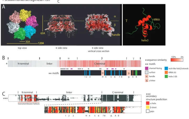
the C-terminal domain (FtsKC) forms the translocation motor involved in chromosome segregation. The general structure and sequence conservation of FtsK is shown in Fig. 1. FtsKNis~200 residues long and poorly conserved at the sequence level. It, however, invariably contains transmembrane helices that tether the protein to the cell membrane specifically at the division septum (Fig. 1) (Dorazi and Dewar, 2000), where it is proposed to interact
Accepted 21 April, 2007. *For correspondence. E-mail francois. [email protected]; [email protected]; Tel. (+33) 561 335 985; Fax (+33) 561 335 886.†Present address: Memorial Sloan-Kettering Cancer Center, 1275 York Ave., New York, NY 10021, USA. © 2007 The Authors
with several other cell division proteins (Di Lallo et al., 2003). Unlike FtsKCwhich forms multimers (see below), the tertiary structure formed by FtsKNis unknown, render-ing a general model for the structure of septum-borne FtsK difficult to draw. An attractive hypothesis is that FtsKN requires other division proteins and/or the process of septum closure itself to oligomerize, which may restrict the formation of active FtsKCmultimers to a certain stage of septum closure, thus controlling FtsK activity temporally. The linker domain (FtsKL) separates FtsKN from FtsKCand extends into the cytoplasm from the divi-sion septum. It shows high sequence and length variability. The longest linkers (~600 aa) are found in proteobacteria, and, in E. coli, it is required for proper
function of FtsKCactivities (Bigot et al., 2004). The longer linkers tend to be rich in proline and glutamine residues, and many adopt coiled coils as predicted secondary struc-tures, suggesting they might participate in the formation of FtsK multimers and/or in interaction with other divisome proteins (not shown; see legend of Fig. 1).
FtsKC, the signature domain of this protein, can be further subdivided into a, b and g subdomains (Yates
et al., 2003). The a and b subdomains form the DNA pump (Massey et al., 2006) while theg subdomain con-trols translocation by recognition of KOPS (see below) motifs in the DNA and interacts with and controls other proteins involved in segregation (i.e. the Xer recombina-tion machine, see below and Yates et al., 2006). The
N-terminal linker C-terminal
*
sequence similarity 100% 0%*
H γL α β γ 1 2 3 H 4 5 6 7 8 γL A B motifs**
recA-like fold β-strands KRKA (A) helix-3 (B) H γL channel-facing surface handle γ-linker motifs
**
α-helix β-sheet gaps secondary structure predicition***
N-terminal linker C-terminal
1 2 3 H 4 5 6 7 8 γL A B
***
Pseudomonas aeruginosa FtsK
C
top view side view side view vertical cross section
30A 120A120A 60A α β handle helix3 KRKA γ
A
B
C
*
*
transmembrane helicesFig. 1. Pseudomonas aeruginosa FtsK domain organization and conservation.
A. Images of the crystal structure of hexameric FtsK from P. aeruginosa (a and b domains) (Massey et al., 2006) and of the NMR structure of theg domain (Sivanathan et al., 2006). The top view shows the six subunits (in unique colours) that form the hexamer. The side views and (B) are colour-coded according to sequence similarity when comparing FtsK across eubacteria with deeper red indicating higher conservation [obtained using ProtSkin (http://www.mcgnmr.ca/ProtSkin)]. It clearly illustrates the cleft between thea and b domains and the conservation of residues lining the central channel (cross-section). The KRKA loop that interacts with XerD and helix-3 that is involved with KOPS recognition are indicated within theg subdomain.
B. A schematic of the general domain organization of FtsK using STRAP (http://www.charite.de/bioinf/strap) superimposed on annotated motifs for P. aeruginosa’s FtsK. Motifs within the C-terminal domain highlight the regions of high- and low-sequence conservation; the residues lining the central channel (upon hexamerization), the RecA foldb-strands and the regulatory domains within g (motifs A and B) are among the highest conserved regions, while the residues on the outer surface of the hexamer, the handle and theg-linker vary considerably.
C. A schematic of an FtsK sequence alignment (Deprez et al., 2005) with secondary structure prediction using. FtsK homologue was selected from aBLASTin all sequenced bacterial genomes with a cut-off at 1.e-100. The three main domains are annotated for P. aeruginosa FtsK. Gaps litter the linker domains (linker andgL), highlighting their variable length. The transmembrane helices of the N-terminal and the a-helices and b-strands of the C-terminal are strongly conserved. The handle region (H) within the C-terminal domain is prevalent only in proteobacteria. Predicted coiled coil structures in the linker domain are restricted to the long linkers and are thus not shown.
FtsK, a literate chromosome segregation machine 1435
© 2007 The Authors
recently solved crystal structure of P. aeruginosa’s FtsK (consisting of only the a and b subdomains) revealed that six FtsKC domains oligomerize to form a ring that can accommodate dsDNA (Massey et al., 2006). The a subdomains form a smaller ring atop a larger b ring (Fig. 1). Note that the crystallized FtsK is truncated of FtsKN, FtsKLand of theg subdomain, and was solved as double head-to-head hexamers that interact via a ‘handle’ domain (Fig. 1A). However, a double hexamer is difficult to reconcile with functional data, in particular because g, which is almost directly linked to b, must contact the DNA and thus be positioned in the vicinity of the central channel. Consistent with this view, the handle domain is not conserved (Fig. 1B and C). Theb subdo-main contains the core RecA-like fold (with Walker P-loop and B motifs) that is common to AAA+ proteins (ATPases Associated with various cellular Activities), and generates the force required for DNA translocation. It is both the conservation of several sequence motifs and distinctb-strand order and structural arrangement within the RecA-like domain (distinct from other P-loop ATPases), as well as the ability to translocate dsDNA that defines the FtsK/SpoIIIE/Tra family of DNA translo-cases (Fig. 1).
FtsK is the fastest known DNA pump, with translocation rates of up to 7 kb s-1 (Saleh et al., 1996; Pease et al., 2005). Contrary to previously described translocases (i.e. Eco124i; Stanley et al., 2006), FtsK does not rotate to track the grooves of dsDNA during translocation, but rather rotates only once per 150 bp translocated (Saleh
et al., 2005). Comparing structures of ATPgS-bound FtsK (in a hexamer) and ADP-bound FtsK monomers suggests a conformational change between the a and b subdo-mains upon ATP hydrolysis that would correspond to a 1.6 bp displacement of DNA (Massey et al., 2006). This displacement would position the DNA helix to contact the same position on the following subunit of the hexamer with only moderate rotation required. Based on these observations, Massey et al. (2006) have proposed a ‘rotary inchworm’ model for translocation in which each subunit of the translocase would hydrolyse ATP sequen-tially around the hexamer (see also Strick and Quessada-Vial, 2006).
At the tail end of FtsKC, the g subdomain forms a winged helix–turn–helix (wHTH) that is attached to theb domain via a flexible linker. wHTH folds are commonly associated with DNA binding, while some participate in protein–protein interactions (Gajiwala and Burley, 2000). The g domain utilizes both functions and acts as a regulatory domain, with loop1 forming an epitope that interacts with the recombinase XerD and helix3 recog-nizing specific DNA motifs, the KOPS (see below) (Ptacin et al., 2006; Sivanathan et al., 2006; Yates et al., 2006).
There are instances of two to three conserved FtsK motor domains (ab domains) occurring within a single ORF (identified from whole genome sequencing). Such arrangement might promote the formation of active motors. Several of these ORF also encode further spe-cialized domains, suggesting that the FtsK motor domains may provide the translocation activity required to assist different processes. Striking examples include FtsK motor domain(s) fused to a phage integrase or a forkhead-associated domain (ORFS SC6A9.34 and Cthe-DRAFT_1197 from Streptomyces coelicolor and
Clostridium thermocellum respectively).
FtsK is part of the divisome
The co-ordinated action of about 15 proteins is neces-sary for E. coli cell division (for review see Goehring and Beckwith, 2005; Vicente et al., 2006). These proteins localize at midcell and assemble into a multiprotein complex termed the septal ring or divisome. Septation then occurs by constriction of the divisome-associated membranes. FtsK is part of the divisome and studies with truncated forms revealed that only FtsKN is essen-tial for septum formation in E. coli; its absence provokes the formation of long cell filaments with no septum con-striction (Draper et al., 1998; Wang and Lutkenhaus, 1998).
FtsK is among the first divisome proteins to localize at midcell and its localization is required for the recruitment of other divisome components (Wang and Lutkenhaus, 1998; Yu et al., 1998a; Chen and Beckwith, 2001). Over-expression of some divisome proteins (FtsQ, FtsN or co-overproduction of FtsZ and FtsQ together with a mutant form of FtsA) partially suppresses the lethality due to a deletion of FtsK, suggesting that it primarily serves to stabilize the divisome prior to septation (Draper et al., 1998; Geissler and Margolin, 2005; Goe-hring et al., 2006). The added fact that neither of the two FtsK homologues in B. subtilis (SpoIIIE and YtpT) is essential (Sharpe and Errington, 1995) further suggests that FtsK does not play a conserved active role during divisome assembly. Nevertheless, overexpression of other cell division proteins in ftsK-deleted cells is not sufficient for normal division in E. coli; suppressed strains still exhibit cell chains (filaments with deep septum constrictions) indicative of a defect in septum closure (Draper et al., 1998; Geissler and Margolin, 2005). SpoIIIE has been proposed to play a role in the late stages of membrane fusion during spore formation in B. subtilis (Sharp and Pogliano, 2003; Liu et al., 2006). FtsK may play an analogous role in E. coli. Although poorly understood, the observations that FtsK interacts genetically with DacA and may interact physi-cally with FtsI, both involved in peptidoglycan synthesis,
may be relevant to this role (Begg et al., 1995; Draper
et al., 1998; Di Lallo et al., 2003).
While not essential for growth, deletion of all or part of FtsKLand of FtsKCalso interferes with septum formation as judged by the appearance of cell filaments and cell chains (Yu et al., 1998b; Recchia et al., 1999; Bigot et al., 2004). These defects cannot be entirely explained by the inactivation of chromosome dimer resolution, indicating that FtsKCand FtsKLboth play a role in cell division (Bigot
et al., 2004).
The early divisome components, including FtsK, are often observed localized at midcell of cells without con-stricted septa (Wang and Lutkenhaus, 1998). This delay between localization of early divisome proteins and septum constriction does not reflect the time required to assemble late divisome proteins. Indeed, all proteins recruited after FtsK assemble simultaneously at the time of septation (Aarsman et al., 2005). This leaves a broad window for action by FtsK towards the end of the cell cycle (Wang et al., 2005). However, chromosome dimer resolu-tion seems to occur late in the cell cycle, concomitantly with septum constriction (Steiner and Kuempel, 1998a). This raises the question of the state of FtsK from its recruitment to midcell to activation of dimer resolution,
and may indicate that FtsK activity is controlled by the late completion of divisome assembly or even by septum constriction.
FtsK sorts sister chromosomes
Segregation of bacterial chromosomes involves multiple processes acting at different stages of the cell cycle on specific chromosome regions (for reviews see Sherratt, 2003; Gitai et al., 2005; Espeli and Boccard, 2006). In
E. coli, FtsK acts in the region where replication
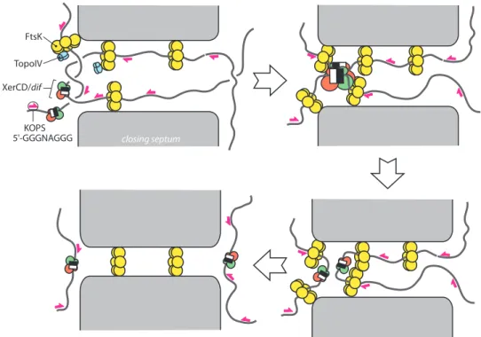
termi-nates (ter) at the last stage of chromosome segregation, which is concomitant with constriction of the division septum (Steiner and Kuempel, 1998a; Steiner et al., 1999). At this stage, two kinds of physical links, intercat-enation links and chromosome dimers, may persist between sister chromosomes. FtsK controls the removal of these links and couples it with cell division (Fig. 2).
Intercatenation links are resolved by topoisomerase IV (Topo IV), a type II topoisomerase composed of two sub-units, ParC and ParE (Adams et al., 1992; Peng and Marians, 1993). The activity of Topo IV is temporally and spatially regulated (Espeli et al., 2003a,b). Active Topo IV is formed preferentially during the last stages of the cell
FtsK KOPS 5'-GGGNAGGG TopoIV XerCD/dif closing septum
Fig. 2. Segregation of the ter region during chromosome dimer resolution. The drawing represents the central part of a dividing cell. The
closing division septum, septum-associated FtsK hexamers, Topo IV, the XerCD/dif complex and some KOPS are indicated. Only FtsKCis represented as hexamers. FtsKN’s and FtsKL’s roles are symbolized by an interaction of FtsKCwith the septum membrane. Top left: sister chromosomes are dimeric and intercatenated and chromosomal DNA is stretched across the septum. FtsK binds to this DNA in an oriented manner by recognizing KOPS and translocates towards XerCD/dif complexes. This process sorts the ter region of sister chromosome to either side of the septum and helps decatenation by Topo IV. Top right: FtsK reaches the XerCD/dif complexes and contacts XerD to induce recombination. Bottom right: the dimer is resolved and FtsK finishes sorting sister chromosomes, allowing septum closure without chromosome damage (bottom left).
FtsK, a literate chromosome segregation machine 1437
© 2007 The Authors
cycle, from termination of replication to cell division. The ParC subunits appear colocalized with the replication machinery. However, it also interacts with FtsKC, stimulat-ing Topo IV activity in vitro. In contrast, the ParE subunits appear distributed in the DNA-free space of the cell. K. Marians and co-workers (Espeli et al., 2003a) have sug-gested that FtsK acts to capture ParC after disassembly of the replisome. Free ParE could then associate with the ParC–FtsK complex to reconstitute active Topo IV. This ensures the spatial and temporal regulation of decatena-tion activity. However, the fact that Topo IV is essential whereas FtsKCis not strongly suggests that decatenation by Topo IV can occur in the absence of FtsK.
In addition to catenation links, the sister chromosome may be dimeric (reviewed in Lesterlin et al., 2004; Fig. 2). In E. coli, chromosome dimers form by homologous recombination between sister chromosomes during repli-cation. This occurs in about 15% of the cells during growth in standard laboratory conditions (Steiner and Kuempel, 1998b; Perals et al., 2000). The dedicated safeguard system, XerCD/dif, consists of two tyrosine recombinases, XerC and XerD, which act at a specific site located in ter,
dif. Dimer resolution depends on FtsKC, which plays at least two distinct role in this process (Fig. 2) (Capiaux
et al., 2002; Yates et al., 2003; Bigot et al., 2004). FtsK
loads onto DNA stretches in the vicinity of the closing septum and translocates DNA towards the duplicated dif sites. This sorts sister chromosomes on either side of the septum and may aid decatenation by Topo IV. Chromo-some mobilization finally allows the formation of a produc-tive recombination synapse between XerCD/dif complexes. This may involved either bringing the two XerCD/dif complexes together in a productive conforma-tion or remodelling a pre-existing synapse to an active conformation. Recombination is then activated by a direct interaction between FtsKCand XerD, which activates XerD catalytic activity (Massey et al., 2004; Yates et al., 2006). This interaction is mediated by the extreme C-terminal subdomain of FtsK, FtsKg (Fig. 1) (Yates et al., 2006). The FtsK-XerCD/dif system may also be directly involved in chromosome decatenation as successive rounds of recombination can remove catenation links in vitro (Ip
et al., 2003). XerCD/dif may also control Topo IV activity as
a preferential region for Topo IV action exists in the imme-diate vicinity of dif (Hojgaard et al., 1999). Surprisingly, this activity depends on XerC and XerD but not on FtsKC. It is thus conceivable that XerCD and Topo IV are parts of a multiprotein complex acting to separate sister ter regions, the formation of which does not strictly depend on FtsK.
FtsK reads the polarity of the chromosome
A key implication of the general model presented in Fig. 2 is that FtsK has to find its way to the dif site. In
vivo data indicate that the loci entrapped in the septum
in the case of a dimer are included in a restricted but rather long part of the chromosome, up to 400 kb around
dif, called the FtsK domain (Corre and Louarn, 2005; C.
Pages and F. Cornet, in preparation). These data strongly suggest two levels of active positioning of the
ter region. The first is global positioning of a large
ter-minal domain close to the septum. This does not require FtsK and is independent of dimer formation. The second involves precise positioning of the XerCD/dif complexes by translocating FtsK. FtsK thus loads onto DNA several kilo base pairs away from the dif site and must translo-cate towards dif to avoid unproductive activity. While it has been known for a long time that dimer resolution requires the correct orientation of the sequences flank-ing dif (Cornet et al., 1996; Kuempel et al., 1996; Corre
et al., 2000; Perals et al., 2000), the demonstration that
this orientation controls FtsK translocation and the iden-tification of the DNA motifs involved are recent discov-eries (Corre and Louarn, 2002; Bigot et al., 2005; Levy
et al., 2005; Pease et al., 2005). FtsK recognizes short
DNA motifs, termed KOPS (FtsK Orienting Polar Sequences), 5′-GGGNAGGG-3′, which are over-represented on the chromosome and strongly biased for their orientation towards dif. This biased distribution of KOPS is conserved in bacteria closely related to E. coli and analyses of other bacterial genomes generally reveal other motifs with KOPS-like distribution, suggest-ing that the control of FtsK by KOPS is conserved in bacteria (Eisen et al., 2000; Levy et al., 2005; Hendrick-son and Lawrence, 2006). Indeed, this is reminiscent of the other skewed sequence whose role has been described thus far, the Chi motif. Chi are recognized by RecBCD complexes translocating from a dsDNA end and switch RecBCD activity from DNA degradation to the creation of RecA-associated single stranded loops that are used for strand exchange during homologous recombination (Taylor et al., 1985; Dixon and Kowalc-zykowski, 1993; Dohoney and Gelles, 2001; Spies et al., 2003). Motifs unrelated to the E. coli Chi motif but with Chi activity have been reported in other bacteria (El Karoui et al., 1998; Sourice et al., 1998; El Karoui et al., 1999). The control of DNA trafficking by short motifs with biased distribution thus appears as a general feature in bacteria. Both KOPS and Chi skews contribute to the global replichores orientation, which accounts for a general organization of bacterial chromosomes following the replication origin to dif axis and now appears as a major player in chromosome structure and dynamics.
Although FtsK may load on any piece of dsDNA tested so far, KOPS are preferred sites of loading and are thought to orient translocation at this step (Fig. 3) (Bigot
et al., 2006). KOPS also block and eventually reverse
the direction of translocation when encountered from
their 3′ end (Fig. 3) (Bigot et al., 2005; Levy et al., 2005). This blockage is not total in in vivo and in vitro assays, strongly suggesting that KOPS recognition by FtsK is stochastic (Bigot et al., 2005). Indeed, it is estimated that FtsK50C, the truncated version of FtsK used in vitro, stops only in 60% of the case when it encounters a single KOPS from its 3′ end. This probability may be close to optimal for FtsK to rapidly locate dif, giving that the orientation bias of the KOPS on the chromosome is not total (Levy et al., 2005). KOPS are recognized by the winged-helix g subdomain (Ptacin et al., 2006; Sivanathan et al., 2006). Notably, this subdomain also contains the KRKA motif that interacts with XerD and is connected to theab motor by a flexible linker (Fig. 1). g may thus binds KOPS-containing DNA to orient loading of the ab motor. Translocating with g at the front end of the motor then positions this subdomain to interact with KOPS or a XerD-bound dif site.
Acknowledgements
We thank Jean-Michel Louarn, David Sherratt and Marcelo Nollmann for helpful discussions. S.B. received a fellowship from the Ministère de la Recherche. Research in F.C. and F.-X.B. groups is funded by the Centre National de la Recher-che Scientifique, the Agence Nationale de la ReRecher-cherRecher-che and the Ministère de la Recherche. Research in the Sherratt group (V.S.) is funded by the Welcome trust.
References
Aarsman, M.E., Piette, A., Fraipont, C., Vinkenvleugel, T.M., Nguyen-Disteche, M., and den Blaauwen, T. (2005) Matu-ration of the Escherichia coli divisome occurs in two steps. Mol Microbiol 55: 1631–1645.
Adams, D.E., Shekhtman, E.M., Zechiedrich, E.L., Schmid, M.B., and Cozzarelli, N.R. (1992) The role of topoi-somerase IV in partitioning bacterial replicons and the structure of catenated intermediates in DNA replication. Cell 71: 277–288.
Aussel, L., Barre, F.X., Aroyo, M., Stasiak, A., Stasiak, A.Z., and Sherratt, D. (2002) FtsK is a DNA motor protein that activates chromosome dimer resolution by switching the catalytic state of the XerC and XerD recombinases. Cell
108: 195–205.
Bath, J., Wu, L.J., Errington, J., and Wang, J.C. (2000) Role of Bacillus subtilis SpoIIIE in DNA transport across the mother cell-prespore division septum. Science 290: 995– 997.
Begg, K.J., Dewar, S.J., and Donachie, W.D. (1995) A new Escherichia coli cell division gene, ftsK. J Bacteriol 177: 6211–6222.
Bigot, S., Corre, J., Louarn, J.M., Cornet, F., and Barre, F.X. (2004) FtsK activities in Xer recombination, DNA mobiliza-tion and cell division involve overlapping and separate domains of the protein. Mol Microbiol 54: 876–886. Bigot, S., Saleh, O.A., Lesterlin, C., Pages, C., El Karoui, M.,
Dennis, C., et al. (2005) KOPS: DNA motifs that control E. coli chromosome segregation by orienting the FtsK translocase. EMBO J 24: 3770–3780.
Bigot, S., Saleh, O.A., Cornet, F., Allemand, J.F., and Barre, F.X. (2006) Oriented loading of FtsK on KOPS. Nat Struct Mol Biol 13: 1026–1028.
Capiaux, H., Lesterlin, C., Perals, K., Louarn, J.M., and Cornet, F. (2002) A dual role for the FtsK protein in Escheri-chia coli chromosome segregation. EMBO Rep 3: 532–536. Chen, J.C., and Beckwith, J. (2001) FtsQ, FtsL and FtsI require FtsK, but not FtsN, for co-localization with FtsZ during Escherichia coli cell division. Mol Microbiol 42: 395– 413.
Cornet, F., Louarn, J., Patte, J., and Louarn, J.M. (1996) Restriction of the activity of the recombination site dif to a small zone of the Escherichia coli chromosome. Genes Dev 10: 1152–1161.
Corre, J., and Louarn, J.M. (2002) Evidence from terminal recombination gradients that FtsK uses replichore polarity to control chromosome terminus positioning at division in Escherichia coli. J Bacteriol 184: 3801–3807.
Corre, J., and Louarn, J.M. (2005) Extent of the activity domain and possible roles of FtsK in the Escherichia coli chromosome terminus. Mol Microbiol 56: 1539–1548. Corre, J., Patte, J., and Louarn, J.M. (2000) Prophage
lambda induces terminal recombination in Escherichia coli by inhibiting chromosome dimer resolution. An orientation-dependent cis-effect lending support to bipolarization of the terminus. Genetics 154: 39–48.
Deprez, C., Lloubes, R., Gavioli, M., Marion, D., Guerlesquin, F., and Blanchard, L. (2005) Solution structure of the E. coli TolA C-terminal domain reveals conformational changes upon binding to the phage g3p N-terminal domain. J Mol Biol 346: 1047–1057.
+
α β 20 bp γ a b+
c+
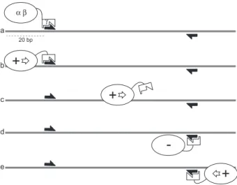
d -eFig. 3. Model for KOPS-directed FtsK loading and translocation
reversal. For clarity, only one FtsK monomer is represented with theab motor and the g subdomains indicated. FtsKNand FtsKLare omitted. The status of the FtsK translocation motor is indicated (+: active; –: inactive). The grey line is a DNA duplex with KOPS indicated by the arrows. (a) FtsK loads onto the DNA by recognizing a KOPS and forms an active hexamer (b). This requires at least 20 bp of DNA upstream the KOPS (Bigot et al., 2006). (c) Upon recognition of a non-permissive KOPS, FtsK stops and the hexamer eventually disassembles (d). (e) The same or another motor then reassembles in the permissive orientation.
FtsK, a literate chromosome segregation machine 1439
© 2007 The Authors
Di Lallo, G., Fagioli, M., Barionovi, D., Ghelardini, P., and Paolozzi, L. (2003) Use of a two-hybrid assay to study the assembly of a complex multicomponent protein machinery: bacterial septosome differentiation. Microbiology 149: 3353–3359.
Dixon, D.A., and Kowalczykowski, S.C. (1993) The recombi-nation hotspot chi is a regulatory sequence that acts by attenuating the nuclease activity of the E. coli RecBCD enzyme. Cell 73: 87–96.
Dohoney, K.M., and Gelles, J. (2001) Chi-sequence recogni-tion and DNA translocarecogni-tion by single RecBCD helicase/ nuclease molecules. Nature 409: 370–374.
Dorazi, R., and Dewar, S.J. (2000) Membrane topology of the N-terminus of the Escherichia coli FtsK division protein. FEBS Lett 478: 13–18.
Draper, G.C., McLennan, N., Begg, K., Masters, M., and Donachie, W.D. (1998) Only the N-terminal domain of FtsK functions in cell division. J Bacteriol 180: 4621–4627. Eisen, J.A., Heidelberg, J.F., White, O., and Salzberg, S.L.
(2000) Evidence for symmetric chromosomal inversions around the replication origin in bacteria. Genome Biol 1: RESEARCH0011.
El Karoui, M., Ehrlich, D., and Gruss, A. (1998) Identification of the lactococcal exonuclease/recombinase and its modu-lation by the putative Chi sequence. Proc Natl Acad Sci USA 95: 626–631.
El Karoui, M., Biaudet, V., Schbath, S., and Gruss, A. (1999) Characteristics of Chi distribution on different bacterial genomes. Res Microbiol 150: 579–587.
Espeli, O., and Boccard, F. (2006) Organization of the Escheri-chia coli chromosome into macrodomains and its possible functional implications. J Struct Biol 156: 304–310. Espeli, O., Levine, C., Hassing, H., and Marians, K.J. (2003a)
Temporal regulation of topoisomerase IV activity in E. coli. Mol Cell 11: 189–201.
Espeli, O., Lee, C., and Marians, K.J. (2003b) A physical and functional interaction between Escherichia coli FtsK and topoisomerase IV. J Biol Chem 278: 44639–44644. Gajiwala, K.S., and Burley, S.K. (2000) Winged helix
proteins. Curr Opin Struct Biol 10: 110–116.
Geissler, B., and Margolin, W. (2005) Evidence for functional overlap among multiple bacterial cell division proteins: compensating for the loss of FtsK. Mol Microbiol 58: 596– 612.
Gitai, Z., Thanbichler, M., and Shapiro, L. (2005) The cho-reographed dynamics of bacterial chromosomes. Trends Microbiol 13: 221–228.
Goehring, N.W., and Beckwith, J. (2005) Diverse paths to midcell: assembly of the bacterial cell division machinery. Curr Biol 15: R514–R526.
Goehring, N.W., Robichon, C., and Beckwith, J. (2006) A role for the non-essential N-terminus of FtsN in divisome assembly. J Bacteriol 189: 646–649.
Hanson, P.I., and Whiteheart, S.W. (2005) AAA+ proteins: have engine, will work. Nat Rev Mol Cell Biol 6: 519–529. Hendrickson, H., and Lawrence, J.G. (2006) Selection for chromosome architecture in bacteria. J Mol Evol 62: 615– 629.
Hojgaard, A., Szerlong, H., Tabor, C., and Kuempel, P. (1999) Norfloxacin-induced DNA cleavage occurs at the dif resolvase locus in Escherichia coli and is the result of
interaction with topoisomerase IV. Mol Microbiol 33: 1027– 1036.
Ip, S.C., Bregu, M., Barre, F.X., and Sherratt, D.J. (2003) Decatenation of DNA circles by FtsK-dependent Xer site-specific recombination. EMBO J 22: 6399–6407.
Iyer, L.M., Makarova, K.S., Koonin, E.V., and Aravind, L. (2004) Comparative genomics of the FtsK-HerA superfam-ily of pumping ATPases: implications for the origins of chromosome segregation, cell division and viral capsid packaging. Nucleic Acids Res 32: 5260–5279.
Kataoka, M., Seki, T., and Yoshida, T. (1991) Five genes involved in self-transmission of pSN22, a Streptomyces plasmid. J Bacteriol 173: 4220–4228.
Kendall, K.J., and Cohen, S.N. (1987) Plasmid transfer in Streptomyces lividans: identification of a kil-kor system associated with the transfer region of pIJ101. J Bacteriol
169: 4177–4183.
Kuempel, P., Hogaard, A., Nielsen, M., Nagappan, O., and Tecklenburg, M. (1996) Use of a transposon (Tndif) to obtain suppressing and nonsuppressing insertions of the dif resolvase site of Escherichia coli. Genes Dev 10: 1162– 1171.
Lesterlin, C., Barre, F.X., and Cornet, F. (2004) Genetic recombination and the cell cycle: what we have learned from chromosome dimers. Mol Microbiol 54: 1151–1160. Levy, O., Ptacin, J.L., Pease, P.J., Gore, J., Eisen, M.B.,
Bustamante, C., and Cozzarelli, N.R. (2005) Identification of oligonucleotide sequences that direct the movement of the Escherichia coli FtsK translocase. Proc Natl Acad Sci USA 102: 17618–17623.
Liu, N.J., Dutton, R.J., and Pogliano, K. (2006) Evidence that the SpoIIIE DNA translocase participates in membrane fusion during cytokinesis and engulfment. Mol Microbiol
59: 1097–1113.
Massey, T.H., Aussel, L., Barre, F.X., and Sherratt, D.J. (2004) Asymmetric activation of Xer site-specific recombi-nation by FtsK. EMBO Rep 5: 399–404.
Massey, T.H., Mercogliano, C.P., Yates, J., Sherratt, D.J., and Lowe, J. (2006) Double-stranded DNA translocation: structure and mechanism of hexameric FtsK. Mol Cell 23: 457–469.
Pease, P.J., Levy, O., Cost, G.J., Gore, J., Ptacin, J.L., Sher-ratt, D., et al. (2005) Sequence-directed DNA translocation by purified FtsK. Science 307: 586–590.
Peng, H., and Marians, K.J. (1993) Decatenation activity of topoisomerase IV during oriC and pBR322 DNA replication in vitro. Proc Natl Acad Sci USA 90: 8571–8575.
Perals, K., Cornet, F., Merlet, Y., Delon, I., and Louarn, J.M. (2000) Functional polarization of the Escherichia coli chro-mosome terminus: the dif site acts in chrochro-mosome dimer resolution only when located between long stretches of opposite polarity. Mol Microbiol 36: 33–43.
Possoz, C., Ribard, C., Gagnat, J., Pernodet, J.L., and Guerineau, M. (2001) The integrative element pSAM2 from Streptomyces: kinetics and mode of conjugal transfer. Mol Microbiol 42: 159–166.
Ptacin, J.L., Nollmann, M., Bustamante, C., and Cozzarelli, N.R. (2006) Identification of the FtsK sequence-recognition domain. Nat Struct Mol Biol 13: 1023–1025.
Recchia, G.D., Aroyo, M., Wolf, D., Blakely, G., and Sher-ratt, D.J. (1999) FtsK-dependent and -independent
ways of Xer site-specific recombination. EMBO J 18: 5724–5734.
Saleh, O.A., Perals, C., Barre, F.X., and Allemand, J.F. (2004) Fast, DNA-sequence independent translocation by FtsK in a single-molecule experiment. EMBO J 23: 2430– 2439.
Saleh, O.A., Bigot, S., Barre, F.X., and Allemand, J.F. (2005) Analysis of DNA supercoil induction by FtsK indicates translocation without groove-tracking. Nat Struct Mol Biol
12: 436–440.
Saleh, A.Z., Yamanaka, K., Niki, H., Ogura, T., Yamazoe, M., and Hiraga, S. (1996) Carboxyl terminal region of the MukB protein in Escherichia coli is essential for DNA binding activity. FEMS Microbiol Lett 143: 211–216.
Sharp, M.D., and Pogliano, K. (2003) The membrane domain of SpoIIIE is required for membrane fusion during Bacillus subtilis sporulation. J Bacteriol 185: 2005–2008.
Sharpe, M.E., and Errington, J. (1995) Postseptational chro-mosome partitioning in bacteria. Proc Natl Acad Sci USA
92: 8630–8634.
Sherratt, D.J. (2003) Bacterial chromosome dynamics. Science 301: 780–785.
Sivanathan, V., Allen, M.D., de Bekker, C., Baker, R., Arciszewska, L.K., Freund, S.M., et al. (2006) The FtsK gamma domain directs oriented DNA translocation by inter-acting with KOPS. Nat Struct Mol Biol 13: 965–972. Smokvina, T., Boccard, F., Pernodet, J.L., Friedmann, A., and
Guerineau, M. (1991) Functional analysis of the Streptomy-ces ambofaciens element pSAM2. Plasmid 25: 40–52. Sourice, S., Biaudet, V., El Karoui, M., Ehrlich, S.D., and
Gruss, A. (1998) Identification of the Chi site of Haemophi-lus influenzae as several sequences related to the Escheri-chia coli Chi site. Mol Microbiol 27: 1021–1029.
Spies, M., Bianco, P.R., Dillingham, M.S., Handa, N., Baskin, R.J., and Kowalczykowski, S.C. (2003) A molecu-lar throttle: the recombination hotspot chi controls DNA translocation by the RecBCD helicase. Cell 114: 647– 654.
Stanley, L.K., Seidel, R., van der Scheer, C., Dekker, N.H., Szczelkun, M.D., and Dekker, C. (2006) When a helicase is not a helicase: dsDNA tracking by the motor protein EcoR124I. EMBO J 25: 2230–2239.
Steiner, W.W., and Kuempel, P.L. (1998a) Cell division is
required for resolution of dimer chromosomes at the dif locus of Escherichia coli. Mol Microbiol 27: 257–268. Steiner, W.W., and Kuempel, P.L. (1998b) Sister chromatid
exchange frequencies in Escherichia coli analyzed by recombination at the dif resolvase site. J Bacteriol 180: 6269–6275.
Steiner, W., Liu, G., Donachie, W.D., and Kuempel, P. (1999) The cytoplasmic domain of FtsK protein is required for resolution of chromosome dimers. Mol Microbiol 31: 579– 583.
Strick, T.R., and Quessada-Vial, A. (2006) FtsK: a groovy helicase. Nat Struct Mol Biol 13: 948–950.
Taylor, A.F., Schultz, D.W., Ponticelli, A.S., and Smith, G.R. (1985) RecBC enzyme nicking at Chi sites during DNA unwinding: location and orientation-dependence of the cutting. Cell 41: 153–163.
Vicente, M., Rico, A.I., Martinez-Arteaga, R., and Mingo-rance, J. (2006) Septum enlightenment: assembly of bac-terial division proteins. J Bacteriol 188: 19–27.
Wang, L., and Lutkenhaus, J. (1998) FtsK is an essential cell division protein that is localized to the septum and induced as part of the SOS response. Mol Microbiol 29: 731–740.
Wang, X., Possoz, C., and Sherratt, D.J. (2005) Dancing around the divisome: asymmetric chromosome segrega-tion in Escherichia coli. Genes Dev 19: 2367–2377. Wu, L.J., and Errington, J. (1994) Bacillus subtilis spoIIIE
protein required for DNA segregation during asymmetric cell division. Science 264: 572–575.
Yates, J., Aroyo, M., Sherratt, D.J., and Barre, F.X. (2003) Species specificity in the activation of Xer recombination at dif by FtsK. Mol Microbiol 49: 241–249.
Yates, J., Zhekov, I., Baker, R., Eklund, B., Sherratt, D.J., and Arciszewska, L.K. (2006) Dissection of a functional inter-action between the DNA translocase, FtsK, and the XerD recombinase. Mol Microbiol 59: 1754–1766.
Yu, X.C., Tran, A.H., Sun, Q., and Margolin, W. (1998a) Localization of cell division protein FtsK to the Escherichia coli septum and identification of a potential N-terminal tar-geting domain. J Bacteriol 180: 1296–1304.
Yu, X.C., Weihe, E.K., and Margolin, W. (1998b) Role of the C terminus of FtsK in Escherichia coli chromosome segregation. J Bacteriol 180: 6424–6428.
FtsK, a literate chromosome segregation machine 1441
© 2007 The Authors