HAL Id: hal-02326935
https://hal-amu.archives-ouvertes.fr/hal-02326935
Submitted on 22 Oct 2019HAL is a multi-disciplinary open access archive for the deposit and dissemination of sci-entific research documents, whether they are pub-lished or not. The documents may come from teaching and research institutions in France or abroad, or from public or private research centers.
L’archive ouverte pluridisciplinaire HAL, est destinée au dépôt et à la diffusion de documents scientifiques de niveau recherche, publiés ou non, émanant des établissements d’enseignement et de recherche français ou étrangers, des laboratoires publics ou privés.
Photodetachment of Deprotonated Aromatic Amino
Acids: Stability of the dehydrogenated radical depends
on deprotonation site
Jennifer Anna Noble, Juan Aranguren-Abate, Claude Dedonder, Christophe
Jouvet, Gustavo Pino
To cite this version:
Jennifer Anna Noble, Juan Aranguren-Abate, Claude Dedonder, Christophe Jouvet, Gustavo Pino. Photodetachment of Deprotonated Aromatic Amino Acids: Stability of the dehydrogenated radical depends on deprotonation site. Physical Chemistry Chemical Physics, Royal Society of Chemistry, 2019, �10.1039/c9cp04302k�. �hal-02326935�
Photodetachment of Deprotonated Aromatic Amino Acids: Stability of the
dehydrogenated radical depends on deprotonation site
Jennifer Anna Noble*, Juan P. Aranguren‐Abate, Claude Dedonder, Christophe Jouvet, Gustavo A. Pino
a‐ PIIM: UMR‐CNRS 7345, Aix‐Marseille Univ. Avenue Escadrille Normandie‐Niémen 13397 Marseille cedex 20, France. jennifer.noble@univ‐amu.fr
b‐ INFIQC: Instituto de Investigaciones en Fisicoquímica de Córdoba (CONICET – UNC) ‐ Haya de la Torre y Medina Allende, Ciudad Universitaria, X5000HUA Córdoba, Argentina. c‐ Departamento de Fisicoquímica, Facultad de CienciasQuímicas– Universidad Nacional de Córdoba – Haya de la Torre y Medina Allende, Ciudad Universitaria, X5000HUA Córdoba, Argentina. d‐ Centro Láser de Ciencias Moleculares ‐ Universidad Nacional de Córdoba ‐ Haya de la Torre s/n, Pabellón Argentina,Ciudad Universitaria, X5000HUA Córdoba, Argentina.
Abstract
While aromatic amino acids in their deprotonated form have been well characterized by IR and photoelectron spectroscopies, no information is available on the neutral dehydrogenated protonated radicals and, in particular, on their stability when the deprotonation site is changed. This is investigated by observing the neutral fragment issued from either simple photodetachment or dissociative photodetachment of the deprotonated aromatic amino acids phenylalanine, tyrosine, and tryptophan. We show that the dehydrogenated radicals of aromatic amino acids produced upon photodetachment of molecules deprotonated on the carbonyl group dissociate without barrier, leading to the formation of CO2 and a radical amine. However, when the system is deprotonated on
functional groups located on the chromophore, the radicals produced by photodetachment are stable, indicating the important photostabilizing role played by functional groups.
Introduction
Though seemingly simple species, fundamental questions regarding the gas‐phase structure of deprotonated amino acids and, in particular, the neutral radicals issued from their electron detachment remain unanswered to date. The stability of small dehydrogenated acid radicals has been studied through coincidence techniques1. In particular, it has been shown that the simplest, acetyloxyl, loses CO2 upon photodetachment, with a small barrier (0.1eV)calculated for CO2 loss2.
The most favorable protonation or deprotonation site in aminoacids is a recurring question in experiments involving electrospray ionization (ESI), addressed by means of IRMPD spectroscopy 34 or photoelectron spectroscopy5. The photodetachment spectrum of deprotonated tryptophan[Trp‐H]‐ has been studied at room temperature by Compagnon et al.6. A threshold at 315nm (3.94eV, 31750cm‐1) was observed and assigned to the carboxylate anion.The photoelectron spectrum obtained by Tian etal.5demonstrated the competition between two deprotonation sites in tyrosine, leading to either the carboxylate or phenoxide anions. More recently, it has been shown that the preferred protonation or deprotonation site is totally dependent on the (uncontrollable) ESI conditions7. More dramatically, it has been shown, in the case of p‐hydroxybenzoic acid produced by ESI, that populations of the thermodynamically less favored gaseous carboxylate anionor the thermodynamically more favored gaseous phenoxide anion can be varied back and forth by changing the probe position, capillary voltage, desolvation‐gas temperature, sample infusion flow rate, and cone voltage8. From these works it can be concluded that there is no clear relationship between the observation of a given isomer and its relative stability; thus this topic will not be discussed in this paper.
The collision induced dissociation (CID)of [Trp‐H]‐has been studied910 and the observed fragments derive mainly from the fragmentation of the peptide side chain.
In a recent paper11 we have shown that in deprotonated naphthoate anions there is a competition between ionic fragmentation in the electronically excited anion and electron detachment. This is not observed in benzoate since the excited states of the deprotonatedbenzoate anion are higher than those of naphthoate and lie above the adiabatic detachment energy (ADE).Once above the photodetachment energy, one can observe the simple photodetachment of the anion leaving the stable radical, but at higher energies the radical loses CO2 through an exit barrier along the C‐CO2
bond which has been measured at 0.5eV11.
Recently, the indolide anion has been characterized and its adiabatic detachment energy (ADE) = 2.43 eV was measured through photoelectron spectroscopy12. Since the excited states of indole are also low in energy compared to those in phenol or benzene, one might expect to see competition between ionic fragmentation and photodetachment, as for naphthoate.
. How does the stability of the dehydrogenated (deprotonated, then electron detached) radical change with the deprotonated site?
. Is there a competition between photofragmentation of the anion and its photodetachment? . Is there a fragmentation in the radical after photodetachment and, if so, is there a barrier to the fragmentation? . What is the Influence of the amino acid side chain on the electron binding energy of the deprotonated chromophore?
Experimental
The experimental setup used for cold ion photofragmentation spectroscopy has been described in previous publications131415and has recently been modified to detect negative ions and neutral particles11.Deprotonated phenylalanine, tyrosine, and tryptophan were produced in the electrospray ionization (ESI) source by injecting a solution of amino acid at 10‐4 M in 5:1 mixture of methanol:water.The ions produced in the ESI are injected into the cryogenic ion trap just after a helium pulse has been introduced. The ions are stored in the cold trap for a few tens of ms, the time necessary for cooling to c.a. 30 K and subsequent reduction of the pressure in the trap. The ions are then extracted and accelerated at 3.0 kV. The voltage of the extracting electrode of the trap and the accelerating grid are adjusted to fulfill the Wiley McLaren focusing conditions16. After the accelerating grid, the ions enter the Gauss tube set at the accelerating grid potential and, once they are inside, the tube is switched to ground. The ions then travel in the field free region of the time of flight (TOF) mass spectrometer with a kinetic energy due to the accelerating voltage and they are referenced to the ground potential. After 1m of flight, they enter the post accelerating/decelerating “box”. The ions (or neutrals) are detected after the “box” with a MCP detector17.
The laser used for photodissociation or photodetachment of the ions is a tunable OPO laser (EKSPLA) (10 Hz repetition rate, 10 ns pulse width, and a spectral resolution of ~ 10 cm‐1). The laser can interact with the ions in two different parts of the setup (see supplementary information): either in the ion trap, which allows monitoring of the photodissociation process leading to a daughter ion and a neutral fragment by extracting the ions after the laser shot, or in the Gauss tube. In the latter case, since the parent ions have been accelerated, the neutral parent radicals produced after photodetachment and/or the neutral daughter fragments can be detected by the MCP. The intact parent radical will arrive at the same time as the anion precursor and the signal will be as narrow as the anion signal. In contrast, the neutral fragments will travel with the kinetic energy of the parent ion plus the kinetic energy (positive or negative) released in the dissociative process, so that they will
be observed at the same time‐of‐flight as the parent but exhibiting peak broadening due to the kinetic energy release.
To discriminate between the parent anion and the parent neutral photodetached radical, two methods can be used:
a) Decelerating the anions by applying high voltages (1.2 kV) in the accelerating/decelerating “box” (when a negative voltage is applied, the ions are decelerated in the box and will travel slower than the neutrals, thus arriving at the detector after the neutrals).
b) Leaving the voltage on the Gauss tube in order to decelerate the anions at the output of the tube and prevent them reaching the detector, while the neutrals are not perturbed. This method minimizes the time and the distance travelled by the parent ions and thus minimizes the background signal from neutral fragments produced by collisions of the parent ions with the residual gas.
Calculations
Simple calculations using the density functional theory (DFT) with the B3LYP functional as implemented in the Turbomole software18 with the aug‐cc‐pVTZbasis set19 were performed to determine the ADE and vertical detachment energy (VDE)as well as the dissociation energy of the photodetached radical.
The ground states of the neutral and the deprotonated and protonated ions20 have previously been calculated in the case of Trp. For the deprotonated anion [Trp‐H]‐, similar calculations have been performed by Compagnon et al.6 and the most stable structure has been determined to be the one in which the carboxylate lies above the indole plane. Similar starting geometries have been used in the case of [Phe‐H]‐ and [TyrH]‐ as the starting point of our calculations (see S.I. for the xyz coordinate of optimized structure).
Results
a) Photodissociation in the ion trap None of the deprotonated aromatic amino acids led to the production of any ionic fragment when the interaction with the laser took place in the ion trap, indicating that there are no optically accessible excited states below the photodetachment threshold, in contrast with the case of naphthoate (Pino et al. 2019).
b) Photodetachment in the Gauss tube [Phe‐H]‐ anion
As presented in the experimental section, intact radicals appear as narrow peaks while fragments appear as broadened peaks. For deprotonated phenylalanine [Phe‐H]‐, only two broad peaks are observed in the TOF mass spectra of neutrals, without any signature of a narrow component, indicating that the phenylalanyl radical is unstable above the photodetachment threshold (Figure1). -1.0 -0.5 0.0 0.5 1.0 0.000 0.005 ne ut ra l sig n a l ne utr a l s ig n a l 235 nm/5.28 eV -1.0 -0.5 0.0 0.5 1.0 0.000 0.005 0.010 295 nm/4.20 eV -1.0 -0.5 0.0 0.5 1.0 0.000 0.005 TOF (µs) 320 nm/3.87 eV -1.0 -0.5 0.0 0.5 1.0 0.000 0.001 0.002 TOF (µs) 345 nm/3.59 eV Figure 1. Time of Flight peak profiles obtained at different excitation energies. The profiles have been symmetrized (see supplementary information) and are fitted with two combinations of erf functions with different widths for the fragments. The peak profiles were symmetrized and fitted assuming two combinations of erf functions for two fragments (see supplementary information for the fitting procedure).The mean widths of the two broad peaks (140 ± 10 ns and 405 ± 35 ns) remain unchanged as a function of the excitation energy(Figure 1 and supplementary information). The ratio of the widths of the two broad peaks should reflect the mass ratio of the two neutral fragments. If we assume that the dissociation channel corresponds to CO2 loss, as in aryl acids11, the ratio of the fragments masses is 0.37. The ratio
Considering the widths of the peaks and the geometry of the TOF mass spectrometer, we can extract the kinetic energy released in the dissociation process, which is 0.21 ± 0.02 eV.(See S.I.)
Since the phenylalanyl radical fragments upon photodetachment, the photodetachment and photofragmentation thresholds are the same. The thresholds were determined to be 3.54 ± 0.02 eV (350±2 nm)by scanning the laser while monitoring the intensity of the neutral fragment peak at mass 120 (Figure 2). 230 240 250 260 270 280 290 300 310 320 330 340 350 360 0.0 0.1 0.2 0.3 0.4 0.5 3.6 3.8 4 4.2 4.4 4.6 4.8 5 5.2 5.4
Excitation energy (eV)
n eut ral s igna l Excitation wavelength (nm) Neutral fragment at mass 120 CO2loss from [Phe‐H]● Figure 2.Excitation spectrum recorded while detecting the neutral fragment at mass 120 [Phe‐H‐ CO2]●. [Tyr‐H]‐ anion
The TOF mass spectra corresponding to neutrals produced after photodetachment of the anion, either intact tyrosyl radicals appearing as a narrow peak or its fragments (broadened peaks), were recorded at different excitation energies (Figure 3).
-1.5 -1.0 -0.5 0.0 0.5 1.0 0.000 0.005 225 nm/5.51 eV -1.0 -0.5 0.0 0.5 1.0 1.5 0.000 0.005 0.010 285 nm/4.35 eV -1.5 -1.0 -0.5 0.0 0.5 1.0 0.000 0.005 0.010 0.015 neu tr a l signa l neut ra l signa l TOF (µs) 315 nm/3.94 eV -1.0 -0.5 0.0 0.5 1.0 1.5 0.000 0.005 TOF (µs) 365 nm/3.40 eV Figure 3. Time of Flight peak profiles obtained at different excitation energies for [Tyr‐H]‐. The profiles have been symmetrized and are fitted with a sum of a Gaussian function for the narrow peak (tyrosil radical) and a combination of two erf functions of different widths for the fragment peaks (see supplementary information for the symmetrization and fitting procedure).
The intensity of the narrow peak decreases relative to that of the broad peaks as the energy increases, indicating that the fragmentation channel yield increases. The peak profiles were symmetrized and fitted assuming two combinations of erf functions for the two fragments and a Gaussian function for the tyrosyl narrow peak (see supplementary information for the fitting
procedure). The width of the narrow peak is the same as the width of the parent anion peak, i.e. 20
ns. As for phenylalanyl, the widths of the broad peaks remain unchanged as a function of the excitation energy. In this case, the mean widths of the broad peaks are (135 ± 15) ns and (480 ± 30) ns, the ratio being (0.28 ± 0.05), in agreement with the mass ratio of 0.32 obtained if we assume that the dissociation channel corresponds to CO2 loss, as for [Phe‐H]‐.From the widths of the fragment
peaks, we can then extract the kinetic energy released in the dissociation process, which is 0.21 ± 0.04 eV.
The photodetachment and fragmentation thresholds were determined by scanning the laser while monitoring the intensity of the narrow and the broad part of the peak profile, respectively. The photodetachment threshold for [Tyr‐H]‐(Figure 4) is observed at 2.58 ± 0.03 eV (480 ± 5 nm) and corresponds to the ADE observed by Tian et al. for the TyrO‐ anion. The fragmentation signal rapidly increases above 3.54 ± 0.05 eV, which corresponds to the experimental ADE for the TyrCO2‐ anion
observed by Tian et al., and there is also a weak fragmentation signal increasing slowly from 3.10 ± 0.05 eV to 3.54 eV. 240 260 280 300 320 340 360 380 400 420 440 460 480 500 0.000 0.005 0.010 0.015 0.020 0.025 0.030 0.035 0.040 0.045 2.5 3 3.5 4 4.5 5 5.5
Excitation energy (eV)
ne utral s ig nal Excitation wavelenth (nm)
narrow peak (tyrosyl radical) broad peak(CO
2 loss fragment at mass 136)
Figure 4. Excitation spectra recorded while scanning the laser with the detection set either on the tyrosyl radical (narrow peak, red trace) or on the fragment at mass 136 (blue trace). [Trp‐H]‐ anion
The TOF mass spectra corresponding to neutrals due to the photodetachment of the anion, either intact tryptophanyl radicals or its fragments, were recorded at different excitation energies (Figure 5). As in the case of [Tyr‐H]‐, at low excitation energies (< 3.7 eV), only the intact parent radical is observed as a narrow peak. At higher excitation energies (> 3.7 eV), the mass spectra show three components corresponding to tryptophanyl radicals (narrow peak) and two fragments (broad peaks).
-1.0 -0.5 0.0 0.5 1.0 -0.01 0.00 0.01 0.02 0.03 0.04 0.05 0.06 0.07 neutral signa l 230 nm/5.4 eV -1.0 -0.5 0.0 0.5 1.0 0.000 0.005 0.010 0.015 0.020 0.025 0.030 0.035 290 nm/4.3 eV -1.0 -0.5 0.0 0.5 1.0 0.000 0.005 0.010 0.015 0.020 0.025 330 nm/3.75 eV neutral sig nal TOF (µs) -1.0 -0.5 0.0 0.5 1.0 0.000 0.002 0.004 0.006 0.008 0.010 0.012 380 nm/3.3 eV TOF (µs) Figure5.Time of Flight peak profiles obtained at different excitation energies for [Trp‐H]‐. The profiles
have been symmetrized and are fitted with a sum of a Gaussian function for the narrow peak (tryptophanyl radical) and two combinations of erf functions of different widths for the wider fragment peaks (see supplementary information for the symmetrization and fitting procedure).
The same fitting procedure as for [Tyr‐H]● and its fragments has been applied to the peak profiles: symmetrization and fitting with two combinations of erf functions for the two fragments and a Gaussian function for the tryptophanyl narrow peak, the width of which is the same as the width of the parent anion peak, i.e. 20 ns. As can be seen in Figure 5, the fragmentation channel yield increases as the excitation energy increases. From the fitting, we extract the mean widths of the two broad peaks (137 ± 15 ns and 465 ± 60 ns), which remain unchanged as a function of the excitation energy. If we assume that the dissociation channel corresponds to CO2 loss, as for phenylalanyl and
tyrosyl, the ratio of the widths of the two broad peaks, 0.29 ± 0.07, reflects the mass ratio of the two neutral fragments CO2 and C10H11N2,i.e.is 0.27. Considering the widths of the peaks and the geometry
of the TOF mass spectrometer, we can extract the kinetic energy released in the dissociation process, which is (0.19±0.02) eV. (See S.I.4)
The photodetachment and fragmentation thresholds were determined by scanning the laser while monitoring the intensity of the narrow and the broad part of the peak profile (Figure 6A). The
photodetachment threshold (Figure 2B) was found at low energy: 2.85 ± 0.03 eV (440 ± 5 nm), while the photofragmentation signal is weak and slowly increasing from 3.35 eV to 3.75 eV, with a more abrupt increase after 3.75 eV (Figure 2A). 240 260 280 300 320 340 360 380 400 0.00 0.05 0.10 3.5 4 4.5 5 5.5
Excitation energy (eV)
ne
utr
a
l signal
Excitation wavelength (nm) broad peak (fragment at mass 160) narrow peak (tryptophanyl)
-0.5 0.0 0.5 A B 340 360 380 400 420 440 460 0.000 0.005 0.010 0.015 2.75 3 3.25 3.5
Excitation energy (eV)
neu tral signal Excitation wavelength (nm) Tryptophanyl radical photodetachment threshold Figure 6.A: excitation spectra recorded by scanning the laser with the detection set either on the tryptophanyl radical (narrow peak, red trace) or on the fragment at mass 160 (blue trace). B: excitation spectrum recorded on the tryptophanyl radical at low energy c) Calculations
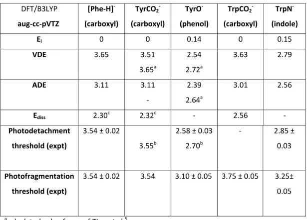
Vertical and adiabatic detachment energies (VDE and ADE) have been calculated for deprotonated aromatic amino acids. In the case of[Tyr‐H]‐ and [Trp‐H]‐, two isomers have been considered: one isomer being deprotonated on the carboxylic acid group (labeled TyrCO2‐ or TrpCO2‐) , the other one
being deprotonated on the phenolic oxygen or indolic nitrogen (TyrO‐ or TrpN‐), respectively. The relative energy of the isomers (Ei)is displayed in Table 1.The calculated ADE and VDE corresponds to
the most stable conformers previously reported 20.
In all three cases, when the aromatic amino acids are deprotonated on the carbonyl group (RCO2‐),
the radical optimization necessary to obtain the ADE leads to CO2 loss. The final energy for the CO2 +
Rychannel Ediss (see Table 1) is referenced to the ground state of the anion and is more than 1 eV
below the VDE. In this case, the ADEs shown in Table 1 correspond to calculations in which the C‐CO2
constraint after this optimization leads to CO2 dissociation, indicating the absence of any barrier in
the CO2 loss channel.
Table 1. Vertical and adiabatic detachment energies (VDE and ADE) for deprotonated aromatic amino acids. For deprotonated tyrosine and tryptophan, two isomers have been considered, one isomer
being deprotonated on the carboxylic acid group (TyrCO2‐ or TrpCO2‐), the other deprotonated on the
phenolic oxygen or indolic nitrogen (TyrO‐ or TrpN‐), respectively. When the molecules are
deprotonated on the carbonyl group, the radical optimization leads to CO2 loss (the final energy for
CO2 + Ry is Ediss) and in this case the ADE values correspond to calculations where the C‐CO2 bond is
maintained at a fixed value, the other coordinates being optimized. All values are in eV. DFT/B3LYP aug‐cc‐pVTZ [Phe‐H]‐ (carboxyl) TyrCO2‐ (carboxyl) TyrO‐ (phenol) TrpCO2‐ (carboxyl) TrpN‐ (indole) Ei 0 0 0.14 0 0.15 VDE 3.65 3.51 3.65a 2.54 2.72a 3.63 2.79 ADE 3.11 3.11 ‐ 2.39 2.64a 3.01 2.56 Ediss 2.30c 2.32c ‐ 2.56 ‐ Photodetachment threshold (expt) 3.54 ± 0.02 3.55b 2.58 ± 0.03 2.70b ‐ 2.85 ± 0.03 Photofragmentation threshold (expt) 3.54 ± 0.02 3.54 3.10 ± 0.05 3.75 ± 0.05 3.25± 0.05 acalculated value from ref.Tian et al 5 b experimental value from ref. Tian et al 5 c referenced to the energy of the ground state of the carbonyl ion
Discussion
[Phe‐H]‐ The phenylalanine case is the simplest since there is only one possibility for removing a proton from phenylalanine, i.e. from the carboxylic acid group. The [Phe‐H]‐ion does not photofragment below the photodetachment threshold, and thus we observe only photodetachment. The phenylalanyl radical produced by photodetachment of [Phe‐H]‐is unstable and loses CO2 as soon as thephotodetachment threshold is reached, at (3.54±0.02) eV, which is slightly lower than the calculated VDE value (3.65 eV) and even lower than the experimental VDE previously reported by other
authors(3.91 ± 0.19 eV)21.In agreement with the experimental data, the geometry optimization process leads directly to CO2 loss with a dissociation energy calculated at 2.3 eV above the ground
state of the anion, i.e. 1.24 eV lower than the photodetachment threshold.
The widths of the fragment peaks do not vary with the excitation energy, which means that the kinetic energy released in the fragmentation process does not depend on the excitation energy, which in turn means that the excess energy is transferred to the electron. The kinetic energy released in the dissociative photodetachment process can be compared to the impulsive model22in which all the atoms except those involved in the chemical bond that breaks are spectators. The kinetic energy release upon CO2 loss is Ekin= (µαβ/µAB)*Eavl, where Eavl is the available energy, here 1.24
eV, µαβ is the reduced mass of atoms α and β (α−β being the bond that breaks), and µAB is the
reduced mass of the two fragments A and B. In the present case, the calculated kinetic energy is Ekin=
0.23 eV, in good agreement with the experimental value of 0.20±0.02 eV.
[Tyr‐H]‐and[Trp‐H]‐
There are two possibilities for removing a proton from tyrosine and tryptophan: either from the carboxylic acid group or from the phenol (OH) or indole (NH) chromophores, which leads to two different isomers for the deprotonated forms of tryptophan and tyrosine (labeled TyrCO2‐ or TrpCO2‐
for deprotonation on the carboxylic acid group and TyrO‐ or TrpN‐ for deprotonation on the chromophore).
The two isomers of deprotonated tyrosine have already been observed via their photoelectron spectroscopy, the observed ADE and VDE being 3.55 and 3.90 eV for TyrCO2‐ and 2.70 and 2.74 eV for
TyrO‐.5Our action spectrum following the intact parent radical looks very much like this previously reported photoelectron spectrum. The photodetachment threshold observed at (2.58±0.03) eV is assigned to the TyrO‐ isomer, in agreement with the calculated VDE value (2.54 eV).In addition, this value is 0.33 eV higher than the photodetachment threshold reported for the phenolate anion (2.2538 eV) 2324.
The action spectrum recorded while detecting the neutral fragments shows a clear and rapid increase of the fragmentation channel at 3.54 eV, which corresponds to fragmentation from the TyrCO2‐ isomer, in agreement with both the photoelectron spectrum reported by Tian et al. and the
calculated VDE of 3.51 eV. There is also a weak and slowly increasing fragmentation from 3.1 to 3.54 eV. The origin of this minor fragmentation signal from the TyrO‐ isomer is unclear. One possible explanation is that fragmentation is induced by either proton transfer from the acid group to the phenolic oxygen leading to concerted CO2‐ rupture and photodetachment in the ion or by a
concerted hydrogen transfer and CO2 rupture in the radical. Such a signal would only be observed if
As in the case of deprotonated phenylalanine, above the fragmentation threshold of the TyrCO2‐
isomer the widths of the fragment peaks do not vary with the excitation energy, which means that the excess energy is transferred to the electron. The dissociation threshold for the CO2
+C8H10NO●channel is calculated at 2.32 eV above the anion ground state energy, i.e. 1.22 eV lower
than the experimental dissociation threshold for the TyrCO2●radical(3.54 eV).The experimental
kinetic energy release of 0.22 ±0.04 eV compares well with the value obtained via the impulsive model22: Ekin=0.22 eV.
Deprotonated tryptophan is very similar to deprotonated tyrosine: the photodetachment threshold is observed at 2.85±0.03 eV while the dissociation channel is observed above 3.75 eV, with two broad bands centered at around 4.4 eV and 5.4 eV. It may be noted that, in the 3.80 eV to 5.5 eV energy region, our spectrum is similar to that recorded by Compagnon et al. from depletion of the parent anion6.However, our photodetachment threshold is lower than their value of 3.94 eV.The photodetachment threshold observed is also quite different from the electron affinity reported in the literature for the [Trp‐H] radical(3.90 ± 0.19 eV)21. It is likely that these observed differences are due to the different experimental methods used to derive the values. Comparing the experimental thresholds with the calculated VDE for the two isomers of [Trp‐H]‐ (2.79 eV for TrpN‐ and 3.63 eV for TrpCO2‐)as well as to the deprotonated tyrosine case, we assign the photodetachment threshold at
2.85 eV to the TrpN‐isomer, and the dissociation threshold at 3.75 eV to the TrpCO2‐ isomer. As in the
case of TyrO‐ compared to the phenoxide, the photodetachment threshold for the TrpN‐ isomer is higher (by 0.42 eV) than the photodetachment energy of the indoline anion.12Additionally, as for Tyr, there is a weak fragmentation component observed to increase very slowly from 3.35 to 3.75 eV below the photofragmentation threshold of TrpCO2‐. Once again, a concerted hydrogen/ proton
transfer from the acid group to the N may be at play.
As in the other cases, the widths of the fragment peaks do not vary with the excitation energy, indicating that the electron removes all excess energy. The dissociation threshold for the CO2
+C10H11N2●channel is calculated at2.56eV above the anion ground state energy, i.e.1.19 eV lower
than the experimental dissociation threshold for the TrpCO2●radical (3.75 eV). The experimental
kinetic energy release is 0.19 ±0.02 eV, in agreement with the value obtained via the impulsive model: Ekin=0.20 eV.
Conclusion
When aromatic amino acids are deprotonated on the carbonyl group, the radicals produced upon photodetachment dissociate without barrier, leading to the formation of CO2 and a radical amine. This is in contrast to our previous results for benzoate and naphthoate, where barriers to CO2 loss were observed. When the system is deprotonated on functional groups located on the chromophore (OH for Tyr or NH for Trp), the radicals produced by photodetachment are stable. The role of the amino acid group on the electron binding energy of the chromophore is quite significant, since it increases the photodetachment threshold by more than 15%compared to the neat chromophores phenol and indole. In no case could ionic dissociation be evidenced, probably because the ionic excited states are higher in energy than the ADE.
Conflicts of interest
There are no conflicts to declare.
Acknowledgments
This work has been conducted within the International Associated Laboratory LEMIR (CNRS/CONICET) and was supported by CONICET, FONCyT, SeCyT‐UNC and the ANR Research Grant (ANR2010BLANC040501‐ESPEM and ANR17CE05000502‐Wsplit). We also acknowledge the use of the computing facility cluster Meso‐LUM of the LUMAT federation (LUMAT FR 2764).
Supplementary information
1) Experimental setup for the study of anions and neutrals2) Symmetrization of the mass peaks
3)Neutral fragment peak analysis
4) Kinetic energy released in the fragmentation process. 5) Xyz coordinates of the geometries of deprotonated anion or radical calculated at the DFT/B3LYP/aug‐cc‐pVTZ levelReferences
1 R. E. Continetti and H. Guo, Chem. Soc. Rev., 2017, 46, 7650–7667. 2 Y. Z. Zhou, S. Li, Q. S. Li and S. W. Zhang, J. Mol. Struct. THEOCHEM, 2008, 854, 40–45. 3 J. Oomens, J. D. Steill and B. Redlich, J. Am. Chem. Soc., 2009, 131, 4310–4319.4 H. Li, Z. Lin and Y. Luo, Chem. Phys. Lett., 2014, 598, 86–90. 5 Z. Tian, X. Bin Wang, L. S. Wang and S. R. Kass, J. Am. Chem. Soc., 2009, 131, 1174–1181. 6 I. Compagnon, A.‐R. Allouche, F. Bertorelle, R. Antoine and P. Dugourd, Phys. Chem. Chem. Phys., 2010, 12, 3334–3335. 7 D. Schröder, M. Buděšínský and J. Roithová, J. Am. Chem. Soc., 2012, 134, 15897–905. 8 H. Xia and A. B. Attygalle, Anal. Chem., 2016, 88, 6035–6043. 9 L. Feketeová, G. N. Khairallah, R. A. J. O’Hair and S. B. Nielsen, Rapid Commun. Mass Spectrom., 2015, 29, 1395–1402. 10 J. H. Bowie, Mass Spectrom. Rev., 1979, 9, 349–379. 11 G. A. Pino, R. A. Jara‐Toro, J. P. Aranguren‐Abrate, C. Dedonder‐Lardeux and C. Jouvet, Phys. Chem. Chem. Phys., 2019, 21, 1797–1804. 12 D. J. Nelson, A. M. Oliveira and W. C. Lineberger, J. Chem. Phys., 2018, 148, 1–8. 13 I. Alata, J. Bert, M. Broquier, C. Dedonder, G. Feraud, G. Grégoire, S. Soorkia, E. Marceca and C. Jouvet, J. Phys. Chem. A, 2013, 117, 4420–7. 14 G. Féraud, C. Dedonder, C. Jouvet, Y. Inokuchi, T. Haino, R. Sekiya and T. Ebata, J. Phys. Chem. Lett., 2014, 5, 1236–1240. 15 M. Berdakin, G. Féraud, C. Dedonder‐Lardeux, C. Jouvet and G. A. Pino, Phys. Chem. Chem. Phys., 2014, 16, 10643–10650. 16 W. C. Wiley and I. . McLaren, Rev. Sci. Instrum., 1955, 26, 1150. 17 M. Barat, J. C. Brenot, J. A. Fayeton and Y. J. Picard, Rev. Sci. Instrum., 2000, 71, 2050. 18 a Dev. Univ. Karlsruhe Forschungazentrum Karlsruhe GmbH, 1989‐2007, Turbomole GmbH, since 2007; available from http//www.turbomole.com Dev. Univ. Karlsruhe Forschungszentrum Karlsruhe GmbH, 1989‐2007, TURBO. 19 D. E. . Woon and T. H. Dunning Jr., J. Chem. Phys., 1993, 98, 1358. 20 U. Purushotham and G. N. Sastry, J. Comput. Chem., 2014, 35, 595–610. 21 R. A. J. O’Hair, J. H. Bowie and S. Gronert, Int. J. Mass Spectrom. Ion Process., 1992, 117, 23– 36. 22 Z. Lu and R. E. Continetti, J. Phys. Chem. A, 2004, 108, 9962–9969. 23 S. J. Kregel and E. Garand, J. Chem. Phys., 2018, 149, 0–9. 24 J. B. Kim, T. I. Yacovitch, C. Hock and D. M. Neumark, Phys. Chem. Chem. Phys., 2011, 13, 17378–17383.
Supplementary information
Experimental setup for the study of anions and neutrals
Ion trap Extraction 300/0V Gauss tube 2.5/0kV V1 V2 MCP 1.2m Dissociation in the trap: ionic fragmentation Excitation after the acceleration in the gauss tube: detection of neutral TOFFigure SI-1: Scheme of the experimental setup. The two regions where the laser interacts with the ion packet are 1) in the cold trap, and 2) in the gauss tube.
Symmetrization of the mass peaks
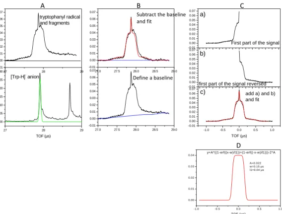
As can be seen in Figure SI‐2 panel A, the raw data anion signal, as well as the neutral signal,is not symmetrical, but broadened towards higher times of flight due to electronic ringing in the detector…. One way to get rid of this deformation is to subtract a baseline, as in panel B, but we have to estimate the baseline, and the resulting fit is not optimal. Another method is to take the first half of the signal (up to the peak maximum), reverse it, and add the two components, as illustrated in panel C. We then obtain a good fit with a three component function: a Gaussian function for the narrow peak ‐C*exp(‐(x2/l2))‐ and two combinations of erf functions for the fragments A*((1‐erf((x‐ w)/l1))+(1‐erf((‐x‐w)/l1)))‐2*A where w represents the half width at half maximum of the
27.0 27.5 28.0 28.5 29.0 -0.01 0.00 0.01 0.02 0.03 0.04 0.05 0.06 0.0727.0 27.5 28.0 28.5 29.0 -0.01 0.00 0.01 0.02 0.03 0.04 0.05 0.06 0.07 27 28 29 0.00 0.05 0.10 0.15 0.20 0.25 0.30 TOF (µs) [Trp-H]- anion 27 28 29 -0.01 0.00 0.01 0.02 0.03 0.04 0.05 0.06 0.07 tryptophanyl radical and fragments Define a baseline Subtract the baseline and fit A B -1.0 -0.5 0.0 0.5 1.0 -0.01 0.00 0.01 0.02 0.03 0.04 0.05 0.06 0.07 TOF (µs) add a) and b) and fit -0.01 0.00 0.01 0.02 0.03 0.04 0.05 0.06 0.07
first part of the signal reversed
-0.01 0.00 0.01 0.02 0.03 0.04 0.05 0.06 0.07 c) b)
First part of the signal
a) C -1.0 -0.5 0.0 0.5 1.0 0.00 0.01 0.02 0.03 0.04 TOF (µs) y=A*((1-erf((x-w)/l1))+(1-erf((-x-w)/l1)))-2*A A=0.022 w=0.15 µs l1=0.04 µs D
Figure SI-2: panel A,raw time of flight mass spectra as recorded with the MCP detector. Panel B, subtraction of a baseline and fit. Panel C, symmetrization by selecting the first half of the peak, reversing it, and adding the two contributions. Panel D, combination of erf functions to fit the fragment profile of the form A*((1-erf((x-w)/l1))+(1-erf((-x-w)/l1)))-2*A, where w represents the half width at half maximum of the function, l1 the slope of the erf functions.
Neutral fragment peak analysis
220 240 260 280 300 320 340 0 100 200 300 400 4 4.5 5 5.5 width of t he T O F dist ribut ion f o r t he ne ut ral f ragment s Excitation wavelength (nm) w1 (light fragment)=0.405 µs w2 (heavy fragment)=0.140 µsFigure SI-3. Variation of the widths of the fragment peaks as a function of the excitation wavelength for the fragments of the [Phe-H]● radical
Figure SI‐4.Kinetic energy released in the fragmentation process. 27.0 27.5 28.0 28.5 TOF detector V0 dv L0 A parent ion with a velocity V0 will arrive at the detectot at time T0= L0/V0 a fragment ion has the initial velocity of the parent plus a relative velocity dv due to the fragmentation process (we assume here that dv is the maximum velocity that can be imparted to the fragment). If dvis in the opposite direction to V0, the fragment will arrive later on the detector at time T0+dt. Conversely, if dvis in the same direction as V0, the fragment will arrive sooner on the detector at time T0‐dt and all the fragments that have an initial velocity in another direction or a smaller velocity will arrive at a time between T0‐dt and T+dt. dv T0=27.87 µs 2*dt V0=L0/T0, V0‐dv=L0/(T0+dt)…. V0‐dv=V0(1‐dt/T0+(dt/T0)2‐…), dt/T0 is small (0.13/27.87) Then dv=V0*dt/T0 and the relative kinetic energy of fragment mfis Ekin=1/2*mf*(V0*dt/T0)2=1/2*mf*(L0/T0)2*(dt/TO)2
Geometries of deprotonated anion or radical calculated at the
DFT/B3LYP/aug‐cc‐pVTZ level
Phenyl alanine aniondeprotonated on CO
21 c 0.16865091238731 ‐0.25415226304652 0.25836602724172 2 c 0.41456381453982 0.44948970492088 2.77697528026225 3 c 2.77341038704134 1.12384844586595 3.73025864917984 4 c 4.92861196611267 1.11035955441249 2.20486114397444 5 c 4.65127661420242 0.40601932015652 ‐0.33256682744145 6 c 2.30000824331672 ‐0.26811573800071 ‐1.28720507391389 7 c 7.48587253491187 1.82319214183266 3.25676844904883 8 c 8.49911402170535 4.42170690584230 2.43469350491675 9 c 9.26969693423235 4.54676326724159 ‐0.44218629418933 10 o 8.87143439177082 6.62351148122967 ‐1.50607463908593 11 h ‐1.66101964217337 ‐0.77927078938879 ‐0.49495765025628 12 n 6.81291789894409 6.48639144721865 3.23425380352489 13 o 10.28177356520158 2.59408846802207 ‐1.32753846628611 14 h 7.17621789691949 7.89780862023176 1.97332836587437 15 h 10.29216824961529 4.66558853561591 3.44649923495727 16 h 7.38566556141583 1.77473789097372 5.32106167240502 17 h 8.88102956512812 0.43863980138080 2.64228774645031 18 h 2.95054261065437 1.67276057903125 5.69550911234131 19 h 6.30954703507718 0.43089801321741 ‐1.53161826116454 20 h ‐1.22553576260103 0.47980879904034 4.00229088004463
Radical –C‐‐‐CO2 fixed
1 c ‐6.09090851359800 1.13531954023667 ‐1.15321827777162 2 c ‐5.63380416686941 0.77801489778018 1.40723064735399 3 c ‐3.39485552174641 ‐0.35359782194246 2.19636125248942 4 c ‐1.57504461066894 ‐1.13851978013159 0.44943495917408 5 c ‐2.06457006619120 ‐0.77658704539202 ‐2.12097317990815 6 c ‐4.30110847242929 0.34912371486595 ‐2.91155708150178 7 c 0.85138164149856 ‐2.35969605630578 1.34169041202110 8 c 3.11120632514979 ‐0.56379940929722 1.65592232198492 9 c 4.61101367896291 0.23899742838096 ‐0.80067137639047 10 o 6.12172172285127 1.94494896331005 ‐0.45028315947106 11 h ‐7.83260254817667 2.00587086150096 ‐1.77592873527155 12 n 2.50389367912626 1.59391781888687 3.05819975706147 13 o 3.99968591170458 ‐1.13399226845655 ‐2.58530151434563 14 h 3.88380164413895 2.91482422992733 3.12705899053461 15 h 4.66713231467567 ‐1.58797575177256 2.56466815166193 16 h 0.52466940034622 ‐3.21450301676702 3.19118047753538 17 h 1.41684963166303 ‐3.85756469042205 0.05549438205285 18 h ‐3.07302581920985 ‐0.66151867356052 4.19514620385332 19 h ‐0.68072764848504 ‐1.37866504556755 ‐3.49746223408055 20 h ‐7.02030086364945 1.36239723196606 2.79183297776067 21 h ‐4.65007653962269 0.60609274999538 ‐4.90984586388169 22 h 0.75284248928950 2.31000809561610 2.78252285105043
C—CO2 free
1 c 0.16507534806649 ‐0.27078463554534 0.21620924081983 2 c 0.19808929064045 0.41330280214110 2.74971363231662 3 c 2.47518830009742 1.01793674400981 3.92425950124909 4 c 4.74798331945495 0.95317485911092 2.59242919206221 5 c 4.68625692837021 0.27466157388837 0.04045143972224 6 c 2.42160649610651 ‐0.33745048620181 ‐1.13608297819163 7 c 7.22803514155176 1.62933408864816 3.86415275256727 8 c 8.26877673604632 4.14522734327818 3.09285016458237 9 c 10.12114916478683 5.33090462235400 ‐2.70353038062186 10 o 9.67054634389591 7.42624855996841 ‐2.22698137932822 11 h ‐1.59828859428247 ‐0.75217382121331 ‐0.69931189334066 12 n 6.74375897638459 6.27065773202729 3.37495352712117 13 o 10.60356753368445 3.26212441389759 ‐3.24980339724940 14 h 7.42060722029110 7.86391774731968 2.57720304223297 15 h 10.27233730638100 4.48463401618785 3.33017575312724 16 h 6.94301671610752 1.55546866677763 5.92339433502590 17 h 8.64094139730366 0.19161564514120 3.42671884368730 18 h 2.48760638724749 1.53289180233159 5.90527559572578 19 h 6.43074493035817 0.22129047147828 ‐1.02682367303239 20 h ‐1.54340701778198 0.47123004332727 3.81983089512267 21 h 2.41624092992967 ‐0.87528170328793 ‐3.10900579834480 22 h 4.90050424097583 6.00578255094250 2.95087289530649
Tyrosine anion deprotonated on phenoxy
1 n 0.08731893607592 ‐0.25244351356949 ‐0.12203400028704 2 h ‐0.15038092767218 0.94065427711042 1.37199782360100 3 c 2.73887415133195 ‐0.10833062897929 ‐0.95170642267331 4 h 2.79815835536698 ‐0.13874521613986 ‐3.01674106579478 5 c 4.12334012769014 ‐2.54893195194012 ‐0.18224548456307 6 o 6.32392124735807 ‐2.94377724033993 ‐0.63127003499251 7 c 4.14030760869025 2.31946946684994 ‐0.02904325539235 8 h 3.69819058575667 3.81919084271027 ‐1.38016765767281 9 h 6.16270261801547 1.94184986607070 ‐0.19176800005810 10 c 3.41846153315479 3.18322706025511 2.58217318756225 11 c 1.61319062914294 5.09738025555072 2.94480392965426 12 h 0.81279115706252 6.01934719956391 1.28846470210290 13 c 4.43350799709272 2.09133843757311 4.78311090559494 14 h 5.85445819548094 0.62001768896691 4.59654065102123 15 c 0.84033501177501 5.87729399312862 5.31641028722067 16 h ‐0.54282292537020 7.37633114879438 5.51499434272217 17 c 3.69497923922455 2.83473244783240 7.17236084721973 18 h 4.52730883738272 1.95929226997194 8.82775578913408 19 c 1.82772928581537 4.78024903389330 7.60930188260323 20 o 1.13892950667281 5.46474474833142 9.79106026544944 21 o 2.63589452769747 ‐4.25673137713968 0.94816089675463 22 h 0.96793665248393 ‐3.41383541792348 1.01026617334631 23 h ‐1.11820661652626 0.28780002411562 ‐1.50074840174992
Tyrosine rad dep onPhenyl
1 n ‐2.34470016461140 2.82955649575025 ‐3.21555965734288 2 h ‐2.76913058401107 2.69111512163324 ‐1.35351916718856 3 c 0.08121694190611 1.63527920292872 ‐3.81544714334473 4 h 0.81939483004206 2.56406827240692 ‐5.50542343612384 5 c ‐0.34722499160123 ‐1.11783597195172 ‐4.65429905057539 6 o 1.34319504875821 ‐2.61972996561951 ‐4.88645660712412 7 c 2.16662580144788 1.82841119156115 ‐1.76703502044393 8 h 2.73141486069690 3.80641901848040 ‐1.63235659169408 9 h 3.78345841130886 0.75385561375231 ‐2.45424114559107 10 c 1.38620005546631 0.88690206505149 0.79878919931263 11 c 0.49637266236483 2.59413655620627 2.64624565752756 12 h 0.45837300151722 4.59385035503084 2.20649164579325 13 c 1.50001696220605 ‐1.71528699422705 1.39761931106822 14 h 2.19840169985107 ‐3.02003997156457 ‐0.01289294215441 15 c ‐0.28058997269830 1.77742707179292 4.97959692830472 16 h ‐0.95929895597406 3.07397962767947 6.40628691216150 17 c 0.74077549221170 ‐2.58587182442128 3.71356426928432 18 h 0.82343857018192 ‐4.57155867561253 4.19092881489361 19 c ‐0.20665615712536 ‐0.87882241023186 5.63105241180275 20 o ‐0.91765771567179 ‐1.65292236611434 7.74924818062116 21 o ‐2.75279268186007 ‐1.66859737195631 ‐5.18998879434182 22 h ‐3.71501819818809 ‐0.12972803128822 ‐4.80386701253443 23 h ‐2.38677697787618 4.67639085244690 ‐3.69813719655978
Tyrosine deprotonated on CO2
Anion. 1 n 0.19109204607645 ‐0.29564895266318 0.24242006035000 2 h 0.29223166938765 0.23861161365828 2.09130489822343 3 c 2.79507994085767 0.00548904675671 ‐0.69304406881894 4 h 3.76421032396840 ‐1.80668612366649 ‐0.41861931681936 5 c 4.41869261276810 1.94553666102485 0.88272378766078 6 o 6.47812685186440 2.60013363346470 ‐0.09526650207409 7 c 2.85790076271253 0.50541113732678 ‐3.55577679399181 8 h 1.99523542371758 ‐1.10247542946640 ‐4.52826641248435 9 h 4.84552691848086 0.58692260157164 ‐4.08780687653902 10 c 1.55247361657096 2.89726045097223 ‐4.41521443280041 11 c ‐0.74557684390784 2.82840799409098 ‐5.70920956881890 12 h ‐1.61417892902189 1.01416927656517 ‐6.09536903590285 13 c 2.61520087172863 5.27346254906997 ‐3.94347240494668 14 h 4.37947378114326 5.36282383931686 ‐2.90954135505346 15 c ‐1.96021880511330 5.01813487542496 ‐6.52063846129266 16 h ‐3.74295826426822 4.93387029675707 ‐7.51903076013455 17 c 1.42244414176583 7.47136656667652 ‐4.73981416086121 18 h 2.27378929612468 9.29522775322143 ‐4.34206715651789 19 c ‐0.86600166603745 7.34949568308305 ‐6.03021972882701 20 o ‐2.11105515543351 9.49279370994813 ‐6.85582953310133 21 o 3.51611127270609 2.56018875003720 2.98272022074423 22 h ‐0.89812420192583 1.06091134846790 ‐0.57039353411658 23 h ‐1.15743366442383 10.94733439432629 ‐6.32802874293679
Radical CC fixed
1 n ‐2.05703875414633 ‐2.74433307978794 3.38752064016210 2 h ‐3.47893494548639 ‐2.50709262471879 4.64251416010559 3 c 0.21312061587459 ‐1.56325869045659 4.05717708813992 4 h 0.98370750500431 ‐2.42981743534226 5.77524826415005 5 c ‐0.36879414145779 1.11308897211048 5.23396987209609 6 o 1.27836535815085 2.64033260033877 4.59868992937803 7 c 2.19956259111908 ‐1.70063456532078 1.94309230784686 8 h 2.81372366695957 ‐3.66800836206979 1.84605486620815 9 h 3.81477923357503 ‐0.57672247698294 2.53352758184692 10 c 1.29801091435380 ‐0.88603443471875 ‐0.64296411748194 11 c 0.42894585434069 ‐2.66751992327122 ‐2.39071598030860 12 h 0.44347934171269 ‐4.65439869946006 ‐1.89508776686574 13 c 1.31066443930996 1.65003979753913 ‐1.37664722207197 14 h 1.97826414858712 3.06998874232331 ‐0.06931096368783 15 c ‐0.42086076375135 ‐1.96648246409955 ‐4.77214379043153 16 h ‐1.07956491046827 ‐3.35992560953260 ‐6.11312113575636 17 c 0.47028819423504 2.37715645979324 ‐3.75250876748621 18 h 0.50143293144303 4.35523881452449 ‐4.28433953432164 19 c ‐0.40555234076097 0.57172841636778 ‐5.45740307497798 20 o ‐1.25299889385378 1.17984665544212 ‐7.81882746580018 21 o ‐2.22882610135704 1.14586479964090 6.59744087373616 22 h ‐2.58893307848929 ‐2.55619477688455 1.56062977555740 23 h ‐1.14550174058174 2.97833644273237 ‐8.06973518650852
Radical Without constrain on the CC bond after 30 step of optimization
1 n 0.04093495372257 ‐0.37395502325248 ‐0.11531545310275 2 h 0.02902241470023 ‐0.55582469774193 1.78196509187458 3 c 2.41885679827831 ‐0.36110634498989 ‐1.23951066032412 4 h 3.74518546449896 ‐1.71106262230362 ‐0.45982770019786 5 c 5.60779579336056 3.09498575369787 2.59353018954085 6 o 7.21117763142793 3.49517691180830 1.14961821588335 7 c 2.60730453337436 0.26780888300662 ‐3.99372554106203 8 h 1.74078795369488 ‐1.23537988032806 ‐5.14271124767680 9 h 4.60509272229448 0.29954666180165 ‐4.50582356237349 10 c 1.38695996054502 2.75881281586243 ‐4.71528844493003 11 c ‐0.98355671547782 2.84515331902129 ‐5.86716116456968 12 h ‐1.95666662371836 1.09407709829207 ‐6.28926155673692 13 c 2.59593610492285 5.05185317652094 ‐4.22714819277896 14 h 4.43980411773682 5.04865284369952 ‐3.34030643652916 15 c ‐2.12615510423825 5.12211339483309 ‐6.50813567653795 16 h ‐3.96164098104702 5.16900093817100 ‐7.40492460015937 17 c 1.48752872329718 7.34269415583679 ‐4.86739399958062 18 h 2.47186086814726 9.09992764618434 ‐4.49074510498900 19 c ‐0.88522070480108 7.38287382258471 ‐6.00866459316969 20 o ‐2.07437253141718 9.58253072386885 ‐6.67484987736033 21 o 4.05468545774235 2.74171152837617 4.10407745408026 22 h ‐1.13148045871467 1.02075701715046 ‐0.68965366143361 23 h ‐1.02179459913585 11.00239544359054 ‐6.24718146719995TRP anion deprotonated on CO2
1 n 0.52768610527851 ‐1.19355049942161 ‐0.06924010476488 2 h 0.91641871424601 ‐2.04873592675984 1.61494450056308 3 c 2.98873411503976 ‐0.27049785513588 ‐0.97332753753218 4 h 3.56862055192035 ‐1.30466994579041 ‐2.67371324642338 5 c 5.14546804414503 ‐0.77495686950151 1.01645593178419 6 o 4.55117190506973 ‐2.10348202325628 2.88417900730597 7 c 2.88554205897985 2.54531281466465 ‐1.73939509108134 8 h 4.69619516738275 2.99822059415026 ‐2.61622153093111 9 h 1.42096326234976 2.77524867020388 ‐3.17900251486940 10 c 2.38497954859575 4.35841534939567 0.38749342274723 11 c 3.94621162866067 4.81291351553098 2.39721004498649 12 h 5.73701887065005 3.93896059136119 2.81794018221910 13 c 0.21532234793748 5.95766413598184 0.74458478196846 14 n 2.88282785428328 6.60614239254147 3.97814282862802 15 h 3.61440309588853 7.15349554227851 5.63738500041925 16 c 0.58689255897863 7.34049733298298 3.02157625278755 17 c ‐2.01570460558809 6.36921268222554 ‐0.62103542438360 18 h ‐2.37034156272606 5.32738246002502 ‐2.34473822730669 19 c ‐1.17910233314498 9.07172704825381 3.92401313126157 20 h ‐0.86422116690819 10.09969335460260 5.66717327740275 21 c ‐3.77592479867278 8.09340497283245 0.26749528832563 22 h ‐5.50562732931782 8.40758696733402 ‐0.78040651028961 23 c ‐3.36504313789008 9.43545236338114 2.52231476011997 24 h ‐4.77525053700531 10.76471190906054 3.17851439880090 25 o 7.23822738315491 0.21683234409946 0.51563881189657 26 h ‐0.14802828955308 ‐2.55056942777157 ‐1.23506607202346Radical
1 n ‐4.06556346565252 ‐2.63977960970772 1.15341218768968 2 h ‐3.19520318189824 ‐1.12283663498732 1.94400408167724 3 c ‐2.76868940459854 ‐3.75091138491116 ‐0.86231006550992 4 h ‐3.60726229589053 ‐5.50645970163668 ‐1.55274845779715 5 c 1.53860824549740 ‐7.20610002022106 1.61718073935416 6 o 0.27548378485659 ‐7.34514619716112 3.42274183691075 7 c ‐1.51727086177122 ‐2.04508022775211 ‐2.76985294059102 8 h ‐0.43585187774907 ‐3.24196820466873 ‐4.07806211853112 9 h ‐2.96225730967751 ‐1.07394658417539 ‐3.94060857905076 10 c 0.21257264830673 ‐0.06572187335156 ‐1.66122215373202 11 c 2.78995458297811 ‐0.26383425669578 ‐1.36849482035894 12 h 4.04067730637719 ‐1.81305911361585 ‐1.86224543709999 13 c ‐0.47525974721533 2.40184966565661 ‐0.70353358788974 14 n 3.74243839438288 1.92000846770549 ‐0.28306817263797 15 h 5.58148975946991 2.24704352356353 0.09171313971783 16 c 1.78775702518153 3.59453531091486 0.14206999840834 17 c ‐2.77703336864379 3.73235646021349 ‐0.50184731366333 18 h ‐4.54165286543969 2.87324679241408 ‐1.12890206172359 19 c 1.80687029578146 6.03445224014749 1.17182746086466 20 h 3.55621319960811 6.92092617820954 1.80626210231110 21 c ‐2.76649523781500 6.15747092800691 0.51824779747366 22 h ‐4.53513006712033 7.20272709859845 0.68108629719110 23 c ‐0.49739084925814 7.29739066954193 1.34874449957369 24 h ‐0.54933488413149 9.19956448264943 2.14016078121323 25 o 2.87994593865503 ‐7.14083581638826 ‐0.13690164653521 26 h ‐4.69630015194929 ‐3.85708249202693 2.48954042361946Radical CC‐fixed
1 n ‐3.58842970896621 ‐2.74249664435268 1.35811565870386 2 h ‐2.44539440061223 ‐1.85529098003560 2.60953582251437 3 c ‐2.29254611192938 ‐4.05476174639724 ‐0.55577480682781 4 h ‐3.61322059308870 ‐5.24691821588126 ‐1.57436658373128 5 c ‐0.17154237913709 ‐5.80954012327845 0.57549754554472 6 o 1.18254291895562 ‐4.80607031372513 2.16190080645818 7 c ‐0.92829031857737 ‐2.30575540687949 ‐2.51885482397093 8 h 0.21430717987383 ‐3.52896798274878 ‐3.71702918123189 9 h ‐2.45255891396254 ‐1.55681749044022 ‐3.69357717478170 10 c 0.59773375512295 ‐0.19098416209867 ‐1.48434355210613 11 c 3.17609202912268 ‐0.16865308855296 ‐1.16514666940217 12 h 4.52671740138320 ‐1.64357814139366 ‐1.54029862760306 13 c ‐0.29470784348326 2.23165663468269 ‐0.62010487654204 14 n 3.93018064627486 2.09477729015143 ‐0.16918718089928 15 h 5.71877862539254 2.54827742251113 0.27211175950195 16 c 1.84489997418823 3.61963072656770 0.19599887048805 17 c ‐2.68170459780957 3.36732845701551 ‐0.46644726983835 18 h ‐4.35273969359930 2.35896751764944 ‐1.07230336851011 19 c 1.66422492293332 6.06329257596236 1.14716379410727 20 h 3.31864770801493 7.09723043732196 1.76166601448458 21 c ‐2.87261125382053 5.79949522705204 0.48442563446587 22 h ‐4.70622650829633 6.69387643875188 0.61358892777636 23 c ‐0.72464230239234 7.13356283235478 1.28294422315113 24 h ‐0.93404283804720 9.03055819896934 2.01399884496711 25 o ‐0.30409240667403 ‐7.91289683843504 ‐0.44083128325068 26 h ‐4.99006107812565 ‐3.70711619557046 2.2185099324286

![Figure SI-3. Variation of the widths of the fragment peaks as a function of the excitation wavelength for the fragments of the [Phe-H] ● radical](https://thumb-eu.123doks.com/thumbv2/123doknet/14371063.504371/20.892.151.602.190.542/figure-variation-fragment-function-excitation-wavelength-fragments-radical.webp)
