HAL Id: tel-03126630
https://tel.archives-ouvertes.fr/tel-03126630
Submitted on 1 Feb 2021HAL is a multi-disciplinary open access archive for the deposit and dissemination of sci-entific research documents, whether they are pub-lished or not. The documents may come from teaching and research institutions in France or abroad, or from public or private research centers.
L’archive ouverte pluridisciplinaire HAL, est destinée au dépôt et à la diffusion de documents scientifiques de niveau recherche, publiés ou non, émanant des établissements d’enseignement et de recherche français ou étrangers, des laboratoires publics ou privés.
Anti-Cancer Immunity : a Study of the Immunological
Potential of PARP inhibitors
Roman Chabanon
To cite this version:
Roman Chabanon. Exploiting DNA Repair Vulnerabilities to Modulate Anti-Cancer Immunity : a Study of the Immunological Potential of PARP inhibitors. Cancer. Université Paris Saclay (COmUE), 2019. English. �NNT : 2019SACLS007�. �tel-03126630�
Exploiting DNA repair vulnerabilities
to modulate anti-cancer immunity:
a study of the immunological potential
of PARP inhibitors.
Thèse de doctorat de l’Université Paris-Saclay
préparée à l’Université Paris-Sud
Unité Inserm U981, Gustave Roussy (Villejuif) Gene Function Laboratory, The Institute of Cancer Research (Londres) École Doctorale N°582 : Cancérologie – Biologie – Médecine – Santé
Disciplines : Sciences de la vie et de la santé Aspects moléculaires et cellulaires de la biologie
Soutenue publiquement le 31 janvier 2019 à Gustave Roussy par
Roman Chabanon
Composition du jury
Pr. Chris Lord
Directeur de Recherche, Institute of Cancer Research, Londres Président Dr. Philippe Pasero
Directeur de Recherche, Institut de Génétique Humaine, Montpellier Rapporteur Pr. Fabrice Barlesi
Professeur des Universités-Praticien Hospitalier, Hôpital Nord, Marseille Rapporteur
Dr. Olivier Delattre
Praticien Hospitalier et Directeur de Recherche, Institut Curie, Paris Examinateur Dr. Jean-Luc Perfettini
Directeur de Recherche, Gustave Roussy, Villejuif Examinateur Dr. Sophie Postel-Vinay
Praticien Hospitalier et Directrice d’Equipe, Gustave Roussy, Villejuif Directeur de thèse Pr. Jean-Charles Soria
Professeur des Universités-Praticien Hospitalier, Gustave Roussy, Villejuif Co-directeur de thèse
Dr. Aurélien Marabelle
Praticien Hospitalier et Directeur d’Equipe, Gustave Roussy, Villejuif Co-directeur de thèse
Th
ès
e
de
d
oc
tor
at
NNT : 2019SACLS007
Avec sincérité et humilité, je souhaiterais dédier ce travail au Dr. Sophie Postel-Vinay, qui a admirablement dirigé mes recherches et sans conteste contribué à leur aboutissement. La confiance qu’elle m’a rapidement accordée et le soutien inconditionnel qu’elle m’a apporté tout au long de mon doctorat ont façonné un cadre de travail extrêmement enrichissant, tant sur le plan scientifique qu’humain. Dans cette atmosphère bienveillante et positive, Sophie m’a à chaque instant poussé à l’excellence ; avec douceur et professionnalisme, elle m’a permis de grandir scientifiquement et humainement et a souvent créé des opportunités pour me mettre en avant à travers mon travail. Grâce à elle, j’ai appris à me lancer, oser faire des hypothèses, expérimenter, et plus que tout, avoir confiance dans mon travail et mes résultats. Sophie m’a également transmis sa passion pour la recherche translationnelle en cancérologie, et m’a aidé à garder à l’esprit que tout ce que nous faisons, nous le faisons pour le bénéfice des patients. Enfin, Sophie a concrétisé mon envie de découvrir un autre environnement de travail, une autre approche de la recherche, en me permettant d’effectuer une partie de mon doctorat à l’ICR à Londres. Pour tout cela, je souhaite la remercier chaleureusement, et lui dire que je ne suis pas peu fier d’avoir été son premier étudiant en thèse !
Je souhaiterais ensuite exprimer ma plus grande gratitude au Pr. Jean-Charles Soria, qui m’a initialement accepté comme étudiant en thèse dans son équipe, et qui, avec une clairvoyance déconcertante, a tout de suite su que je pourrais m’épanouir sous la supervision de Sophie. En tant que directeur puis co-directeur de thèse, Jean-Charles m’a soutenu tout au long de mon doctorat et je suis aujourd’hui très heureux qu’il puisse faire le déplacement depuis les Etats-Unis pour assister à ma soutenance de thèse.
Un grand merci également au Dr. Aurélien Marabelle, qui m’a accompagné avec beaucoup de bienveillance et de gentillesse durant ces trois années, et m’a donné un autre regard sur la recherche et sur les parcours possibles au-delà de la thèse.
mais plus que cela, ils ont chacun à leur manière enrichit ma personnalité, et sont pour moi des modèles inspirants pour mon futur parcours professionnel.
I would like to warmly thank Prof. Chris Lord, for having welcomed me in his lab at the ICR in London. I am very grateful for his high-quality scientific input on my project, and for the confidence he has placed in me since our very first meetings. A big thank you also goes to his team members, Steve, Drago, Ilirjana, Rachel, Feifei, Ger, Marta and all the others that made my time at the ICR a really enriching experience.
Merci aussi à toutes les personnes à Gustave Roussy qui m’ont, de près ou de loin, aidé à mener mon projet de recherche durant mes deux premières années de thèse : je pense tout d’abord à Marlène et Nicolas, mais aussi Isabelle, Kariman, Daphné, Chloé et Mei-Shue.
Je voudrais aussi remercier mes parents, Chantal et Christian, et mes grands-parents, Raymonde et René, qui ont toujours cru en moi et m’ont soutenu sans relâche, depuis toujours.
Last but not least, je voudrais remercier de tout mon cœur ma petite chérie, Laetitia, pour le soutien sans faille qu’elle m’a apporté depuis le début de cette aventure, pour sa compréhension face à mes craintes, mes inquiétudes et mes moments de doutes, pour son aide précieuse dans la mise en forme de ce manuscrit de thèse, et pour tout le bonheur qu’elle m’a donné, adoucissant un quotidien parfois rude. Sans elle, je n’en serais pas là aujourd’hui.
Poly(ADP-ribose) polymerase inhibitors (PARPi) selectively target cancer cells with DNA repair deficiencies such as BRCA1/2 mutations or ERCC1 defects. Clinically, several PARPi are currently approved for the treatment of BRCA-mutant or platinum-sensitive advanced ovarian and breast cancers, and ongoing clinical trials are investigating the efficacy of PARPi in platinum-sensitive Non-Small Cell Lung Cancer (NSCLC). While PARPi constitute potent targeted therapies for the treatment of DNA repair-deficient malignancies, an increasing number of clinical trials are also evaluating their efficacy in combination with immune checkpoint inhibitor (ICI) in various populations. In this context, it is of critical importance to better understand how PARPi might modulate immune responses against cancer, and to investigate the inherent immunological potential of these agents.
In this study, we show that ERCC1-defective NSCLC cells exhibit an enhanced type I interferon (IFN) transcriptomic signature and that low ERCC1 expression correlates with increased lymphocytic infiltration in human NSCLC tumours. Using isogenic cell lines and patient-derived xenografts, we further demonstrate that several clinical PARPi, including olaparib and rucaparib, display cell-autonomous immunomodulatory properties in ERCC1-defective NSCLC and BRCA1-mutant triple-negative breast cancer (TNBC) models. Mechanistically, PARPi generate cytoplasmic chromatin fragments with micronuclei characteristics; this activates the cGAS/STING pathway and elicits downstream type I IFN signalling and CCL5 secretion. Importantly, these effects are suppressed in BRCA1-reverted TNBC cells and ERCC1-rescued NSCLC cells, suggesting that DNA repair defects exacerbate the innate immunity-related phenotypes triggered by PARPi. Similarly, these effects are totally abrogated in PARP1-null TNBC cells, supporting the on-target effect of PARPi in mediating such phenotypes.
Besides this potential to activate tumour cell-autonomous immunity through cGAS/STING and type I IFN signalling, we also observed that PARPi synergize with type II IFN to induce PD-L1 expression in NSCLC cell lines and fresh patient tumour cells, especially in the ERCC1-deficient setting. Moreover, we show that lethal concentrations of some PARPi independently activate the key damage-associated molecular patterns dictating the immunogenicity of cancer cell death, including calreticulin exposure at the tumour cell surface, ATP secretion and HMGB1 release in the extracellular compartment.
Together, these preclinical data suggest that PARPi have intrinsic immunomodulatory properties that activate anti-cancer immune responses; this could be exploited clinically in combination with ICI in appropriately molecularly-selected populations.
Publications included in this thesis
Roman M. Chabanon, Gareth Muirhead, Dragomir B. Krastev, Julien Adam, Daphné Morel, Marlène Garrido, Clémence Hénon, Nicolas Dorvault, Rebecca Marlow, Ilirjana Bajrami, Marta Llorca Cardeñosa, Asha Konde, Benjamin Besse, Alan Ashworth, Stephen J. Pettitt, Syed Haider, Aurélien Marabelle, Andrew N.J. Tutt, Jean-Charles Soria, Christopher J. Lord and Sophie Postel-Vinay. ERCC1-deficiency exacerbates tumor cell-intrinsic immunity in response to PARP inhibitors in non-small cell lung cancer. The Journal of Clinical Investigation, 2018.
Roman M. Chabanon, Marion Pedrero, Céline Lefebvre, Aurélien Marabelle, Jean-Charles Soria, and Sophie Postel-Vinay. Mutational landscape and sensitivity to immune checkpoint blockers. Clinical Cancer Research, 2016.
Contribution to other publications
Mehdi Touat, Tony Sourisseau, Nicolas Dorvault, Roman M. Chabanon, Marlène Garrido, Daphné Morel, Dragomir B. Krastev, Ludovic Bigot, Julien Adam, Jessica R. Frankum, Sylvère Durand, Clement Pontoizeau, Sylvie Souquère, Mei-Shiue Kuo, Sylvie Sauvaigo, Faraz Mardakheh, Alain Sarasin, Ken A. Olaussen, Luc Friboulet, Frédéric Bouillaud, Gérard Pierron, Alan Ashworth, Anne Lombès, Christopher J. Lord, Jean-Charles Soria, et Sophie Postel-Vinay. DNA repair deficiency sensitizes lung cancer cells to NAD+ biosynthesis blockade. The Journal of Clinical Investigation, 2018.
Ruth Pidsley, Mitchell G. Lawrence, Elena Zotenko, Birunthi Niranjan, Aaron Statham, Jenny Song, Roman M. Chabanon, Wenjia Qu, Hong Wang, Michelle Richards, Shalima S. Nair, Nicola J. Armstrong, Hieu T. Nim, Melissa Papargiris, Preetika Balanathan, Hugh French, Timothy Peters, Sam Norden, Andrew Ryan, John Pedersen, James Kench, Roger J. Daly, Lisa G. Horvath, Phillip Stricker, Mark Frydenberg, Renea A. Taylor, Clare Stirzaker, Gail P.
microenvironment. Genome Research, 2018.
Presentations
November 2018: 30th EORTC/AACR/NCI conference, Dublin, Ireland. Poster presentation. PARP inhibitors activate cancer cell-intrinsic immunity via cGAS/STING in ERCC1- and BRCA1-defective contexts. Roman M. Chabanon, Gareth Muirhead, Dragomir B. Krastev, Julien Adam, Marlène Garrido, Daphné Morel, Nicolas Dorvault, Thomas Eychenne, Clémence Hénon, Rebecca Marlow, Christophe Massard, Alan Ashworth, Stephen J. Pettitt, Syed Haider, Aurélien Marabelle, Andrew N.J. Tutt, Jean-Charles Soria, Christopher J. Lord and Sophie Postel-Vinay.
September 2018: Institute of Cancer Research Science Bites, London, United Kingdom. Oral presentation. Potential of PARP inhibitors to modulate tumour-intrinsic immunity. Roman M. Chabanon.
May 2017: Journées Scientifiques de l’Ecole Doctorale de Cancérologie, Roscoff, France. Poster presentation. Beyond DNA repair: Bringing the immunological potential of PARP inhibition to light. Roman M. Chabanon, Aurélien Marabelle, Jean-Charles Soria, and Sophie Postel-Vinay.
April 2017: Gustave Roussy Research Days, Tours, France. Oral presentation. Beyond DNA repair: Bringing the immunological potential of PARP inhibition to light. Roman M. Chabanon.
Contribution to other presentations
January 2019: Keynote Symposium “DNA Replication and Genome Instability: From Mechanism to Disease”, Snowbird, United States of America. Oral presentation. Combining DNA damage response (DDR) inhibitors with immunotherapy: The next step change in cancer therapy? Sophie Postel-Vinay.
Acknowledgements ... 1
Abstract ... 5
Publications and Presentations ... 7
Publications included in this thesis ... 7
Contribution to other publications ... 7
Presentations ... 8
Contribution to other presentations ... 8
Table of Contents ... 9
List of Figures ... 14
List of Tables ... 21
List of Abbreviations ... 23
List of Genes and Proteins ... 26
Chapter I.
Introduction ... 30
A.
Cancer and immunity: the emergence of a paradigm ... 30
1. From cancer immunosurveillance to cancer immunoediting ... 30
2. When oncology meets immunology: the cancer-immunity cycle ... 33
3. Mechanisms of immune escape in cancer ... 35
a.
Immune escape through reduction of tumour immunogenicity ... 35
b.
Immune escape through restriction of immune effectors activity ... 39
c.
Immune escape through corruption of suppressive immune cells ... 42
d.
Immune escape through modulation of immune checkpoints ... 42
B.
Immunotherapy: a novel generation of cancer therapeutics ... 46
1. The many faces of immunotherapy ... 46
a.
Passive immunotherapy approaches ... 47
b.
Active immunotherapy approaches ... 50
2. The advent of ICI: a revolution in cancer treatment ... 51
a.
Principle and mechanism of action ... 51
b.
Clinical development and initial successes ... 53
c.
Current clinical impact of ICI ... 53
C.
Key determinants of response to ICI ... 56
1. Tumour-related factors influencing response to ICI ... 56
a.
Tumour mutational burden and neo-antigen burden ... 56
b.
Tumour PD-L1 expression is a biomarker of responses to anti-PD(L)1 therapy ... 60
2. Microenvironment-related factors influencing response to ICI ... 61
a.
T cell infiltration determines response to ICI ... 62
b.
Role of immune checkpoints expression in TILs ... 63
4. Cancer-immune phenotypes and the cancer–immune set point ... 67
D.
The DNA damage response determines anti-cancer immunity ... 69
1. Mutational processes control genomic instability in cancer ... 69
2. The extent of DNA repair alterations in cancer ... 71
3. DNA repair pathway alterations are associated with response to ICI ... 75
a.
MMR-deficiency predicts response to ICI ... 75
b.
POLE/POLD1 proofreading mutations are associated with exceptional responses to ICI 76
c.
Defects in HR correlate with markers of immune activation ... 77
4. The cGAS/STING pathway: another interface between the DNA damage response and innate immunity ... 79
a.
Function of the cGAS/STING pathway ... 80
b.
The cGAS/STING pathway is activated in the context of DDR deficiency ... 82
5. Other neo-antigen-independent mechanisms connecting the DDR and immunity .... 84
E.
Targeting DDR deficiencies to modulate anti-cancer immunity ... 84
1. Immunogenic properties of cytotoxic chemotherapy ... 84
a.
ICD elicited by chemotherapeutics ... 85
b.
Other immunogenic effects of chemotherapy ... 89
c.
Combinatorial approaches of chemotherapy with immunotherapy ... 90
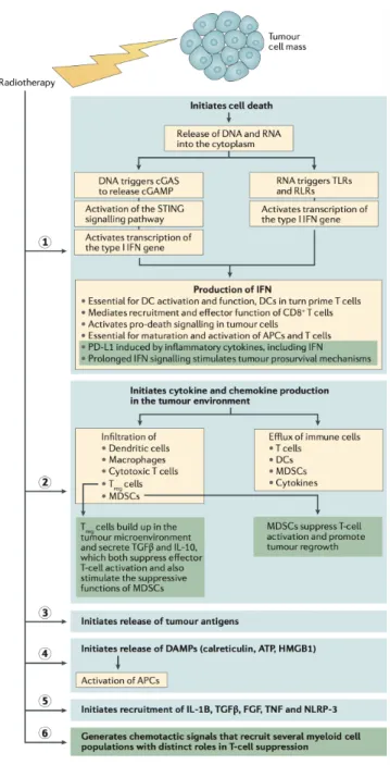
2. Radiotherapy enhances anti-tumour immune responses ... 91
a.
The multiple immunogenic properties of radiotherapy ... 91
b.
The abscopal effect ... 94
c.
Combinatorial approaches of radiotherapy with IO ... 94
3. DNA repair-targeted therapies: another class of immunomodulatory agents? ... 95
a.
PARPi: the advent of synthetic lethal approaches in the clinic ... 95
b.
PARPi plus ICI: a beneficial combination? ... 100
F.
Aims and approaches ... 101
Chapter II.
Materials and Methods ... 103
A.
Reagents ... 103
1. General chemicals and solutions ... 103
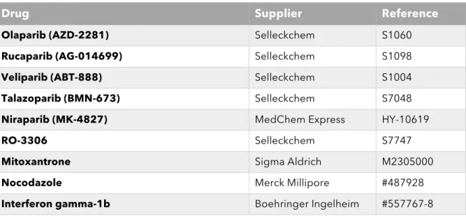
2. Drugs and chemotherapeutics ... 104
3. Antibodies ... 105
4. siRNA oligonucleotides ... 107
5. RT-qPCR probes ... 107
B.
Biological material ... 108
1. Tumour cell lines ... 108
2. Fresh pleural effusion samples ... 108
3. Archival tumour samples ... 109
C.
Protocols ... 109
1. Tissue culture ... 109
2. RNAi and transfections ... 109
3. Short-term drug survival assays ... 110
4. Protein manipulation ... 111
a.
Whole-cell protein extraction ... 111
b.
Subcellular protein fractionation ... 111
b.
RT-qPCR and gene expression measurements ... 112
6. Immunofluorescence and image analysis ... 113
7. Flow cytometry analyses ... 113
8. ELISA detection ... 115
9. ATP secretion assays ... 115
10. Immunohistochemistry and pathological scoring ... 116
11. Cytoblock preparation and immunocytochemistry ... 118
12. Transcriptomic analyses ... 118
a.
RNA-seq ... 118
b.
RNA-seq data analysis ... 119
c.
Nanostring® ... 120
d.
Nanostring® data analysis ... 120
13. TCGA data analyses ... 121
14. In vivo studies ... 121
a.
Generation of PDX models ... 121
b.
In vivo assessment of olaparib immunomodulatory potential ... 122
c.
Evaluation of rucaparib potential to induce ICD in vaccination assays ... 123
D.
Statistical analyses ... 123
1. General statistical analyses ... 123
2. Drug dose-response curves ... 124
Chapter III.
ERCC1 deficiency elicits cancer cell-autonomous immune
phenotypes in NSCLC ... 125
A.
Introduction ... 125
B.
Results ... 127
1. Isogenic NSCLC model of ERCC1 deficiency ... 127
2. ERCC1-deficiency in NSCLC drives activation of immune signalling in a cell-autonomous fashion ... 130
a.
RNA-seq of isogenic ERCC1-deficient A549 cells: general experimental approach ... 130
b.
RNA-seq results ... 130
3. Loss of ERCC1 correlates with increased lymphocytic infiltration in NSCLC patients’ tumours ... 139
4. ERCC1 dysfunction is associated with higher TMB in human tumours ... 143
5. Loss of ERCC1 associates with spontaneous re-expression of STING in isogenic NSCLC cells ... 146
C.
Discussion ... 148
Chapter IV.
PARPi exacerbate cancer cell-autonomous immunity through
cGAS/STING in DDR-deficient cells ... 150
A.
Introduction ... 150
B.
Results ... 151
1. PARPi induce formation of CCF in an ERCC1-dependent manner in NSCLC cells ... 151
a.
Cytoplasmic DNA: a peculiar phenomenon linked to genomic instability ... 151
b.
Choice of appropriate detection and quantification approaches for the evaluation of cytoplasmic DNA ... 152
e.
PARPi enhance ERCC1-dependent formation of CCF in H1975 cells ... 158
2. PARPi induce formation of CCF in a BRCA1-dependent manner in TNBC cells ... 160
a.
Isogenic TNBC models of BRCA1 deficiency and PARP1-deficiency ... 160
b.
PARPi generate CCF in a BRCA1-dependent fashion in SUM149 cells ... 162
c.
PARPi-mediated formation of CCF results from an on-target effect of PARPi on PARP1 164
3. PARPi-mediated generation of CCF is cell cycle-dependent ... 165
a.
CCF generated by PARPi have micronuclei characteristics ... 165
b.
PARPi generate dose-dependent formation of micronuclei ... 170
4. PARPi-induced CCF are detected by cGAS ... 172
a.
cGAS mediates the detection of CCF in PARPi-treated NSCLC cells ... 172
b.
cGAS mediates the detection of CCF in PARPi-treated TNBC cells ... 176
5. PARPi activate cGAS/STING signalling in a DDR-defects-dependent manner ... 179
a.
PARPi trigger TBK1 phosphorylation in an ERCC1-dependent manner in NSCLC cells . 179
b.
cGAS and STING are required for the activation of TBK1 by PARPi ... 182
c.
Progression through the cell cycle is required for PARPi-mediated TBK1 activation ... 184
d.
PARPi trigger TBK1 phosphorylation and downstream STING signalling in a BRCA1-dependent manner in TNBC cells ... 185
e.
Specificity of activation of the cGAS/STING pathway by PARPi ... 187
6. Activation of cGAS/STING by PARPi triggers secretion of CCL5 in a DDR-defects-dependent manner ... 189
a.
ERCC1-deficient NSCLC cells secrete CCL5 in response to PARPi ... 190
b.
BRCA1-deficient TNBC cells secrete CCL5 in response to PARPi ... 193
7. PARPi activate type I IFN signalling in ERCC1-deficient cells ... 196
8. PARPi exert immunomodulatory properties in vivo ... 202
a.
Experimental approach ... 202
b.
Genetic characteristics of the PDX models ... 205
c.
Verification of the tissue specificity of the Nanostring® assay ... 209
d.
PARPi-treated tumours exhibit enhanced expression of type I IFN genes ... 210
e.
PARPi upregulate MHC components in NSCLC cell lines in vitro ... 213
C.
Discussion ... 214
Chapter V.
PARPi modulate PD-L1 expression in tumour cells ... 217
A.
Introduction ... 217
B.
Results ... 219
1. PARPi synergise with IFN-γ to induce PD-L1 expression in NSCLC cells ... 219
a.
Experimental approach and controls ... 220
b.
Several clinical PARPi potentiate IFN-γ-mediated PD-L1 upregulation in NSCLC cells .. 222
c.
PARPi-mediated PD-L1 upregulation is dose-dependent, specific, and results from an on-target effect of PARPi ... 222
d.
PARPi induce PD-L1 expression in patient-derived NSCLC cells ... 226
2. ERCC1 deficiency exacerbates PARPi-mediated PD-L1 upregulation ... 228
3. PARPi-mediated PD-L1 upregulation is independent from cGAS/STING signalling activation ... 230
4. PARP1 activity is linked to PD-L1 expression in cancer cells ... 232
C.
Discussion ... 235
B.
Results ... 239
1. In vitro detection of ICD: study design and experimental choices ... 239
2. PARPi generate apoptosis of NSCLC cells ... 242
3. PARPi induce ER stress in NSCLC cells ... 246
a.
Rucaparib induces phosphorylation of the ER factor eIF2α ... 246
b.
Rucaparib and talazoparib trigger CALR exposure ... 248
4. PARPi stimulate autophagy and promote ATP secretion in NSCLC cells ... 252
a.
ATP is secreted in response to rucaparib exposure ... 253
b.
Rucaparib and talazoparib trigger LC3 activation ... 256
5. PARPi generate HMGB1 release in NSCLC cells ... 259
6. In vivo study of the potential of rucaparib to generate ICD ... 264
a.
Design of a pilot vaccination assay ... 264
b.
Vaccination assay: results ... 266
C.
Discussion ... 269
Chapter VII.
Final discussion and perspectives ... 272
A.
Critical findings presented in this thesis ... 274
B.
Mechanisms controlling cytosolic DNA accumulation in response to PARPi 276
C.
Biological implications of PARPi-mediated stimulation of the cGAS/STING pathway ... 280
1. Immunological impact of cGAS/STING signalling activation ... 280
2. Deleterious effects of chronic cGAS/STING pathway activation ... 281
D.
Potential determinants of cancer cell-autonomous immune responses elicited by PARPi ... 281
1. Epigenetic determinants of cGAS/STING-mediated immune responses ... 281
2. Multiple DDR defects might trigger tumour cell-intrinsic immunity ... 282
E.
Challenges in exploiting PARPi potential to induce ICD ... 283
F.
Complementary approaches to assess the immunomodulatory properties of PARPi ... 285
G.
Clinical implications of the immunological potential of PARPi ... 287
H.
Final conclusion ... 289
References ... I
Synthèse en français ... XXXIX
Figure I.1. The three phases of cancer immunoediting. ... 32
Figure I.2. The cancer-immunity cycle. ... 34
Figure I.3. Mechanisms of immune escape in the tumour microenvironment. ... 36
Figure I.4. Classes of human tumour antigens that are recognized by T lymphocytes. ... 37
Figure I.5. Processing of tumour antigens that are recognized by CD8+ T cells. ... 38
Figure I.6. Impact of oncogenic signalling on immune inhibitory pathways and cell populations. ... 41
Figure I.7. Multiple co-stimulatory and co-inhibitory interactions regulate T cell responses. ... 43
Figure I.8. Two general mechanisms of expression of immune-checkpoint ligands on tumour cells. ... 45
Figure I.9. Derivation of TCRs and CARs for the genetic modification of T cells. ... 49
Figure I.10. Mechanistic nodes in immune checkpoint pathways. ... 53
Figure I.11. Pipeline for the identification of immune-relevant neo-antigens. ... 58
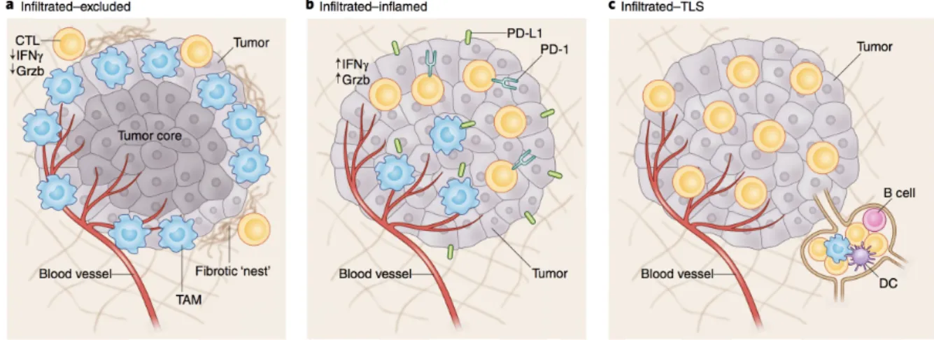
Figure I.12. General classes of TME. ... 61
Figure I.13. Cancer-immune phenotypes. ... 68
Figure I.14. The prevalence of somatic mutations across human cancer types. ... 70
Figure I.15. DNA repair defects and their association with anti-PD-(L)1 efficacy in solid tumours. ... 78
Figure I.17. Mechanisms of chemotherapy-driven ICD. ... 88
Figure I.18. Radiation-induced effects on tumour cells. ... 93
Figure I.19. A model describing PARP1 catalytic cycle. ... 96
Figure I.20. Clinical PARP inhibitors. ... 97
Figure III.1. Generation of an isogenic model of ERCC1-deficiency in the A549 NSCLC cell line. ... 129
Figure III.2. Differential expression analysis of A549-ERCC1 isogenic cell lines. ... 131
Figure III.3. GSEA of the REACTOME pathway Interferon Alpha Beta Signalling in A549-ERCC1WT/WT compared with A549-ERCC1-/- cells. ... 135
Figure III.4. GSEA of the REACTOME pathway Cytokine Signalling in Immune System in the A549-ERCC1WT/WT compared with A549-ERCC1-/- cells. ... 136
Figure III.5. GSEA of REACTOME pathways in A549-c295 cells (A), A549-c375 cells (B) and A549-ERCC1+/- cells (C) compared with A549-ERCC1WT/WT cells. ... 138
Figure III.6. Low ERCC1 expression correlates with high levels of TILs in human NSCLC tumours. ... 140
Figure III.7. Evaluation of the correlation between ERCC1 gene copy number and expression of immune-related markers in the TCGA lung adenocarcinoma cohort. 141 Figure III.8. Evaluation of the correlation between ERCC1 gene copy number and expression of immune-related markers in the TCGA lung squamous cell carcinoma cohort. ... 142
Figure III.9. Deleterious mutations of ERCC1 are associated with increased TMB in human tumours. ... 144
Figure IV.1. Computational image analysis pipeline used for the identification of CCF, micronuclei and cytoplasmic cGAS foci in fluorescence microscopy images. ... 153 Figure IV.2. ERCC1-deficient cells exhibit enhanced sensitivity to PARPi. ... 155 Figure IV.3. PARPi induce formation of CCF in an ERCC1-dependent manner in A549 cells.
... 158 Figure IV.4. PARPi induce formation of CCF in an ERCC1-dependent manner in H1975 cells.
... 159 Figure IV.5. Generation of isogenic models of BRCA1-deficiency and PARP1-deficiency in the SUM149 TNBC cell line. ... 161 Figure IV.6. PARPi induce formation of CCF in a BRCA1-dependent manner in SUM149 cells. ... 164 Figure IV.7. PARPi generate cytoplasmic chromatin in SUM149 cells via an on-target effect on PARP1. ... 165 Figure IV.8. The cell cycle blockers 5-FU and hydroxyurea prevent PARPi-mediated formation of CCF in A549 cells. ... 167 Figure IV.9. The CDK1 inhibitor RO-3306 prevents PARPi-mediated formation of CCF. in A549 and SUM149 cells. ... 168 Figure IV.10. PARPi-induced CCF have micronuclei characteristics. ... 170 Figure IV.11. PARPi generate dose-dependent formation of micronuclei. ... 171 Figure IV.12. PARPi induce formation of cGAS foci in an ERCC1-dependent manner in A549 cells. ... 174 Figure IV.13. PARPi induce formation of cGAS foci in an ERCC1-dependent manner in H1975 cells. ... 176
SUM149 cells. ... 178 Figure IV.15. PARPi trigger TBK1 phosphorylation in a dose-dependent manner in
ERCC1-deficient A549 cells. ... 180 Figure IV.16. PARPi trigger TBK1 phosphorylation in a dose-dependent manner in H1975 cells. ... 181 Figure IV.17. PARPi-mediated phosphorylation of TBK1 is dependent on cGAS and STING activity. ... 183 Figure IV.18. PARPi-mediated phosphorylation of TBK1 is cell cycle-dependent. ... 184 Figure IV.19. PARPi trigger TBK1 phosphorylation in a dose-dependent manner in
BRCA1-mutated SUM149 cells. ... 186 Figure IV.20. PARPi do not trigger RLR or TLR signalling pathways activation in NSCLC cells.
... 188 Figure IV.21. PARPi trigger CCL5 secretion via cGAS/STING in ERCC1-deficient A549 cells.
... 191 Figure IV.22. PARPi activate CCL5 transcription via cGAS/STING in A549 cells. ... 192 Figure IV.23. PARPi induce CCL5 and IFNB1 transcription in BRCA1-mutated SUM149 cells.
... 194 Figure IV.24. PARPi induce secretion of CCL5 but not IFN-β in BRCA1-mutated SUM149 cells. ... 195 Figure IV.25. GSEA of the REACTOME pathway Interferon Alpha Beta Signalling in talazoparib- vs DMSO- treated A549-ERCC1-/- cells. ... 199 Figure IV.26. GSEA of the REACTOME pathway Interferon Alpha Beta Signalling in talazoparib- vs DMSO- treated A549-ERCC1WT/WT cells. ... 201
tumours. ... 205 Figure IV.28. Heatmap showing all significantly differentially expressed genes in
olaparib-treated vs vehicle-olaparib-treated BTBC456 tumours. ... 211 Figure IV.29. Heatmap showing all significantly differentially expressed genes in
olaparib-treated vs vehicle-olaparib-treated BX102 tumours. ... 212 Figure IV.30. PARPi induce cell-surface expression of MHC class I components in NSCLC cells. ... 213 Figure IV.31. A proposed model to explain cGAS/STING activation following PARPi exposure in tumour cells harbouring DDR defects. ... 215 Figure V.1. Details of the flow cytometry analysis used to detect PD-L1 cell surface expression in NSCLC cells. ... 221 Figure V.2. PARPi synergize with IFN-γ to induce PD-L1 cell surface expression in NSCLC cells. ... 223 Figure V.3. PARPi-mediated induction of PD-L1 is dose-dependent. ... 224 Figure V.4. PARPi induce cell surface expression of PD-L1 but not TLR4 in H1975 cells. 225 Figure V.5. PD-L1 induction results from an on-target effect of PARPi on PARP1. ... 226 Figure V.6. PARPi induce PD-L1 expression in patient-derived tumour cells. ... 227 Figure V.7. ERCC1-deficient cells present an enhanced potential to induce PD-L1 expression in response to PARPi and IFN-γ. ... 229 Figure V.8. PARPi does not induce PD-L1 expression via cGAS/STING signalling activation.
... 231 Figure V.9. Low PARylation levels correlate with high PD-L1 expression in human NSCLC tumours. ... 233
PD-L1 expression in immune cells in human NSCLC tumours. ... 234 Figure V.11. PARP1-deficient SUM149 cells express higher baseline expression levels of PD-L1 compared to their PARP1-wildtype isogenic counterparts. ... 234 Figure V.12. PARPi potentiate IFN-γ-mediated phosphorylation of STAT1 in normally cycling ERCC1-proficient cells. ... 237 Figure VI.1. PARPi have distinct pharmacological properties and exert different cytotoxic effects in NSCLC cell lines. ... 240 Figure VI.2. Diagram depicting the experimental pipeline developed for the evaluation of ICD in response to PARPi in NSCLC cell lines. ... 242 Figure VI.3. PARPi induce apoptosis and subsequent secondary necrosis in A549-ERCC1WT/WT cells. ... 244 Figure VI.4. PARPi induce apoptosis and subsequent secondary necrosis in H1975-ERCC1WT/WT cells. ... 245 Figure VI.5. Rucaparib induces intense phosphorylation of eIF2α in A549-ERCC1WT/WT and H1975-ERCC1WT/WT cells. ... 247 Figure VI.6. PARPi rucaparib and talazoparib induce CALR exposure in A549-ERCC1WT/WT cells. ... 250 Figure VI.7. A549-ERCC1WT/WT cells undergo CALR exposure in response to treatment with talazoparib. ... 251 Figure VI.8. Rucaparib triggers ATP secretion in A549-ERCC1WT/WT cells. ... 254 Figure VI.9. Rucaparib triggers ATP secretion in H1975-ERCC1WT/WT cells. ... 255 Figure VI.10. Rucaparib and talazoparib promote the formation of LC3-decorated autophagic puncta in A549-ERCC1WT/WT cells. ... 258
Figure VI.12. Rucaparib and talazoparib trigger HMGB1 nucleo-cytoplasmic translocation in A549-ERCC1WT/WT cells. ... 261 Figure VI.13. HMGB1 is released in response to rucaparib in NSCLCcells. ... 262 Figure VI.14. Schematic of the differential induction of ICD-associated DAMPs by PARPi in NSCLC cells. ... 263 Figure VI.15. Cytotoxic effects of PARPi in CT26 cells. ... 265 Figure VI.16. Schematic of the pilot vaccination assay designed to evaluate the potential of rucaparib to trigger ICD in vivo. ... 266 Figure VI.17. Rucaparib induces apoptosis, ER stress and autophagy in CT26 cells. ... 267 Figure VI.18. Rucaparib does not protect syngeneic mice against CT26 tumours. ... 268
Table I.1. Clinical trials of anti-PD-(L)1 and their results in NSCLC. ... 54 Table I.2. Immune-related biomarkers for anti-PD-(L)1 therapy. ... 65 Table I.3. Type and frequency of DNA repair alterations in solid tumours. ... 73 Table II.1. Summary of the drugs used in this study. ... 104 Table II.2. Summary of antibodies used for WB and IF in this study. ... 105 Table II.3. Summary of antibodies used for IHC and ICC in this study. ... 106 Table II.4. Summary of antibodies used for flow cytometry in this study. ... 106 Table II.5. Summary of the siRNAs used in this study. ... 107 Table II.6. Summary of the RT-qPCR probes used in this study. ... 107 Table III.1. GSEA of REACTOME pathways in A549-ERCC1-/- cells compared with
A549-ERCC1WT/WT cells. ... 132 Table III.2. GSEA of REACTOME pathways in A549-ERCC1-/- cells compared with
A549-ERCC1WT/WT cells. ... 132 Table III.3. Differential expression analysis of A549-ERCC1-/- cells compared with
A549-ERCC1WT/WT cells for various immune-related genes. ... 137 Table III.4. Characteristics of the cohorts used for the pan-cancer analysis of TMB according to ERCC1 mutation status. ... 145 Table III.5. Differential expression analysis of A549-ERCC1-/- cells compared with
compared with DMSO-treated A549-ERCC1-/- cells. ... 197 Table IV.2. GSEA of REACTOME pathways in talazoparib-treated A549-ERCC1-/- cells compared with DMSO-treated A549-ERCC1-/- cells. ... 198 Table IV.3. GSEA of REACTOME pathways in talazoparib-treated A549-ERCC1WT/WT cells compared with DMSO-treated A549-ERCC1WT/WT cells. ... 200 Table IV.4. Genetic characteristics of the BTBC456 PDX model. ... 206 Table IV.5. Genetic characteristics of the BX102 PDX model. ... 207 Table IV.6. Genes of the nCounter® PanCancer immune panel whose probe sequence has shown alignment with the mouse genome. ... 209 Table VII.1. Summary of the ongoing clinical trials evaluating PARPi plus anti-PD-(L)1 agents for the treatment of cancer. ... 273
5-FU 5-Fluorouracil
7-AAD 7-aminoactinomycin D
ADCC Antibody-Dependent Cellular Cytotoxicity
ALL Acute Lymphoid Leukaemia
APC Antigen-Presenting Cells
ATCC American Type Culture Collection
BCG Bacillus Calmette-Guérin
CAR T cells Chimeric Antigen Receptor T cells
CCF Cytoplasmic Chromatin Fragments
CDC Complement-Dependent Cytotoxicity
CDK1i CDK1 inhibitor
cGAMP Cyclic GMP-AMP
CI Confidence Interval
CLL Chronic Lymphoid Leukaemia
CRC Colorectal Carcinoma
DAMPs Damage-Associated Molecular Patterns
DAPI 4',6-diamidino-2-phenylindole
DC Dendritic Cells
DEGs Differentially Expressed Genes
DSB Double-Strand Break
dsDNA Double-Stranded DNA
ELISA Enzyme-Linked Immunosorbent Assay
EMT Epithelial–Mesenchymal Transition EpCAM Epithelial Cell Adhesion Molecule
ER Endoplasmic Reticulum
FACS Fluorscence-Activated Cell Sorting
FDR False Discovery Rate
FSC Forward Scatter
GSEA Gene Set Enrichment Analysis
HCC Hepatocellular Carcinoma
HGSOC High-Grade Serous Ovarian Cancer
HLA Human Leucocyte Antigen
HNSCC Head and Neck Squamous Cell Carcinoma
HR Homologous Recombination
HRP Horseradish Peroxidase
HU Hydroxyurea
ICC Immunocytochemistry
IHC Immunohistochemistry
IO Immunotherapy
IR Ionizing Radiation
IRF Interferon Regulatory Factor LAK Lymphokine-Activated Killer
LFC log2 Fold-Change
Mb Megabase
mCRPC Metastatic Castration-Resistant Prostate Cancer
MDSC Myeloid-derived Suppressor Cells
MFI Mean Fluorescence Intensity MHC Major Histocompatibility Complex
miRNA Micro-RNA
MMR Mismatch-Repair
mRNA Messenger Ribonucleic Acid
MSI Microsatellite-Instability
MTX Mitoxantrone
NCI National cancer Institute
NCZ Nocodazole
NER Nucleotide Excision Repair
NES Normalized Enrichment Score
NGS Next-Generation Sequencing
NHEJ Non-Homologous End Joining
Nira Niraparib
NK Natural Killer
NKT Natural Killer T
NSCLC Non-Small Cell Lung Cancer
nsSNV Non-Synonymous Single-Nucleotide Variants
Ola Olaparib
ORR Overall Response Rate
OS Overall Survival
PAMP Pathogen-Associated Molecular Pattern
PBMC Peripheral Blood Mononuclear Cells
PDX Patient-Derived Xenograft
PE Phycoerythrin
PFS Progression-Free Survival
PMSF Phenylmethylsulfonyl Fluoride
PRR Pattern Recognition Receptors
PS Phosphatidyl-Serine
RCC Renal Cell Carcinoma
RNA-seq RNA sequencing
RT-qPCR Quantitative Reverse Transcription Polymerase Chain Reaction
siRNA Small-Interfering RNA
ssDNA Single-Stranded DNA
Talazo Talazoparib
TAM Tumour-Associated Macrophages
TAN Tumour-Associated Neutrophils
TCGA The Cancer Genome Atlas
TCR T Cell Receptor
TILs Tumour-Infiltrating Lymphocytes
TLR Toll-Like Receptors
TLS Tertiary Lymphoid Structures
TMB Tumour Mutational Burden
TME Tumour Microenvironment
TNB Tumour Neo-antigen Burden
TNBC Triple-Negative Breast Cancer
Treg Regulatory T cells
UBC Urothelial Bladder Carcinoma
UPR Unfolded Protein Response
WB Western Blot
ANXA1 Annexin A1
ATF6 Activating Transcription Factor 6 ATM Ataxia Telangiectasia Mutated
ATR Ataxia Telangiectasia and Rad3 related
B2M Beta-2 Microglobulin
BARD1 BRCA1 Associated RING Domain 1
BLM Bloom Syndrome RecQ Like Helicase BRCA1/2 Breast Cancer Susceptibility Gene ½
BRIP1 BRCA1 Interacting Protein C-Terminal Helicase 1
CALR Calreticulin
CCL5 C-C Motif Chemokine Ligand 5
CD4 T-Cell Surface Glycoprotein CD4
CD47 Antigenic Surface Determinant Protein OA3 CD8 T-Cell Surface Glycoprotein CD8
CD91/LRP1 LDL Receptor Related Protein 1
CDK12 Cyclin Dependent Kinase 12
cGAS Cyclic GMP-AMP Synthase
CHK1/2 Checkpoint Kinase 1/2
CTLA-4 Cytotoxic T-Lymphocyte Associated Protein 4 CXCL5/9/10/12 C-X-C Motif Chemokine Ligand 5/9/10/12
CXCR4 C-X-C Motif Chemokine Receptor 4
DNASE2 Deoxyribonuclease 2, Lysosomal
ERCC1 Excision Repair Cross-Complementation Group 1
EXO1 Exonuclease 1
FANCA Fanconi Anemia Complementation Group A
FANCD2 Fanconi Anemia Complementation Group D2
FANCF Fanconi Anemia Complementation Group F
FAS-L FAS Ligand
FRP1 Formyl Peptide Receptor 1
Gal9 Galectin-9
GM-CSF Granulocyte/Macrophage Colony-Stimulating Factor
HER2 Human Epidermal growth factor Receptor-2
HMGB1 High-Mobility Group Box 1
HSP70/90 Heat Shock Protein 70 kDa/90 kDa
IDO Indoleamine 2,3-Dioxygenase
IFN-γ Interferon Gamma
IKK Inhibitor of Nuclear Factor Kappa B Kinase Subunit Beta
IL Interleukin
IRE1α Inositol-Requiring Enzyme 1 Alpha KLRK1/NKG2D Killer Cell Lectin Like Receptor K1 LKB1/STK11 Serine/Threonine Kinase 11
MLH1/3 MutL Homolog 1/3
MRE11 MRE11 Homolog, Double Strand Break Repair Nuclease
MSH2/3/6 MutS Homolog 2/3/6
mTOR Mammalian Target Of Rapamycin
MUS81 MUS81 Structure-Specific Endonuclease Subunit MYC MYC Proto-Oncogene, BHLH Transcription Factor
NF-κB Nuclear Factor-kappa B
NFKB1/2 Nuclear Factor Kappa B Subunit 1/2
NSB1/NBN Nibrin
PALB2 Partner And Localizer of BRCA2
PANX1 Pannexin 1
PARP1/2 Poly(ADP-Ribose) Polymerase 1/2
PD-1 Programmed Cell Death 1
PD-L1/CD274 Programmed Cell Death 1 Ligand 1
PDIA3/ERp57 Protein Disulfide Isomerase family A member 3
PERK Protein Kinase R-like ER Kinase
PGE2 Prostaglandin E2
PMS2 PMS1 Homolog 2, Mismatch Repair System Component
POLD1 DNA Polymerase Delta 1, Catalytic Subunit POLD3 DNA Polymerase Delta 3, Accessory Subunit POLE/Pol-ε DNA Polymerase Epsilon
pTBK1 Phosphorylated TBK1
PTEN Phosphatase And Tensin Homolog
RAD17 RAD17 Checkpoint Clamp Loader Component
RAD50 RAD50 Double Strand Break Repair Protein
RAD51C RAD51 Paralog C
RAD54L RAD54 Like
RAGE Advanced Glycosylation End product-specific Receptor
RELB RELB Proto-Oncogene, NF-KB Subunit
RNASEH2 Ribonuclease H2
SAMHD1 SAM And HD Domain Containing Deoxynucleoside Triphosphate STING Stimulator of Interferon Genes
TAP Transporter associated with Antigen Processing
TBK1 TANK Binding Kinase 1
TGF-β Transforming Growth Factor beta
TNF-α Tumour Necrosis Factor alpha
TRAIL Tumour necrosis factor–Related Apoptosis-Inducing Ligand
TREX1 Three Prime Repair Exonuclease 1
VEGF Vascular Endothelial Growth factor
XPF Xeroderma Pigmentosum Group F-Complementing Protein
Chapter I. Introduction
A. Cancer and immunity: the emergence of a paradigm
Cancer is a genetic disease. It arises from alterations in genes controlling key biological processes, collectively responsible for the maintenance of cellular homeostasis. Both gain-of-function mutations in proto-oncogenes and loss-gain-of-function mutations in tumour suppressor genes can lead to the transformation of normal cells into malignant cells, thereby initiating tumour development. This genetic-based description of carcinogenesis has enabled the identification of many oncogenic drivers involved in tumour development, among which some of them are currently exploited as actionable targets for the treatment of cancer. Beyond the cancer cell itself, the contribution of the tumour microenvironment (TME) — and notably the immune system — to carcinogenesis has been recognized for more than a century, but has only very recently led to significant therapeutic advances.
1. From cancer immunosurveillance to cancer immunoediting
The involvement of immunity in tumour recognition and control was first hypothesized by the German physician and scientist Paul Ehrlich in 1909. He postulated that cancer occurred spontaneously in human, but that in the majority of people, the host immune system was able to prevent neoplastic cells from developing into tumours (1). In the late 1950s, the idea of a host immune protection against cancer was revisited through the concept of cancer immunosurveillance by two prominent immunologists: Frank Macfarlane Burnet and Lewis Thomas. Burnet’s pioneering theory stated that tumour cells-specific neo-antigens could challenge host immune tolerance and trigger an effective anti-tumour immunological reaction capable of eliminating nascent neoplasms (2,3). Thomas’s theory was shaped by an evolutionary point of view and suggested that long-lived organisms must possess protecting mechanisms against cancer, similar to those mediating allograft rejection. Both
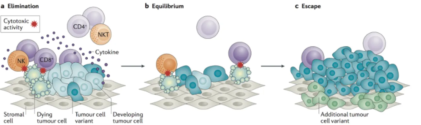
theories were supported by the functional demonstration of mouse tumour-specific antigens (4) and converged towards the concept of cancer immunosurveillance, which defined the critical role of sentinel thymus-dependent immune cells in constantly monitoring the appearance of early neoplasms in host tissues. This concept gained recognition, but was rapidly challenged by studies showing no differences in primary tumour development between athymic nude mice and syngeneic wild-type mice (5,6). Later discoveries demonstrated that lymphocytes and interferon-gamma (IFN-γ) play a beneficial role in the protection of hosts against the development of carcinogen-induced sarcomas and spontaneous epithelial carcinomas, but that they also promote selection of tumour cells with reduced immunogenicity, capable of escaping immune recognition and destruction (7). This process of immunoselection of tumour cells could explain the apparent paradox of tumour formation in immunologically competent individuals, by raising the possibility of escape from immunosurveillance. These findings were the basis for the development of the immunoediting theory in 2002 (8), which described for the first time how both innate and adaptive immunity contribute to tumour development through a dynamic process of Darwinian immunoselection of tumour cell variants. This process classically consists of three distinct steps (Figure I.1):
(1) Elimination: the innate and adaptive compartments of the immune system coordinately drive immune rejection;
(2) Equilibrium: through a clonal selection process, the dynamic balance between tumour and immune cells results in the emergence of specific tumour cell variants with increased resistance, which take advantage of acquired mutations;
(3) Escape: the immune-resistant clones freely expand, circumventing both innate and adaptive immune responses.
Cancer immunoediting describes the intricate relationship between a tumour and its infiltrating immune system, during which genetic instability and tumour heterogeneity increase and immune selection of tumour cell variants occurs. (A) The first phase encompasses the classical concept of cancer immunosurveillance. Tumour invasion generates inflammatory signals and recruitment of innate immune cells to the tumour site such as NK cells, γδ T cells, macrophages, and dendritic cells (DCs). Tumour antigens expressed by malignant cells are recognized by infiltrating NK or γδ T cells, which are in turn stimulated for the production of IFN-γ. This key soluble cytokine primarily induces the production of other mediators by the tumour cells themselves — such as the lympho-attractant CXC-chemokine ligand CXCL9, CXCL10, and CXCL11 —, and also directly triggers tumour cell death through the activation of angiostatic, anti-proliferative or apoptotic mechanisms (9). While chemokines produced during this escalating inflammatory process recruit more NK cells and macrophages to the tumour, apoptotic tumour cells and debris are engulfed by local DCs; these migrate to lymph nodes and activate tumour-specific CD4+ T helper cells expressing IFN-γ, that in turn stimulate the proliferation of tumour-specific cytotoxic CD8+ T cells. Tumour-infiltrating NK cells and macrophages destroy more tumour cells through the production of tumour necrosis factor-related apoptosis-inducing ligand (TRAIL) and perforin. In parallel, newly activated CD4+ and CD8+ T cells migrate to the tumour site, and cytotoxic CD8+ T cells eradicate the remaining antigen-bearing tumour cells whose immunogenicity has been enhanced by IFN-γ. Hence, in the elimination phase, both innate and adaptive immunity cooperatively participate to cancer rejection. (B) During the intermediate phase, the host immune system and surviving tumour cell variants enter into a dynamic equilibrium. Indeed, T infiltrating lymphocytes (TILs) and IFN-γ cooperate to exert a potent selection pressure that is sufficient to contain, but not fully eliminate, a bulk of genetically unstable and highly mutating tumour cells. Occurring over a long period of time, this Darwinian selection eventually leads to the extinguishment of originally escaping variants, and concomitant emergence of new variants bearing different mutations that confer them both a proliferative advantage and an increased resistance to immune aggression. (C) The ultimate phase corresponds to the expansion of surviving tumour variants that have acquired resistance to immunological detection or rejection through mutations or epigenetic changes. This leads to aggressive tumour development and clinically observable malignant disease.
Figure and legend adapted from Van der Burg et al., Nature Reviews, 2016.
The immune system therefore plays a dual role of (i) orchestrating tumour rejection, and (ii) sculpting the molecular profile and immunogenicity of developing tumours, by favouring tumour cell variants with low immunogenicity through a T cell-dependent immunoselection — which eliminates clones with strong rejection antigens (10) — and a T cell-independent immunoselection mediated by innate immune cells (11). Type I and type II IFN further play a key role in the coordination of tumour-immune interactions during immunoediting (12), and in particular, IFN-γ was recently described as an essential mediator of cytotoxic T cell-dependent tumour genome immunoediting (13).
Besides immunoselection, other immune-independent mechanisms, such as neutral evolution (14) — that is, the accumulation of passenger mutations without selective sweeps —, have been described to contribute to tumour heterogeneity in some cancers. A recent study showed using an in vivo model of microsatellite-instable (MSI) colorectal carcinoma (CRC), that immunoediting effects were weak and dominated by neutral accumulation of mutations (15) during CRC tumour development. Interestingly, the use of immune checkpoint inhibitors (ICI) in this model effectively potentiated immunoediting through increased immunoselective pressure, thereby inducing changes in the clonal and subclonal composition of the tumour, and changing the evolutionary dynamics from neutral to selective evolution. This underlines the plasticity of cancer immunoediting processes, and further suggests that modulation of the tumour-immune interactions via anti-cancer therapies having immunomodulatory properties can favour immunoediting, and potentially facilitate immune evasion through acquired resistance to therapy.
2. When oncology meets immunology: the cancer-immunity cycle
The cancer immunoediting approach describes a continuous evolution of the interactions between cancer and immunity which eventually favours tumour development despite the initiated immune rejection. How the immune system actually eliminates tumour cells during the elimination phase of cancer immunoediting is a well-known process, which has been recapitulated and nicely illustrated with the concept of cancer-immunity cycle (16).
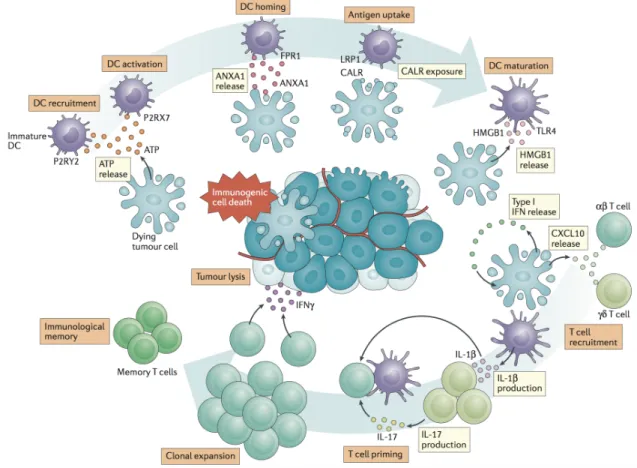
(1) Rapidly growing tumours display high levels of tumour cell death and apoptosis because of their elevated growth rate. This generates neo-antigens that are captured by DCs upon release in the TME. In addition, dying tumour cells produce immunogenic signals, such as pro-inflammatory cytokines, that stimulate DCs and further activate other innate immunity effectors such as macrophages. (2) Stimulated DCs migrate to the lymph nodes and efficiently process tumour neo-antigens to present them on major histocompatibility complex (MHC) Class II to T cells through cross-presentation. (3) These neo-antigens are detected as “non-self” because of their specificity to cancer cells or because the central tolerance has been incomplete towards them. This results in the priming and activation of CD4+ helper T cells and CD8+ effector T cells, and eventually leads to the expansion of tumour cell-specific T cell clones, that constitute the adaptive arm of anti-tumour immunity. At this stage, the nature of the immune response is already determined, especially since the critical balance between effector T cells and regulatory T cells (Treg) is defined. (4) CD8+ effector T cells traffic through blood circulation to the tumour site. (5) T cells infiltrate the tumour bed, and become the so-called tumour-infiltrating lymphocytes (TILs). (6) TILs specifically recognize and bind tumour cells through the interaction between their T cell receptor (TCR) and the cognate neo-antigen presented on MHC class I by tumour cells. (7) This results in killing of the cancer cells, a process which releases additional neo-antigens and eventually increases the breadth and depth of the immune response in subsequent revolutions of the cycle. Abbreviations: APCs, antigen presenting cells; CTLs, cytotoxic T lymphocytes.
Figure and legend adapted from Chen and Mellman, Immunity, 2013.
This concept describes a series of seven sequential events required for the establishment of an effective anti-tumour immunity (detailed in Figure I.2). Importantly, the cycle involves various immune cell types circulating from and towards distinct anatomic locations, and constitutes a self-propagating process. However, failure in any step of the cycle leads to evasion of the tumour from immunological control, and eventually cancer progression.
3. Mechanisms of immune escape in cancer
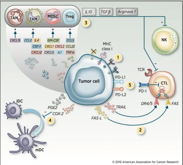
Immune evasion is a hallmark of cancer (17), which arises from genetic or epigenetic alterations of cancer cells (Figure I.3), and involves various mechanisms: (i) mechanisms that affect tumour immunogenicity; (ii) mechanisms that restrain anti-tumour activity of immune effectors; (iii) mechanisms that corrupt suppressive immune effectors; (iv) mechanisms that involve modulation of immune checkpoints.
a. Immune escape through reduction of tumour immunogenicity
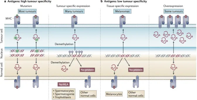
Tumour immunogenicity is primarily determined by the antigenicity of tumour cells, which derives from their ability to generate and present tumour-associated antigens, recognized as “non-self” by the cells of the immune system, and responsible for the activation of anti-tumour immunity. Tumour-associated antigens originate from various sources, and are classically comprised by two distinct types of antigens (18,19) (Figure I.4):
S Mutated tumour-specific antigens, also known as neo-antigens, are exclusively expressed by tumour cells and directly originate from the large number of somatic mutations that are found in human tumours; most of the time, point mutations are responsible for the expression of mutant peptides that are not tolerated by the immune system, and eventually trigger anti-tumour immune responses. Such neo-antigens may be newly displayed at the surface of tumour cells because a mutation increases the binding affinity of a peptide to major histocompatibility complex (MHC) molecules. Alternatively, the mutation can alter the T cell receptor (TCR)-exposed area of a peptide that is also presented by MHC in its non-mutated form.
Several mechanisms, involving multiple immune components, contribute to tumour immune escape. (1) Immune recognition can be impaired following reduced expression of MHC class I molecules in malignant cells, resulting in decreased antigen presentation and consequently reduced detection by cytotoxic CD8+ T lymphocytes. (2) Cancer cells can activate immunosuppressive mechanisms by inducing apoptosis of immune cells through the expression of death signals (including FAS- and TRAIL-ligands). (3) Tumour cells release in the microenvironment a variety of immunomodulatory molecules that inhibit the immune system by inducing immunosuppressive cells. (4) This cytokine imbalance, combined with the secretion of TGF-β, cyclooxygenase-2 (COX-2), and prostaglandin E2 (PGE2), inhibits DCs differentiation and maturation, thereby affecting antigen presentation and recognition by T cells. (5) Disrupted expression of immune checkpoint ligands by cancer cells provides co-inhibitory signals to CD4+ and CD8+ T lymphocytes. Abbreviations: CCL, chemokine ligand; CXCL, chemokine (C-X-C motif) ligand; FAS-L, FAS-ligand; GM-CSF, granulocyte macrophage colony-stimulating factor; iDC, immature dentritic cell; IDO, indoleamine-2,3-deoxygenase; mDC, mature dentritic cell; MDSC, myeloid-derived suppressor cell; PD-1, programmed cell death 1; PD-L, programmed cell death ligand; TAN, tumour-associated neutrophil; TCR, T cell receptor; Treg, regulatory T cells.
Figure and legend adapted from Chabanon et. al, Clinical Cancer Research, 2016.
S Non-mutated self-antigens, by contrast, are not exclusively expressed by tumour cells, but also by other cells in a restricted set of cell types. These include (i) “cancer-testis antigens” whose expression is normally restricted to male germline cells in the testis: in tumour cells of many cancer types, these derive from the abnormal expression of cancer-germline genes due to transcriptional regulation defects or demethylation events; (ii) “tissue-differentiation antigens”, which emanate from the expression of tissue-specific genes in tumour cells, but whose expression is shared with cells of the tissue they originated from; (iii) “protein-overexpression antigens”, that derive from proteins that are overexpressed in tumours, but are also expressed in healthy tissues (e.g. HER2).
While most neo-antigens are very likely to trigger anti-tumour immunity because of their tumour-specificity, non-mutated self-antigens do not always activate immune responses.
A. Tumour antigens with high tumour specificity usually originate from point mutations.
Cancer-testis antigens can also be considered as tumour-specific because of their selective expression in tumours — germline cells, which lack HLA molecules, do not express them. B. Antigens with low tumour specificity arise from tissue-specific gene expression or overexpression of particular proteins. Only HLA class I molecules are represented, but the genetic processes shown can also lead to the presence on tumour cells of antigenic peptides that are presented by MHC class II molecules to CD4+ T cells.
Figure and legend adapted from Coulie et. al, Nature Reviews, 2014.
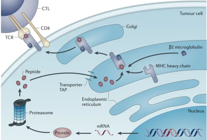
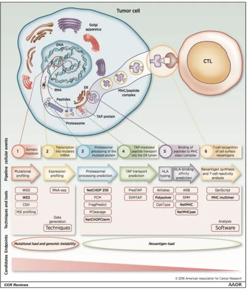
The process of neo-antigen presentation in tumour cells involves six successive steps, illustrated in Figure I.5 (19). As this multistep process involves various independent but complementary components, a defect in any of them is sufficient to impede the whole antigen presentation machinery, reduce tumour immunogenicity and eventually favour immune escape. For example, TAP1 protein deficiency in tumour cells has been associated with evasion from immune surveillance and increased tumourigenesis (20). Similarly, immunoproteasome deficiency has been linked to reduced antigen presentation, immune escape and poorer outcome in non-small cell lung cancer (NSCLC) (21).
(1) A somatic mutation occurs in the genome of the cancer cell; this leads to (2) the transcription of a mutated messenger RNA (mRNA) and subsequent translation into a mutated, presumably non-functional protein. (3) The mutated protein is processed through the proteasome and degraded into multiple peptides, that are subsequently (4) transported into the endoplasmic reticulum (ER) lumen by transporter associated with antigen processing (TAP) proteins, and (5) loaded on MHC Class I. MHC-bound peptides are then translocated to the cell-surface and eventually (6) presented to a TCR-matching T cell.
Figure and legend adapted from Coulie et. al, Nature Reviews, 2014.
But the most frequent and best documented cause of immune escape through reduced tumour immunogenicity is MHC Class I deficiency. Loss of the MHC Class I components human leukocyte antigens (HLA)-A, -B and -C has been reported in a number of cancers, including approximately 96% of breast carcinomas, 63% of melanomas, 87% of CRC, 70% of head and neck squamous cell carcinomas (HNSCC), 39% of pancreatic carcinomas, and 88% of papillary thyroid cancer (22). This can result from point mutations, gene deletions or loss-of-heterozygosity (LOH) on the locus of HLA genes in chromosome 6, or defects in their transcription (23). Alternatively, loss of beta-2 microglobulin (B2M), a necessary component of MHC Class I, has also been associated with lack of cell-surface expression of MHC Class I (24–27). In either case, reduced or impaired MHC Class I expression was shown to facilitate immune evasion. Recent studies have also found that genetic or epigenetic alterations of the key MHC Class I transactivator NLRC5 [NOD-like receptor family, caspase recruitment (CARD) domain containing 5], promote immune evasion through inhibition of MHC Class I genes transcription (28,29), thus reinforcing this line of evidence.
Interestingly, some genetic characteristics of human cancers also modulate tumour immunogenicity: for example, tumour aneuploidy, also known as tumour somatic copy number alterations, has recently been found to correlate with markers of immune evasion and reduced CD8+ T cells infiltration (30), potentially following weakened antigen presentation.
In totality, reduction of tumour immunogenicity is an important mechanism of immune evasion, that is clearly favoured by cancer immunoediting: as T cells primarily destroy tumour cells with high immunogenicity, they maintain a tumour bed predominantly made of clones with low immunogenicity that are more likely to evade immunosurveillance.
b. Immune escape through restriction of immune effectors activity
Tumour cells are known to develop a myriad of stratagems to prevent immune effectors activity.
First, they express death molecules such as Fas ligand (FAS-L) or TNF-related apoptosis-inducing ligand (TRAIL) that directly mediate apoptosis of tumour-infiltrating lymphocytes
(TILs) (Figure I.3, (31)). Recent studies have showed that FAS-L expression was also induced in cells of the TME following increased IFN-γ, leading to the suppression of TILs and associated tumour progression (32,33).
Second, tumour cells release immunosuppressive chemokines and express cell-surface receptors which negatively regulate the function of innate and adaptive immune cells. For example, expression of the “don’t eat me signal” receptor CD47 has been shown to prevent macrophages activity through the engagement of the signal-regulatory protein alpha (SIRPα), which serves as an inhibitory receptor on these cells (34,35). Release of transforming growth factor beta (TGF-β) has recently been shown to promote immune evasion via T cell exclusion in CRC (36), but other immunosuppressive chemokines, such as vascular endothelial growth factor (VEGF), interleukin 10 (IL-10) or prostaglandin E2 (PGE2) are also known to inhibit the function, proliferation or differentiation of immune effectors (Figure I.3, (31,37)).
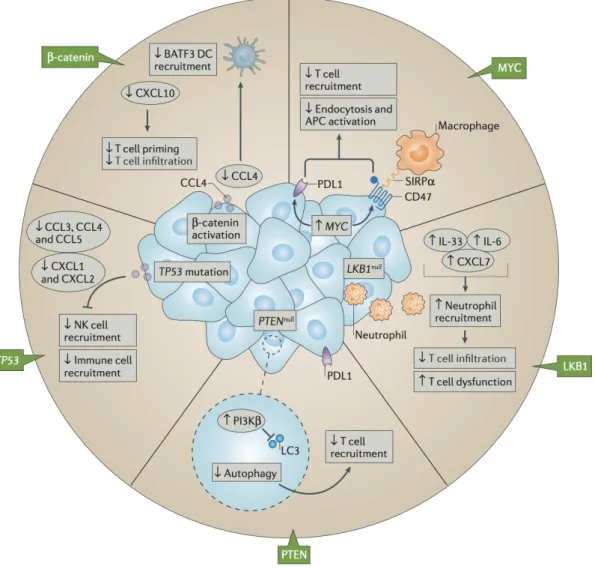
Third, tumour cells often exhibit activation of one or few oncogenic pathways, which are known to promote immune evasion via restriction of immune cells activity. A recent article nicely reviewed the various aspects of tumour-immune interactions that can be altered by specific oncogenic pathways, and described how these pathways indirectly mediate T cell exclusion from the TME and block recruitment of other immune cells to the tumour site (Figure I.6, (38)).
Finally, tumour cells often have functional defects in key immune pathways due to the deletion of immune genes in their genome. A recent study demonstrated that deletion of a number of genes in tumour necrosis factor (TNF) signalling, or IFN-γ signalling provides protection of tumour cells from CD8+ T cell-mediated killing. In addition, defects in the TNF signalling pathway also provide resistance to killing from primary NK cells (39). This demonstrates that the function of these pathways in tumour cells is key to allow immune-mediated tumour surveillance. Another example of tumour immune dysfunction leading to immune escape is the downregulation of IFN-alpha and -beta receptor subunit 1 (IFNAR1), frequently found in CRC, which is detrimental to TILs activity and survival, and associated with a poor prognosis in CRC patients (40).