A Dietary Regimen of Caloric Restriction or Pharmacological
Activation of SIRT1 to Delay the Onset of Neurodegeneration
The MIT Faculty has made this article openly available. Please share
how this access benefits you. Your story matters.
Citation
Graff, J. et al. “A Dietary Regimen of Caloric Restriction or
Pharmacological Activation of SIRT1 to Delay the Onset of
Neurodegeneration.” Journal of Neuroscience 33, 21 (May 2013):
8951–8960 © 2013 The Authors
As Published
http://dx.doi.org/10.1523/JNEUROSCI.5657-12.2013
Publisher
Society for Neuroscience
Version
Final published version
Citable link
http://hdl.handle.net/1721.1/112704
Terms of Use
Creative Commons Attribution 4.0 International License
Neurobiology of Disease
A Dietary Regimen of Caloric Restriction or Pharmacological
Activation of SIRT1 to Delay the Onset of Neurodegeneration
Johannes Gra¨ff,
1,2Martin Kahn,
1Alireza Samiei,
1Jun Gao,
1Kristie T. Ota,
1Damien Rei,
1,2and Li-Huei Tsai
1,2,31Picower Institute for Learning and Memory, Department of Brain and Cognitive Sciences and2Howard Hughes Medical Institute, Massachusetts Institute
of Technology, Cambridge, Massachusetts 02139, and3Broad Institute of Harvard University and Massachusetts Institute of Technology, Cambridge,
Massachusetts 02142
Caloric restriction (CR) is a dietary regimen known to promote lifespan by slowing down the occurrence of age-dependent diseases. The
greatest risk factor for neurodegeneration in the brain is age, from which follows that CR might also attenuate the progressive loss of
neurons that is often associated with impaired cognitive capacities. In this study, we used a transgenic mouse model that allows for a
temporally and spatially controlled onset of neurodegeneration to test the potentially beneficial effects of CR. We found that in this model,
CR significantly delayed the onset of neurodegeneration and synaptic loss and dysfunction, and thereby preserved cognitive capacities.
Mechanistically, CR induced the expression of the known lifespan-regulating protein SIRT1, prompting us to test whether a
pharmaco-logical activation of SIRT1 might recapitulate CR. We found that oral administration of a SIRT1-activating compound essentially
repli-cated the beneficial effects of CR. Thus, SIRT1-activating compounds might provide a pharmacological alternative to the regimen of CR
against neurodegeneration and its associated ailments.
Introduction
Caloric restriction (CR), the reduction in the consumption of
calories, is an effective regimen to slow aging in a variety of
or-ganisms ranging from yeast, nematodes, and fruitflies to
mam-mals (
Bishop and Guarente, 2007
;
Fontana et al., 2010
). CR acts
on a broad spectrum of tissues including liver, muscle, fat,
pan-creatic
-cells, and, interestingly, also the brain (
Bordone and
Guarente, 2005
,
2007
). There, CR has been shown to lead to an
attenuation of age-associated ailments, in particular, Alzheimer’s
disease (AD)-related pathologies: CR reduced the load of
amyloid-
, a pathological hallmark of AD, in different mouse
models of AD (
Patel et al., 2005
;
Wang et al., 2005
;
Qin et al.,
2006b
,
Halagappa et al., 2007
;
Wu et al., 2008b
;
Mouton et al.,
2009
) and in aged squirrel monkeys (
Qin et al., 2006a
), and in the
3xTg AD mouse model, CR also reduced the phosphorylation of
tau (
Halagappa et al., 2007
), another pathological aggregation in
AD. Furthermore, CR was shown to rescue against memory
def-icits in several of these models (
Halagappa et al., 2007
;
Wu et al.,
2008b
), and similar effects were also observed in wild-type mice
(
Fonta´n-Lozano et al., 2007
) and in aged humans (
Witte et al.,
2009
). Last, in an endpoint study with rhesus monkeys, CR was
found to have significantly attenuated brain atrophy by
preserv-ing gray matter volume (
Colman et al., 2009
). However, whether
CR can delay the onset of neurodegeneration and the
mecha-nisms of this process currently remain unknown.
One potential mechanism that might mediate the beneficial
effects of CR on brain health is the activation of the nicotinamide
adenine dinucleotide (NAD)-dependent protein deacetylase
SIRT1 (
Guarente and Picard, 2005
;
Bishop and Guarente, 2007
;
Bordone et al., 2007
;
Chen and Guarente, 2007
;
Lavu et al., 2008
).
SIRT1 was shown to at least partly mediate the beneficial effects
of CR on AD-related pathologies (
Qin et al., 2006b
), on
inflam-matory responses following neurotoxic insults (
Chen et al.,
2008
), and on enhancing learning and memory capacities (
Fusco
et al., 2012
). Moreover, SIRT1 overexpression or activation in
parallel to CR has been shown to reduce amyloid-
 deposition
(
Chen et al., 2005
;
Donmez et al., 2010
), tau phosphorylation
(
Min et al., 2010
), and microglia and neurotoxicity-dependent
inflammation (
Chen et al., 2005
;
2008
), and to improve learning,
memory, and synaptic plasticity (
Gao et al., 2010
;
Micha´n et al.,
2010
). Furthermore, SIRT1 activation reduced axonal
degenera-tion in the Wallerian degeneradegenera-tion mouse model (
Araki et al.,
2004
) and protected against neuronal loss in mouse models of
AD and amyotrophic lateral sclerosis (
Kim et al., 2007
).
How-ever, the potentially beneficial implication of SIRT1 activation
following CR against neurodegeneration has not been
investi-gated.
For this study, we hypothesized that CR delays the onset of
neurodegeneration and neurodegeneration-associated
patholo-Received Dec. 11, 2012; revised March 6, 2013; accepted March 30, 2013.
Author contributions: J. Gra¨ff, D.R., and L.-H.T. designed research; J. Gra¨ff, M.K., A.S., J. Gao, K.T.O., and D.R. performed research; L.-H.T. contributed unpublished reagents/analytic tools; J. Gra¨ff, M.K., J. Gao, K.T.O., and L.-H.T. analyzed data; J. Gra¨ff and L.-H.T. wrote the paper.
This work was supported by a Swiss National Science Foundation Grant for Prospective Researchers (J. Gräff), the Theodor and Ida Herzog-Egli Foundation (M.K.), and the Glenn Foundation for Medical Research and NIH Grant PO1 AG027916 (L.-H.T.). L.-H.T. is an HHMI investigator. We thank Vipin X. Suri and Jim Ellis from Sirtris-GSK for the SRT3657 compound, Maria Ericsson for the electron microscopy experiments, Katie Schlieper and Mali Taylor for mouse colony maintenance and the oral gavage, Alison Mungenast for text editing, and Matthew M. Dobbin for fruitful scientific discussions. Sirtris Pharmaceuticals contributed to this study in providing and characterizing SRT3657 under a material transfer agreement.
L.-H.T. is a scientific advisor of Sirtris-GSK.
Correspondence should be addressed to Li-Huei Tsai at the above address. E-mail: lhtsai@mit.edu. K. T. Ota’s present address: Laboratory of Molecular Psychiatry, Yale University School of Medicine, New Haven, CT 06508.
DOI:10.1523/JNEUROSCI.5657-12.2013
Copyright © 2013 the authors 0270-6474/13/338951-10$15.00/0
gies such as brain atrophy, loss of synaptic plasticity, and
cogni-tive deficits via the activation of SIRT1. Furthermore, we
reasoned that if this was the case, SIRT1-activating compounds
(STACs) should be able to recapitulate the beneficial effects of
CR and provide a pharmacological alternative to this dietary
regimen.
Materials and Methods
Animals. Adult (3-month-old) double-transgenic CK-p25 mice of either sex (Cruz et al., 2003,2006) and their single-transgenic or nontransgenic littermates were used for all experiments. All animal work was approved by the Committee for Animal Care of the Division of Comparative Med-icine at the Massachusetts Institute of Technology.
Experimental manipulations. CR treatment on CK-p25 mice consisted of 30% CR, representing 70% of the normal ad libitum caloric consump-tion. Mice were first accustomed to this diet during a 6 week period and then maintained on it for another 6 weeks, for a total duration of 3 months CR. During the last half of this treatment, p25 was induced by doxycycline removal from the diet. Control treatment consisted of ad libitum feeding of age- and sex-matched CK-p25 mice during the same time. Ad libitum feeding allowed for unrestricted continuous access to solid food and water. In total, the CR regimen was repeated two indepen-dent times.
Treatment of CK-p25 mice with SRT3657关tert␣-butyl 4-((2-(2-(6-(2-(tert-butoxycarbonyl(methyl)amino)ethylamino)-2-butylpyrimidine-4- carbox-amido)phenyl)thiazolo关5,4-b兴pyridin-6-yl)methoxy)piperidine-1-carboxylate兴 [Sirtris-GSK; compound 10 in the study byDai et al. (2010)] was performed by daily oral gavage with 30 mg/kg SRT3657 for 6 weeks. SRT3657 was diluted in Vitamin E TPGS (D-␣-tocopheryl polyethylene glycol 1000 succi-nate; Cognis) as vehicle. Control treatment consisted of daily oral gavage with vehicle alone. Injection volume was 0.1 ml per animal. At this formu-lation, the brain/plasma ratio of SRT3657 at 3 mg/kg was 0.13.
Behavior. Behavioral experiments were essentially conducted as de-scribed previously (Fischer et al., 2005;Gao et al., 2010). Open-field behavior was monitored using the VersaMax system (Accuscan) during 20 min. For fear conditioning, mice were put in the conditioning cham-ber (TSE Systems) for 3 min, after which a constant tone was played during 30 s, the last 2 s of which were paired with a one-time 2 s footshock (0.8 mA). Animals were then left in the box for another 30 s. Twenty-four hours later, mice were first tested for their contextual fear memory by putting them into the same training chamber for 3 min, during which their freezing behavior was scored. For assessing their cued fear memory, which took place at least 4 h after the contextual memory task, mice were put into a visually (gray instead of transparent) and contextually (flat instead of gridded surface) different box with a different smell (vanilla scented instead of ethanol scented) for a total of 3 min, the first half of which no tone was played, which was taken to measure baseline freezing levels, and the second half of which the same tone as during training was played, which served to measure their cued memory performance. Freez-ing was scored manually by an experimenter blind to the treatment group, by measuring freezing as the absence of movement except briefing every 10 s for 2 s. For the novel object recognition test, mice were habit-uated to the open testing arena (41.3⫻ 41.3 ⫻ 30.5 cm) over 2 d, for four trials per day, lasting 5 min each. During training, mice were exposed to two unknown objects that they were allowed to explore during four sessions of 5 min each. The objects had previously been put into a dirty cage to avoid olfactory preferences. Object memory was tested 24 h later by replacing one of the objects with a novel one and measuring the time with which the animals explored the novel object during a 10 min explo-ration session. The location of the object was counterbalanced between mice. Object exploration was scored by an automated software (Ethovision XT) and expressed as a discrimination ratio, DR⫽ (tnovel⫺ tfamiliar1)/ttotal
with tnoveland tfamiliarbeing the time spent with the novel and the
familiar objects, respectively, and ttotalbeing the total time spent with
both objects.
Electrophysiology. Mice were killed by cervical dislocation, and the brains were rapidly removed and transferred to ice-cold artificial CSF (ACSF) equilibrated with 95% O2/5% CO2. Transverse hippocampal
slices (400m thick) were cut with a Leica VT1000S vibratome and recovered in a holding chamber at 32–34°C for 30 min, and were then allowed to return to room temperature for at least another 60 min before recording. The ACSF used for cutting contained the following (in mM): 75 sucrose, 87 NaCl, 2.5 KCL, 1.25 NaH2PO4, 21.4 NaHCO3, 0.5 CaCl2, 7 MgCl2, 1.3 ascorbic acid, and 20D-glucose. The same ACSF was used
for both incubation and recording, and contained the following (in mM): 119 NaCl, 2.5 KCl, 2.5 CaCl2, 1 NaH2PO4, 1.3 MgSO426.2 NaHCO3, and
11D-glucose, saturated with 95% O2/5% CO2, pH 7.4. For extracellular
recordings in the CA1 region of the hippocampus, a bipolar platinum– iridium stimulating electrode was placed in the Schaffer collateral axons to elicit field population responses. The field EPSPs (fEPSPs) were re-corded via a glass micropipette filled with ACSF (1–3 M⍀) placed in the stratum radiatum region of the CA1. Stimuli (0.1 ms duration) were delivered every 30 s. Data were acquired using a HEKA EPC 10 amplifier and analyzed by Patchmaster software (HEKA). Peak stimulation-induced fEPSP amplitudes were required to be at least 0.7 mV, and stim-ulus intensity was set to produce 30 –50% of the maximal response. Responses were allowed to stabilize, then a baseline was recorded for 30 min. Long-term potentiation (LTP) was induced by theta-burst stimula-tion (TBS), which was delivered as four trains of four pulses at 100 Hz separated by 200 ms. Recordings were made at baseline test intensity for an additional hour after LTP induction. In all experiments, the initial slope of the fEPSP was measured, and LTP for each time point was expressed as a percentage of the preinduction baseline. For analysis, mea-surements of evoked responses during the last 10 min of the recording session were compared with the last 5 min of the baseline period using Student’s t tests.
Immunohistochemistry. For immunohistochemistry, mice were per-fused with 10% paraformaldehyde under deep anesthesia (ketamine, xy-lazine) and their brains sectioned at 40m thickness using a vibratome (Leica). Slices were permeabilized with 0.1% Triton X-100, blocked, and incubated overnight with 0.1% Triton X-100/10% fetal bovine serum in PBS containing primary antibodies: NeuN (Millipore), GFP (Aves Labs), synaptophysin (SVP; Sigma), SIRT1 (Abcam), SIRT3 (Santa Cruz Bio-technology), and histone 3 lysine 56 acetylation (AcH3K56; Epitomics). Primary antibodies were visualized with Alexa-Fluor 488, Cy3, and Cy5 antibodies (Invitrogen) in 1⫻ PBS, and neuronal nuclei with Hoechst 33342 (Invitrogen). Note that the specificity of the SIRT1 antibody was confirmed in SIRT1F/F animals (Cheng et al., 2003) (data not shown). Slices were covered with Fluoromount G (Electron Microscopy Sci-ences). Images were acquired using a confocal microscope (LSM 510; Zeiss) and analyzed using ImageJ 1.42q. Three or four animals per group were used.
Electron microscopy. Animals were perfused with 2.5% glutaralde-hyde/2% paraformaldehyde in 0.1Msodium cacodylate buffer, pH 7.4. The hippocampus was dissected out, sliced into 1 mm slices, washed in 0.1Mcacodylate buffer, and postfixed with 1% osmiumtetroxide (OsO4)/
1.5% potassiumferrocyanide (KFeCN6) for 1 h, washed in water three times, and incubated in 1% aqueous uranyl acetate for 1 h followed by two washes in water and subsequent dehydration in grades of alcohol (10 min each at 50, 70, and 90%; two times for 10 min at 100%). The samples were then placed in propyleneoxide for 1 h and subsequently infiltrated overnight in a 1:1 mixture of propyleneoxide and TAAB Epon (Marivac Canada). The following day, the samples were embedded in TAAB Epon and polymerized at 60 nm for 48 h. Ultrathin sections (about 60 nm) were cut on a Reichert Ultracut-S microtome, placed onto copper grids, stained with uranyl acetate and lead citrate and examined in a TecnaiG2
Spirit BioTWIN. Images were recorded with an AMT 2k CCD camera. Fifteen distinct apical regions (stratum radiatum) of CA1,⬃50–100M from the cell body layer of the hippocampus were imaged per animal. A synapse was defined as an electron dense postsynaptic density area jux-taposed to a presynaptic terminal filled with synaptic vesicles. The num-ber of synapses was determined by an experimenter blind to treatment and genotype.
Golgi staining. Mice were perfused with 10% paraformaldehyde, and their entire brains Golgi-Cox stained using the Rapid Golgistain Kit (FD NeuroTechnologies) as per the manufacturer’s instructions. Brains were cut at 60m thickness using a Leica vibratome. The number of apical and
basal spines on hippocampal CA1 pyramidal neurons was counted by an experimenter blind to treatment group and genotype. Only mature spines with a clear mushroom-like body and a distinctive shaft were included. For each experimental group, a minimum of 10 cells from three or four animals each was analyzed.
Immunoprecipitation and Western blot. Immunoprecipitation (IP) was performed by homogenizing whole-tissue hippocampal lysates in 500l of sterile-filtered 50 mMTris, 120 mMNaCl, and 0.5% NP-40 containing proteinase inhibitors (Roche), followed by 15 min centrifugation at 14,000 rpm at 4°C and collection of the supernatant, which was incu-bated 60 –90 min with 1–2g of p53 (Cell Signaling Technologies). After incubation, BSA-precleared protein G beads (GE Healthcare) were added for 45 min at 4°C, and the immune complexes were collected at 8000 rpm for 3 min, by one wash each in high-salt homogenization buffer (containing 50 mMTris, 500 mMNaCl, and 1% NP-40) and regular homogenization buffer (see above). For Western blot (WB),
whole-tissue lysates were homogenized in 400l 1⫻ RIPA buffer containing proteinase inhibitors (Roche). IP or WB samples were loaded on 10 – 12% SDS gels, transferred onto PVDF mem-branes, and incubated with primary antibodies against acetylated lysine, p53 (Cell Signaling Technologies), IgG, synaptophysin (Sigma), or SIRT1 (Millipore) in 1⫻ TBST, and visualized with HRP-conjugated secondary antibodies (GE Healthcare) and Western Lightning ECL solutions (PerkinElmer). Band intensities were quantified using ImageJ 1.42q.
SIRT1 activity assay. SIRT1 activity was mea-sured by a Fluor-de-Lys enzymatic assay (Enzo Life Sciences) with the following specifications. In vitro, 1.5l of the Fluor-de-Lys substrate was incubated with 1l of recombinant hu-man SIRT1 protein in 30l 1⫻ assay buffer (50 mMTris-Cl, pH8.0, 1 mMDTT, 137 mMNaCl, 2.7 mMKCl, 4 mMMgCl2, 0.1 mMEDTA, 1 mM
NAD⫹, and 10% glycerol in ddH2O) in the
presence or absence of 1l 50 mM nicotin-amide or 1l 0.5 g/100 l suramin sodium. Fluorescence was measured in transparent polystyrol plates (Corning) by excitation wave-length 355 nm and emission wavewave-length 460 nm using an infinite 200Pro microplate reader (Tecan). In vivo, the protein content of whole-tissue lysates was extracted as described under immunoprecipitation (see above) and incu-bated overnight with 1–2g of SIRT1 antibody (Millipore). After incubation, BSA-precleared protein A beads (GE Healthcare) were added for 45 min at 4°C, and the immune complexes were collected by centrifugation at 8000 rpm for 3 min and washed twice with high-salt ho-mogenization buffer (see Immunoprecipita-tion and Western blot, above). The immune complexes were then redissolved in 30l 1⫻ assay buffer together with 1.5l of the Fluor-de-Lys substrate for 30 min at 37° shaking, and the supernatant was used for the fluorescent measurement. SIRT1 activity was normalized to SIRT1 protein levels.
Statistics. Statistical analyses were performed using GraphPad Prism 5. One-way ANOVAs followed by Tukey’s post hoc analyses or one-tailed Student’s t tests were used. All data are represented as mean⫾ SEM. Statistical signif-icance was set at pⱕ 0.05.
Results
CR rescues against neuronal loss and
brain atrophy
To examine whether CR can protect against neuronal loss, we
used the CK-p25 mouse model of neurodegeneration (
Cruz et
al., 2003
,
2006
;
Fischer et al., 2005
). These mice allow for a
time-controlled onset of neurodegeneration in the forebrain by
doxycycline-mediated inducible activation of the CdK5
coactiva-tor p25 (coupled to GFP) under the CamKII
␣ promoter. At 6
weeks of p25 induction, these mice display substantial
neuro-nal loss, reduced dendritic arborization, reduced synaptic
density and plasticity, as well as learning and memory deficits.
CK-p25 mice were subjected to 3 months of 30% calorie
re-striction (CK-p25,CR) (
Fig. 1 A
), the typical regimen of food
reduction found to increase rodent lifespan (
Bishop and
Guarente, 2007
). After 6 weeks of CR, when the animals’
re-Figure 1. A regimen of 3 month 30% CR reduces neurodegeneration. A, Schematic of the experimental timeline. Three groups of mice were used: CON,AL, CK-p25,AL, and CK-p25,CR. B, Weight development over the experimental timeline of all three groups of mice used in this study. C, CR-mediated preservation of neuronal integrity in CK-p25,CR animals. Immunohistochemical images depicting the number of neurons as evidenced by NeuN-staining and the number of p25-expressing cells by GFP-staining. Scale bars, 200m. D, Quantification of NeuN-positive cells under C. One-way ANOVA (F(2,9)⫽ 10.01, p ⱕ 0.01), followed by Tukey’s
post hoc tests; n⫽ 4 mice each. E, Quantification of GFP-positive cells under C (one-tailed t test, t(4)⫽ 3.56; n ⫽ 3 each). F,
CR-mediated preservation of overall brain mass. One-way ANOVA F(2,30)⫽ 102.6, p ⱕ 0.0001, followed by Tukey’s post hoc tests;
n⫽ 12 for CON,AL and CK-p25,AL; n ⫽ 9 for CK-p25,CR. *p ⱕ 0.05; **p ⱕ 0.01; ***p ⱕ 0.001 for Tukey’s post hoc comparisons. All values are mean⫾ SEM.
duction in body weight reached
asymp-totic levels (
Fig. 1 B
), we induced
transgene production by removing
doxycycline from the diet (
Fig. 1 A
).
p25,CR animals were compared to
CK-p25 mice fed ad libitum (CK-CK-p25,AL)
and to control mice fed ad libitum
(CON,AL) (see Materials and Methods)
(
Fig. 1 A
,B).
Following the regimen of CR, we
found that CK-p25,CR animals had a
sim-ilar number of neurons in hippocampus
area CA1 compared to CON,AL animals
as evidenced by NeuN
immunohisto-chemistry, despite the profound neuronal
loss displayed by CK-p25,AL animals (
Fig.
1C,D
) (
Fischer et al., 2007
). The number
of neurons in CK-p25,CR animals was
significantly higher than in CK-p25,AL
animals (
Fig. 1C,D
), an effect that was also
detectable by measuring the amount of
GFP-positive p25-expressing cells in
this brain area (
Fig. 1 E
). This suggests
that CR not only rescues against
neuro-nal death in p25-positive neurons, but
in a non cell-autonomous way
through-out this brain area. These neuroprotective
effects of CR were also observed by an
attenuated loss of brain mass in
CK-p25,CR animals compared to CK-p25,AL
mice (
Fig. 1 F
). Therefore, a regimen of
30% CR effectively rescued against
p25-driven neurodegeneration.
CR preserves synaptic density
To assess whether CR also preserves
struc-tural synaptic integrity in CK-p25 mice, we
measured synaptic density by
immunohis-tochemical labeling of SVP, a marker of
pre-synaptic terminals. SVP labeling was
markedly reduced in the stratum radiatum
of CK-p25,AL animals compared to
CON,AL mice (
Fig. 2A
,B) (
Fischer et al.,
2007
). In contrast, in CR-treated CK-p25
mice the levels of SVP were comparable to CON,AL mice, and
sig-nificantly elevated over those in CK-p25,AL animals (
Fig. 2A
,B).
Similar results were obtained by Western blot analysis of SVP levels
(data not shown). Thus, CR rescued against the loss of presynaptic
terminals.
Next, to more precisely determine the effect of CR on synaptic
integrity, we examined the number of functional synapses by
transmission electron microscopy. We defined a synapse as an
electron-rich postsynaptic density area juxtaposed to a
presynap-tic terminal filled with synappresynap-tic vesicles (see Materials and
Meth-ods). We found that in the stratum radiatum of CK-p25,CR
animals, the number of functional synapses was markedly
ele-vated over CK-p25,AL mice, and not significantly different from
that in CON,AL animals (
Fig. 2C
,D). In addition to preserving
presynaptic terminals, CR was thus also effective to prevent the
loss of functional synapses that normally occurs during
p25-mediated neurodegeneration (
Fischer et al., 2005
).
Another aspect of synaptic integrity is the number of dendritic
spines, which reflects the degree of connectivity between neurons
and thereby synaptic strength (
Fiala et al., 2002
). To explore
whether CR affects dendritic spine density, we used Golgi
im-pregnation of CA1 pyramidal neurons. We found that whereas
CK-p25,AL mice had lower spine density than their control
lit-termates (CON,AL), CK-p25 mice that underwent CR had a
sim-ilar number of spines per given length to CON,AL animals, a
number that was significantly higher when compared to
CK-p25,AL mice (
Fig. 2 E
,F ). Together, these data indicate that, in
addition to rescuing against neurodegeneration, CR is also
effec-tive in maintaining synaptic density.
CR preserves synaptic plasticity and memory capacities
As structural synaptic integrity was rescued by the CR regimen,
we next questioned if such beneficial effects would also translate
into ameliorated synaptic plasticity and enhanced memory
ca-pacities. Six-week induced CK-p25 mice show impairments in
hippocampal LTP and associative and spatial memory tasks (
Fi-scher et al., 2005
). We therefore asked whether CR had a positive
effect on LTP and these types of memory. We first measured
Figure 2. CR protects structural synaptic and dendritic integrity. A, CR-mediated preservation of presynaptic terminals in CK-p25,CR animals as evidenced by SVP immunohistochemistry in the stratum radiatum of hippocampal area CA1. B, Quantifica-tion of A [one-way ANOVA (F(2,8)⫽8.99,pⱕ0.01)followedbyTukey’sposthoctests;n⫽3forCON,ALandCK-p25,CR;n⫽5for
CK-p25,AL]. C, CR-mediated preservation of the number of synapses in the hippocampus of CK-p25,CR animals as evidenced by transmission electron microscopy. D, Quantification of C. One-way ANOVA (F(2, 6)⫽24.84,pⱕ0.01)followedbyTukey’sposthoc
tests; n⫽ 3 mice each. E, CR-mediated preservation of the number of spines per given dendritic length in the hippocampus of CK-p25,CR animals as evidenced by Golgi impregnation. F, Quantification of E [one-way ANOVA (F(2,22)⫽ 23.31, p ⱕ 0.01)
followed by Tukey’s post hoc tests; n⫽ 7–10 dendrites per animal, 3 animals each]. *p ⱕ 0.05; **p ⱕ 0.01; ***p ⱕ 0.001 for Tukey’s post hoc comparisons. All values are mean⫾ SEM. Scale bars: A, 100m; C, 0.5 m; E, 2 m.
fEPSPs following theta-burst stimulation of Schaffer collaterals.
While this stimulation was not capable of evoking LTP in
CK-p25,AL animals, it did so in CK-p25,CR animals to the extent that
fEPSPs of CK-p25,CR mice were undistinguishable from
CON,AL animals (
Fig. 3A
). Importantly, basal synaptic
transmis-sion as measured by the synaptic input– output relationship was
similar between all three groups, albeit slightly reduced in
CK-p25,AL and CK-p25,CR animals (
Fig. 3B
). Thus, CR rescued
against neurodegeneration-mediated synaptic plasticity deficits.
Then, we measured the animals’ memory performance on a
cued and contextual version of fear conditioning and on an object
recognition paradigm to assess associative
and spatial memories for objects,
respec-tively. On the cued fear conditioning task,
CK-p25,CR animals remembered the
tone significantly better than CK-p25,AL
animals 24 h after training, at levels
indis-tinguishable from those of CON,AL
ani-mals (
Fig. 3C
, After). Importantly, the fear
response of CK-p25,CR mice upon
reex-posure to the conditioning chamber
alone, i.e., before the onset of the tone,
was comparable between all three groups
(
Fig. 3C
, Before), suggesting a selective
enhancement of the trained memory, and
not an overall increase of the fear reaction.
A similar rescue in memory performance
was observed in the contextual fear
condi-tioning paradigm 24 h after training:
CK-p25,CR animals performed significantly
better than CK-p25,AL mice, at levels
comparable to CON,AL animals (
Fig.
3D
). Last, on the novel object recognition
task, CK-p25,AL mice spent significantly
less time exploring the novel object than
CON,AL mice 24 h after training,
indicat-ing impaired object memory (
Fig. 3E
).
CK-p25,CR mice, however, spent
signifi-cantly more time with the novel object
than CK-p25,AL mice, at levels
indistin-guishable from CON,AL animals (
Fig.
3E
), suggesting that CR was capable of
preserving object memory. There was no
difference in object preference during
training (data not shown).
Importantly, CR treatment per se did
not alter overall locomotor activity as
as-sessed by total distance moved and time
spent moving in an open-field test, and
CR did further not affect the animals’
anx-iety as measured by the time spent in the
center of an open arena, nor their
ex-ploratory behavior, indicated by their
time spent rearing (data not shown).
Therefore, without affecting the
ani-mals’ general behavior, the regimen of
CR specifically improved synaptic
plas-ticity as well as associative and object
recognition memories.
CR activates SIRT1 and SIRT1-mediated
neuroprotective mechanisms
One potential mechanism that might
me-diate the beneficial effects of CR against neurodegeneration and
its associated structural and behavioral impairments involves the
activation of SIRT1, since SIRT1 overexpressing mice were
shown to recapitulate the majority of the systemic effects of CR
(
Bordone et al., 2007
), and because SIRT1 expression was found
to be increased in the brains of CR-treated animals (
Qin et al.,
2006b
;
Chen et al., 2008
). Based on this, we first measured the
levels of hippocampal SIRT1 by immunohistochemistry. In
agreement with previous reports (
Qin et al., 2006b
;
Chen et al.,
2008
), we found SIRT1 protein expression to be markedly
in-creased in this brain area in CK-p25,CR animals compared to
Figure 3. CR maintains synaptic plasticity and memory. A, Preservation of long-term potentiation by CR. fEPSPs (expressed in percentage of maximal amplitude) in hippocampal area CA1 of CON,AL, CK-p25,AL and CK-p25,CR animals (n⫽ 3–4 slices from 3– 4 mice each) before and after TBS. *pⱕ0.05byANOVAfortheeffectoftreatment.Sampletracesabovethelinechartrepresent fEPSPs at 1 min before (black) and 1 h after (colored) TBS. B, Input– output relationship of comparable baseline synaptic trans-mission between CON,AL, CK-p25,AL, and CK-p25,CR animals. C, Cued fear conditioning. Left, Freezing responses before the onset of the tone were comparable between CON,AL, CK-p25,AL and CK-p25,CR animals 24 h after training. Right, Freezing responses after the onset of the tone show comparable memory retention between CON,AL and CK-p25,CR animals, but reduced memory for CK-p25,AL mice. One-way ANOVA (F(2,30)⫽ 6.29, p ⱕ 0.01) followed by Tukey’s post hoc tests; n ⫽ 10–12 mice each. D,
Contextual fear conditioning. Freezing responses 24 h after training on a contextual fear conditioning task show comparable memory retention between CON,AL and CK-p25,CR animals, but reduced memory for CK-p25,AL mice [one-way ANOVA (F(2,30)⫽
7.52, pⱕ 0.01) followed by Tukey’s post hoc tests; n ⫽ 10–13 mice each]. E, Novel object recognition. Discrimination ratio 24 h after training revealing a significantly enhanced object memory in CK-p25,CR compared to CK-p25,AL animals [one-way ANOVA (F(2,19)⫽ 7.16, p ⱕ 0.01) followed by Tukey’s post hoc tests; n ⫽ 7–8 mice each]. *p ⱕ 0.05; **p ⱕ 0.01 for Tukey’s post hoc
comparisons. All values are mean⫾ SEM.
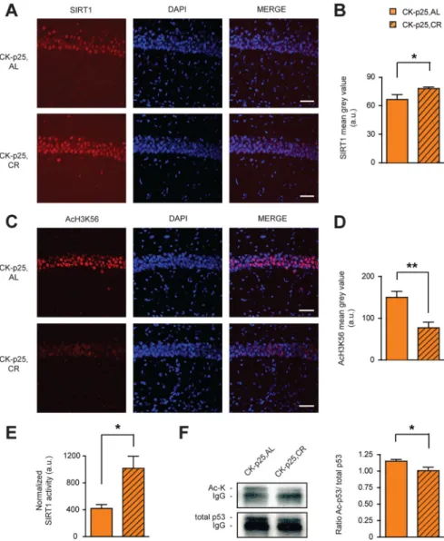
CK-p25,AL mice (
Fig. 4 A
,B).
Concomi-tant to the increase of SIRT1, we observed
a significant reduction in acetylation of
H3K56, a target of SIRT1’s deacetylase
ac-tivity (
Kong et al., 2011
), in CK-p25,CR
compared to CK-p25,AL mice (
Fig.
4C
,D). As SIRT1 levels in CK-p25 animals
have been found previously to be elevated
over those in control animals, and as this
upregulation confers protection against
neurotoxicity (
Kim et al., 2007
), CR is
likely to further potentiate the
neuropro-tective effects of SIRT1. In contrast, the
levels of SIRT3, a sirtuin with antioxidant
characteristics that is also known to be
re-sponsive to CR (
Someya et al., 2010
),
re-mained unchanged (data not shown).
Next, we measured the activity of
SIRT1 in CK-p25,CR and CK-p25,AL
an-imals. We immunoprecipitated SIRT1
out of hippocampal lysates and then
fluo-rometrically assessed its enzymatic
activ-ity (see Materials and Methods). To assure
that this method does indeed measure the
deacetylase activity of SIRT1, we first
per-formed an in vitro test reaction, in which
we combined recombinant SIRT1
to-gether with a deacetylase substrate and the
known SIRT1-specific inhibitors
nicotin-amide or suramin sodium. The
fluoro-metric assay reflected a complete in vitro
inhibition of recombinant SIRT1
activ-ity [recombinant SIRT1 plus substrate,
42426 fluorescent activity in arbitrary
units (f.a.a.u.); recombinant SIRT1 plus
substrate plus nicotinamide, 7327 f.a.a.u.;
recombinant SIRT1 plus substrate plus
suramin sodium, 7547 f.a.a.u.; substrate
alone, 4649 f.a.a.u.]. Using this assay in
vivo, we observed that CR significantly
in-creased the activity of SIRT1 in
CK-p25,CR compared to CK-p25,AL animals
(
Fig. 4 E
). Therefore, the CR regimen in
CK-p25 mice not only increased SIRT1
protein levels, but also its activity.
One of the downstream mechanisms
by which SIRT1 increases cellular lifespan
is p53 (
Chen and Guarente, 2007
), a proapoptotic transcription
factor that promotes cellular senescence (
Oren, 2003
). Upon
SIRT1-mediated deacetylation, p53 becomes inactivated (
Luo et
al., 2001
;
Vaziri et al., 2001
). Based on this knowledge, we
mea-sured p53 acetylation in hippocampal extracts of CK-p25
ani-mals following CR or AL treatment by immunoprecipitation
of p53 coupled to Western blot analysis. We observed that the
acetylation of p53 was significantly reduced following CR
treatment (
Fig. 4 F
), suggesting that p53 becomes inactivated
following CR. Together, these results indicate that CR
upregu-lates SIRT1 and activates its deacetylase function, which
co-occurs with a deacetylation of p53 and likely p53 inactivation.
As a reduction of p53 has been shown to be neuroprotective
(
Kim et al., 2007
), this SIRT1-p53 pathway constitutes a
po-tential mechanism by which CR might rescue against
neuro-degeneration.
SRT3657, a small-molecule activator of SIRT1
Since SIRT1 expression and activity were increased by CR, we
reasoned that by using small-molecule STACs we might be able to
obtain the same beneficial effects against neurodegeneration as
under the CR regimen. To test this, we administered a STAC to
CK-p25 mice during the 6 weeks of p25 transgene induction (
Fig.
5A
). This timeline was chosen to most closely recapitulate the
asymptotic phase of CR during p25-transgene induction (
Fig.
1 B
). CK-p25 mice were given a known SIRT1-specific STAC,
SRT3657 (CK-p25,SRT3657) (
Dai et al., 2010
) by oral gavage, or
its vehicle alone (CK-p25,VEH) (
Fig. 5A
). As shown in
Figure 5B
,
SRT3657 treatment significantly upregulated the activity of
SIRT1 in hippocampal extracts of CK-p25 animals. Such
in-creased SIRT1 activity was also reflected by dein-creased H3K56
acetylation (
Fig. 5C
,D). Thus, SRT3657 treatment effectively
ac-tivated SIRT1 in vivo.
Figure 4. SIRT1 activation by the CR regimen. A, Increased SIRT1 expression following CR in hippocampal area CA1 as evidenced by SIRT1 immunohistochemistry. B, Quantification of A (one-tailed t test, t(4)⫽2.68;n⫽3each).C,DecreasedAcH3K56following
CR in hippocampal area CA1 as evidenced by AcH3K56 immunohistochemistry. D, Quantification of C (one-tailed t test, t(4)⫽3.84;
n⫽ 3 each). E, Increased SIRT1 deacetylase activity following CR. SIRT1 deacetylase activity in CK-p25,AL and CK-p25,CR hip-pocampi. One-tailed t test, t(5)⫽ 2.03; n ⫽ 3 for CK-p25,AL; n ⫽ 4 for CK-p25,CR. F, Decreased acetylation of p53 following CR.
Left, Representative images of Western blot analysis following immunoprecipitation of acetylated (top) and total (bottom) p53 levels in CK-p25,AL and CK-p25,CR hippocampi. Right, Quantification of the ratio of acetylated/total p53 protein levels (one-tailed t test, t(13)⫽2.04;n⫽6forCK-p25,AL;n⫽9forCK-p25,CR).*pⱕ0.05;**pⱕ0.01forone-tailedttests.Allvaluesaremean⫾
If a STAC is to recapitulate CR in vivo, it should activate SIRT1
downstream mechanisms similar to the CR treatment. Therefore,
we next tested whether the acetylation of the proapoptotic
pro-tein p53 might be altered by SRT3657 treatment. Similar to the
CR regimen, we found the acetylation of p53 to be significantly
reduced in the hippocampus of CK-p25,SRT3657 mice
com-pared to CK-p25,VEH animals (
Fig. 5E
). This suggests that
sim-ilar neuroprotective pathways might be activated by the SRT3657
treatment.
SRT3657 recapitulates the neuroprotective potential of CR
To further assess a potential neuroprotective role of SRT3657,
we examined synaptic density by SVP immunohistochemistry.
Compared to CK-p25,VEH animals, CK-p25,SRT3657 mice
displayed higher synaptic density in the
stratum radiatum of the hippocampus
at levels indistinguishable from CON,
VEH animals (
Fig. 6A
,B), indicating
pre-served synaptic integrity. Then, to account
for the extent of neurodegeneration, we
ex-amined the number of neurons by
NeuN-immunohistochemistry in hippocampal
area CA1. We found that the number of
NeuN-positive cells was significantly
in-creased in CK-p25,SRT3657 animals
com-pared to CK-p25,VEH mice, to a number
comparable to CON,VEH animals (
Fig.
6C
,D), demonstrating that
neurodegenera-tion can be at least partially prevented by the
STAC SRT3657. Intriguingly, this was
fur-ther evidenced by a partial rescue in overall
brain loss (
Fig. 6E
), similar to the effect
ob-served under the CR regimen (
Fig. 1F
).
Last, to evaluate whether the
so-main-tained neuronal and synaptic integrity
might translate into behavioral
improve-ments, we tested CK-p25,SRT3657 and
control littermates for fear memory.
Twenty-four hours after training,
CK-p25,SRT3657 mice froze significantly more
than CK-p25,VEH mice on the cued fear
conditioning task after the onset of the tone,
but not before (
Fig. 6
F ), indicating
en-hanced memory retention. Likewise,
CK-p25,SRT3657 mice outperformed their
VEH-treated counterparts on the
contex-tual fear memory test (
Fig. 6
G).
Impor-tantly, both cued and contextual fear
memory retention were comparable
be-tween CK-p25,SRT3657 and CON,VEH
animals. Furthermore, SRT3657 treatment
per se did not affect overall locomotor and
exploratory activity, nor did it alter the
ani-mals’ anxiety levels (data not shown).
To-gether, these data indicate that SRT3657
treatment prevented synaptic and neuronal
loss and effectively rescued against
neurode-generation-driven memory impairments.
For that reason, SRT3657 treatment might
constitute a pharmacological alternative
to CR.
Discussion
In the current study, we have shown
that a regimen of CR effectively delays the onset of
neurode-generation, preserves structural and functional synaptic
plasticity as well as memory capacities, and activates the
expression and activity of SIRT1, a known promoter of
neu-ronal lifespan (
Tang, 2009
;
Zhang et al., 2011
). Importantly,
mimicking the dietary regimen by oral administration of the
STAC SRT3657 recapitulated the beneficial effects of CR
against neurodegeneration-associated pathologies and thus
might constitute a pharmacological alternative to CR against
neurodegeneration.
Expanding the favorable effects of CR on memory
perfor-mance (
Halagappa et al., 2007
;
Fonta´n-Lozano et al., 2008
;
Wu et
al., 2008a
;
Witte et al., 2009
) and against AD-related pathologies
Figure 5. SRT3657 activates SIRT1 in CK-p25 mice. A, Schematic of the experimental timeline. Two groups of mice were used: CK-p25,VEH and CK-p25,SRT3657. B, Increased SIRT1 activity following SRT3657 treatment. SIRT1 deacetylase activity in CK-p25,VEH and CK-p25,SRT3657 hippocampi (one-tailed t test, t(8)⫽ 2.65; n ⫽ 4 for CK-p25,VEH; n ⫽ 6 for
CK-p25,SRT3657). C, Decreased AcH3K56 following SRT3657 treatment in hippocampal area CA1 as evidenced by AcH3K56 immunohistochemistry. Scale bar, 100m. D, Quantification of C. One-tailed t test, t(8)⫽ 2.61; n ⫽ 4 for CK-p25,VEH; n ⫽
6 for CK-p25,SRT3657. E, Decreased acetylation of p53 following SRT3657 treatment. Top, Representative images of Western blot analysis following immunoprecipitation of acetylated (top) and total (bottom) p53 levels in CK-p25,VEH and CK-p25,SRT3657 hippocampi. Bottom, Quantification of the ratio of acetylated/total p53 protein levels (one-tailed t test, t(8)⫽ 2.61; n ⫽ 4 for CK-p25,VEH; n ⫽ 6 for CK-p25,SRT3657). *p ⱕ 0.05 for one-tailed t tests. All values are
mean⫾ SEM.
such as amyloid- accumulation (
Patel et
al., 2005
;
Wang et al., 2005
;
Qin et al.,
2006a
,
b
;
Halagappa et al., 2007
;
Wu et al.,
2008b
;
Mouton et al., 2009
) and tau
hy-perphosphorylation (
Halagappa et al.,
2007
), this study newly shows that a
regi-men of CR can also delay the onset of
neu-rodegeneration
per
se.
It
thereby
complements results in rhesus monkeys,
where CR was shown to preserve gray
matter volume (
Colman et al., 2009
), and
in Caenorhabditis elegans, where it was
found to prevent the destruction of
dopa-minergic neurons in a Parkinson’s disease
model (
Jadiya et al., 2011
). One question
that remains to be answered is whether
the regimen of CR would also prove
ef-fective against neurodegeneration when
started later, i.e., after the onset of
neu-rodegeneration. Likewise, future studies
will need to assess whether the beneficial
effects of CR are permanent or whether
neurodegeneration regains the upper
hand once the regimen of CR is stopped.
Mechanistically, the CR regimen used
here was found to activate SIRT1. This is
in line with previous reports showing that
CR increases hippocampal SIRT1
expres-sion (
Qin et al., 2006b
;
Chen et al., 2008
)
and that, conversely, a fat-rich diet
re-duces hippocampal SIRT1 expression
(
Heyward et al., 2012
). Although we
can-not determine that the beneficial effects
of CR are solely mediated by SIRT1, nor
that SIRT1 acts in parallel to CR, it is
nevertheless likely that such activation
of SIRT1 does contribute to the positive
effects of CR: overexpression of SIRT1
has been shown previously to not only
improve memory performance (
Gao et
al., 2010
;
Micha´n et al., 2010
), but also
to protect against neurodegeneration
(
Araki et al., 2004
;
Kim et al., 2007
),
whereas RNAi-mediated knockdown of
SIRT1 levels precluded the
neuropro-tective effects of CR in C. elegans (
Jadiya
et al., 2011
).
This possibility is further supported
by our findings that, using the STAC
SRT3657, the beneficial effects of CR
could be essentially recapitulated. Such
treatment represents the first direct
ev-idence of a STAC as a pharmacological
alternative to a dietary regimen against neurodegeneration.
Since dietary changes do not always produce the anticipated
beneficial outcomes (
Mattison et al., 2012
), STAC treatments
might be preferable over CR. While we did not observe any
adverse effects following SRT3657 treatment, more refined
studies are still needed to determine the overall safety and
tolerability of this STAC. Moreover, similar questions as
raised by the results obtained with the CR treatment
concern-ing the timconcern-ing of administration as well as the persistence of
the beneficial effects are also applicable here. In addition,
it would be interesting to assess the combined effects of
phar-macological SIRT1 activation and CR on the progression of
neurodegeneration. Such experiments would also help to
dis-entangle the question as to whether SIRT1 activation acts via
the same pathway as CR.
Last, although we identified p53 deactivation as a potential
target of the SRT3657 and CR regimen, other neuroprotective
pathways such as the Forkhead box protein O transcription
fac-tors, insulin/IGF-signaling, PGC1␣, or NfB might also be
acti-vated following both STAC and CR treatments (
Gan and Mucke,
Figure 6. SRT3657 recapitulates the beneficial effects of CR. A, SRT3657-mediated increase in presynaptic terminals in the stratum radiatum of hippocampal area CA1 in CK-p25,SRT3657 animals compared to CK-p25,VEH mice as evidenced by SVP immunohistochemistry. B, Quantification of A [one-way ANOVA (F(2,11)⫽ 5.77, p ⱕ 0.05) followed by Tukey’s post hoc
tests; n⫽ 4 –5 mice each]. C, SRT3657-mediated increase in the number of neurons in hippocampal area CA1 in CK-p25,SRT3657 animals compared to CK-p25,VEH mice as revealed by NeuN-immunohistochemistry. D, Quantification of C [one-way ANOVA (F(2,12)⫽ 29.99, p ⱕ 0.0001) followed by Tukey’s post hoc tests; n ⫽ 3– 6 mice each]. E,
SRT3657-mediated preservation of overall brain mass. One-way ANOVA (F(2,11)⫽ 19.68, p ⱕ 0.001) followed by Tukey’s post hoc
tests; n⫽ 4 –7 mice each. F, Cued fear conditioning. Left, Freezing responses before the onset of the tone were comparable between CK-p25,VEH and CK-p25,SRT3657 animals 24 h after training. Right, Freezing responses after the onset of the tone show improved memory retention in CK-p25,SRT3657 mice [one-way ANOVA (F(2,23)⫽ 7.47, p ⱕ 0.01) followed by
Tukey’s post hoc tests; n⫽ 8 –10 mice each]. G, Contextual fear conditioning. Freezing responses 24 h after training on a contextual fear conditioning task show improved memory retention in CK-p25,SRT3657 mice [one-way ANOVA (F(2,22)⫽
5.28, pⱕ 0.05) followed by Tukey’s post hoc tests; n ⫽ 8 –10 mice each]. *p ⱕ 0.05; **p ⱕ 0.01; ***p ⱕ 0.001 for Tukey’s post hoc comparisons. All values are mean⫾ SEM. Scale bars: A, 50m; C, 20 m.
2008
;
Lavu et al., 2008
). These limitations notwithstanding, the
present study proposes the STAC SRT3657 as an attractive
alter-native route to convey the beneficial effects of CR against
neuro-degeneration, without the need for CR itself.
References
Araki T, Sasaki Y, Milbrandt J (2004) Increased nuclear NAD biosynthe-sis and SIRT1 activation prevent axonal degeneration. Science 305: 1010 –1013.CrossRef Medline
Bishop NA, Guarente L (2007) Genetic links between diet and lifespan: shared mechanisms from yeast to humans. Nat Rev Genet 8:835– 844.
CrossRef Medline
Bordone L, Guarente L (2005) Calorie restriction, SIRT1 and metabolism: understanding longevity. Nat Rev Mol Cell Biol 6:298 –305.CrossRef Medline
Bordone L, Cohen D, Robinson A, Motta MC, van Veen E, Czopik A, Steele AD, Crowe H, Marmor S, Luo J, Gu W, Guarente L (2007) SIRT1 trans-genic mice show phenotypes resembling calorie restriction. Aging Cell 6:759 –767.CrossRef Medline
Chen D, Guarente L (2007) SIR2: a potential target for calorie restriction mimetics. Trends Mol Med 13:64 –71.CrossRef Medline
Chen D, Steele AD, Hutter G, Bruno J, Govindarajan A, Easlon E, Lin SJ, Aguzzi A, Lindquist S, Guarente L (2008) The role of calorie restriction and SIRT1 in prion-mediated neurodegeneration. Exp Gerontol 43: 1086 –1093.CrossRef Medline
Chen J, Zhou Y, Mueller-Steiner S, Chen LF, Kwon H, Yi S, Mucke L, Gan L (2005) SIRT1 protects against microglia-dependent amyloid-beta toxicity through inhibiting NF-kappaB signaling. J Biol Chem 280: 40364 – 40374.CrossRef Medline
Cheng HL, Mostoslavsky R, Saito S, Manis JP, Gu Y, Patel P, Bronson R, Appella E, Alt FW, Chua KF (2003) Developmental defects and p53 hy-peracetylation in Sir2 homolog (SIRT1)-deficient mice. Proc Natl Acad Sci U S A 100:10794 –10799.CrossRef Medline
Colman RJ, Anderson RM, Johnson SC, Kastman EK, Kosmatka KJ, Beasley TM, Allison DB, Cruzen C, Simmons HA, Kemnitz JW, Weindruch R (2009) Caloric restriction delays disease onset and mortality in rhesus monkeys. Science 325:201–204.CrossRef Medline
Cruz JC, Tseng HC, Goldman JA, Shih H, Tsai LH (2003) Aberrant Cdk5 activation by p25 triggers pathological events leading to neurodegenera-tion and neurofibrillary tangles. Neuron 40:471– 483.CrossRef Medline
Cruz JC, Kim D, Moy LY, Dobbin MM, Sun X, Bronson RT, Tsai LH (2006) p25/cyclin-dependent kinase 5 induces production and intra-neuronal accumulation of amyloid beta in vivo. J Neurosci 26:10536 – 10541.CrossRef Medline
Dai H, Kustigian L, Carney D, Case A, Considine T, Hubbard BP, Perni RB, Riera TV, Szczepankiewicz B, Vlasuk GP, Stein RL (2010) SIRT1 activation by small molecules: kinetic and biophysical evidence for direct interaction of enzyme and activator. J Biol Chem 285:32695– 32703.CrossRef Medline
Donmez G, Wang D, Cohen DE, Guarente L (2010) SIRT1 suppresses beta-amyloid production by activating the alpha-secretase gene ADAM10. Cell 142:320 –332.CrossRef Medline
Fiala JC, Spacek J, Harris KM (2002) Dendritic spine pathology: cause or consequence of neurological disorders? Brain Res Brain Res Rev 39: 29 –54.CrossRef Medline
Fischer A, Sananbenesi F, Pang PT, Lu B, Tsai LH (2005) Opposing roles of transient and prolonged expression of p25 in synaptic plasticity and hippocampus-dependent memory. Neuron 48:825– 838. CrossRef Medline
Fischer A, Sananbenesi F, Wang X, Dobbin M, Tsai LH (2007) Recovery of learning and memory is associated with chromatin remodelling. Nature 447:178 –182.CrossRef Medline
Fontana L, Partridge L, Longo VD (2010) Extending healthy life span–from yeast to humans. Science 328:321–326.CrossRef Medline
Fonta´n-Lozano A, Sa´ez-Cassanelli JL, Inda MC, de los Santos-Arteaga M, Sierra-Domínguez SA, Lo´pez-Lluch G, Delgado-García JM, Carrio´n AM (2007) Caloric restriction increases learning consolidation and facilitates synaptic plasticity through mechanisms dependent on NR2B subunits of the NMDA receptor. J Neurosci 27:10185–10195.CrossRef Medline
Fonta´n-LozanoA, Romero-Granados R, Troncoso J, Mu´nera A, Delgado-García JM, Carrio´n AM (2008) Histone deacetylase inhibitors improve learning consolidation in young and in KA-induced-neurodegeneration
and SAMP-8-mutant mice. Mol Cell Neurosci 39:193–201.CrossRef Medline
Fusco S, Ripoli C, Podda MV, Ranieri SC, Leone L, Toietta G, McBurney MW, Schutz G, Riccio A, Grassi C, Galeotti T, Pani G (2012) A role for neu-ronal cAMP responsive-element binding (CREB)-1 in brain responses to calorie restriction. Proc Natl Acad Sci U S A 109:621– 626.CrossRef Medline
Gan L, Mucke L (2008) Paths of convergence: sirtuins in aging and neuro-degeneration. Neuron 58:10 –14.CrossRef Medline
Gao J, Wang WY, Mao YW, Gra¨ff J, Guan JS, Pan L, Mak G, Kim D, Su SC, Tsai LH (2010) A novel pathway regulates memory and plasticity via SIRT1 and miR-134. Nature 466:1105–1109.CrossRef Medline
Guarente L, Picard F (2005) Calorie restriction—the SIR2 connection. Cell 120:473– 482.CrossRef Medline
Halagappa VK, Guo Z, Pearson M, Matsuoka Y, Cutler RG, Laferla FM, Mattson MP (2007) Intermittent fasting and caloric restriction amelio-rate age-related behavioral deficits in the triple-transgenic mouse model of Alzheimer’s disease. Neurobiol Dis 26:212–220.CrossRef Medline
Heyward FD, Walton RG, Carle MS, Coleman MA, Garvey WT, Sweatt JD (2012) Adult mice maintained on a high-fat diet exhibit object location memory deficits and reduced hippocampal SIRT1 gene expression. Neu-robiol Learn Mem 98:25–32.CrossRef Medline
Jadiya P, Chatterjee M, Sammi SR, Kaur S, Palit G, Nazir A (2011) Sir-2.1 modulates ‘calorie-restriction-mediated’ prevention of neurodegenera-tion in Caenorhabditis elegans: implicaneurodegenera-tions for Parkinson’s disease. Biochem Biophys Res Commun 413:306 –310.CrossRef Medline
Kim D, Nguyen MD, Dobbin MM, Fischer A, Sananbenesi F, Rodgers JT, Delalle I, Baur JA, Sui G, Armour SM, Puigserver P, Sinclair DA, Tsai LH (2007) SIRT1 deacetylase protects against neurodegeneration in models for Alzheimer’s disease and amyotrophic lateral sclerosis. EMBO J 26: 3169 –3179.CrossRef Medline
Kong S, Kim SJ, Sandal B, Lee SM, Gao B, Zhang DD, Fang D (2011) The type III histone deacetylase Sirt1 protein suppresses p300-mediated his-tone H3 lysine 56 acetylation at Bclaf1 promoter to inhibit T cell activa-tion. J Biol Chem 286:16967–16975.CrossRef Medline
Lavu S, Boss O, Elliott PJ, Lambert PD (2008) Sirtuins—novel therapeu-tic targets to treat age-associated diseases. Nat Rev Drug Discov 7:841– 853.CrossRef Medline
Luo J, Nikolaev AY, Imai S, Chen D, Su F, Shiloh A, Guarente L, Gu W (2001) Negative control of p53 by Sir2alpha promotes cell survival under stress. Cell 107:137–148.CrossRef Medline
Mattison JA, Roth GS, Beasley TM, Tilmont EM, Handy AM, Herbert RL, Longo DL, Allison DB, Young JE, Bryant M, Barnard D, Ward WF, Qi W, Ingram DK, de Cabo R (2012) Impact of caloric restriction on health and survival in rhesus monkeys from the NIA study. Nature 489:318 –321.CrossRef Medline
Micha´n S, Li Y, Chou MM, Parrella E, Ge H, Long JM, Allard JS, Lewis K, Miller M, Xu W, Mervis RF, Chen J, Guerin KI, Smith LE, McBurney MW, Sinclair DA, Baudry M, de Cabo R, Longo VD (2010) SIRT1 is essential for normal cognitive function and synaptic plasticity. J Neurosci 30:9695–9707.CrossRef Medline
Min SW, Cho SH, Zhou Y, Schroeder S, Haroutunian V, Seeley WW, Huang EJ, Shen Y, Masliah E, Mukherjee C, Meyers D, Cole PA, Ott M, Gan L (2010) Acetylation of tau inhibits its degradation and contributes to tauopathy. Neuron 67:953–966.CrossRef Medline
Mouton PR, Chachich ME, Quigley C, Spangler E, Ingram DK (2009) Ca-loric restriction attenuates amyloid deposition in middle-aged dtg APP/ PS1 mice. Neurosci Lett 464:184 –187.CrossRef Medline
Oren M (2003) Decision making by p53: life, death and cancer. Cell Death Differ 10:431– 442.CrossRef Medline
Patel NV, Gordon MN, Connor KE, Good RA, Engelman RW, Mason J, Morgan DG, Morgan TE, Finch CE (2005) Caloric restriction attenuates Abeta-deposition in Alzheimer transgenic models. Neurobiol Aging 26: 995–1000.CrossRef Medline
Qin W, Chachich M, Lane M, Roth G, Bryant M, de Cabo R, Ottinger MA, Mattison J, Ingram D, Gandy S, Pasinetti GM (2006a) Calorie restric-tion attenuates Alzheimer’s disease type brain amyloidosis in Squirrel monkeys (Saimiri sciureus). J Alzheimer’s Dis 10:417– 422.Medline
Qin W, Yang T, Ho L, Zhao Z, Wang J, Chen L, Zhao W, Thiyagarajan M, MacGrogan D, Rodgers JT, Puigserver P, Sadoshima J, Deng H, Pedrini S, Gandy S, Sauve AA, Pasinetti GM (2006b) Neuronal SIRT1 activation as a novel mechanism underlying the prevention of Alzheimer disease am-Gra¨ff et al.• Setback of Neurodegeneration by Caloric Restriction J. Neurosci., May 22, 2013•33(21):8951– 8960 • 8959
yloid neuropathology by calorie restriction. J Biol Chem 281:21745– 21754.CrossRef Medline
Someya S, Yu W, Hallows WC, Xu J, Vann JM, Leeuwenburgh C, Tanokura M, Denu JM, Prolla TA (2010) Sirt3 mediates reduction of oxidative damage and prevention of age-related hearing loss under caloric restric-tion. Cell 143:802– 812.CrossRef Medline
Tang BL (2009) Sirt1’s complex roles in neuroprotection. Cell Mol Neuro-biol 29:1093–1103.CrossRef Medline
Vaziri H, Dessain SK, Ng Eaton E, Imai SI, Frye RA, Pandita TK, Guarente L, Weinberg RA (2001) hSIR2(SIRT1) functions as an NAD-dependent p53 deacetylase. Cell 107:149 –159.CrossRef Medline
Wang J, Ho L, Qin W, Rocher AB, Seror I, Humala N, Maniar K, Dolios G, Wang R, Hof PR, Pasinetti GM (2005) Caloric restriction attenuates beta-amyloid neuropathology in a mouse model of Alzheimer’s disease. Faseb J 19:659 – 661.Medline
Witte AV, Fobker M, Gellner R, Knecht S, Floel A (2009) Caloric restriction
improves memory in elderly humans. Proc Natl Acad Sci U S A 106:1255– 1260.CrossRef Medline
Wu J, Basha MR, Brock B, Cox DP, Cardozo-Pelaez F, McPherson CA, Harry J, Rice DC, Maloney B, Chen D, Lahiri DK, Zawia NH (2008a) Alzhei-mer’s disease (AD)-like pathology in aged monkeys after infantile expo-sure to environmental metal lead (Pb): evidence for a developmental origin and environmental link for AD. J Neurosci 28:3–9.CrossRef Medline
Wu P, Shen Q, Dong S, Xu Z, Tsien JZ, Hu Y (2008b) Calorie restriction ameliorates neurodegenerative phenotypes in forebrain-specific presenilin-1 and presenilin-2 double knockout mice. Neurobiol Aging 29:1502–1511.CrossRef Medline
Zhang F, Wang S, Gan L, Vosler PS, Gao Y, Zigmond MJ, Chen J (2011) Protective effects and mechanisms of sirtuins in the nervous system. Prog Neurobiol 95:373–395.CrossRef Medline