HAL Id: hal-01911957
https://hal-amu.archives-ouvertes.fr/hal-01911957
Submitted on 26 May 2020
HAL is a multi-disciplinary open access
archive for the deposit and dissemination of sci-entific research documents, whether they are pub-lished or not. The documents may come from teaching and research institutions in France or abroad, or from public or private research centers.
L’archive ouverte pluridisciplinaire HAL, est destinée au dépôt et à la diffusion de documents scientifiques de niveau recherche, publiés ou non, émanant des établissements d’enseignement et de recherche français ou étrangers, des laboratoires publics ou privés.
laboratory and pilot scale from sugarcane vinasse for
potential biological new peptides production
Grecia Montalvo, Vanete Soccol, Luciana P.S. Vandenberghe, Júlio Carvalho,
Craig Faulds, Emmanuel Bertrand, Maria R.M. Prado, Sandro J.R. Bonatto,
Carlos Soccol
To cite this version:
Grecia Montalvo, Vanete Soccol, Luciana P.S. Vandenberghe, Júlio Carvalho, Craig Faulds, et al.. Arthrospira maxima OF15 biomass cultivation at laboratory and pilot scale from sugarcane vinasse for potential biological new peptides production. Bioresource Technology, Elsevier, 2019, 273, pp.103-113. �10.1016/j.biortech.2018.10.081�. �hal-01911957�
Version postprint
Arthrospira maxima OF15 biomass cultivation at laboratory and pilot scale from
sugarcane vinasse for potential biological new peptides production Grecia E. Barriga Montalvo, Vanete Thomaz-Soccol, Luciana P.S.
Vandenberghe, Júlio C. Carvalho, Craig B. Faulds, Emmanuel Bertrand, Maria R.M. Prado, Sandro J.R. Bonatto, Carlos R. Soccol
PII: S0960-8524(18)31513-X
DOI: https://doi.org/10.1016/j.biortech.2018.10.081
Reference: BITE 20645
To appear in: Bioresource Technology
Received Date: 19 October 2018 Revised Date: 27 October 2018 Accepted Date: 29 October 2018
Please cite this article as: Montalvo, E.B., Thomaz-Soccol, V., Vandenberghe, L.P.S., Carvalho, J.C., Faulds, C.B., Bertrand, E., Prado, M.R.M., Bonatto, S.J.R., Soccol, C.R., Arthrospira maxima OF15 biomass cultivation at laboratory and pilot scale from sugarcane vinasse for potential biological new peptides production, Bioresource
Technology (2018), doi: https://doi.org/10.1016/j.biortech.2018.10.081
This is a PDF file of an unedited manuscript that has been accepted for publication. As a service to our customers we are providing this early version of the manuscript. The manuscript will undergo copyediting, typesetting, and review of the resulting proof before it is published in its final form. Please note that during the production process errors may be discovered which could affect the content, and all legal disclaimers that apply to the journal pertain.
Version postprint
Arthrospira maxima OF15 biomass cultivation at laboratory
and pilot scale from sugarcane vinasse for potential
biological new peptides production
Grecia E. Barriga Montalvo1, Vanete Thomaz-Soccol1, Luciana P.S. Vandenberghe1, Júlio C.
Carvalho1, Craig B. Faulds2, Emmanuel Bertrand2, Maria R. M. Prado3, Sandro J. R. Bonatto4,
Carlos R. Soccol1*
1Bioprocess Engineering and Biotechnology Department, Federal University of Parana (UFPR)
81531-990 Curitiba-PR, Brazil.
2 Aix Marseille Univ, INRA, BBF, 13009 Marseille, France.
3 Faculdades Pequeno Príncipe, 80230-020 Curitiba-PR, Brazil.
4 Instituto de Pesquisa Pelé Pequeno Príncipe de Pesquisa, 80250-060 Curitiba, PR, Brazil
Version postprint
Abstract
An environmental friendly process was developed to produce Arthrospira maxima's biomass
from sugarcane vinasse, which was generated in a bioethanol production chain, at laboratory and
pilot scale. Peptides fractions were than obtained from enzymatically hydrolyzed biomass. High
microalgae biomass productivities were reached (0.150 g L-1day-1) coupled with a significant
reduction of BOD and COD (89.2 and 81%, respectively). Three peptide fractions were obtained
from microalgae biomass through single or sequential enzymatic hydrolysis. Antioxidant,
antimicrobial, anti-inflammatory, and/or anti-collagenase activities of biopetides’ fractions were
observed. The PHS showed multi-biological activities. The three peptides fractions could be
potential candidates for different applications in pharmaceutical, cosmetic and food industry.
Keywords: peptides, sugarcane liquid residue, microalgae, antioxidant, antimicrobial, anti-inflammatory.
Version postprint
1. Introduction
In the process of sugarcane transformation into bioethanol, the liquid waste, denominated
vinasse, is generated in large volumes. This effluent is originated in the distillation stage of
bioethanol production from sugarcane fermentation (Fig. 1) and it is usually seen as the effluent
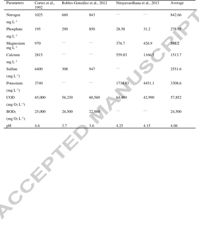
with the greatest environmental impact. Vinasses’s physico-chemical composition is variable
(Table 1) and depends on the nature and composition of the raw material, the system used in the
preparation of the must, the type of must, the fermentation method, the variety of yeast and type
of distillation apparatus (Christofoletti et al., 2013). It presents a high organic content, with
macronutrients such as carbon, nitrogen, phosphorus and potassium (Santana et al., 2017).
Generally, sugarcane vinasse has a dark color, strong odor and a water content of approximately
93%.
Fig.1
According to Leme and Seabra (2017) for each liter of ethanol produced, around 10 to
15 L of effluent are generated. Consequently, between 2014 and 2015, approximately 336
million m3 of sugarcane vinasse were generated in Brazil. The effluent was mainly used in
processes of fertigation without prior treatment. Nonetheless, there is an increasing concern
about the environmental impacts that can be generated with the application of effluents to the
soil, such as salinization, contamination of aquifers, depletion in the concentration of oxygen in
the soil, acidification, contamination with nitrates, chlorides and metals such as lead and zinc
(Fuess and Garcia, 2014). According to Plaza (1999), fertilization with sugarcane vinasse
partially replaces the use of chemical fertilizers, but it greatly increases the content of Ca, K, P
and N in the soil (Table 1).
Version postprint
The cyanobacteria Arthrospira (synonymie Spirulina) has been used by humans as food
since ancient times due to its high protein content (43-70%), which can be hydrolyzed into
biopeptides ( Yu et al., 2016). It has been experimentally proven to have some activities in health
conditions, such as anti-inflammatory, antioxidant, antiviral, anti-bacterial, hypertensive,
immunomodulatory, anticancer (Jang and Park, 2016; Ovando et al., 2018). Furthermore, the
pharmaceutical industry has shown a great interest in Arthrospira for its nutritional and
biotechnological properties, as well as its GRAS (Generally Regarded as Safe) status by the FDA
(Food and Drug Administration) (Oliveira et al., 2013).
The importance of novel bioactive compounds significantly increased in the last years.
Bioactive peptides (BP) are specific protein fragments, they have a positive impact on body
functions and may influence health (Singh et al., 2014). BP can be obtained from diverse raw
materials, such as plants, macroalgae, microalgae, seafood, and fungi (Hayes, 2013).
Recently, there has been great interest in the use and evaluation of peptides that show
biological activities. Antioxidant peptides are important, because of their protective effect in
lessening the severity of diseases; considering that in our body oxidative stress can cause serious
damage to proteins, lipids, and DNA by subtracting electrons (Nurdiani et al., 2016). These
peptides act by preventing binding of other molecules to oxygen, and by the inhibition of
free-radicals action (Kang et al., 2011). On the other hand, the antioxidant peptides can present
aminoacids with nucleophilic sulfur-containing side chains (Cys and Met) or aromatic side
chains (His, Tyr and Met), which can easily donate hydrogen atoms (Hayes, 2013). Also,
iron-chelating peptides have action in the metabolic pathways of autoxidation mechanisms and have
Version postprint
These peptides may present a structure that contains Met, Gln, Lys and Arg (de Castro and Sato,
2015).
The antimicrobial peptides (AMPs) are of enormous interest because of their inhibitory
activity against several pathogens and their ability as stimulators of the human immune system.
AMPs are known as host defense peptides due to their innate presence in the immune system in
animals, insect, plants, and humans with the role of defending them against the diversity of
bacterial, fungal, viral, and other pathogenic agents (Zhang and Gallo, 2016). Furthermore, it is
known that AMPs have the capacity to play other important roles in such processes as
angiogenesis, attraction of leukocytes, inflammation, and inhibition of cell proliferation (Phoenix
et al., 2013).The AMPs present common characteristics such as high proportion of hydrophobic
residues like Leucine (Leu), Isoleucine (Ile), Valine (Val), Phenylalanine (Phe), and Tryptophan
(Trp) (Haney and Hancock, 2014).
In the same way, anti-inflammatory peptides have received much attention due to their
potential in the therapeutic treatment for several health problems (cancer, aging, allergy, asthma,
autoimmune diseases, and coeliac disease) (Vo et al., 2013). These peptides act in the inhibition
of hyaluronidase enzyme and prevent hydrolysis of hyaluronic acid, helping in the regeneration,
proliferation, and repair of tissues. Besides, they can increase elasticity and decrease the loss of
moisture on the skin (Prado et al., 2016). On the other hand, the anti-collagenase peptides
prevent degradation of the extracellular matrix (ECM) by blocking the action of collagenase
(Chattuwatthana and Okello, 2015). These peptides can present a sequence, which resembles the
cleavage site in native collagen. Their structure can be composed of hydrophobic residues such
as Leu, Ile, Val, and Phe (Aureli et al., 2008). Thus, the objective of this study is the generation
Version postprint
with the valorization and decrease of environmental impact of sugarcane vinasse. Biopeptides
were produced through a one-step or sequential biomass enzymatic hydrolysis with further
separation and purification, and in vitro bioactivity tests to evaluate antioxidant, antimicrobial,
anti-inflammatory, and/or anti-collagenase biological activities.
2. Material and methods
The main steps of biomass peptides production and analyses are presented in the following
schema (Fig. 2).
Fig.2 2.1 Materials
All solvents used were of analytical grade. 1,1-diphenyl-2- picrylhydrazyl (DPPH),
2,2′-azino-bis (3-ethylbenzthiazoline-6-sulfonic acid (ABTS),
3-(2-Pyridyl)-5,6-diphenyl-1,2,4-triazine-p,p′-disulfonic acid monosodium salt hydrate (Ferrozine), Iron(II) chloride, Disodium
ethylenediaminetetraacetate dihydrate (EDTA-Na2), Subtilisin A from Bacillus licheniformis
(EC 3.4.21.62), Pepsin from porcine gastric mucosa (EC 3.4.23.1), hyaluronidase from bovine
testes (EC 3.2.1.35) and a Collagenase Activity Colorimetric Assay Kit were all purchased from
Sigma-Aldrich, St. Louis, MO, USA.
2.2 Analysis of sugarcane vinasse composition
Sugarcane vinasse, from Central Energética Moreno Açúcar e Álcool – Luis Antônio-S,
Brazil, was analyzed for its physical and chemical parameters before and after biomass
Version postprint
concentration were determined. COD and BOD were analyzed through spectrophotometry, and
the total organic nitrogen was evaluated using the Kjeldahl method according to the standard
method of the American Public Health Association (APHA, 1989). Total nitrogen was evaluated
by the elemental analysis performed in a CHNS Vario Macro analyser (Elementar, Germany).
Nitrate/nitrite and phosphate were analyzed spectrophotometrically by the standard tests for
effluent analysis from AOAC (Horwitz, William, Latimer, 2005) for Nitrate/Nitrite (AOAC
935.48).
2.3 Optimization of Arthrospira maxima OF15 biomass production
Arthrospira maxima OF15 inoculum was prepared in 125 mL of vinasse using 250 mL
flasks with forced aeration (0.5 vvm) during 5 days at room temperature. The effect of medium
dilution (10, 30 and 50%) on Arthrospira maxima biomass production was investigated. An
assay with Zarrouk medium was also conducted as a control. Batch culture experiments were
carried out in 2 L Erlenmeyer flasks with 1.8 L of working volume and constant air flow 0.5
vvm, at 30°C with 2500 Lux and photoperiod of 12 h. The illumination consisted of 32W
fluorescent lights, with a photon flux density (PFD) of 31 µmol photons.m-2.s-1 and a
photoperiod of 12 h (light:dark). For open cultures, conducted from september to march, in
Ribeirão Preto-Sp, Brazil (21.29ºS, 47.75ºW), where the average irradiation was 5.57kWh.m
-2.day-1, or 1061 µmol photons.m-2.s-1 for an average day length of 12.75h and an average
pluviometric index of 170 mm/month. Cultures, with an initial cellular concentration of 0.20 g/L,
were conducted during 15 days, without additional control of pH. Biomass concentration
Version postprint
2.4 Arthrospira maxima OF15 pilot scale biomass production
Arthrospira maxima OF15 biomass was produced at pilot scale in Ouro Fino
Agribusiness, Ribeirão Preto, São Paulo, Brazil. Inoculum was prepared in 250 mL flasks with
forced aeration (0.5 vvm, volume of air per volume of medium, or 125 mL min-1) during 5 days
at room temperature. The microalgal cultures were then successively scaled up to 2.5 L
(Erlenmeyer flasks, 0.5 vvm of air), 25 L (carboys, 0.5 vvm of air), 250 L (mini-raceway tanks,
agitated by paddle wheels to 20 cm s-1 of superficial velocity) up to a 14 m3 raceway bioreactor
(12 x 3 x 0.4 m, also agitated to 20 cm s-1 using paddle wheels). The medium for all steps used
vinasse diluted with water at 30% v/v and pH adjusted to 9.5-10 with CaCO3. Biomass
production was carried out for 15 days at 30°C in small scale, at pilot scale (raceway bioreactors)
in environmental conditions with temperature that varied between 21 to 31°C.
After cultivation, biomass was separated using a filter press. The fresh paste was placed
in plastic bags, for transportation and extruded to 50 x 70 cm trays and sun dried for 12 h, then
transported to a drying chamber for another 12 h at 60°C. Dried biomass was used in the next
steps for protein extraction and further enzymatic hydrolysis.
2.5. Proximate composition of A. maxima biomass
Analyses of A. maxima OF15 biomass were performed to determine its proximate
composition. Analyses of total protein, ashes, carbohydrate and lipids composition were carried
out according to the methods described by the Association of Official Analytical Chemists
(Horwitz, William, Latimer, 2005). Total protein was estimated by the Khjeldhal method
Version postprint
984.13), ashes were determined by calcination at 600°C for 2h (AOAC 942.05), lipids were
determined by exhaustive extraction with ethyl ether, followed by drying and weighing the
residue (AOAC 920.39), and carbohydrates were estimated by difference.
2.6. Protein extraction from Arthrospira maxima
Soluble protein extraction was performed as described previously (Wang and Zhang,
2016), with modifications. A. maxima OF15 powder (100 g) was suspended in 1 L pure water.
The suspension was frozen at −20°C for 4 h and thawed at 37°C, with a total of 4 freeze– thaw
cycles. After homogenization (2800 x g 30 s, 11000 x g 1 min, 2800 x g 30 s), the mixture was
ultrasonicated under 160 W power for 25 min (every 10 s with 13 s interval) in an ice bath.
Afterwards the extract solution was centrifuged at 10,000 x g and 4oC for 15 min. The protein
content of the supernatant was determined by Bradford protein assay.
2.7. Enzymatic hydrolysis of A. maxima biomass protein
The protein extract of A. maxima OF15 was diluted to 3% in 0.1 M citrate phosphate buffer
in pH 7 and pH 3. Enzymatic hydrolysis was carried out with endopeptidases (pepsin and
subtilisin A under specific conditions, individually or both sequentially (Fig. 3). The first
hydrolysate was prepared using subtilisin A at an enzyme/substrate (E/S) ratio of 4% w/w, 60oC,
pH 7 and during 5 h. The second hydrolysate was obtained using pepsin at an enzyme to
substrate ratio (E/S) of 3% w/w, 37oC, pH 2 and during 5 h. The third hydrolysate was prepared
using both enzymes sequentially. In this case, the protein solution was first treated using
Version postprint
then hydrolyzed by pepsin, under the conditions described above, during 3 h. The reactions were
stopped by heating the solution in a boiling water bath for 10 min. The obtained hydrolysates
were centrifuged at 6000 x g for 10 min. The liquid fraction was conserved and stored at -18ºC
for further analyses.
Fig. 3
2.6 Determination of the degree of hydrolysis
The method used to determine the degree of hydrolysis (DH) was performed as described
by Hoyle and Merritt, 1994. The three hydrolysates obtained before were evaluated: 1 mL
aliquots were denatured by the addition of 9 mL of 6.25% (w/v) trichloroacetic acid (TCA)
solution and left to settle for 10 min. The solution was then centrifuged for 5 min at 3000 x g and
the precipitate removed. The soluble proteins content from the supernatant was determined using
the Bradford (1976) method. DH was calculated as shown in Eq. 1:
𝐷𝐻 (%) = (𝑃𝑆𝑡𝑖−𝑃𝑆𝑡0
𝑃𝑡𝑜𝑡𝑎𝑙 ) 𝑥 100 (Eq. 1)
Where PSto, corresponds to the amount of soluble protein in TCA 6.25% w/v before
enzymatic hydrolysis; PSti, is the protein soluble after enzymatic hydrolysis and Ptotal is the total
amount of protein in the sample.
Version postprint
Peptides obtained from enzymatic hydrolysis were purified by ultrafiltration in a Vivaflow
200 Sartorius (tangential filtration) system. First the hydrolysate was microfiltrated using a 0.22
µm membrane. Then, it was ultrafiltrated using a membrane of 10 kDa molecular weight cut off
(MWCO). The permeate fraction containing molecules below 10 kDa was collected and stored at – 80oC, before lyophilization.
2.8 Peptide lyophilization and quantification
Peptide fractions were lyophilized (Freeze Dryer, ModulyoD, Thermo Scientific, USA)
during 24 h at -45ºC to a pressure of 50 mbar. The lyophilized peptide fraction (<10 kDa) of A.
maxima was solubilized in ultrapure water at a concentration of 1 mg mL-1. Peptide
concentration was determined using the Micro BCA Protein Assay Kit (Thermo Fisher) .
2.9 Mass spectrophotometry analysis
Peptide extracts from A. maxima were analyzed by MALDI-TOF in the Unit of Protein
Chemistry and Mass Spectrometry (Uniprote-MS) of the Federal university of Rio Gande do Sul.
Samples were prepared according to Udeshi et al. (2013) and Villén and Gygi (2008).
2.10 DPPH Radical Scavenging -in vitro assay
The capacity of the peptides for sequestering the free radical
2,2-diphenyl-1-picryl-hidrazol (DPPH) was performed as described previously (Yu et al., 2016). For the preparation of
the DPPH reagent, 4 mg DPPH (Sigma-Aldrich, St. Louis, MO, USA) were dissolved in 100 mL
Version postprint
used in the assay of each peptide fraction. Vitamin C (0.1 mg/mL) was used as a positive control.
A 96-well microplate was used to determine the scavenging activity, where 100 µl of the
samples or standard were mixed to 100 µL of DPPH reagent, and incubated for 30 min in the
dark at room temperature. After this time the absorbance was measured in a PowerWave XS
Microplate Spectrophotometer (BioTek Instruments, Inc., Winooski, USA) at 517 nm. The
percentage of DPPH radical scavenging was calculated as shown in Eq. 2:
𝐷𝑃𝑃𝐻 𝑟𝑎𝑑𝑖𝑐𝑎𝑙 𝑠𝑐𝑎𝑣𝑒𝑛𝑖𝑛𝑔 (%) = [𝐴0−𝐴1
𝐴0 ] 𝑥 100 (Eq. 2)
Where A0 is the absorbance of the control; A1 is the absorbance of the sample.
2.11 ABTS radical scavenging- in vitro assay
The 2,2’-azinobis-3-etilbenzothiazoline-6-sulfonic acid (ABTS) radical scavenging assay
was performed as described previously (Lee et al., 2015). The ABTS reagent was prepared by
mixing 5 mL of 7 mM ABTS (Sigma-Aldrich, St. Louis, MO, USA) with 88 L of 140 mM potassium persulfate, and reacting for 16 h at room temperature in the dark. After this time, the
ABTS reagent was diluted to 1:45 with ethanol (99%) until reaching an absorbance of 0.700,
which was measured in the spectrophotometer at 734 nm. Trolox
(6-hydroxy-2,5,7,8-tetramethylchroman-2-carboxylic acid) (Sigma-Aldrich, St. Louis, MO, USA) was prepared
from a stock solution (1 mM) in the concentration range of 2, 5, 10, 25, 50, 100 and 200 µM.
Peptide fractions and the positive control (vitamin C) were tested at 2, 5, 10, 25, 50 and 100 µg
Version postprint
the samples or standard were mixed with 100 L of ABTS reagent, in the dark at room
temperature. The absorbance was measured in a PowerWave XS Microplate Spectrophotometer
(BioTek Instruments, Inc., Winooski, USA) at 734 nm. The percentage of ABTS radical
scavenging was calculated as shown in Eq. 3:
𝐴𝐵𝑇𝑆 𝑟𝑎𝑑𝑖𝑐𝑎𝑙 𝑠𝑐𝑎𝑣𝑒𝑛𝑖𝑛𝑔 (%) = [𝐴0−𝐴1
𝐴0 ] 𝑥 100 (Eq. 3)
Where A0 was the absorbance control, A1 was the absorbance of the sample.
2.12 Ferrous ion-chelating activity - in vitro assay
The ferrous ion-chelating activity was performed as described previously (Wang et al.,
2009). Peptide fraction (100 L) was added in the following concentrations: 1.25, 2.5,5,10 and 25 µg mL-1, and mixed with 135 L of distilled water and 5 L of 2 mM FeCl2 in the microplate.
The reaction was initiated by the addition of 10 L of 5 mM ferrozine, and incubated for 10 min at room temperature. After incubation, the absorbance was measured at 562 nm with a
PowerWave XS Microplate Spectrophotometer. Distilled water (100 µL) instead of sample was
used as the control. For the blank, distilled water (10 µL) instead of ferrozine was used.
EDTA-Na2 was used as reference standards. All measurements were performed in triplicate. The ferrous
ion-chelating activity was calculated as shown in Eq. 4:
Ferrous ion-chelating activity (%) = [ 𝐴0− (𝐴1−𝐴2)
𝐴0 ] 𝑥 100 (Eq. 4)
Where A0 was the absorbance control, A1 was the absorbance of the sample or standard and
Version postprint
2.13 Antimicrobial activity - in vitro assay
The determination of minimal inhibitory concentration (MIC) of a substance was carried out
by the Broth microdilution method (EUCAST, 2003).
Minimum Inhibitory Concentration (MIC) Determination
This assay was carried out to determine the antimicrobial potential of the peptide fraction.
A 96-well microplate assay was used to determine the MIC: 80 µL of Mueller Hinton broth
(MHB) were added to the wells with 100 µL of peptides fractions at different concentrations
(0.13, 0.63, 1.25, 6.25 mg mL-1) and inoculated with 20 µL of each bacterial suspension (1.0x
107 UFC mL-1) (Bacillus subtilis (ATCC 6633), Staphylococcus aureus (ATCC 25923),
Salmonella typhi (ATCC 14028), Escherichia coli (ATCC 35218)). Chloramphenicol
(Sigma-Aldrich, St. Louis, MO, USA) was used as the positive control, and the culture medium without
the peptide fraction was the negative control. The microplate was sealed and incubated at 37oC
for 24 h. After incubation, the absorbance was measured at 600 nm. The percentage of growth
inhibition was calculated as shown in Eq. 7:
Grow inhibition (%) = 1 – ( 𝐴𝑐
𝐴0) 𝑥 100 (Eq. 7)
Where Ac is the absorbance of the sample, Ao is the absorbance of the control. Finally,
the microplate was colored with 30 µl of resazurin indicator solution (0.1%), and incubated for 2
h.
Version postprint
After MIC determination of the peptide fractions, an aliquot of 5 μL from all microplate
assay wells was seeded in Mueller Hinton Agar (MHA) plates. The plates were then incubated at
37°C for 24 h. The MBC endpoint was defined as the lowest concentration of antimicrobial
agent that kills >99.9% of the initial bacterial population where no visible growth of the bacteria
was observed on the MHA plates.
2.14 Anti-inflammatory activity - in vitro assay
The anti-inflammatory activity was evaluated by the inhibition of the enzyme
hyaluronidase (Type IV), as described previously (Prado et al., 2016), with few modifications.
Briefly, the three lyophilized peptide isolates (<10 kDa) of A. maxima were used in different
concentrations (3.3, 10, 33,100, 333 µg mL-1). The propolis commercial fraction (Bitmel, São
José do Rio Preto, SP) was used as positive control. 100 µL of peptide fraction or the positive
control was added to 500 µl of the potassium salt of hyaluronic acid (Sigma-Aldrich, St. Louis,
MO, USA), and incubated for 5 min at 37oC. Then, 350 units of the enzyme hyaluronidase type
IV-S were added (Sigma-Aldrich, St. Louis, MO, USA), and incubated at 37oC for 40 min. The
reaction was inactivated by adding 10 mL of sodium hydroxide solution (4 N) and 100 µL of
potassium tetraborate at 0.8 M, and incubated for 3 min at 100oC. Afterwards, 3 mL of
4-dimethylaminobenzaldehyde (DMAB) were added to the tubes, mixed and transferred to a water
bath for 20 min at 37oC. Finally, the absorbance was measured in a spectrophotometer at 585
nm. The percentage of inhibition was calculated as shown in Eq. 5.
Hyaluronidase inhibition activity (%)= 𝐴𝑚
Version postprint
Where: Am is the absorbance of the sample after enzymatic reaction, and Ac corresponds
to the absorbance of the control.
2.15 Collagenase inhibition - in vitro assay
This assay was performed according to the descriptive instructions supplied with the
Collagenase Activity Colorimetric Assay Kit (Sigma). The principles of this assay are based on
the enzyme-substrate interaction between collagenase from Clostridium histolyticum and the
synthetic N-[3-(2- furyl)acryloyl]-Leu-Gly-Pro-Ala (FALGPA). Peptide fractions (10 µL) were
added in different concentrations (10, 25, 50, 75 µg/mL). Negative controls consisted of water
and positive control consisted of 10-Phenanthroline. Absorbance was measured at 345 nm. All
measurements were performed in triplicate. The collagenase inhibition activity was calculated as
shown in Eq. 6:
Collagenase inhibition activity (%) = [𝐴0− 𝐴1
𝐴0 ] 𝑥 100 (Eq. 6)
Where A0 was the absorbance of the control, A1 was the absorbance of the sample.
2.16 Statisticalanalysis
Statistical analysis was performed using one-way and two-way ANOVA tests with the
support of Graphpad Prism 305 software.
3 Results and discussion
Version postprint
Arthrospira maxima OF15 biomass production was tested in different conditions using
vinasse at three concentrations. It was observed that the microalgae showed some difficulties to
adapt in the initial phases of cultivation. Best results were reached when vinasse was diluted at
30% with a biomass production of 3.015 g L-1 after 15 days of culture with a productivity of
0.201 g L-1day-1 at laboratory scale (Table 2). With Zarrouk medium, which is one of the
reference mediums for microalgae of Arthrospira genus cultivation, the production was similar
3.090 g L-1 of culture with a productivity of 0.206 g L-1day-1. Increasing productivities at
laboratory scale were attained when CO2 was injected continuously in the media at different
concentrations of 5, 10 and 15% with corresponding productivities of 0.206, 0.221 and 0.279 g
L-1day-1, respectively. This fact happened possibly because the microalgae assimilation of CO2,
depends on the medium pH, and the balance of CO2/CO32-.
In the literature, there are reports of successful cultivation of microalgae strains using
vinasse. However, the use of this effluent at high concentrations seems to inhibit algal growth.
Spirulina maxima biomass dry weight productivities ranging from 0.240 to 1 g L-1day-1 were
reported by Barrocal et al. (2010) using synthetic media supplemented with 1 to 7 g L-1 of beet
vinasse. Ramirez et al. (2014) cultivated Scenedesmus sp. in synthetic media supplemented with
sugarcane vinasse at a concentration of 50%, although they reached lower productivities of
biomass dry weight (0.024 g L-1day-1). In complete study of the influence of different vinasse
concentrations, Santana et al. (2017) cultivated Micractinium sp. Embrapa|LBA32 and
Chlamydomonas biconvexa Embrapa|LBA40 strains (isolated from a sugarcane vinasse
stabilization pond) in 50% diluted vinasse or 100% clarified vinasse obtaining average
productivities of 0. 222 g L-1day-1 of biomass dry weight. In their work, dos Santos et al., (2016)
Version postprint
with photoautotrophic condition (12 h, 70 μmol photons m2 s-1) followed by a heterotrophic
condition during the dark phase of the photoperiod (12 h, 3.0% v/v vinasse). The CTSC strategy,
separated by autotrophic rest periods of few days between CTSC cycles, shows an increase of
biomass concentration between 0.495 g L-1 and 0.609 g L-1 at the 7th day of each cycle and high
protein content (between 74.3 and 77.3% w/w).
In this work, the results obtained with the selected strain Arthrospira maxima OF15 in
terms of biomass productivities was considered promising for the use in pilot scale using as
culture media only vinasse diluted in water at 30% and CaCO3 to increase the pH to 9-10.
3.2 A. maxima cultivation in sugarcane vinasse at pilot scale
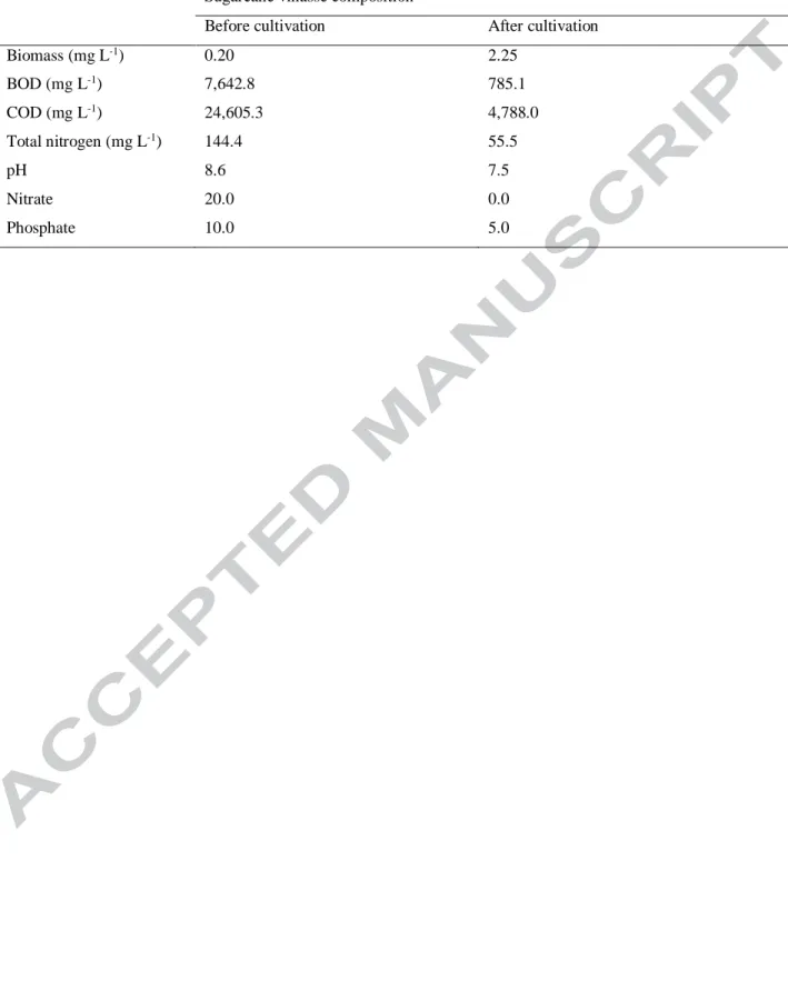
Microalgae biomass production was carried out in a raceway bioreactor with filtered
diluted vinasse at 30% (v/v). Biomass reached 2.25 g L-1 after 15 days (a productivity of 0.150 g
L-1day-1, and average specific growth rate of 0.23 day-1). These values are very promising when
compared to those reported in the literature with the use of synthetic media at laboratory scale,
and the final biomass is superior to other reports for vinasse use as culture medium. Barrocal et
al. (2010) produced Spirulina maxima OF15 0.150 g L-1day-1 using Schlösser medium
supplemented with beet vinasse (5 g/L). About 1 g.L-1 and 0.177 g L-1day-1 of biomass were
produced by dos Santos et al. (2016). Santana et al. (2017) obtained Chlamydomonas biconvexa
Embrapa|LBA40 biomass productivities of 0.182 g L-1day-1 and 0.222 g L-1day-1 with the use of
vinasse medium (50%) and clarified vinasse (100%), respectively.
The developed process also reduces the environmental pollution impact of sugarcane
Version postprint
BOD and COD were also observed, of 89.2% and 81%, respectively. Nitrate and phosphate
contents were also significantly reduced in 100% and 50%, respectively (Table 2).
Table 2
3.1 A. maxima biomass proximate composition
The composition of A. maxima OF15 produced in pilot scale was defined: 57.04±0.031%
(w/w) proteins, 5.65±0.276% (w/w) ash, 10.67±0.12% (w/w) carbohydrates and 11.2±0.36%
(w/w) lipids. Biomass proximate composition was similar to that reported in the literature (Bills
and Kung, 2014). As a protein-rich material (57% of protein), the biomass can be used as a
source for new peptides production with functional and biological activities.
3.2 Production of peptide fractions
About 80% of proteins from A. maxima OF15 were fractionated through water extraction,
freeze-thawing, homogenization, and ultrasonication. The protein isolates were subjected to
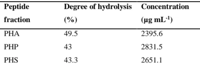
single-step and two-step hydrolysis, under controlled conditions. Three enzymatic procedures, in
a single or combined hydrolysis process, resulted in three different peptides’ fractions: the first
one was obtained by enzymatic hydrolysis with subtilisin A (PHA), the second was obtained by
pepsin hydrolysis (PHP) and the third one was produced after the hydrolysis with both enzymes
(PHS). The three fractions showed a degree of hydrolysis between 43-50% (Table 3), thus
generating a large amount of peptides. According to previous research, the same enzymes have
Version postprint
Chlorella vulgaris, whey and others, which showed antioxidant, anticancer, anti-microbial and
ACE-inhibitory, among other activities (Kang et al., 2011).
Table 3
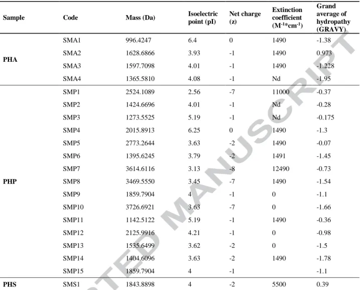
Peptides from different enzymatic hydrolysates (PHA, PHP and PHS) are shown in Table
5. They were analyzed using MALDI-TOF showing very different molecular masses. The single
enzymatic hydrolysis using subtilisin A generated 4 peptides. The hydrolysis with pepsin
resulted in the liberation of 15 peptides and the sequential hydrolysis (pepsin/subtilisin A)
showed only one peptide (data not shown). Subsequently, the physicochemical properties of each
peptide sequence were analyzed with a database (www.pepcalc.com) and the details are
summarized (Table 4). These peptides presented a small molecular weight, and GRAVY values
between -2 to +2, which can indicate that they have hydrophobic chains. According to previous
research, peptides that have a low molecular weight and are hydrophobic can be absorbed into
our body through two mechanisms, the paracellular pathway, and the transcellular pathway
(Maestri et al., 2015). In addition, as it can be seen, most peptides present negative charges,
which means that they can interact with the surfaces of the cellular membranes, and can be used
as antimicrobial and anti-cancer agents (Andersson et al., 2016; Perumal and Pandey, 2013). For
example the Microplusin from Rhipicephalus (Boophilus) microplus showed antimicrobial
activity against Gram-positive bacteria (Silva et al., 2009). The PopuDef (anionic peptide) from
Polypedates puerensis presented antimicrobial activity against Gram-positive and Gram-negative
(Wei et al., 2015).
Version postprint
3.3 Evaluation of peptides’ fractions biological activities
DPPH radical scavenging
All peptides fractions exhibited the capacity of sequestering the free radical DPPH (Fig.
4A). PHA showed a percentage of sequestering of 78±0.44% at a concentration of 0.1 g mL-1,
PHS of 78±0.21% and PHP of 77±0.71%. The IC50 values for DPPH scavenging by three
fractions are summarized in Table 5. PHS fraction demonstrated a strongest radical scavenging
activity IC50 of 1.79 mg/ mL if compared with Vitamin C that presented only an IC50 value of 1.2
mg mL-1. These results indicate that PHS contained peptides with a high potential DPPH
scavenging activity. Previously, Yu et al., (2016) reported the A. platensis hydrolysate showed
antioxidant activity 85.21 ± 1.59% at 10 mg mL-1. Lisboa et al., (2016) reported an antioxidant
activity between 48.5 and 73.2% at 2.5 mg mL-1 of Spirulina sp. LEB 18 hydrolysate. Compared
to the literature, PHS fraction showed better antioxidant activity using lower concentration.
Fig. 4
Table 5
ABTS radical scavenging
The antioxidant ability of peptides fractions to scavenge the blue-green colored ABTS+
radical cation was measured according to the radical scavenging ability of Trolox and IC50 (Fig.
4B). As shown in Table 5, the results clearly indicate that PHS and PHA have a good reducing
Version postprint
mL-1, respectively. Lisboa et al., (2016) reported a total antioxidant capacity expressed as TEAC
value of 248 μM of Trolox /g sample, using peptides of Arthrospira sp. Norzagaray-Valenzuela
et al. (2017), reported the enzymatic hydrolysis of three species of microalgae, Dunaliella
tertiolecta, Tetraselims suecica and Nannochloropsis sp. obtaining fractions with a TEAC value
of 437.01±1.34 Trolox µM g-1 protein hydrolysate, 696.99±1.82 Trolox µM g-1 protein
hydrolysate, and 519.44±4.46 Trolox µM g-1 protein hydrolysate, respectively. Comparing the
peptide fractions PHS and PHA with those previously reported in the literature, it is possible to
observe that they have a better total antioxidant capacity. This fact would be probably due to the
presence of cysteine (Cys) and methionine (Met) in their structure, or aromatic side chains with
amino acids histidine (His) and tyrosine (Tyr), which can easily donate hydrogen atoms.
Furthermore, based on these results, it can be said that PHS and PHA fractions have the ability of
scavenging free radicals, and to prevent oxidative damage to proteins, lipids, and DNA (Nurdiani
et al., 2016).
Iron-chelating activity
Peptide fractions (PHP, PHS, and PHA) were assayed for their Fe2+chelating activity at
five different concentrations and their activity was compared with the chelating activity of the
synthetic metal chelator EDTA. PHA fraction chelated more iron (97.3%) than PHP and PHS at
25 µg mL-1 and IC50 of 0.007 mg/mL (Table 6). The EDTA-Na2 presented a chelating activity of
61% at 25 µg mL-1. These values indicated that PHA was more efficient than the commercial
chelator Na2-EDTA. This results suggested that PHA fraction could be used as an agent to
preserve foods with high lipid content, a catalyst of metal ions for reducing cell damage and a
Version postprint
al., 2012; Wu et al., 2012). Previously, Kim et al., (2014) reported that the peptide Threonine
(Thr)-Aspartic acid (Asp)-Proline (Pro)- (Ile(Leu)-Alanine(Al)-Al-Cys-Ile(Leu), which was
obtained from Arthrospira sp., showed 80% of iron-chelating activity. Wu et al., (2012) reported
that hydrolysates from anchovy showed IC50 of 0.048 and 0.086 mg mL-1. Comparing PHA
fraction with those previously reported, it is possible to observe that it has a better chelating
activity, suggesting that the peptides can present in their composition the following amino acids:
Met, glutamine (Gln), lysine (Lys) and arginine (Arg) (de Castro and Sato, 2015).
Antimicrobial activity
The three peptide fractions were evaluated at different concentrations, from 0.13 to 6.25
mg/mL, for their antimicrobial activity against human pathogenic bacteria (E. coli, S. typhi, B.
subtilis and S. aureus). The three peptide fractions (PHP, PHS, and PHA) showed antimicrobial
activity (Table 6). The PHP displayed better values of MIC and IC50 values against B. subtilis, S.
aureus and S. typhi. Additionally, this peptide fraction is unique in that it showed bactericidal
action against all four pathogenic bacteria used in this experiment. PHS was the most efficient
against E. coli. The PHP showed activity against both Gram positive and Gram negative bacteria,
this fraction present a promising potential to be used as an antibiotic in the future. The research
of new antimicrobial substances is important because these pathogens mutations increase
resistance to existing drugs (Allen et al., 2014). The Arthrospira genre is renowned for having
antibacterial activity through the production of phycocyanins and carotenoids, but the existence
of antibacterial peptides was rarely reported (Ozdemir et al., 2004). Previously, Sun et al.,
(2016), reported antimicrobial activity against E. coli and S. aureus with a peptide from A.
Version postprint
that reported in the literature, PHP exhibits a better antimicrobial activity against E. coli and S.
aureus. In addition, with respect to B. subtilis and S. typhi A, there are no previous reports.
Table 6
Anti-inflammatory activity
For anti-inflammatory activity, the inhibition by PHA, PHP and PHS of hyaluronidase
Type IV was evaluated. The three peptide fractions isolates showed anti-inflammatory activity.
PHS showed the higher anti-inflammatory activity with 38.8± 1.1% at 333 µg mL-1 (Fig. 5) and
lowest IC50 0.92 mg mL-1. Meanwhile, PHA and PHS showed values of inhibition <32% and
IC50 1.63 mg mL-1, and 1.66 mg mL-1, respectively. Furthermore, the control + (propolis)
showed a IC50 23.61 mg mL-1. These results suggest that the peptides generated by the sequential
action of both proteases (PHS) have a higher anti-inflammatory activity at lower doses than
propolis ethanol fraction, in addition, to help maintain the elasticity in the skin and increased
proliferation, tissue regeneration, and repair by inhibiting hyaluronic acid hydrolysis (Prado et
al., 2016). Norzagaray-Valenzuela et al., (2017) reported that Dunaliella tertiolecta exhibited a
IC50 of 5.542 mg mL-1 and Tetraselmis suecica showed a IC50 of 5.907 mg mL-1. This research
showed that PHS is a potent anti-inflammatory agent when compared with similar compounds
reported in the literature. The anti-hyaluronidase activity of peptides from Arthrospira has not
been reported in the scientific literature until the present date.
Fig. 5
Version postprint
Collagen, which is the major component of the skin, is degraded by the enzyme collagenase
provoking the aging of the skin. The peptide fractions (PHP, PHS, PHA) were assayed at
different concentrations for their ability to inhibit collagenase. The anti-collagenase activity was
compared with the synthetic inhibitor 10-Phenanthroline. PHS at 75 µg/mL showed the best
anti-collagenase activity (92.5 %) and an IC50 of 32.5 µg mL-1 (Fig. 6), while PHP and PHA showed
values <70% at the same concentration and IC50 values of 43.9 and 96.7 µg mL-1, respectively.
The 10-Phenanthrolinepresented an inhibition activity of 57.13±1.9% at 75 µg mL-1. Based on
these results, it can be said that PHS showed higher collagenase inhibitory activity compared
with the synthetic inhibitor (10-Phenanthroline). It can also be suggested that it has the ability to
prevent the cutting of collagen, delaying the process of pre-collagen fibers formation and the loss
of strength, flexibility, and elasticity of the skin (Chattuwatthana and Okello, 2015). Combined
enzymatic hydrolysis with both proteases is a potential alternative for producing bioactive
peptides for anti-aging treatment. To our knowledge no existing scientific reports about the
potentialities of peptides from Arthrospira that display anti-aging activity.
Fig. 6
Conclusions
This study presented the development of a highly advantageous process for sugarcane vinasse
valorization together with significant reduction of its environmental impact. The microalgae
Arthrospira maxima OF15 was cultivated in sugarcane vinasse with high biomass productivities
at laboratory and pilot scale. A very performant enzymatic hydrolysis of microalgae biomass was
conducted to generate biopeptides. Noticeable biological activities of three biopeptides’ fractions
Version postprint
antimicrobial, antioxidant, anti-hyaluronidase and anti-collagenase activities were identified. Biopeptides’ fractions from A. maxima OF15 are certainly potential candidates for pharmaceutic,
cosmetic and food industry.
Acknowledgements
This work was supported by the Coordination of Improvement of Higher Level Personnel
(CAPES), the Graduate Program in Bioprocess Engineering and Biotechnology
(PPGEBB-UFPR), National Council for Scientific and Technological Development (CNPq) and Pelé
Pequeno Príncipe Research Institute, Brazil.
References
1. Allen, H.K., Trachsel, J., Looft, T., Casey, T.A., 2014. Finding alternatives to antibiotics. Ann. N. Y. Acad. Sci. 1323, 91–100. doi:10.1111/nyas.12468
2. Andersson, D.I., Hughes, D., Kubicek-Sutherland, J.Z., 2016. Mechanisms and
consequences of bacterial resistance to antimicrobial peptides. Drug Resist. Updat. 26, 43–57. doi:10.1016/j.drup.2016.04.002
3. Aureli, L., Gioia, M., Cerbara, I., Monaco, S., Fasciglione, G., Marini, S., Ascenzi, P., Topai, A., Coletta, M., 2008. Structural bases for substrate and inhibitor recognition by matrix metalloproteinases. Curr. Med. Chem. 15, 2192–2222.
doi:10.2174/092986708785747490
4. Barrocal, V.M., García-Cubero, M.T., González-Benito, G., Coca, M., 2010. Production of biomass by Spirulina maxima using sugar beet vinasse in growth media. N.
Biotechnol. 27, 851–856.
5. Bradford, M.M., 1976. A rapid and sensitive method for the quantitation microgram quantities of protein utilizing the principle of protein-dye binding. Anal. Chem. 72, 248– 254.
Version postprint
C., Alaiz, M., Girón-Calle, J., Vioque, J., Dávila-Ortiz, G., 2012. Antioxidant and metal chelating activities of peptide fractions from phaseolin and bean protein hydrolysates. Food Chem. 135, 1789–1795. doi:10.1016/j.foodchem.2012.06.016
7. Chattuwatthana, T., Okello, E., 2015. Anti-collagenase, anti-elastase and antioxidant activities of Pueraria candollei var. mirifica root extract and Coccinia grandis fruit juice extract: an in vitro study. European J. Med. Plants 5, 318–327.
doi:10.9734/EJMP/2015/14129
8. Christofoletti, C.A., Escher, J.P., Correia, J.E., Marinho, J.F.U., Fontanetti, C.S., 2013. Sugarcane vinasse: Environmental implications of its use. Waste Manag. 33, 2752–2761. doi:10.1016/j.wasman.2013.09.005
9. Cortez, L., Magalhães, P., Happi, J., 1992. Principais subprodutos da agroin-dústria canavieira e sua valorização. Rev. Bras. Energ. 2, 1-17.
10. de Castro, R.J.S., Sato, H.H., 2015. Biologically active peptides: Processes for their generation, purification and identification and applications as natural additives in the food and pharmaceutical industries. Food Res. Int. 74, 185–198.
doi:10.1016/j.foodres.2015.05.013
11. dos Santos, R.R., Araújo, O. de Q.F., de Medeiros, J.L., Chaloub, R.M., 2016. Cultivation of Spirulina maxima in medium supplemented with sugarcane vinasse. Bioresour. Technol. 204, 38–48.
12. European Committee on Antimicrobial Susceptibility Testing, 2003. Determination of minimum inhibitory concentrations (MICs) of antibacterial agents by broth dilution. Clin. Microbiol. Infect. 9, 1–7. doi:10.1046/j.1469-0691.2003.00790.x.
13. Fuess, L.T., Garcia, M.L., 2014. Implications of stillage land disposal: A critical review on the impacts of fertigation. J. Environ. Manage. 145, 210–229.
doi:10.1016/j.jenvman.2014.07.003
14. Haney, E., Hancock, R., 2014. Peptide design for antimicrobial and immunomodulatory applications. Biopolymers 100, 572–583. doi:10.1002/bip.22250.
15. Hayes, M., 2013. Biological activities of proteins and marine-derived peptides from byproducts and seaweeds, in: marine proteins and peptides. John Wiley & Sons, Ltd, Chichester, UK, pp. 139–165. doi:10.1002/9781118375082.ch7
16. Horwitz, William; Latimer, G., 2005. Official methods of analysis, 222nd ed. Association of Official Analytical Chemists, Washington, DC.
Version postprint
(Clupea harengus). J. Food Sci. 59, 76–79. doi:10.1111/j.1365-2621.1994.tb06901.x
18. Jang, I.-S., Park, S.J., 2016. A Spirulina maxima-derived peptide inhibits HIV-1 infection in a human T cell line MT4. Fish. Aquat. Sci. 19, 37. doi:10.1186/s41240-016-0039-3 19. Kang, K.H., Ryu, B.M., Kim, S.K., Qian, Z.J., Kim, S.K., 2011. Characterization of
growth and protein contents from microalgae Navicula incerta with the investigation of antioxidant activity of enzymatic hydrolysates. Food Sci. Biotechnol. 20, 183–191. doi:10.1007/s10068-011-0025-6
20. Kim, N.H., Jung, S.H., Kim, J., Kim, S.H., Ahn, H.J., Song, K. Bin, 2014. Purification of an iron-chelating peptide from Spirulina protein hydrolysates. J. Korean Soc. Appl. Biol. Chem. 57, 91–95. doi:10.1007/s13765-013-4211-5
21. Lee, K.J., Oh, Y.C., Cho, W.K., Ma, J.Y., 2015. Antioxidant and anti-inflammatory activity determination of one hundred kinds of pure chemical compounds using offline and online screening hplc assay 2015. doi:10.1155/2015/165457
22. Leme, R.M., Seabra, J.E.A., 2017. Technical-economic assessment of different biogas upgrading routes from vinasse anaerobic digestion in the Brazilian bioethanol industry. Energy 119, 754–766. doi:10.1016/j.energy.2016.11.029
23. Lisboa, C.R., Pereira, A.M., Alberto, J., Costa, V., 2016. Biopeptides with antioxidant activity extracted from the biomass of Spirulina sp. LEB 18. African J. Microbiol. Res. 10, 79–86. doi:10.5897/AJMR2015.7760
24. Maestri, E., Marmiroli, M., Marmiroli, N., 2015. Bioactive peptides in plant-derived foodstuffs. J. Proteomics 147, 140–155. doi:10.1016/j.jprot.2016.03.048
25. Nitayavardhana, S., Issarapayup, K., Pavasant, P., Khanal, S. K., 2013. Production of protein-,rich fungal biomass in an airlift bioreactor using vinasse as substrate. Bioresour. Technol. 133, 301–306. doi:10.1016/j.biortech.2013.01.073
26. Norzagaray-Valenzuela, C.D., Valdez-Ortiz, A., Shelton, L.M., Jiménez-Edeza, M., Rivera-López, J., Valdez-Flores, M.A., Germán-Báez, L.J., 2017. Residual biomasses and protein hydrolysates of three green microalgae species exhibit antioxidant and anti-aging activity. J. Appl. Phycol. 29, 189–198. doi:10.1007/s10811-016-0938-9
27. Nurdiani, R., Vasiljevic, T., Yeager, T., Donkor, K.T., Singh N., O., 2016. Bioactive peptides with radical scavenging and cancer cell cytotoxic activities derived from Flathead (Platycephalus fuscus) by-products. Eur. Food Res. Technol.
doi:10.1007/s00217-016-2776-z
Version postprint
Spirulina. Assoc. Bras. Nutr. 7527, 52–59.
29. Ovando, C.A., Carvalho, J.C. de, Vinícius de Melo Pereira, G., Jacques, P., Soccol, V.Thomaz-Soccol, C.R., 2018. Functional properties and health benefits of bioactive peptides derived from Spirulina: A review. Food Rev. Int. 34, 34–51.
doi:10.1080/87559129.2016.1210632
30. Ozdemir, G., Ulku Karabay, N., Dalay, M.C., Pazarbasi, B., 2004. Antibacterial activity of volatile component and various extracts of Spirulina platensis. Phyther. Res. 18, 754– 757. doi:10.1002/ptr.1541
31. Perumal, P., Pandey, V.P., 2013. Antimicrobial peptides : The role of hydrophobicity in the alpha helical structure. J. Pharm. Pharmacogn. Res. 1, 39–53.
32. Phoenix, D.A., Dennison, S.R., Harris, F., 2013. Antimicrobial Peptides. Wiley-VCH Verlag GmbH & Co. KGaA, Weinheim, Germany. doi:10.1002/9783527652853
33. Plaza, C., 1999. Tecnologia da Digestão Anaeróbia da Vinhaça e Desenvolvimento Sustentável Tecnologia da Digestão Anaeróbia da Vinhaça e Desenvolvimento Sustentável. Universidade Estadual de Campinas.
34. Prado, M.R.M., Boller, C., Zibetti, R.G.M., de Souza, D., Pedroso, L.L., Soccol, C.R., 2016. Anti-inflammatory and angiogenic activity of polysaccharide extract obtained from Tibetan kefir. Elsevier 108, 29–33. doi:https://doi.org/10.1016/j.mvr.2016.07.004
35. Ramirez, N.N.V., Farenzena, M., Trierweiler, J.O., 2014. Growth of microalgae Scenedesmus sp in ethanol vinasse. Brazilian Arch. Biol. Technol. 57, 630–635.
36. Rice, E.W., Baird, R.B., Clesceri, L.S, 2012. Standard method for the examination of water and wastewater, 22nd ed. Washington, American Public Health Association. 37. Robles-González V, Galíndez-Mayer J, Rinderknecht-Seijas N, Poggi-Varaldo H., 2012.
Treatment of mezcal vinasses: a review. J Biotechnol. 157, 521–546. doi: 10.1016/j.jbiotec.2011.09.006.
38. Santana, H., Cereijo, C.R., Teles, V.C., Nascimento, R.C., Fernandes, M.S., Brunale, P., Campanha, R.C., Soares, I.P., Silva, F.C.P., Sabaini, P.S., Siqueira, F.G., Brasil,
B.S.A.F., 2017. Microalgae cultivation in sugarcane vinasse: Selection, growth and biochemical characterization. Bioresour. Technol. 228, 133–140.
doi:10.1016/j.biortech.2016.12.075
39. Silva, F.D., Rezende, C.A., Rossi, D.C.P., Esteves, E., Dyszy, F.H., Schreier, S., Gueiros-Filho, F., Campos, C.B., Pires, J.R., Daffre, S., 2009. Structure and mode of action of microplusin, a copper ii-chelating antimicrobial peptide from the cattle tick
Version postprint
Rhipicephalus (Boophilus) microplus. J. Biol. Chem. 284, 34735–34746. doi:10.1074/jbc.M109.016410
40. Singh, B.P., Vij, S., Hati, S., 2014. Functional significance of bioactive peptides derived from soybean. Peptides 54, 171–179. doi:10.1016/j.peptides.2014.01.022
41. Sun, Y., Chang, R., Li, Q., Li, B., 2016. Isolation and characterization of an antibacterial peptide from protein hydrolysates of Spirulina platensis. Eur. Food Res. Technol. 242, 685–692. doi:10.1007/s00217-015-2576-x
42. Udeshi, N.D., Mertins, P., Svinkina, T., Carr, S.A., 2013. Large-scale identification of ubiquitination sites by mass spectrometry. Nat. Protoc. 8, 1950–1960.
doi:10.1038/nprot.2013.120
43. Villén, J., Gygi, S.P., 2008. The SCX/IMAC enrichment approach for global phosphorylation analysis by mass spectrometry. Nat. Protoc. 3, 1630–1638. doi:10.1038/nprot.2008.150
44. Vo, T.S., Ryu, B., Kim, S.K., 2013. Purification of novel anti-inflammatory peptides from enzymatic hydrolysate of the edible microalgal Spirulina maxima. J. Funct. Foods 5, 1336–1346. doi:10.1016/j.jff.2013.05.001
45. Wang, T., Jónsdóttir, R., Ólafsdóttir, G., 2009. Total phenolic compounds, radical scavenging and metal chelation of extracts from Icelandic seaweeds. Food Chem. 116, 240–248. doi:10.1016/j.foodchem.2009.02.041
46. Wang, Z., Zhang, X., 2016. Inhibitory effects of small molecular peptides from Spirulina (Arthrospira) platensis on cancer cell growth. Food Funct . 7, 781–788.
doi:10.1039/c5fo01186h
47. Wei, L., Che, H., Han, Y., Lv, J., Mu, L., Lv, L., Wu, J., Yang, H., 2015. The first anionic defensin from amphibians. Amino Acids 47, 1301–1308. doi:10.1007/s00726-015-1963-8
48. Wu, H., Liu, Z., Zhao, Y., Zeng, M., 2012. Enzymatic preparation and characterization of iron-chelating peptides from anchovy (Engraulis japonicus) muscle protein. Food Res. Int. 48, 435–441. doi:10.1016/j.foodres.2012.04.013
49. Yu, J., Hu, Y., Xue, M., Dun, Y., Li, S., Peng, N., Liang, Y., Zhao, S., 2016. Purification and Identification of Antioxidant Peptides from Enzymatic Hydrolysate of Spirulina platensis. J. Microbiol. Biotechnol. 26, 1216–1223. doi:10.4014/jmb.1601.01033
50. Zhang, L., Gallo, R.L., 2016. Antimicrobial peptides. Curr. Biol. 26, R14–R19. doi:10.1016/j.cub.2015.11.017
Version postprint
Figure Captions
Fig. 1. Simplified process of bioethanol production and vinasse generation
Fig. 2. Steps of Arthrospira maxima OF15 biomass and peptides’ production from sugarcane vinasse
Fig. 3. Enzymatic hydrolysis of Arthrospira maxima OF15 proteins
Fig 4. PHA, PHP, PHS peptide fractions biological activity essays: (A) Percentage of radical scavenging using DPPH assay; (B) Percentage of radical scavenging using ABTS assay
Fig. 5. Percentage of inhibition of hyaluronidase enzyme by PHS, PHA and PHP at different concentrations
Fig. 6. Percentage of inhibition of collagenase enzyme by PHP, PHA and PHS peptide fractions at different concentrations (10, 20, 50 and 75 µg mL-1) (p=<0.0001)
Version postprint
Version postprint
Version postprint
Version postprint
Version postprint
Version postprint
Version postprint
Table 1. Average physico-chemical composition of sugarcane vinasse generated from bioethanol producing chain
Parameters Cortez et al., 1992
Robles-González et al., 2012 Nitayavardhana et al., 2013 Average
Nitrogen mg L–1 1025 660 843 ––– ––– 842.66 Phosphate mg L–1 195 290 850 28.58 31.2 278.95 Magnesium mg L–1 970 ––– ––– 376.7 426.9 591.2 Calcium mg L–1 2815 ––– ––– 559.83 1166.5 1513.7 Sulfate (mg L–1) 6400 308 947 ––– ––– 2551.6 Potassium (mg L–1) 3740 ––– ––– 1734.83 4451.1 3308.6 COD (mg O2 L–1) 65,000 56,230 60,560 64,480 42,990 57,852 BOD5 (mg O2 L–1) 25,000 26,500 22,000 ––– ––– 24,500 pH 4.6 3.7 3.6 4.25 4.15 4.06
Version postprint
Table 2. Sugarcane vinasse composition before and after Arthrospira maxima OF15 biomass cultivation at pilot scale
Sugarcane vinasse composition
Before cultivation After cultivation
Biomass (mg L-1) 0.20 2.25 BOD (mg L-1) 7,642.8 785.1 COD (mg L-1) 24,605.3 4,788.0 Total nitrogen (mg L-1) 144.4 55.5 pH 8.6 7.5 Nitrate 20.0 0.0 Phosphate 10.0 5.0
Version postprint
Table 3. Characterization of Arthrospira maxima OF15 protein hydrolysates
Peptide fraction Degree of hydrolysis (%) Concentration (µg mL-1) PHA 49.5 2395.6 PHP 43 2831.5 PHS 43.3 2651.1
Version postprint
Table 4. Characterization of peptide fractions’ physicochemical properties
Sample Code Mass (Da) Isoelectric
point (pI) Net charge (z) Extinction coefficient (M-1*cm-1) Grand average of hydropathy (GRAVY) PHA SMA1 996.4247 6.4 0 1490 -1.38 SMA2 1628.6866 3.93 -1 1490 0.973 SMA3 1597.7098 4.01 -1 1490 -1.228 SMA4 1365.5810 4.08 -1 Nd -1.95 PHP SMP1 2524.1089 2.56 -7 11000 -0.37 SMP2 1424.6696 4.01 -1 Nd -0.28 SMP3 1273.5525 5.19 -1 Nd -0.175 SMP4 2015.8913 6.25 0 1490 -1.3 SMP5 2773.2644 3.63 -2 1490 -0.07 SMP6 1395.6245 3.79 -2 1491 -1.45 SMP7 3614.6116 3.13 -8 12490 -0.73 SMP8 3469.5550 3.45 -7 1490 -1.54 SMP9 1859.7904 4 -1 0 -1.1 SMP10 3726.6921 3.63 -7 0 -1.66 SMP11 1142.5122 5.19 -1 1490 -0.36 SMP12 2125.9916 4.21 -1 0 -0.98 SMP13 1535.6499 3.62 -2 0 -1.5 SMP14 1404.6096 3.63 -2 1490 -1.78 SMP15 1859.7904 4 -1 -1.1 PHS SMS1 1843.8898 4 -2 5500 0.39
Version postprint
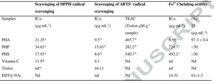
Table 5. Antioxidant and iron-chelating activities of different peptide fractions from Arthrospira maxima OF15
Scavenging of DPPH radical scavenging
Scavenging of ABTS+ radical
scavenging Fe2* Chelating activity Samples IC50 (µg mL-1) IC50 (µg mL-1) TEAC (Trolox µM g-1 sample) IC50 (µg mL-1) (%) 25 (µg mL-1) PHA 21.25 a 9.5 b 465.7 b 6.98 c 97.3 ± 0.4 PHP 34.63 a 15.63 b 282.2 b 724.7 c <30 PHS 17.93 a 8.6 b 540.7 b 492.2 c <30 Vitamin C 11.97 6.1 Nd nd Nd Trolox nd* 44.11 Nd nd Nd EDTA-NA2 Nd nd Nd 14.31 61±1.3
a Analysis of variance showed р=0.006 (PHA vs PHP; PHA vs PHS; PHP vs PHS). b Analysis of variance showed р=0.0113 (PHA vs PHP; PHA vs PHS; PHP vs PHS). c Analysis of variance showed р=<0.0001 (PHA vs PHP; PHA vs PHS; PHP vs PHS)
Version postprint
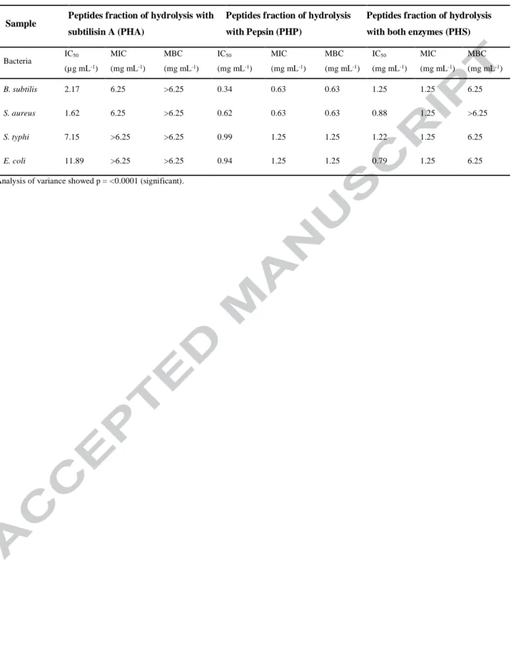
Table 6. Antimicrobial activity of peptides from Arthrospira maxima determined by MIC, MBC and IC50
Sample Peptides fraction of hydrolysis with
subtilisin A (PHA)
Peptides fraction of hydrolysis with Pepsin (PHP)
Peptides fraction of hydrolysis with both enzymes (PHS)
Bacteria IC50 (µg mL-1) MIC (mg mL-1) MBC (mg mL-1) IC50 (mg mL-1) MIC (mg mL-1) MBC (mg mL-1) IC50 (mg mL-1) MIC (mg mL-1) MBC (mg mL-1) B. subtilis 2.17 6.25 >6.25 0.34 0.63 0.63 1.25 1.25 6.25 S. aureus 1.62 6.25 >6.25 0.62 0.63 0.63 0.88 1.25 >6.25 S. typhi 7.15 >6.25 >6.25 0.99 1.25 1.25 1.22 1.25 6.25 E. coli 11.89 >6.25 >6.25 0.94 1.25 1.25 0.79 1.25 6.25 Analysis of variance showed p = <0.0001 (significant).
Version postprint
Peptides from
Arthrospira maxima
Pilot Scale
Production of Arthrospira maxima in vinasse Biomassa of A. maxima Extraction of A. maxima protein Enzymatic hydroliyses Pepsin Tangential filtration MALDI-TOF Biological activities Antioxidant Iron-chelating Antimicrobial Anti-inflamatory Anti-collagenase Subtilisin A Subtilisin A and pepsin Proximate composition of A.maxima PHP PHA Peptides PHSLaboratory
Scale
Version postprint
• High titers of Arthrospira maxima biomass cultivated at Laboratory and pilot scale
• Microalgae produced using sugarcane vinasse with BOD and COD reduction
• Peptide fractions obtained through biomass enzymatic hydrolysis