The MIT Faculty has made this article openly available.
Please share
how this access benefits you. Your story matters.
Citation
Strasser, Geraldine A, Nazimah Abdul Rahim, Kristyn E VanderWaal,
Frank B Gertler, and Lorene M Lanier. “Arp2/3 Is a Negative
Regulator of Growth Cone Translocation.” Neuron 43, no. 1 (July 8,
2004): 81-94. Copyright © 2004 Cell Press
As Published
http://dx.doi.org/10.1016/j.neuron.2004.05.015
Publisher
Elsevier
Version
Final published version
Citable link
http://hdl.handle.net/1721.1/83506
Terms of Use
Article is made available in accordance with the publisher's
policy and may be subject to US copyright law. Please refer to the
publisher's site for terms of use.
of Growth Cone Translocation
al., 1998; Zheng et al., 1996). These filopodia are
dy-namic structures comprised of long, bundled actin
fila-ments. Between the filopodia lie thin regions known
Geraldine A. Strasser,
1Nazimah Abdul Rahim,
2Kristyn E. VanderWaal,
2Frank B. Gertler,
1and Lorene M. Lanier
2,*
as lamellipodial veils. The transition region contains a
1
Biology Department
dense network of actin filaments, actin arcs, and motile
Massachusetts Institute of Technology
actin structures known as intrapodia (Dent and Kalil,
Cambridge, Massachusetts 02139
2001; Rochlin et al., 1999; Schaefer et al., 2002). The
2
Department of Neuroscience
central region contains a relatively sparse network of
University of Minnesota
actin filaments and abundant microtubules. These
mi-Minneapolis, Minnesota 55455
crotubules are often bundled or looped, but they also
stochastically explore the peripheral region, where they
contribute to pathfinding (Buck and Zheng, 2002; Dent
Summary
and Kalil, 2001; Lin and Forscher, 1993; Tanaka et al.,
1995; Zhou et al., 2002). Directed growth cone motility
Arp2/3 is an actin binding complex that is enriched
in response to extracellular cues is produced by the
in the peripheral lamellipodia of fibroblasts, where it
coordinated regulation of actin and microtubule
net-forms a network of short, branched actin filaments,
works (Dent and Kalil, 2001; Suter et al., 1998; Zhou et
generating the protrusive force that extends
lamelli-al., 2002).
podia and drives fibroblast motility. Although it has
Studies in motile cells such as fibroblasts have
identi-been assumed that Arp2/3 would play a similar role
fied a large variety of factors that are involved in the
in growth cones, our studies indicate that Arp2/3 is
organization and modification of the actin cytoskeleton
enriched in the central, not the peripheral, region of
(Pollard and Borisy, 2003), and in some cases these
growth cones and that the growth cone periphery
con-proteins have been shown to be functionally important
tains few branched actin filaments. Arp2/3 inhibition
in growth cones (Dickson, 2002). Actin binding proteins
in fibroblasts severely disrupts actin organization and
are classified according to their activities. Capping
pro-membrane protrusion. In contrast, Arp2/3 inhibition in
teins like CapZ and anticapping proteins like the Ena/
growth cones minimally affects actin organization and
VASP proteins bind the polymerizing ends of actin
fila-does not inhibit lamellipodia protrusion or de novo
ments and regulate filament length (Bear et al., 2002;
filopodia formation. Surprisingly, Arp2/3 inhibition
sig-Caldwell et al., 1989; Cooper and Schafer, 2000; Lanier
nificantly enhances axon elongation and causes
de-et al., 1999; Wills de-et al., 1999). Bundling and crosslinking
fects in growth cone guidance. These results indicate
proteins like fascin and filamin bind to preformed
fila-that Arp2/3 is a negative regulator of growth cone
ments and regulate their organization and stability
(Co-translocation.
han et al., 2001; Kureishy et al., 2002; Stossel et al.,
2001). Severing proteins like gelsolin and ADF/cofilin
Introduction
bind actin filaments and cause them to depolymerize
(Cooper and Schafer, 2000; Gungabissoon and
Bam-During development of the nervous system, axons and
burg, 2003; Lu et al., 1997). Membrane linking proteins
dendrites extend over significant distances to form
syn-like those of the Ezrin/Radixin/Moesin family (ERMs) link
aptic connections. To make these connections
accu-actin filaments to the plasma membrane (Bretscher et
rately, growth cones must detect a vast array of
diffus-al., 2002; Paglini et diffus-al., 1998). Finally, nucleating proteins
ible and surface bound guidance molecules present in
like formins and Arp2/3 induce the formation of new
the extracellular environment (Tessier-Lavigne and
actin filaments (Kobielak et al., 2004; Pollard, 2002;
Pol-Goodman, 1996; Yu and Bargmann, 2001). Binding of
lard and Beltzner, 2002). Differential activation of these
these guidance cues to receptors on the growth cone
actin binding proteins by multiple signaling pathways is
surface activates intracellular signaling cascades whose
key to cell motility.
downstream targets include proteins that regulate the
Arp2/3 is one of the most extensively studied actin
growth cone cytoskeleton (Dickson, 2002; Koleske,
binding proteins (Weaver et al., 2003). The Arp2/3
com-2003; Luo, 2002). Modification of the cytoskeleton re-
plex is composed of seven evolutionarily conserved
pro-sults in selective protrusion and retraction of the growth
teins, known as Arp2, Arp3, p41-Arc, p34-Arc, p21-Arc,
cone, driving growth cone motility and guiding axons
p20-Arc, and p16-Arc in mammals (Machesky et al.,
and dendrites to their appropriate synaptic targets.
1997; Welch et al., 1997a). Arp2/3 has been shown to
The growth cone has been operationally divided into
nucleate the polymerization of actin monomers in vitro
three main compartments: the distal peripheral region,
by binding to pre-existing actin filaments and inducing
the transition region, and the proximal central region.
the formation of a new filament as a branch (Amann
The peripheral region is an actin-rich region containing
and Pollard, 2001). Ultrastructural studies of fibroblast
abundant filopodia that play a crucial role in pathfinding
lamellipodia show that Arp2/3 is found at characteristic
(Bentley and Toroian-Raymond, 1986; Chien et al., 1993;
Y-shaped 70
⬚ branches in the actin cytoskeleton
(Svit-Gomez and Letourneau, 1994; Kim et al., 2002; Kuhn et
kina and Borisy, 1999). In vivo, Arp2/3 induces the
forma-tion of a network of short, branched filaments, called
a “dendritic array.” Polymerizing dendritic arrays can
generate the protrusive force that drives many types of
inhibition of Arp2/3 in neurons had little affect on growth
actin-based motility. For example, formation of a den-
cone morphology or filopodia formation and appeared
dritic array is essential for generating the actin-based
to enhance axon elongation. In fibroblasts, inhibition of
“comet tails” that propel intracellular bacteria such as
Arp2/3 severely disrupted the lamellipodial actin
cy-Listeria monocytogenes through the cytoplasm of in-
toskeleton, inhibited actin tail formation by Listeria
mo-fected cells (May et al., 1999; Welch et al., 1997b). In
nocytogenes, and altered lamellipodial protrusion. Our
fibroblasts, keratocytes, and a variety of other cell types,
findings suggest that in the growth cone, Arp2/3 is a
Arp2/3 is enriched in the leading edge, where rapid actin
negative regulator of neurite outgrowth and may be
im-filament polymerization generates the protrusive force
portant for the pathfinding role of the cytoskeleton.
that drives lamellipodia extension and forward motility
(Machesky et al., 1997; Welch et al., 1997a). Inhibition of
Results
Arp2/3 by microinjection of a function-blocking antibody
blocks membrane protrusion, indicating that Arp2/3 is
Arp2/3 Is Enriched in the Central Region
essential for lamellipodia protrusion in fibroblasts (Bailly
of the Growth Cone
et al., 2001).
Although Arp2/3 is a key player in many types of
actin-The Arp2/3 complex has a low level of constitutive
based motility, its role in the formation and motility of
activity and must undergo a conformational change in
the growth cone remains unclear. Both the dendritic
order to become fully activated. This conformational
array model of lamellipodia protrusion and the
conver-change can be mediated by binding to the C-terminal
gent extension model of filopodia formation require
domain of members of the WASP/WAVE family of pro-
Arp2/3 to be enriched at the periphery of the fibroblast.
teins in vitro and in vivo (Miki et al., 1996; Symons et
If either model is directly applicable to growth cone
al., 1996; Weaver et al., 2001, 2003; Weed et al., 2000).
motility, Arp2/3 should be enriched in an analogous
re-This C-terminal domain contains three regions: the ver-
gion in the growth cone. Surprisingly,
immunofluores-polin-homology (V) actin binding region (Miki and Take-
cence localization revealed that Arp2/3 is enriched in
nawa, 1998); the cofilin-homology or central (C) region;
the neurite shaft and central region of the growth cone
and the acidic (A) Arp2/3 binding region (Marchand et
but not the peripheral region of the growth cone (Figure
al., 2001). Constructs containing only the VCA regions
1). Similar results were obtained with antisera to Arp3,
are potent stimulators of Arp2/3-mediated actin poly-
p34-Arc, and p21-Arc (Figures 1A–1C) in growth cones
merization in vitro but fail to localize and therefore inhibit
from hippocampal neurons (Figure 1) and dorsal root
Arp2/3 function in vivo (Machesky et al., 1999; Rohatgi
ganglia (DRG) neurons (data not shown). Staining of
et al., 1999; Winter et al., 1999). A peptide containing
Rat2 fibroblasts with the same antibodies under
identi-only the CA domains has been shown to bind to Arp2/3
cal fixation conditions confirmed that, as expected,
with an affinity similar to VCA but fails to activate Arp2/3
Arp2/3 is enriched in the fibroblast
periphery/lamelli-activity in vitro (Hufner et al., 2001; Rohatgi et al., 1999).
podia (Figures 1H–1J).
The CA peptide blocks activation of the Arp2/3 complex
To verify the immunostaining results, Arp2/3 was
lo-by VCA or full-length WASP/Scar proteins in vitro and
calized using a p21-EGFP fusion protein. As with the
has been used in vivo to inhibit Arp2/3 activity (Falet et
antisera staining, p21-EGFP was highly enriched in the
al., 2002; Hufner et al., 2002).
fibroblast periphery (Figure 1K) but was restricted to
Arp2/3 is regarded as a key player in most models of
the neurite shaft and central region of the growth cone
actin-based motility. Although Arp2/3 has been shown
(Figures 1D–1G). Similar Arp2/3 localization was seen in
to be essential for embryonic CNS axon morphology in
small (Figures 1A and 1D) and large (Figures 1B and 1F)
Drosophila (Zallen et al., 2002), the role of Arp2/3 actin
growth cones, in terminal (Figures 1A, 1B, and 1D) and
nucleating activity in the formation and motility of the
collateral (Figures 1F and 1G) branch growth cones,
growth cone has not been studied. It has been assumed
and in dendrite growth cones (Figure 1E). Since small
that the primary role of Arp2/3 in growth cones would
dynamic growth cones are likely to be translocating,
be to generate a dendritic actin array that drives
lamelli-while large growth cones are likely to be paused (Kim
podial veil protrusion. Growth cones are, however,
domi-et al., 1991; Mason and Erskine, 2000; Szebenyi domi-et al.,
nated by filopodia containing unbranched actin
fila-2001; Tosney and Landmesser, 1985), this indicates that
ments, and the Arp2/3 complex is generally absent from
Arp2/3 localization does not change with motility. Under
filopodia (Svitkina and Borisy, 1999). Recently, Svitkina
no circumstances was Arp2/3 enrichment detected in
and colleagues proposed a “convergent elongation
the growth cone periphery. Finally, Arp2/3 localization
model” of filopodia formation that suggests a role for
in the growth cone central region was resistant to
pre-Arp2/3 in filopodia formation. In this model, filopodia
permeabilization (data not shown), suggesting that
are derived from rearrangement of elongating actin
fila-Arp2/3 in the central region of the growth cone is
associ-ments into bundles (Svitkina et al., 2003; Vignjevic et
ated with the cytoskeleton.
al., 2003). The role of Arp2/3 is limited to the initial
nucle-ation of filaments into a dendritic array; a subset of these
Actin Organization in the Growth Cone Periphery
filaments then elongate and are subsequently bundled
Arp2/3 activity is essential for the formation of the highly
together by fascin to form a filopodium.
branched dendritic array of actin filaments found at the
Given its key role in other types of actin-based motility,
edge of the protruding fibroblast lamellipodia. If Arp2/3
we sought to determine if Arp2/3 plays a similar role in
plays a similar role in the growth cone, we would expect
growth cone morphology and motility. To do this, we
to see similar branched actin arrays in the peripheral
used the N-WASP CA or Wave-1 VCA peptides to block
supramolecu-Figure 1. Arp2/3 Localization in Growth Cones
(A–C) Embryonic hippocampal neurons la-beled with antibodies to Arp3 (A⬘), p34-Arc (B⬘), or p21-Arc (C⬘), anti-tubulin  III (A–C), and phalloidin to visualize filamentous actin (A″–C″). Arp2/3 and phalloidin staining are merged and overlap appears yellow (A–C). Arp2/3 is enriched in the central regions of small growth cones (A–A″, arrows), of large growth cones with looped microtubules (B–B″, arrows), and in filopodia-rich regions of the neurite shaft (C⬘, C″, arrow), where de-creased tubulin staining (C, arrow) correlates with branch emergence.
(D–G) Embryonic hippocampal neurons in-fected with recombinant adenovirus express-ing p21-EGFP (D–G) and stained with phalloi-din (D⬘–G⬘). Cells were also labeled with anti-tubulin  III to identify neurons (data not shown). p21-EGFP is enriched in the central region of small axon growth cones (D, arrow), dendrite growth cones (E, arrow), large collat-eral branch growth cones (F), and filopodia-rich regions of the neurite shaft (G and G⬘, arrows).
(H–K) Control Rat2 fibroblasts labeled with antibodies to Arp3 (H), p34-Arc (I), or p21-Arc (J), and expressing p21-EGFP (K). In fibro-blasts, Arp2/3 is enriched in the peripheral lamellipodia (arrows in H–K). There is also a detergent-extractable cytoplasmic pool (visi-ble here because cells were not treated with detergent before fixation). Scale bars equal 10m for all growth cones (as shown in A) and 10m for all fibroblasts (as shown in K).
lar organization of the actin filaments in the growth cone,
other, we identified very few structures resembling
clas-sic Arp2/3-generated Y branch structures (Svitkina and
we used the platinum replica method of Svitkina et al.
(1995), modified to preserve the overall growth cone
Borisy, 1999). As with the localization studies, similar
results were obtained in both large and small growth
morphology and visualize the actin filaments. As
ex-pected, the fibroblast periphery contains a dense den-
cones (Figures 2B and 2C, respectively) and in dendrite
growth cones (data not shown). Furthermore, analysis
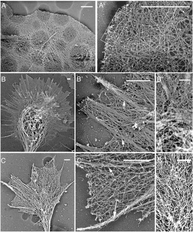
dritic array (Figures 2A–2A
⬘).
In contrast, the actin network in the growth cone pe-
of lamellipodia veils between growth cone filopodia
re-vealed that neither concave nor convex growth cone
riphery appears to be dominated by thick bundles of
filaments organized into filopodia, with an array of very
lamellipodia (Figures 2B
⬘ and 2C⬘) contained structures
resembling the Arp2/3-dependent dendritic arrays
long filaments underlying the lamellipodial veil (Figures
2B and 2C). The veil region is quite dense with long
formed in fibroblasts (Figure 2A
⬘). Together, these
find-ings suggest that, in contrast to fibroblasts, Arp2/3 may
filaments, and although they frequently cross over each
Figure 2. Comparison of Actin Filament Organization in Fibroblasts and Neuronal Growth Cones
The actin cytoskeleton is visualized in Rat2 fibroblasts (A) and growth cones from embry-onic hippocampal neurons (B and C) using platinum replica electron microscopy. Lower-magnification images reveal the overall mor-phology and relative size (A–C). Higher-mag-nification images reveal details of the actin organization in the periphery (A⬘–C⬘). Individ-ual filaments can be seen in (A⬘)–(C⬘). The dense dendritic array is visible in the boxed region of the fibroblast (A⬘). Prominent actin bundles are visible in the growth cone (arrows, B⬘ and C⬘). High-magnification im-ages of the growth cone central region (B″ and C″) show the dense network of actin fila-ments and microtubules. Scale bars equal 1m.
not be a major determinant of actin organization at the
expressing EGFP-CA by EM revealed an acute
disrup-tion of the dendritic array (Figures 4A–4D). Actin
fila-growth cone periphery.
ments were still present but were often extremely long,
unbranched, and oriented parallel to the cell membrane
Inhibition of Arp2/3 Dramatically Alters Fibroblast
(Figure 4C) or organized into bundles (Figure 4D). By
Actin Ultrastructure and Lamellipodial Dynamics
contrast, a normal dendritic array morphology could
To test the requirement for Arp2/3 in vivo, Arp2/3 activity
been seen in the lamellipodia of uninfected cells (Figure
was functionally inhibited by expressing amino acids
4A) and cells expressing EGFP alone (Figure 4B). Finally,
468–505 of the N-WASP CA domains fused to enhanced
as previously shown using Arp2/3 function blocking
anti-green fluorescent protein (EGFP-CA). Residues 468–505
bodies (Bailly et al., 2001), expression of EGFP-CA
al-of the bovine N-WASP include the entire sequence re-
tered membrane protrusion and lamellipodia formation
quired for full-strength binding to Arp2/3 (Zalevsky et
(Figures 4E and 4F).
al., 2001) and the “KRSK” sequence that was originally
described as the “cofilin homology” domain and which
was shown to play a role in neurite extension in PC12
Inhibition of Arp2/3 Minimally Affects Growth
Cone Actin Organization and Dynamics
cells (Banzai et al., 2000). Constructs containing this
sequence bind to, but do not activate, Arp2/3 and do
To determine the effect of Arp2/3 inhibition on growth
cone dynamics and translocation, we used cultured
pri-not bind to actin (Hufner et al., 2001; Rohatgi et al.,
1999). Similar constructs have been shown to inhibit the
mary embryonic hippocampal neurons, which form a
single axon and multiple dendrites in vitro, just as they
actin-based motility of Listeria monocytogenes (May et
al., 1999).
do in vivo (Goslin and Banker, 1998). Because it is
diffi-cult to obtain high-transfection efficiency in neurons
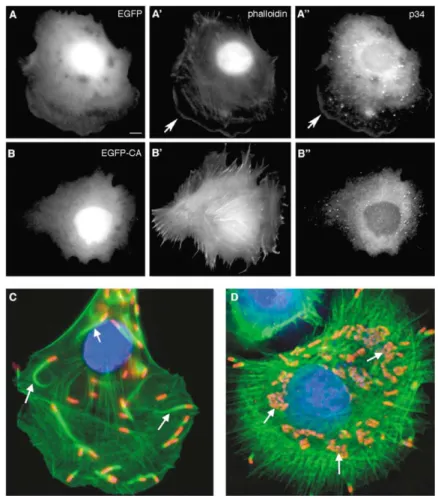
As predicted, the EGFP-CA construct displaced
Arp2/3 from the fibroblast lamellipodia and altered the
using standard techniques, replication-defective
re-combinant adenoviruses were used to express proteins
actin structures (Figures 3A and 3B). The displaced
Arp2/3 was sensitive to detergent extraction (see Sup-
in fibroblasts and neurons. As previously demonstrated
for other adenoviral vectors (Le Gal La Salle et al., 1993;
plemental Figure S1 at http://www.neuron.org/cgi/
content/full/43/1/81/DC1), indicating that it is no longer
Meberg and Bamburg, 2000), our replication-defective
adenoviruses gave consistent, high level expression
associated with the cytoskeleton. The in vivo efficacy
of EGFP-CA was further demonstrated by its ability to
with low cytotoxicity in neurons. For these and
subse-quent studies, infection was monitored by EGFP
expres-inhibit Listeria motility in fibroblasts (Figure 3D). Control
cells expressing EGFP appeared normal and supported
sion and only neurons expressing moderate levels of
EGFP were analyzed.
Figure 3. Controls for EGFP-CA Efficacy as an Arp2/3 Inhibitor
EGFP-CA was tested for its ability to displace Arp2/3 from the cell periphery and to inhibit
Listeria motility.
(A and B) Rat2 fibroblasts infected with a re-combinant adenoviruses expressing control EGFP (A) or EGFP-CA (B) and labeled with phalloidin (A⬘ and B⬘) and antibodies to p34-Arc (A″ and B″). In control EGFP-expressing cells, p34-Arc (A″) colocalizes with actin (A⬘) at the leading edge (arrows). In EGFP-CA-expressing cells, actin structures are altered (B⬘) and p34-Arc is no longer enriched at the periphery (B″).
(C and D) 25 hr after infection with adenovir-uses, Rat2 cells were infected with Listeria
monocytogenes and labeled with antiserum
to Listeria (red), phalloidin (green), and DAPI (blue). EGFP is not shown in these images. In the control expressing EGFP, Listeria gen-erate actin comet tails (arrows) that propel them through the cytoplasm (C). Expression of EGFP-CA inhibits tail formation, so the
Lis-teria divide and form microcolonies (D,
arrows); a few Listeria “escapees” are seen at the periphery.
If Arp2/3 has a similar role in fibroblasts and growth
Inhibition of Arp2/3 Enhances Axon Elongation
There are two main components to growth cone motility:
cones, then we would expect that inhibition of Arp2/3
would cause dramatic changes in the growth cone actin
dynamic protrusion/retraction and translocation. The
dynamic protrusion and retraction of filopodia and
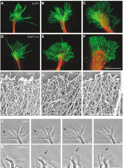
la-cytoskeleton. Surprisingly, growth cones of neurons
ex-pressing EGFP-CA were indistinguishable from controls
mellipodia veils occurs in all growth cones, including
paused growth cones. Translocation (i.e., forward
move-by immunofluorescence (Figures 5A–5F). EGFP and
EGFP-CA are found in the central region of the growth
ment) occurs only in elongating neurites. Our previous
results suggest that Arp2/3 is not essential for dynamic
cone and in the neurite shaft (data not shown), making
it difficult to detect delocalization of the Arp2/3 in EGFP-
protrusion and retraction in growth cones.
Surprisingly, inhibition of Arp2/3 by EGFP-CA induced
CA neurons; however, the EGFP-CA was sensitive to
detergent extraction, indicating that EGFP-CA and any
a significant increase in axon length (Figures 6A–6E). As
an additional control for the specificity of the CA peptide,
bound Arp2/3 are no longer associated with the
cy-toskeleton (data not shown). At the EM level, actin orga-
we expressed EGFP fused to a truncated peptide
con-taining only the “C” domain of N-WASP, which does
nization in control EGFP (Figure 5G) and
EGFP-CA-expressing (Figures 5H and 5I) growth cones was
not bind to Arp2/3 (Hufner et al., 2001). As expected,
expression of this construct had no effect on axon length
virtually indistinguishable, though filament density was
sometimes reduced in EGFP-CA-expressing growth
(Figures 6B and 6E). As a positive control, the “VCA”
region of Wave-1 (another Arp2/3 activator), was
ex-cones (Figure 5I). Populations of control and
EGFP-CA-expressing neurons both contained small growth cones
pressed with a nonfused EGFP under a second CMV
promoter. The VCA peptide contains an actin binding
(Figures 5A and 5D), large growth cones (Figures 5B
and 5E), and large paused growth cones with looped
“V” domain in addition to the “CA” domains and, like
CA alone, binds to Arp2/3. Although VCA will activate
microtubules (Figures 5C and 5F). This is in contrast to
fibroblasts expressing even moderate levels of EGFP-
Arp2/3 in vitro, it disrupts Arp2/3 localization and acts
as a dominant inhibitor in vivo (Machesky and Insall,
CA, in which dramatic changes in the cytoskeleton were
visible at both the light and EM levels (Figures 4). Consis-
1998). As in the case of EGFP-CA, VCA expression
sig-nificantly increased axon length compared to controls
tent with the apparently normal actin cytoskeleton,
growth cones expressing EGFP-CA retained the ability
expressing either EGFP-C or EGFP alone (Figures 6A–
6E). In addition, VCA did not cause any obvious changes
to form protrusive filopodia and lamellipodia (Figures 5J
Figure 4. Inhibition of Arp2/3 Alters Fibroblast Actin Organization and Membrane Protrusion
(A–D) EM visualization of actin organization in Rat2 fibroblasts that were uninfected (A) or expressing EGFP (B) or EGFP-CA (C and D). In the controls (A and B), the periphery is dominated by short, branched filaments. In contrast, cells expressing EGFP-CA (C and D) exhibit abnormal organization, characterized by a dramatic increase in long filaments that were oriented parallel to the membrane (C, arrowheads indicate one such filament). These long filaments were occasionally bundled (D, arrows).
(E and F) Time-lapse images of Rat2 cells expressing control EGFP (E) or EGFP-CA (F) demonstrate the effect of EGFP-CA on membrane protrusion. The control (E) forms a smooth, convex protruding lamellipodium. The cell expressing EGFP-CA only forms spiky, filopodia-like structures. Star and arrow mark fixed points. Scale bars equal 0.1m for (A)–(D) and 10 m for (E) and (F).
immunofluorescence (data not shown). Because axon
growth cone, a region that is also rich in microtubules.
Previous studies using pharmacological agents to alter
length is directly proportional to net growth cone
trans-location, these results suggest that Arp2/3 may be a
actin and microtubule dynamics have shown that actin
polymerization is critical for growth cone response to
negative regulator of growth cone translocation.
It is interesting to note that compared to the effect
guidance signals, that microtubule polymerization drives
axon elongation, and that coordination of actin and
mi-on axmi-on length, expressimi-on of EGFP-CA or VCA did not
cause a significant change in dendrite length (Figure
crotubule dynamics is critical (Bamburg et al., 1986;
Bentley and Toroian-Raymond, 1986; Dent and Kalil,
6F). As Arp2/3 is enriched in the central region of both
axonal and dendritic growth cones (Figure 1), this sug-
2001; Marsh and Letourneau, 1984; Rochlin et al., 1999;
Suter et al., 1998). To assess the effect of Arp2/3
inhibi-gests that its activity is modulated by differential
expres-sion or activity of Arp2/3 activators. At least two Arp2/3
tion on growth cone microtubules, we used antisera to
posttranslational modifications that correlate with
mi-activators, cortactin and N-WASP, are expressed in
hip-pocampal neurons (Figure 6G). Previous studies have
crotubule stability. Newly polymerized microtubules are
tyrosinated, but as microtubules “age,” they are
detyro-shown that before neurons differentiate an axon,
cortac-tin is enriched in the central region of all neurite growth
sinated and become acetylated (Arce et al., 1978;
L’Her-nault and Rosenbaum, 1985).
cones (Du et al., 1998). At this stage, N-WASP is found
in neurite shafts but is barely detectable in the growth
Although Arp2/3 inhibition did not cause an overall
change in microtubule distribution in the growth cone,
cones (Figure 6H). As the neurons differentiate an axon
and dendrites, N-WASP becomes preferentially en-
immunolabeling revealed that the ratio of tyrosinated
to acetylated microtubules was increased when Arp2/3
riched in the distal portion of the axon and the axon
growth cone, but not in dendrite growth cones (Figures
was inhibited (Figures 7A–7E). The effect of EGFP-CA
on microtubules was observed specifically in smaller
6I and 6J).
growth cones (Figure 7E). This is consistent with the
observation that Arp2/3 inhibition leads to increased
Growth Cone Microtubules Are Affected
by Arp2/3 Inhibition
axon length because translocating growth cones tend
to be smaller (Kim et al., 1991; Mason and Erskine, 2000;
We have shown that Arp2/3 appears to regulate axon
Figure 5. Arp2/3 Is not a Major Determinant of Growth Cone Actin Organization and Mem-brane Protrusion
(A–F) Growth cones expressing control EGFP (A–C) or EGFP-CA (D–F) were labeled with phalloidin to visualize actin (green) and with anti-tubulin III (red). At this level, EGFP ex-pressing growth cones are indistinguishable from those expressing EGFP-CA; both popu-lations of growth cones contain small growth cones (A and D), large growth cones (B and E), and large paused growth cones with looped microtubules (C and F).
(G–I) At the EM level, actin organization in control EGFP (G) and EGFP-CA-expressing (H and I) growth cones is virtually indistin-guishable, though filament density was sometimes reduced in EGFP-CA-expressing growth cones (I).
(J and K) Consistent with the apparently nor-mal peripheral actin cytoskeleton, time-lapse images at 15 s intervals show that EGFP- (J) and EGFP-CA- (K) expressing growth cones are equally capable of dynamic membrane protrusion and retraction. Arrows indicate ar-eas of protruding lamellipodia.
Scale bars equal 10m for (A)–(F), 1 m for (G)–(I), and 10m for (J) and (K).
and enriched in tyrosinated microtubules (Bamburg et
strates, axons of EGFP-CA-expressing neurons were
more likely than controls to cross an inhibitory substrate
al., 1986), while stabilization of microtubules results in
shorter axons (Rochlin et al., 1996).
(Figures 7F and 7G). To control for variation in the
striping and in the infection rate, we compared the
per-cent of total cells (crossing and noncrossing) that were
Arp2/3 and Growth Cone Guidance
When growth cones turn to avoid an inhibitory guidance
infected and the percent of crossing cells that were
infected (Figure 7H). If a treatment has no effect on the
cue, filopodia collapse and dynamic microtubules are
retracted from the side of the growth cone closest to
ability of neurons to cross the inhibitory stripe, then
these two values should be similar. This is the case
the inhibitory cue (Buck and Zheng, 2002; Challacombe
et al., 1997; Tanaka and Kirschner, 1995; Zhou et al.,
for neurons expressing EGFP alone. In contrast, if the
treatment alters the ability of neurons to cross, then these
2002). If actin is destabilized by application of low
con-centrations of cytochalasin B, growth cone turning is
values will be disproportionate. EGFP-CA-expressing
neurons made up approximately 30% of crossing
neu-altered and dynamic microtubules fail to retract from
the side of the growth cone that is in contact with the
rons when only about 15% of the total population was
infected, suggesting that inhibition of Arp2/3 altered the
inhibitory cue (Challacombe et al., 1997). These and
other studies have led to a model of growth cone turning
ability of the growth cone to respond to the inhibitory
substrate.
in which actin structures direct the reorientation and
stabilization of microtubules in the direction of turning.
As discussed, the immediate response to repulsive
and attractive guidance signals involves the dynamic
Consistent with this model, we found that
Arp2/3-dependent actin structures may play a role in growth
retraction and extension of filopodia. Because Arp2/3
has been implicated in filopodia formation in other cell
cone turning. When neurons were plated on coverslips
Figure 6. Inhibition of Arp2/3 Enhances Axon Elongation
(A–D) Examples of embryonic hippocampal neurons expressing EGFP (A and A⬘), EGFP-C (B and B⬘), EGFP-CA (C and C⬘), or VCA and EGFP (D and D⬘) that have axons near the mean length (A–D) or the longest length (A⬘–D⬘) for each treatment. Cultures in (A)–(E) were grown without glia conditioning and visualized by EGFP fluorescence (shown in black and white and inverted such that EGFP appears black). Staining with anti-tubulin III was used to identify neurons (data not shown). y axis indicates length in microns.
(E) Analysis of the effect of EGFP, EGFP-C, EGFP-CA, and VCA on axon length. Error bars indicate⫾1 standard deviation. EGFP-CA and VCA are both significantly greater than EGFP and EGFP-C (pⱕ 0.0067), but there is no significant difference between EGFP and EGFP-C or between EGFP-CA and VCA.
(F) Breakdown of the effect of Arp2/3 inhibition on axons and dendrites of neurons grown with glia conditioning.
(G) Western blot analysis of the relative levels of different actin binding proteins in hippocampal neurons and Rat2 fibroblasts. Results are presented as a ratio of the level of the protein in the neurons relative to the level in the fibroblasts, such that a value greater than 1 indicates relatively more protein in neurons.
(H–J) N-WASP localization (red) in primary hippocampal neurons at different stages of development. N-WASP levels are relatively low in growth cones of undifferentiated neurites (H) and in growth cones of short axons (I). In longer axons, N-WASP becomes increasingly enriched in the distal axon and growth cone (J, arrow).
it is required for filopodia formation in neurons. Analysis
of filopodia within 40 min of application (Dent et al.,
2004; Lebrand et al., 2004). Application of 600 ng/ml
of fixed neurons revealed that neurons expressing
EGFP-CA had at least as many filopodia as control neu-
recombinant mouse Netrin1 to hippocampal neurons
expressing EGFP or EGFP-C lead to an increase in the
rons expressing EGFP or EGFP-C (Figure 8A; the slight
increase in EGFP-CA compared to controls was not
number of filopodia after 40 min (Figures 8B–8D). This
response was not inhibited by expression of EGFP-CA
statistically significant). Surprisingly, neurons
express-ing VCA had significantly more filopodia than both con-
or VCA, indicating that Arp2/3 is not required for de
novo filopodia formation under these circumstances.
trols and EGFP-CA-expressing neurons. Whether this
reflects a function of the V region or an increase in the
efficacy of VCA as compared to CA remains to be deter-
Discussion
mined.
To test the requirement for the Arp2/3 complex in de
Arp2/3 Is a Negative Regulator Role of Growth
Cone Translocation
novo filopodia formation in response to a guidance cue,
we examined the response of neurons to the guidance
In cell types where Arp2/3 plays an essential role in
generating the actin-based protrusive force that drives
molecule Netrin-1, which is an attractive signal to these
neurons. Bath application of Netrin-1 to cultured pyrami-
motility, its function appears to be tightly coupled to its
localization at the periphery (Bailly et al., 1999, 2001;
dal neurons elicits a significant increase in the number
Figure 7. Arp2/3 Plays a Role in Regulating Growth Cone Microtubule Dynamics and Guidance
(A–D) Representative images of growth cones expressing control EGFP (A and B) or EGFP-CA (C and D) that were double labeled with antibodies to tyrosinated tubulin and acetylated tubulin. In the merged images, tyrosinated tubulin is green, acetylated tubulin is red, and overlap appears yellow. The dashed lines trace the growth cone perimeter. Scale bar equals 10m.
(E) The pixel intensity within the growth cone is presented as a ratio of the intensity in the tyrosinated channel to that in the acetylated channel. There is a significant difference in tubulin tyrosination between small growth cones (⬍125 m2) treated with EGFP and EGFP-CA (p⬍ 0.03)
and between small and large growth cones treated with EGFP-CA (p⬍ 0.007).
(F and G) Embryonic hippocampal neurons expressing EGFP (F) or EGFP-CA (G) were grown on coverslips coated with alternating stripes of poly-l-lysine plus Semaphorin 3A (Sema3A, inhibitory stripe [⫹]) and poly-l-lysine alone (permissive stripe [⫺]). Images are inverted so the signal (i.e., EGFP or EGFP-CA) appears dark. Cultures were also stained with anti-tubulin III to visualize all the neurons (data not shown). Positioning of the stripes was visualized as described (see Experimental Procedures) and is indicated by plus and minus signs between (F) and (G).
(H) Quantitation of the effect of EGFP-CA on neuron growth on/across inhibitory stripes.
Machesky and Insall, 1998; Svitkina and Borisy, 1999;
the growth cone cytoskeleton and observed that the
peripheral region is primarily composed of two
popula-Welch et al., 1997b). It was widely assumed that Arp2/3
would play a similar role in the growth cone periphery;
tions of actin filaments: long filaments oriented parallel
to the membrane and shorter, randomly oriented
fila-however, we found that Arp2/3 is enriched in the central
region of the growth cone and that there is relatively
ments. Neither group detected a dendritic array of short
Y-branched filaments indicative of Arp2/3 activity.
little Arp2/3 at the growth cone periphery. Similar results
were obtained in hippocampal and DRG neurons using
If Arp2/3 is not a major determinant of actin
organiza-tion in the growth cone periphery, then funcorganiza-tional
inhibi-multiple different antisera to Arp2/3 and using
p21-EGFP-tagged Arp2/3, suggesting that Arp2/3 function
tion should have little effect on the actin organization in
this region. Indeed, growth cones expressing the Arp2/3
is likely to be most important in the central region of the
growth cone.
inhibitor (EGFP-CA) were indistinguishable from
con-trols by immunofluorescence (Figures 5A–5F). At the EM
Although it is possible that Arp2/3 acts at very low
levels to generate a dendritic actin array at the growth
level, there was no detectable change in actin filament
organization (Figures 5G–5I), but there was sometimes
cone periphery, EM analysis revealed relatively few
branched filaments in the growth cone periphery (Figure
a reduction in filament density (Figure 5I). These results
contrast sharply with the dramatic changes in actin
fila-2). Instead, this region appears to be dominated by long
actin filaments that are roughly perpendicular to the
ment organization seen in Rat2 fibroblasts expressing
EGFP-CA (Figure 4). Similar results were obtained with
distal edge of the growth cone. Similar actin organization
was seen in growth cones of all shapes and sizes and
a construct containing the VCA domains of Wave-1 (data
not shown). Although the high number of glia makes it
in both convex and concave areas of the growth cone,
which are typically associated with membrane protru-
difficult to do experiments with adenoviruses in DRG
cultures, the similar localization of Arp2/3 in the central
sion and retraction, respectively (Figure 2). These data
are in agreement with previous studies (Letourneau,
region of hippocampal and DRG growth cones suggests
that Arp2/3 has a similar function in both populations
1983; Lewis and Bridgman, 1992; Schaefer et al., 2002)
Figure 8. Arp2/3 Is not Required for Filo-podia Formation
The requirement for Arp2/3 for filopodia for-mation was examined in both fixed (A) and living (B–D) neurons.
(A) Analysis of filopodia number in fixed cells. Neurons were infected with viruses 12–24 hr after plating. Approximately 48 hr after plat-ing, neurons were fixed and stained with anti-tubulin III and phalloidin. Infected cells were identified by EGFP expression and the num-ber of filopodia counted. There was no signifi-cant difference between EGFP and EGFP-C. There was a slight, but not statistically signifi-cant, increase in the number of filopodia in EGFP-CA-expressing neurons relative to the EGFP and EGFP-C controls. VCA-expressing neurons showed a very significant (p ⱕ 0.0001) increase in filopodia number relative to EGFP and EGFP-C and significant increase (pⱕ 0.034) relative to EGFP-CA.
(B) Analysis of filopodia induction in living neurons. Neurons were infected with viruses 12–24 hr after plating. Approximately 36 hr after plating, dishes were transferred to the microscope incubator and imaged at 25 s in-tervals for 10 min to determine that the growth cone was dynamically changing. Netrin1 (600 ng/ml) was then added directly to the bath and imaging was continued for an additional 60 min. Data represent the average number of filopodia over the 5 min proceeding Netrin1 addition (T⫽ ⫺5) or from 40 to 45 min after Netrin1 addition (T⫽ ⫹40). Error bars indicate ⫾1 standard deviation.
(C and D) Representative images of neurons expressing EGFP (C) or VCA (D) 5 min before (⫺5 min) or 40 min after (⫹40 min) addition of Netrin1. Black arrows indicate filopodia that formed de novo in regions where there were no filopodia at⫺5 min (white arrows). Scale bar equals 10m.
In fibroblasts, inhibition of Arp2/3 activity alters mem-
Arp2/3 removes this regulation, which enhances
micro-tubule dynamics and in turn enhances neurite
elonga-brane protrusion (Bailly et al., 2001), indicating that
Arp2/3-dependent actin filament branching activity
tion. This inappropriate tendency toward elongation
may indirectly render the growth cone unable to obey
plays an essential role in generating the force that drives
membrane protrusion. Our results indicate that in growth
a repulsive cue in a timely manner.
Although the density of actin and microtubules in the
cones, Arp2/3 is not essential for membrane protrusion,
but it does play a critical role in regulating elongation in
central region of the growth cone makes visualization
of this region difficult (Figure 2), it is likely that Arp2/3
actively translocating axon growth cones. In Drosophila,
null mutants in Arp2/3 and the Scar exhibit defects in
nucleates the formation of Y branches in growth cones,
just as in fibroblasts. These Arp2/3-dependent actin
many tissues, including the nervous system (Zallen et
al., 2002). Flies with neuron-specific deletion of Arp2/3
structures may regulate microtubule dynamics either
by serving as a type of barrier or “net” that retards
or Scar elaborate apparently normal axons, further
sup-porting our conclusion that Arp2/3 is not required for
microtubule advance, or by physical association with
microtubules. One study in particular found a specific
axon outgrowth per se (J. Ng and L. Luo, personal
com-munication).
role for the growth cone actin “meshwork” (the F-actin
not incorporated into the actin bundles) in retarding
dy-namic microtubule advance into the peripheral region
Mechanism of Arp2/3 Function in Growth Cones
Recent studies have shown that there is a direct correla-
(Zhou et al., 2002), suggesting that Arp2/3-dependent
actin structures may act as a barrier to microtubule
tion between the levels of tyrosinated microtubules and
the rate of axon elongation (Bamburg, 2003; Rochlin et
advance. If the effect on microtubule dynamics involves
physical interaction, it is likely that other protein(s) link
al., 1996). Other studies have implicated actin structures
in the regulation of microtubule dynamics, which in turn
the actin and microtubules, though it is formally possible
that there are proteins that link Arp2/3 directly to
micro-affects growth cone motility, pausing, pathfinding, and
axon branching (Dent et al., 1999; Dent and Kalil, 2001;
tubules.
The fact that Arp2/3 inhibition does not block filopodia
Rodriguez et al., 2003; Zhou et al., 2002). In this context,
our finding that inhibition of Arp2/3 activity in neurons
induction suggests that neurons are able to generate
the immediate filopodia response to guidance cues
in-alters the relative levels of tyrosinated microtubules and
results in pathfinding defects suggests that Arp2/3-
dependently of Arp2/3. Whether Arp2/3 function is
re-quired for later steps in growth cone turning toward
dependent actin structures normally function to regulate
growth cone microtubules. This activity is particularly
Netrin remains to be determined. Given that it is an
integral part of the cytoskeleton, we predict that
inhibi-important during growth cone turning. Inhibition of
tively, as previously described (Lanier et al., 1999). Except for the
tion of Arp2/3 would alter growth cone turning and
elon-stripe assays, approximately 2⫻ 104cells were plated on each 12
gation in response to both attractive and repulsive
mm coverslip. After 4 hr, the media was replaced with either neuronal
signals.
growth medium (MEM with Earle’s salts, B27 supplement, 10 mM HEPES, and 0.6% D-glucose) that had been conditioned on glia for
Determinants of Growth Cone
48 hr immediately prior to use or with unconditioned Neurobasal media with B27 supplement, 0.5 mM glutamine, and 25Mgluta-Morphology and Motility
mate. Rat2 cells were grown in DME/5% fetal calf serum.
Our results indicate that inhibition of Arp2/3 affects
growth cone translocation and pathfinding without
sig-Adenovirus Construction and Infection
nificantly altering overall growth cone morphology. The
Replication-deficient recombinant adenoviruses were produced
us-dendritic array and convergent-elongation models of
ing the AdEasy system (He et al., 1998). EGFP, EGFP-C (residueslamellipodia and filopodia formation imply that whether
450–486 of bovine N-WASP), EGFP-CA (residues 468–505 of bovinecells form a lamellipodium dominated by a dendritic
N-WASP), and p21-EGFP were cloned by PCR and inserted into pShuttle CMV. The myc-tagged VCA (residues 444–559 of humanarray (as in the fibroblast) or a structure dominated by
Wave-1) was cloned by PCR and inserted into pAdTrack-CMV. The
filopodia (like a growth cone) will depend on the relative
resulting plasmid expresses VCA under a CMV promoter, with a
concentrations and types of actin binding activities
second CMV promoter expressing the EGFP marker. Virus was
puri-present; the most abundant actin binding proteins will
fied on a CsCl gradient and titered on 293HEK cells. Cells were
determine the overall morphology of the motile struc-
infected with a multiplicity of infection (m.o.i.) of 25–50 infectiousture, while less abundant proteins may have modula-
viruses per cell.tory roles.
Listeria Infection
Comparative Western blot analysis (Figure 6G) and
Listeria monocytogenes strain 10435 was a gift from Darren Higgins.
immunolocalization studies (Cohan et al., 2001; Lanier
Listeria cultures were grown at 30⬚C in brain heart infusion broth
et al., 1999) have shown that compared to fibroblasts,
(BHI, Difco). Overnight cultures were centrifuged, rinsed once in
growth cones have relatively high levels of fascin
PBS, and resuspended in a 1/10thvolume of PBS and applied tobundling and Ena/VASP anticapping activities, which
Rat2 cultures. After 1 hr at 37⬚C for, Rat2 cultures were rinsed twicewould be predicted to favor filopodia formation. We pro-
with the appropriate media and given fresh media supplemented with 10g/ml gentamicin. After 4 hr, cultures were fixed andpro-pose that abundant proteins such as fascin and Ena/
cessed for immunofluorescence. For studies on the effect of EGFP
VASP are the major determinants of growth cone
mor-or EGP-CA expression, cells were infected with adenovirus 24 hr
phology and membrane protrusion, while the less
abun-prior to infection with Listeria. Because CNS neurons are extremely
dant Arp2/3 complex plays an important role in
coordi-refractive to Listeria infection (Dramsi et al., 1998), we were unable
nating actin structures that can regulate microtubule
to use EGFP-CA to inhibit Listeria motility in hippocampal neurons.dynamics and growth cone translocation. Such a model
is supported by the observations that altering fascin
ImmunofluorescenceThe following antisera were used: rabbit anti-p21-Arc, p34-Arc, and
bundling or Ena/VASP anticapping activity dramatically
Arp3 (a gift from Matthew Welch); affinity purified rabbit
anti-p34-alters growth cone morphology and actin organization.
Arc (a gift from Laura Machesky); mouse anti-Tubulin III (Promega);
Treatments that decrease fascin bundling activity lead
rat anti-tyrosinated tubulin MAB1864 (Chemicon); mouse
anti-ace-to a rapid loss of actin bundles in growth cone filopodia
tylated tubulin clone 6-11B-1 (Sigma); C4 anti-actin (Boehringer(Cohan et al., 2001). Functional inhibition of Ena/VASP
Mannheim); rabbit anti-N-WASP antisera (gifts from M. Kirschnerproteins inhibits filopodia formation and increases the
and T. Takenawa); and rabbit Listeria O (Difco). Secondary anti-sera were from Jackson ImmunoResearch and phalloidins were fromdensity of disorganized actin filaments in the growth
Molecular Probes. Fixation and permeabilization conditions were
cone periphery, while increasing Ena/VASP activity
optimized for each antiserum but were identical for Rat2 cells and
leads to an increase in the number and length of growth
neurons labeled with a given set of antisera. Cells were fixed for 30
cone filopodia and actin bundles (Lebrand et al., 2004).
min at 37⬚C in PPS (4% paraformaldehyde in PHEM buffer [60 mMOur experiments clearly demonstrate that Arp2/3 is
PIPES (pH 7.0), 25 mM HEPES (pH 7.0), 10 mM EGDT, 2 mM MgCl2]
not required for lamellipodia protrusion or de novo filo-
with 0.12 M sucrose). After rinsing in PBS, coverslips were incubated in 10% fatty acid free bovine serum albumin (BSA) in PBS for 30 min,podia formation in neurons. In these experiments,
how-permeabilized for 10 min in 0.2% triton/PBS, rinsed, and reblocked in
ever, Arp2/3 was inhibited after neurites were formed,
10% BSA/PBS for 30 min. For anti-p21 and anti-p34 antisera
pro-so it remains possible that Arp2/3 is required for neurite
vided by M. Welch, cells were fixed with PPS as described above,
initiation. While there is clearly a need for actin-nucleat-
denatured with cold methanol for 3 min, then permeabilized anding activity in the motile growth cone, it may be fulfilled
blocked as described above. In neurons, similar results wereob-by other nucleating proteins such as the formins, which
tained with and without methanol, so the methanol step was omitted in order to allow phalloidin staining. For double labeling withanti-have been implicated in the formation of unbranched
tyrosinated and anti-acetylated tubulin, cultures were
simultane-actin filaments in vitro and in keratinocytes (Kobielak,
ously fixed and permeabilized in PPS supplemented with 0.25%
2003). These findings reveal a surprising new role for
glutaraldehyde, 10M Taxol, 1.3 M phalloidin, and 0.1% triton. This
Arp2/3 in growth cone translocation and guidance and
mixture stabilizes microtubules and actin filaments, while extractingprovide further evidence for the importance of crosstalk
soluble tubulin dimers and actin monomers (Dent and Kalil, 2001).between the actin and microtubule cytoskeletons in
Incubations with primary and secondary antisera were done in the presence of 1% BSA/PBS, and coverslips were mounted with 2.5%this process.
1,4-Diazabicyclo-[2.2.2]Octane/150 mM Tris (pH 8.0)/30% glycerol to reduce photo bleaching. Images were captured on a Nikon TE200 Experimental Procedures
using DeltaVision software (API). Cell Culture
All media and supplements were purchased from GIBCO-BRL. Poly- Platinum Replica Electron Microscopy
Correlative platinum replica electron microscopy was performed D-lysine and fibronectin were purchased from Sigma. Hippocampal
modifications. Briefly, dissociated embryonic hippocampal neurons capping protein2 subunit mAb3F2.3 (Iowa Developmental Studies Hybridoma studies bank); rabbit anti-gelsolin (a gift from W. Witke), were cultured as described above on coverslips coated with a gold
locator grid. EGFP-positive cells were located by live cell fluores- rabbit anti-cofilin (Cytoskeleton); mouse anti-␣-actinin mAb BM-75.2 (Sigma); mouse anti-fascin mAb3582 (Chemicon International); cence microscopy, then immediately extracted for 4.5 min with 1%
Triton X-100 in PEM buffer (100 mM PIPES [pH 6.8], 1 mM EGTA, monoclonal anti-zyxin (a gift from J. Wehland); rabbit anti-zyxin and mouse anti-paxilin (a gift from K. Burridge); mouse anti-vinculin and 1 mM MgCl2) containing 10M phalloidin, 10 M Taxol, 0.2%
gluter-aldehyde, and 4.2% sucrose as an osmotic buffer. Coverslips were anti-paxillin (a gift from R. Salgia); rabbit anti-vinculin (a gift from K. Burridge); mouse anti-vinculin hVinc1 (Sigma); and rabbit anti-Mena, washed with PEM containing 1M phalloidin, 1 M Taxol, and 1%
sucrose, fixed in 0.1 M Na-cacodylate buffer (pH 7.3), 2% gluteralde- EVL, and VASP (produced in the Gertler lab). Secondary antibodies were from Jackson Labs. Signal was detected using the ECL reagent hyde, and 1% sucrose and processed for electron microscopy. Cells
previously identified as EGFP positive were relocated using the (Amersham-Pharmacia). Relative concentrations were determined using the NIH Image Gel Scanning macro. Each antiserum was gold grid.
tested at least twice on two different batches of extract and results are presented as an average ratio.
Measurement of Axon and Dendrite Length
Cultures were fixed with PPS as described above and stained with
Acknowledgments mouse anti-Tubulin III (Promega) and Alexa 594 phalloidin
(Molecu-lar Probes). Infected cells were identified by EGFP expression, and
We are grateful to Drs. Matt Welch, Tadaomi Takenawa, Keith Bur-images were taken using either a 20⫻ (for axon and dendrite length
ridge, Jurgen Wehland, Ravi Salgia, and Mark Kirschner for their measurements) or 40⫻ (for filopodia measurements) plan-neofluar
generous gifts of antibodies, to Dr. Laura Machesky for the gift of objective and Openlab software (Improvision). Images were saved
antibodies and DNA constructs, and to Dr. Burt Vogelstein for the as tiff files and assigned coded names, and neurites were manually
gift of AdEasy cloning vectors. We thank Drs. Julian Ng and Liqun traced using NIH Image 1.62 and the Neurite Labeling Macro v1.1
Luo for sharing unpublished data. This work was supported by NIH (available at ftp://rsbweb.nih.gov/pub/nih-image/user-macros/). For
grant #6895154 and a W.M. Keck Distinguished Young Scholar axon and dendrite length measurements, data represent the average
Award to F.B.G. and Minnesota Medical Foundation grant #333-of at least 50 different neurons from at least two different
experi-9238-03 and Academic Health Center Seed grant #2003-39 to L.M.L. ments. For filopodia counts in fixed cells, filopodia were identified
by phalloidin staining and data represent the average of at least 25
neurons from two different experiments. Data were analyzed using Received: September 12, 2003 Statview and significance was determined by ANOVA with Sheffe’s Revised: March 10, 2004
post hoc analysis. Accepted: May 18, 2004
Published: July 7, 2004 Live Cell Imaging and Filopodia Analysis
Rat2 cell were plated directly on fibronectin-coated Bioptechs References dishes, while neuronal cultures were plated on 35 mm tissue culture
dishes fitted with German glass coverslips (“special dishes;” Goslin Amann, K.J., and Pollard, T.D. (2001). The Arp2/3 complex nucleates and Banker, 1998) and coated with 1 mg/ml poly-D-lysine. Rat2 actin filament branches from the sides of pre-existing filaments. cells were grown in a low-bicarbonate medium as previously de- Nat. Cell Biol. 3, 306–310.
scribed (Bear et al., 2000). Neuronal cultures were plated as de- Arce, C.A., Hallak, M.E., Rodriguez, J.A., Barra, H.S., and Caputto, scribed above. For filopodia analysis, special dishes were trans- R. (1978). Capability of tubulin and microtubules to incorporate and ferred to a microscope humidified stage incubator that maintained to release tyrosine and phenylalanine and the effect of the incorpora-the cultures at 5% CO2. Time-lapse movies were made at 25 s tion of these amino acids on tubulin assembly. J. Neurochem. 31,
intervals for 70 min. Filopodia on growth cones and on the distal- 205–210. most 100 microns of the axon shaft were counted in successive
Bailly, M., Macaluso, F., Cammer, M., Chan, A., Segall, J.E., and images. Data represent the average over a 5 min interval from 2–4
Condeelis, J.S. (1999). Relationship between Arp2/3 complex and movies and at least 2 different experiments and significance was
the barbed ends of actin filaments at the leading edge of carcinoma determined by ANOVA and Sheffe’s post hoc analysis. For filopodia
cells after epidermal growth factor stimulation. J. Cell Biol. 145, induction by Netrin 1, the number of filopodia was normalized to
331–345. the starting value.
Bailly, M., Ichetovkin, I., Grant, W., Zebda, N., Machesky, L.M., Seg-all, J.E., and Condeelis, J. (2001). The F-actin side binding activity Stripe Assay
of the Arp2/3 complex is essential for actin nucleation and lamellipod Stripe matrices were produced using a silicon matrix as described
extension. Curr. Biol. 11, 620–625. (Vielmetter et al., 1990). Alkaline-phosphatase tagged Semaphorin
Bamburg, J.R. (2003). Introduction to cytoskeletal dynamics and 3A (Sema 3A) was expressed in HEK293 cells and harvested from
pathfinding of neuronal growth cones. J. Histochem. Cytochem. the tissue culture media as previously described (Kobayashi et al.,
51, 407–409.
1997) For stripe assays, 18 mm coverslips were coated with 1 mg/ml
poly-D-lysine, rinsed, and air dried. The coverslip was then inverted Bamburg, J.R., Bray, D., and Chapman, K. (1986). Assembly of mi-onto the stripe maker and a mixture of Sema 3A and rabbit IgG crotubules at the tip of growing axons. Nature 321, 788–790. perfused though the matrix, forming alternating 90 micron wide Banzai, Y., Miki, H., Yamaguchi, H., and Takenawa, T. (2000). Essen-stripes of poly-lysine alone and poly-lysine with Sema3A (and rabbit
tial role of neural Wiskott-Aldrich syndrome protein in neurite exten-IgG). As a control, BSA was substituted for Sema 3A at the same sion in PC12 cells and rat hippocampal primary culture cells. J. Biol. concentration. Approximately 5⫻ 105neurons were plated on each
Chem. 275, 11987–11992. coverslip. After fixation, the Sema3A stripes were identified by
label-Bear, J.E., Loureiro, J.J., Libova, I., Fassler, R., Wehland, J., and ing with a fluorescent anti-rabbit secondary.
Gertler, F.B. (2000). Negative regulation of fibroblast motility by Ena/ VASP proteins. Cell 101, 717–728.
Western Blot Analysis
Bear, J.E., Svitkina, T.M., Krause, M., Schafer, D.A., Loureiro, J.J., To prepare extracts, cells were rinsed with PBS, lysed in extraction
Strasser, G.A., Maly, I.V., Chaga, O.Y., Cooper, J.A., Borisy, G.G., buffer (10 mM Tris [pH 8.0], 1% NP40, 150 mM NaCl supplemented
and Gertler, F.B. (2002). Antagonism between Ena/VASP proteins with a protease inhibitor cocktail; Boehringer Mannheim), and
centri-and actin filament capping regulates fibroblast motility. Cell 109, fuged 10 min at 10,000⫻ g. Concentration of the extract was
deter-509–521. mined using the BCA protein assay (Pierce). SDS-PAGE and Western
blot analysis were performed using standard techniques. In addition Bentley, D., and Toroian-Raymond, A. (1986). Disoriented pathfind-ing by pioneer neurone growth cones deprived of filopodia by cyto-to the antisera used for immunofluorescence, the following antisera


