HAL Id: hal-00632294
https://hal.archives-ouvertes.fr/hal-00632294
Submitted on 14 Oct 2011
HAL is a multi-disciplinary open access
archive for the deposit and dissemination of sci-entific research documents, whether they are pub-lished or not. The documents may come from teaching and research institutions in France or abroad, or from public or private research centers.
L’archive ouverte pluridisciplinaire HAL, est destinée au dépôt et à la diffusion de documents scientifiques de niveau recherche, publiés ou non, émanant des établissements d’enseignement et de recherche français ou étrangers, des laboratoires publics ou privés.
ALEXANDRIUM TAMARENSE COMPLEX
(DINOPHYCEAE) IN A MEDITERRANEAN
LAGOON FACILITATED BY SEMI-MULTIPLEX PCR
Benjamin Genovesi, Mi-Sun Shin-Grzebyk, Daniel Grzebyk, Mohamed Laabir,
Pierre-Alexandre Gagnaire, André Vaquer, Annie Pastoureaud, Bernard
Lasserre, Yves Collos, Patrick Berrebi, et al.
To cite this version:
Benjamin Genovesi, Mi-Sun Shin-Grzebyk, Daniel Grzebyk, Mohamed Laabir, Pierre-Alexandre Gag-naire, et al.. ASSESSMENT OF CRYPTIC SPECIES DIVERSITY WITHIN BLOOMS AND CYST BANK OF THE ALEXANDRIUM TAMARENSE COMPLEX (DINOPHYCEAE) IN A MEDITER-RANEAN LAGOON FACILITATED BY SEMI-MULTIPLEX PCR. Journal of Plankton Research, Oxford University Press (OUP), 2010, �10.1093/plankt/FBQ127�. �hal-00632294�
For Peer Review
ASSESSMENT OF CRYPTIC SPECIES DIVERSITY WITHIN BLOOMS AND CYST BANK OF THE ALEXANDRIUM
TAMARENSE COMPLEX (DINOPHYCEAE) IN A MEDITERRANEAN LAGOON FACILITATED BY
SEMI-MULTIPLEX PCR
Journal: Journal of Plankton Research Manuscript ID: JPR-2010-164.R1
Manuscript Type: Original Article Date Submitted by the
Author: 29-Aug-2010
Complete List of Authors: Genovesi, Benjamin; Université Montpellier 2, UMR5119 Laboratoire Ecolag
Shin-Grzebyk, Mi-Sun; Université Montpellier 2, UMR5119 Laboratoire Ecolag
Grzebyk, Daniel; CNRS, UMR5119 Laboratoire Ecolag
Laabir, Mohamed; Université Montpellier 2, UMR5119 Laboratoire Ecolag
Gagnaire, Pierre-Alexandre; Université Montpellier 2, UMR 5554 Institut des Sciences de l’Evolution
Vaquer, André; CNRS, UMR5119 Laboratoire Ecolag Pastoureaud, Annie; IFREMER, LER/LR
Lasserre, Bernard; Université Montpellier 2, UMR5119 Laboratoire Ecolag
Collos, Yves; CNRS, UMR5119 Laboratoire Ecolag
Berrebi, Patrick; CNRS, MR 5554 Institut des Sciences de l’Evolution Masseret, Estelle; Université Montpellier 2, UMR5119 Laboratoire Ecolag
Keywords: Alexandrium catenella, Alexandrium tamarense, PSP, ITS, Thau lagoon
For Peer Review
Assessment of cryptic species diversity within blooms and cyst bank of the Alexandrium
tamarense complex (Dinophyceae) in a Mediterranean lagoon facilitated by semi-multiplex PCR
Benjamin Genovesi1,2, Mi-Sun Shin-Grzebyk1, Daniel Grzebyk1, Mohamed Laabir1, Pierre-Alexandre Gagnaire2, André Vaquer1, Annie Pastoureaud3, Bernard Lasserre1, Yves Collos1, Patrick Berrebi2, Estelle Masseret1
1 Université Montpellier 2, UMR 5119 UM2-CNRS-IFREMER-IRD, Ecosystèmes
Lagunaires, équipe Efflorescences Toxiques et Diversité Algale, CC093, place E. Bataillon, 34095 Montpellier cedex 05, France.
2 Université Montpellier 2, UMR 5554 UM2-CNRS-IRD, Institut des Sciences de l’Evolution,
CC065, place E. Bataillon, 34095 Montpellier cedex 05, France.
3 IFREMER Laboratoire Environnement Ressources /LR, BP 171, 34203 Sète, France.
Corresponding authors: benjamin.genovesi@gmail.com and estelle.masseret@univ-montp2.fr
Running title: Co-occurrence of Alexandrium spp. blooms
Keywords: Alexandrium catenella, Alexandrium tamarense, PSP, ITS, Thau lagoon. 3 4 5 6 7 8 9 10 11 12 13 14 15 16 17 18 19 20 21 22 23 24 25 26 27 28 29 30 31 32 33 34 35 36 37 38 39 40 41 42 43 44 45 46 47 48 49 50 51 52 53 54 55 56 57 58 59 60
For Peer Review
ABSTRACT
The occurrence of Alexandrium catenella related to paralytic shellfish poisoning in the French Mediterranean Thau lagoon has been known since 1998. Blooms are recurrent and usually occur each year in spring and/or autumn. Taxonomic diversity of resting cysts and vegetative cells has been studied through morphological examination and molecular typing of 558 clonal strains sampled in 2004 and 2007. Sequencing the nuclear rRNA fragment including ITS1, the 5.8S rRNA gene, ITS2, and the D1/D2 28S rRNA gene enabled two species to be determined, A. catenella and A. tamarense, which are difficult to distinguish
morphologically (cryptic species). In order to carry out extensive and accurate molecular determinations, an original semi-multiplex PCR method, using new ribotype-specific primers targeting the 18S-28S rRNA ITS region has been developed. The relative abundance of each species was then established in seawater in 2007 and in the sediment collected in 2004. The co-occurrence of A. catenella (Group IV), which is known as the main species responsible for toxic PSP events since 1998 and of A. tamarense (Group III) (non-toxic), which was not formally recognized by microscopic observation since 1995, was examined for several months. 3 4 5 6 7 8 9 10 11 12 13 14 15 16 17 18 19 20 21 22 23 24 25 26 27 28 29 30 31 32 33 34 35 36 37 38 39 40 41 42 43 44 45 46 47 48 49 50 51 52 53 54 55 56 57 58 59 60
For Peer Review
INTRODUCTION
Since the 1970s, Harmful Algal Blooms (HABs) have been recognised as one of the main ecological problems in coastal regions worldwide (Maso and Garces 2006). Among HAB species, some Dinophyceae are toxic and cause neurological and human gastric disorders after consumption of contaminated filter-feeding shellfish (Zingone and Enevoldsen 2000; Smayda and Reynolds 2003; Glibert et al. 2005).
In the Mediterranean Sea many species involved in such outbreaks are listed as exotic or non-indigenous phytoplankton (Streftaris et al. 2005, Gomez 2008, Molmar et al. 2008) and tagged by the International Union for the Conservation of Nature (IUCN). The Mediterranean sea harbours the highest number of reported invasive toxic and non-toxic Alexandrium
species (Fraga et al. 2004), with the exception of the autochtonous Mediterranean
Alexandrium tamarense described by John et al. (2003a) belonging to the group II (Lilly et al. 2007). These species spread in the Western Mediterranean region along French, Spanish, Italian and Maghrebian coasts (Penna et al. 2005, 2008, Frehi et al. 2007, Turki and Balti 2008). Among them many taxa are linked to the paralytic shellfish-poisoning (PSP) syndrome.
Based on the LSU rRNA gene sequence analysis, Scholin et al. (1994) delineated eight distinct phylogeographic clusters or “ribotypes” within the genus Alexandrium. Among them, the Alexandrium tamarense complex consists of three morphospecies Alexandrium catenella (Whedon & Kofoid) Balech, A. tamarense (Lebour) Balech and A. fundyense (Balech). This species complex is distributed into five geographic ribotypes (Scholin et al. 1994). These species have been observed worldwide since the 1970's and include both non-toxic and toxic strains (Cordova et al. 2003, Ichimi et al. 2001, Haya et al. 2003). Representatives of the
Alexandrium tamarense species complex are often difficult to identify, especially for taxa such as A. catenella, A. tamarense and A. fundyense, which can be considered as cryptic species. Indeed, the main classical features such as chain-forming ability, cell shape, and presence/absence of a ventral pore between plates 1’ and 4’ (Balech 1995) have been demonstrated to be somewhat inconsistent (Lilly et al. 2007, Penna et al. 2008, Wang et al. 2008).
Regarding the Thau lagoon (France), which is one of the largest Western Mediterranean lagoons, the occurrence of Alexandrium species involved in significant algal blooms and PSP has been known since 1995 and 1998, respectively. The first attempts to accurately
discriminate species involved in PSP in the Thau lagoon were inconsistent. Thecal
morphology and toxin content analyses of fixed cells from seawater sampled in autumn 1998 3 4 5 6 7 8 9 10 11 12 13 14 15 16 17 18 19 20 21 22 23 24 25 26 27 28 29 30 31 32 33 34 35 36 37 38 39 40 41 42 43 44 45 46 47 48 49 50 51 52 53 54 55 56 57 58 59 60
For Peer Review
first suggested that both A. tamarense and A. catenella were present (Masselin et al. 2001). Lilly et al. (2002) analyzed two clonal strains (ATTL01 and ATTL02) established from seawater samples also collected during the autumn of 1998. Strain DNA sequences of the D1– D2 domains of LSU rDNA revealed the Japanese Temperate Asia (TA) ribotype of the A.
tamarense complex, i.e. tamarense/catenella/fundyense species (Scholin and Anderson 1996). This evidence was morphologically supported by the thecal analyses of isolated strains
(Laabir et al. 2004). Until now, rRNA gene sequencing of strains (88) isolated from 2001 to 2004 showed that A. catenella was the only taxon blooming in the Thau lagoon (Masseret et
al. 2009). However, vegetative cells characterized by unusual size and morphology were periodically observed in weekly-collected samples. In addition, high concentrations of
Alexandrium cells were frequently unrelated to toxin detection (unpublished data). Thus, these observations (1) questioned the evidence that the toxic A. catenella was the only taxon involved in the Thau lagoon blooms; and (2) prompted us to follow the evolution of densities of toxic and non-toxic Alexandrium species in order to adapt scientific surveys of toxins in shellfish.
In the present survey, a large-scale sampling was carried out in the water column from May to October 2007, in order to assess the taxonomic diversity within and between Alexandrium populations periodically blooming in the Thau lagoon. In addition, germination of sexual cysts, followed by isolation of sibling strains, was performed from sediment sampled in 2004 and conserved at 4°C (Genovesi et al. 2007) in order to explore taxonomic diversity among the Alexandrium sp. cyst bank. The main results show the co-occurrence of A. tamarense and
A. catenella belonging respectively to Group III and Group IV. Consequently, a rapid and selective method based on ribotyping was developed to discriminate both species through an extensive strain determination. Subsequently, to avoid confusion we use the most recent designation to discriminate the geographic ribotypes of both species, i.e Group III and Group IV (Lilly et al. 2007) instead of Temperate Asian and Western European clades (Scholin and Anderson 1996).
METHOD
Field survey, sample collection and cultures
The Thau lagoon is located on the French Mediterranean coast (43°24’N-3°36’E). It covers 75 km2, one-fifth of which is occupied by shellfish farming structures. This lagoon is connected to the Mediterranean Sea by three channels, one of which opens into the Sète international harbour. Sampling was carried out in the Crique-de-l’Angle, which is a shallow 3 4 5 6 7 8 9 10 11 12 13 14 15 16 17 18 19 20 21 22 23 24 25 26 27 28 29 30 31 32 33 34 35 36 37 38 39 40 41 42 43 44 45 46 47 48 49 50 51 52 53 54 55 56 57 58 59 60
For Peer Review
semi-enclosed area at the north of the lagoon (Fig. 1). For the survey program of Alexandrium sp. population dynamics, a sampling grid (100 m) including 27 geo-referenced stations was applied in the Crique-de-l’Angle (Fig. 1). Since 1998, water samples were systematically collected by using a pump system during periods favourable for the development of
Alexandrium sp. blooms (spring and autumn). Samples were at once fixed in formaldehyde (2% final concentration) for the microscopic enumeration of vegetative cells according to the Utermöhl method. Clonal cultures in enriched natural seawater were based on: (1) single vegetative cells isolated from seawater during the bloom period in 2004, and from May to November 2007; (2) sexual cyst germination isolated from sediment collected in 2004 after a week of incubation at 20°C, under an irradiance of 100 µmoles photons m-2 s-1 on a 12:12 h Light/Dark cycle (Table I) (Genovesi et al. 2009).
Morphological analyses
Morphological examination was performed on cells from 30 A. tamarense (Group III) and 22
A. catenella (Group IV) clonal strains established in 2004 and 2007. All the strains observed were genotyped. For examination and for each culture, more than 30 vegetative cells were selected at exponential phase and stained with Calcofluor white (20 µg m L-1) according to the method of Fritz and Triemer (1985). Observations were carried out using an
epi-fluorescence microscope (Olympus AX70) and photographs were taken with a CCD camera. The morphometric analysis (length and width) of fixed vegetative cells (formalin 0.5% final concentration v/v) was performed using a light microscope (Zeiss Axiovert 25) on at least 30 cells of each strain.
DNA extraction
Genomic DNA was extracted from centrifuged culture strains using a modified guanidine buffer method (Matz 2003). After centrifugation, cells were re-suspended in guanidine buffer (4 M guanidine thiocyanate; 30 mM disodium citrate; 30 mM b-mercaptoethanol; pH 8.0-8.5). In order to improve the extraction efficiency, cells were exposed to ultrasound for 20 seconds. Chloroform purification was then performed after an incubation step at 65°C lasting 10 min. After ethanol precipitation, the DNA was finally re-suspended in TE buffer (10 mM Tris-HCl; pH8.0; 0.1 mM EDTA).
PCR amplification of the (ITS1-5.8SrDNA-ITS2) rRNA region
3 4 5 6 7 8 9 10 11 12 13 14 15 16 17 18 19 20 21 22 23 24 25 26 27 28 29 30 31 32 33 34 35 36 37 38 39 40 41 42 43 44 45 46 47 48 49 50 51 52 53 54 55 56 57 58 59 60
For Peer Review
The ribotype of 308 Alexandrium spp. strains originating from the Thau lagoon (Table I) was resolved by sequencing the rRNA fragment spanning the region including ITS1, the 5.8S rRNA gene, ITS2, and the D1/D2 28S rRNA gene (totalising 1,246 bp). PCR amplifications were performed with 5-10 ng of genomic DNA template using the primers 18S-ITS1-Ac-F 5’-CTTAGAGGAAGGAGAAGTCG-3’ and 28S-D3B-R 5’-TCGGAGGGAACCAGCTACTA-3’. The PCR conditions consisted of 95°C for 2 min, 40 cycles of 95°C for 40 s, 55°C for 4 min, 72°C for 1.4 min and a final extension at 72°C for 5 min. The PCR products were purified using ethanol precipitation and were used as templates for direct sequencing with the forward PCR primers and an internal reverse sequencing primer. Sequencing was performed by the Macrogen Company (Seoul, South Korea) using an ABI 3730XL genetic analyzer (Applied Biosystems).
DNA sequence analyses
Sequences were assembled using the software package BioEdit (Hall 1999). Nucleotide sequence alignments were performed using ClustalX (Thompson et al. 1997) with reference sequence data retrieved from Genbank. The rDNA regions from 308 strains were aligned with 51 sequences of various species of Alexandrium retrieved from GenBank. The sequences of two isolates belonging to A. catenella (Group IV) and A. tamarense (Group III) have been deposited under the accession numbers FR686536 and FR686537, respectively.
Development of an allele specific semi-multiplex PCR for species discrimination
An allele specific semi-multiplex PCR (Gagnaire et al. 2007) using new ribotype-specific primers targeting the 18S-28S rRNA ITS region was developed in this study. These new primers were designed from the alignment of the sequences from the 308 strains studied. Forward primers FACAT28 TGATATTGTGGGCAACTGTAA-3') and FATAM143 (5'-TGGTAATTCTTCATTGATTACAATG-3') were used respectively for A. catenella (Group IV) and A. tamarense (Group III) whereas a common reverse primer RACATAM269 (5'-AACATCTGTTAGCTCACGGAA-3') was designed in a region conserved for both ribotypes. Because 114 bp separate the sites of FACAT28 and FATAM143 in the 18S-28S rRNA-ITS region, the two specific amplification products should differ in their size (126 bp for A. tamarense and 240 bp for A. catenella), enabling species determination after agarose gel migration. PCR amplifications were performed in 10 µL reaction mixtures containing 5-10 ng of genomic DNA template, 2.0 mM of MgCl2, 0.2 mM each deoxynucleoside
triphosphates, 1X buffer provided with the polymerase, 0.5 µM of each primer and 1U of Taq 3 4 5 6 7 8 9 10 11 12 13 14 15 16 17 18 19 20 21 22 23 24 25 26 27 28 29 30 31 32 33 34 35 36 37 38 39 40 41 42 43 44 45 46 47 48 49 50 51 52 53 54 55 56 57 58 59 60
For Peer Review
polymerase (AmpliTaq, Qiagen). Cycling conditions were as follows: 2 min at 95°C, 35 cycles of 40s at 95°C, 1 min and 20s at 54°C and 1 min at 72°C, and 5 min at 72°C. In order to check the specificity of the newly designed primers, the PCR products obtained from 35 Alexandrium spp. strains were sequenced by GenoScreen (Lille, France) using an ABI 3730XL genetic analyzer (Applied Biosystems).
The ratio between both species was determined for all collected samples, i.e. sediment collected in 2004, seawater collected in autumn 2004, and all dates of sampling from the bloom that occurred from May to October 2007. In total, 568 clonal strains were identified using molecular methods: sequencing (308 strains) and semi-multiplex PCR (260 strains).
RESULTS
Seasonality of Alexandrium blooms
Since 1995, Alexandrium blooms have occurred mainly during autumn, except for the years 2000, 2007 and 2008 that had significant spring blooms (Fig. 2). Throughout the past 13 survey years, autumnal blooms were not observed in 2002 and 2006. Maximal vegetative cell densities reached 4.5 × 10⁶ cells L-1 and 16 × 106 cells L-1 in October 2003 and November 2004, and resulted in water discolouration during these periods. Such cell concentrations were usually related to blooms of which Alexandrium spp. may represent more than 80-90% of the phytoplankton. Under suitable environmental conditions, i.e. temperature around 20-21°C related to calm waters (Laania et al. 2008), Alexandrium spp. usually needs 10 to 20 days to reach maximum cell density. However, a relatively low cell concentration (from 1000 to 50000 cells L-1) could be observed during several weeks before the growth peak. During the survey program in 2007, Alexandrium spp. cells were microscopically observed at 8 sampling stations A00, A3, A5, A9, B3, B5, C3 and C5 from May 10 till November 8 (Fig. 1, Table 2). Cell density varied widely within the sampling period and among sampling stations (Table 2). The highest densities were observed on May 24 in C3, C5 and A3, i.e. nearly 2 × 10⁶ cells L-1. In contrast, during the autumnal bloom, the highest densities were recorded on October 15 at B3, A5 and A3 and were lower than 0.2 x 10⁶ cells L-1.
Morphological examination
Vegetative cells of A. tamarense strains were solitary or in pairs. In contrast, A. catenella showed solitary cells but also chains of 2, 4 and 8 cells, mainly observed during the mid-exponential phase. Concerning cell size, there is no significant difference (t-test, p>0.05) between the two morphospecies: 22.20 µm (5.15 s.d.) (H) × 24.82 µm (6.33 s.d.) (W) for A. 3 4 5 6 7 8 9 10 11 12 13 14 15 16 17 18 19 20 21 22 23 24 25 26 27 28 29 30 31 32 33 34 35 36 37 38 39 40 41 42 43 44 45 46 47 48 49 50 51 52 53 54 55 56 57 58 59 60
For Peer Review
tamarense; and 23.09 µm (3.78 s.d.) (H) × 25.48 µm (4.84 s.d.) (W) for A. catenella. All strains examined of both species exhibit the typical plate formula of the A. tamarense
complex P0, 4’, 6’’, 6C, 8S, 5’’’, 2’’’’. Among several morphological characteristics (Apical Pore Complex, Posterior sulcal plate, etc.), the presence of a ventral pore (Balech 1995) on the first apical plate was considered, here, to be the most reliable character to separate A.
tamarense (Group III) from A. catenella (Group IV). Our results show the constant presence of a small ventral pore located halfway along the right margin of the first apical plate (1’) of the A. tamarense vegetative cells.
Development and test of the rapid molecular determination assay
The ribotype-specific semi-multiplex PCR method using the newly designed primers allowed the resolution of specific amplicons according to their size: 126 bp for A. tamarense (Group III) vs. 240 bp for A. catenella (Group IV) (Fig. 3). The reliability of this method was tested on 96 strains whose ribotype was known by sequencing. The results showed that the accuracy of the method reached 100%. The sequencing of amplicons obtained by semi-multiplex PCR was performed on 34 Alexandrium spp. strains and confirmed the accurate specificity of these new primers. 240 bp and 126 bp amplicons were obtained for 22 A. catenella and 12 A.
tamarense strains respectively.
Diversity of Alexandrium spp. clonal cultures
Analysis of the ITS1-5.8SrDNA-ITS2 rRNA region sequences from 308 monoclonal strains revealed the presence of both species: A. catenella (Group IV) and A. tamarense (Group III) according to Scholin et al. (1994) and Lily et al. (2007). Within each ribotype, the sequences of all isolates were totally identical. 23 clonal cultures were obtained from resting cysts isolated from sediment sampled in 2004. The results showed that 13 and 10 strains belonged to A. catenella and A. tamarense, respectively. This demonstrated the co-occurrence of both species in the sediment sampled in April 2004. On the other hand, the A. tamarense ribotype was not detected in 79 clonal cultures established from vegetative swimming cells during the bloom of autumn 2004. In contrast, both ribotypes were isolated from May to September 2007. In October 2007, mainly isolated A. catenella cells were able to generate viable monoclonal cultures (Fig. 4).
DISCUSSION
Morphological characteristics and ribotype
3 4 5 6 7 8 9 10 11 12 13 14 15 16 17 18 19 20 21 22 23 24 25 26 27 28 29 30 31 32 33 34 35 36 37 38 39 40 41 42 43 44 45 46 47 48 49 50 51 52 53 54 55 56 57 58 59 60
For Peer Review
The main morphological characteristics such as chain-forming ability, cell shape and ventral pore (Balech 1995) are neither discrete nor stable (Taylor 1975, Lilly et al. 2007).
Consequently, the chain-forming ability or absence of the ventral pore are considered plesiomorphic characters and may not be suitable for use as taxonomic markers (John et al. 2003a, Leaw et al. 2005). For the first time we performed extensive observations on a large number of strains targeting the most accurate morphological characteristic distinguishing the two morphospecies A. tamarense and A. catenella, i.e. the presence/absence of the ventral pore (Balech 1995) on the first apical plate. The samples examined consisted of ≥ 30 cells for 31 and 28 strains of A. tamarense and A. catenella, respectively. As previously recorded in cell observations on natural samples from Thau, both morphospecies are characterized by variations in cell size according to previous reports (Wang et al. 2008). For all strains examined, we show a correspondence of A. tamarense and A. catenella ribotypes and
morphospecies designation (Scholin et al. 1994) based on the presence/absence of the ventral pore on plate 1’ and chain-formation, which is related to exponential growth phase in A.
catenella (Toulza et al. 2010). However, intraspecific variations have been observed
worldwide in cells with and without a ventral pore (Cembella and Taylor 1986, Orlova et al. 2007, Gayoso and Fulco 2006, Wang et al. 2008). Regarding these contradictory
observations, several characters (such as morphology, toxicity and genetics) seem to be required to accurately discriminate specific levels into cryptic/morphospecies (this study, Lilly et al. 2007).
The genetic analyses of Alexandrium sp. strains isolated in this study place them within Group IV which contains A catenella toxic isolates from Temperate Asia and within Group III including A. tamarense non-toxic isolates from Scottish, Irish and English waters (Scholin et al. 1994 and 1995, Higman et al. 2001, John et al. 2003b, Lilly et al. 2007, Collins et al. 2009).
The succession and the dominance of one and/or the other species could have a significant impact on the toxicity level measured, through survey programs, in the shellfish during HABs. In Thau lagoon, red tides are not related to toxic events according to the Ifremer survey programme (unpublished data). The rapid and reliable semi-multiplex method described in this study should allow the detection of both toxic A. catenella (Group IV) and non-toxic A. tamarense (Group III). Moreover, the determination of the ecological conditions favouring each species is an important field of research to be developed in order to estimate the risk of development of a toxic or non-toxic event.
3 4 5 6 7 8 9 10 11 12 13 14 15 16 17 18 19 20 21 22 23 24 25 26 27 28 29 30 31 32 33 34 35 36 37 38 39 40 41 42 43 44 45 46 47 48 49 50 51 52 53 54 55 56 57 58 59 60
For Peer Review
Identification of cryptic species with the ribotype-specific-PCR method
In this study, the distinction between A. tamarense (Group III) and A. catenella (Group IV) could be made using morphological features, such as the presence or absence of a ventral pore on the apical plate. However, such morphological examinations are time-consuming and might be questionable (Cembella and Taylor 1986, Lilly et al. 2007, Wang et al. 2008). Thus, the sequencing of ITS or LSU seems to be the most accurate method to discriminate
individuals at the species level within the Alexandrium tamarense species complex. However, sequencing is not adapted for routine species discrimination from established culture strains. The original semi-multiplex PCR method developed in this study promptly and accurately discriminates A. tamarense (Group III) and A. catenella (Group IV) in the laboratory cultures library. This rapid method which only demands PCR amplification followed by agarose gel electrophoresis is moreover more straightforward compared with previous methods, such as the RFLP method (Adachi et al. 1994, Lilly et al. 2002) or real-time PCR (Hosoi-Tanabe and Sako 2005).
Cultures reveal co-occurrence of two Alexandrium species in sediment and seawater
Sequencing and the new semi-multiplex ribotype-specific PCR revealed the co-occurrence of two ribotypes in the water column in 2007 and in the sediments of the Thau lagoon collected in 2004: A. catenella (Group IV), which is known as the main species responsible for toxic PSP events in the Thau lagoon since 1998, and A. tamarense (Group III), which was not formally recognized by microscopic observation since 1995.
The ribotyping of cyst-derived strains demonstrates the presence of both species in the sediment sampled in April 2004 and also the ability of the resting cysts to grow after 4 to 5 years of storage at 4°C in darkness with a culturing success of approximately 25%. However, the A. tamarense ribotype is not detected within 79 ribotyped vegetative cell strains
established during the bloom of November 2004 (Masseret et al., 2009, 2010), which can be due to a reduced proportion of this species in the lagoon, but probably also to the sampling and cultivation bias. On the other hand, in 2007, results reveal the presence of these two species within cultures derived from vegetative cells. A prevalence of A. tamarense is
observed in the cultures grown from May to September while A. catenella is the main species isolated from seawater sampled in October 2007. The co-occurrence of A. catenella and A.
tamarense in the same area has been previously reported in the Ofunato bay in Japan (Hosoi-Tanabe and Sako 2005).
3 4 5 6 7 8 9 10 11 12 13 14 15 16 17 18 19 20 21 22 23 24 25 26 27 28 29 30 31 32 33 34 35 36 37 38 39 40 41 42 43 44 45 46 47 48 49 50 51 52 53 54 55 56 57 58 59 60
For Peer Review
Co-occurrence of both cryptic species and their possible succession during blooms may indicate behaviour linked to their life-history traits (e.g. growth and adaptive strategies) and related to environmental conditions. However, our results depend on the isolation and cultivation of microorganisms that are selective and lead to a bias in the description of the diversity and genetic structure of the bloom. During the vegetative pelagic stage in November 2004, genotypes belonging solely to A. catenella have been isolated compared to the available bank of genotypes present in the sediment as resting cysts (Fig 4). Environmental conditions seemed to be not suitable for A. tamarense cyst germination and vegetative reproduction. Three main processes may also exert selective pressure in culturing phytoplankton: isolation, establishment and maintenance (Lakeman et al. 2009). In our study, the isolation and
cultivation success were better in October than from May to September. Some of the isolated cells were not taken into account because they did not grow after the beginning of their isolation in enriched seawater and some of them died after a few divisions. This is also the case for cyst-derived strains that exhibited a low culturing success of 25%. After one month, abortive cultures became scarce and very few losses were recorded. It is evident that selective bottlenecks may occur during these steps. A small proportion of tolerant individuals,
benefiting from phenotypic properties with capacity for survival in laboratory culture conditions (in Enriched natural seawater, at 20°C, 100 µmoles photons m-2 s-1, 12:12 h Light/Dark cycle), grew successfully and for a long period of time. Moreover, an unforeseen result occurred from ribotype analysis performed on clonal cultures of cells isolated from the same sample. During the isolation period, three operators microscopically isolated single swimming cells from field samples. Operator 1 isolated solely A. catenella ribotypes whereas operator 2 isolated mainly A.tamarense ribotypes.
The key role of cyst banks in bloom outbreaks and maintenance
The hypothesis of the former presence of both species in the Thau lagoon is now supported by the significant occurrence of A. tamarense resting cysts in the cyst bank of the sediment sampled in April 2004, i.e. >40% of cyst-derived strains belonged to A. tamarense. The cyst densities recorded in the Crique-de-l’Angle (i.e. 200 cysts cm-3 in the C7 station (Fig. 1)) (Genovesi et al. 2008) underline the arrival and/or production of cysts by A. tamarense before 2004. The vertical profiles of cyst distribution in sediments represent a useful proxy to
estimate the history of bloom development (Mudie et al. 2002). In Sequim Bay (Puget Sound, USA), the occurrence of hundreds of A. catenella cysts down to a 40 cm sediment depth demonstrates that the presence of the species goes back 30 years at least (Cox et al. 2008). In 3 4 5 6 7 8 9 10 11 12 13 14 15 16 17 18 19 20 21 22 23 24 25 26 27 28 29 30 31 32 33 34 35 36 37 38 39 40 41 42 43 44 45 46 47 48 49 50 51 52 53 54 55 56 57 58 59 60
For Peer Review
the sediment of the Thau lagoon, the cyst profiles showed that resting cysts of Alexandrium sp. are mostly concentrated in the upper sediment layers (97% down to 12 cm depth), particularly in the first 3 cm where 30% of the overall cysts were observed (Genovesi et al. 2007). In addition, the mean density of Alexandrium sp. cysts in the Thau lagoon (< 20 cysts g-1 dry sediment) is very low (Genovesi et al. 2009) compared to other sites where
Alexandrium sp. blooms (involved in PSP) are often associated with hundreds to thousands cysts per gram of sediment (Yamaguchi et al. 1996, Hallegraeff et al. 1998, Gayoso 2001, Anderson et al. 2005). Therefore, the vertical profiles and the low cyst densities recorded in the Thau lagoon support the hypothesis of recent proliferation of species belonging to the A.
tamarense species complex.
CONCLUSION
It is already of well known that the current expansion of several Alexandrium species along the NW Mediterranean coasts may be due to human-mediated transport. The occurrence of toxic and non-toxic strains of A. catenella (Group IV) and A. tamarense (Group III) in the Thau lagoon complicates the HAB monitoring performed by Ifremer, in particular for risk management in the shellfish industry. The newly developed semi-multiplex PCR
amplification enables the detection of these species. It will thus ease the characterisation of in
situ genetic diversity of Alexandrium blooms and promote the understanding of the specific ecological conditions responsible for species succession. The large-scale strain isolation shows the bias of the diversity description from cultures. Exhaustive and precise population genetic studies of HABs must include many samples that are often overlooked because they fail to grow. In the future, the bias of the diversity description from cultures will be
minimized by the development of routine single-cell analyses from natural samples. Finally, the regional understanding of Alexandrium expansion will require the resolution of the geographic origins of the two species, which could be certainly achieved by a population genetic approach using microsatellite markers.
ACKNOWLEDGMENTS
This study was supported by grants from the French National Programme “Ecosphère Continentale & Côtière” (EC2CO-PNEC) and from the Agence Nationale de la Recherche (ANR-06-BLAN-0397 GenoSynTox). The monitoring survey and field sampling was allowed by the ALCAT programme of IFREMER. We thank Jérémy Béguin and Boram Lee (the latter in the frame of a CNRS researcher exchange project with South Korea) for help in the
3 4 5 6 7 8 9 10 11 12 13 14 15 16 17 18 19 20 21 22 23 24 25 26 27 28 29 30 31 32 33 34 35 36 37 38 39 40 41 42 43 44 45 46 47 48 49 50 51 52 53 54 55 56 57 58 59 60
For Peer Review
ribotyping of monoclonal cultures. We have a thought for Patrick Gentien (Ifremer DYNECO-Brest), we will miss his valuable support. We thank the assistance of two anonymous reviewers who contributed to improving this manuscript.
REFERENCES
Adachi, M., Sako, Y. and Ishida, Y. (1994) Restriction fragment length polymorphism of ribosomal DNA internal transcribed spacer and 5.8S regions in Japanese Alexandrium species (Dinophyceae). J. Phycol. 30, 857--863.
Bolch, C. J. S. and de Salas, M. F. (2007) A review of molecular evidence for ballast water introduction of the toxic dinoflagellates Gymnodinium catenatum and the Alexandrium “tamarensis complex” to Australia. Harmful Algae 6, 465--485.
Cembella, A. D. and Taylor, F. J. R. (1986) Electrophoresis variability within the
Protogonyaulax tamarensis/catenella species complex: pyridine linked dehydrogenases. Biochem. Syst. Ecol. 14, 311--321.
Cordova, J. L., Vega, M. P. and Lembeye, G. S. (2003) Intracellular damage and death caused by protease inhibitors on Alexandrium catenella cysts and vegetative cells. Harmful
Algae 2, 173--181.
Cox, A. M., Shull, D. H. and Horner, R. A. (2008) Profiles of Alexandrium catenella cysts in Puget Sound sediments and the relationship to paralytic shellfish poisoning events.
Harmful Algae 7, 379--388.
Erard-Le-Denn, E., Desbruyeres, E. and Olu, K. (1993) Alexandrium minutum: resting cyst distribution in the sediments collected along the Brittany coast, France. In Smayda T. J., Shimizu Y. (eds.), Toxic Phytoplankton in the Sea, pp. 109--114.
Fraga, S., Bravo, I., González, R. et al. (2004) Diversity of the dinoflagellate genus
Alexandrium in the Mediterraneran Sea. Rapp. Comm. Int. Mer. Me´dit. 37, 358--358. Frehi, H., Couté, A., Mascarell, G. et al. (2007) Dinoflagellés toxiques et/ou responsables de
blooms dans la baie d'Annaba (Algérie). Compte Rendus de Biologie 330, 615--28. (in French)
Fukuyo, Y. (1985) Morphology of Protogonyaulax tamarensis (Lebour) Taylor and
Protogonyaulax catenella (Whedon and Kofoid) Taylor from Japanese Coastal Waters.
Bull. Mar. Sci. 37, 529--537.
Gagnaire, P. A., Tsukamoto, K., Aoyama, J. et al. (2007) RFLP and semi-multiplex PCR-based identification of four eel species from the south-western Indian Ocean region. J.
Fish Biol. 71, 279--287. 3 4 5 6 7 8 9 10 11 12 13 14 15 16 17 18 19 20 21 22 23 24 25 26 27 28 29 30 31 32 33 34 35 36 37 38 39 40 41 42 43 44 45 46 47 48 49 50 51 52 53 54 55 56 57 58 59 60
For Peer Review
Genovesi-Giunti, B., Laabir, M. and Vaquer, A. (2006) The benthic resting cyst: a key actor in harmful dinoflagellate blooms – a review. Vie Milieu - Life & Environment 56, 327--337.
Genovesi, B., Laabir, M., Vaquer, A. et al. (2009) Dormancy and excystment in resting cysts of Alexandrium catenella (Dinophyceae) from the French Mediterranean. J. Plankton
Res. 31, 1209--1224.
Genovesi-Giunti, B., Vaquer, A., Laabir, M. et al. (2008) Bottom cell clusters as inocula for bloom initiation of Alexandrium catenella in a shallow lagoon (Thau, Southern France). In Moestrup, Ø. et al. (eds.), Proceeding of Harmful Algae XII (4-8 September 2006) Copenhagen, Denmark), pp. 117--119.
Genovesi, B., Mouillot, D., Vaquer, A. et al. (2007) Towards an optimal sampling strategy for
Alexandrium catenella (Dinophyceae) benthic resting cysts. Harmful Algae 6, 837--848.
Gescher, C., Metfies, K. and Medlin L. K. (2008) The ALEX CHIP-Development of a DNA chip for identification and monitoring of Alexandrium. Harmful Algae 7, 485-494. Glibert, P. M., Anderson, D. M., Gentien, P. et al. (2005) The global complex phenomena of
harmful algal blooms. Oceanography 18, 130--141.
Hall, T.A. (1999) BioEdit: a user-friendly biological sequence alignment editor and analysis program for Windows 95/98/NT. Nucleic Acids Symp. Ser. 41, 95--98.
Hallegraeff, G. M. (1998) Transport of toxic dinoflagellates via ship’s ballast water: bioeconomic risk assessment and efficacy of possible ballast water management strategies. Mar. Ecol. Progr. Ser. 168, 297--309.
Haya, K., Martin, J. L., Robinson, S. M. C. et al. (2003) Does uptake of Alexandrium
fundyense cysts contribute to the levels of PSP toxin found in the sea scallop,
Placopecten magellanicus. Harmful Algae 2, 75--81.
Higman, W. A., Stone, D. M. and Lewis, J. M. (2001) Sequence comparisons of toxic and non-toxic Alexandrium tamarense (Dinophyceae) isolates from UK waters.
Phycologia 40, 256--262.
Hosoi-Tanabe, S. and Sako, Y. (2005) Species-specific detection and quantification of toxic marine dinoflagellates Alexandrium tamarense and A. catenella by real-time PCR assay. Marine Biotechnology 7, 506--514.
Ichimi, K., Yamasaki, M., Okumura, Y. et al. (2001) The growth and cyst formation of a toxic dinoflagellate, Alexandrim tamarense, at low water temperature in northeastern Japan. J. Exp. Mar. Biol. Ecol. 261, 17--29.
3 4 5 6 7 8 9 10 11 12 13 14 15 16 17 18 19 20 21 22 23 24 25 26 27 28 29 30 31 32 33 34 35 36 37 38 39 40 41 42 43 44 45 46 47 48 49 50 51 52 53 54 55 56 57 58 59 60
For Peer Review
John, U., Fensome, R. A. and Medlin, L. K. (2003a) The application of a molecular clock based on molecular sequences and the fossil record to explain biogeographic distributions within the Alexandrium tamarense “species complex” (Dinophyceae).
Mol. Biol. Evol. 20, 1015--1027.
John, U., Cembella, A., Hummert, C. et al. (2003b) Discrimination of the toxigenic dinoflagellate species Alexandrium tamarense and Alexandrium ostenfeldii in co-occurring natural populations from Scottish coastal waters. Eur. J. Phycol. 38, 25--40. Ki, J. S. and Han, M. S. (2006) A low-density oligonucleotide array study for parallel
detection of harmful algal species using hybridization of consensus PCR products of LSU rDNA D2 domain. Biosens. Bioelectron. 21, 1812--1821.
Laabir, M., Genovesi-Giunti, B., Barré, N. et al. (2004) The resting cyst of Alexandrium
catenella, a dinoflagellate responsible for harmful algal blooms in Thau lagoon (Western French Mediterranean coast). In Steidinger, K. A. et al. (Eds.), Proceeding
of Harmful Algae X. Florida Fish and Wildlife Conservation Commission, Florida Institute of Oceanography, and Intergovernmental Oceanographic Commission of UNESCO, pp. 26--28.
Laania, N., Vaquer, A., Fiandrino, A. et al. (2008) Wind and temperature controls on the occurrence of Alexandrium blooms in Thau lagoon (Western Mediterranean). Poster in
Harmful Algae XIII (3-7 November 2008) Hong Kong.
Lakeman, M. B., von Dassow, P. and Cattolico, R. A. (2009) The strain concept in phytoplankton ecology. Harmful Algae 8, 746--758.
Leaw, C.P., Lim, P.T., Cheah, M.Y. et al. (2005) Phylogenetic analysis of Alexandrium spp. and Pyrodinium bahamense using a combined molecular and morphological approach.
Phycologia 44, 550--565.
Lilly, E. L., Halanych K. M. and Anderson D. M. (2007) Species boundaries and global biogeography of the Alexandrium tamarense complex (Dinophyceae). J. Phycol. 43, 1329--1338.
Lilly, E. L., Kullis, D. M., Gentien P. (2002) Paralytic shellfish poisoning toxins in France linked to a human-introduced strain of Alexandrium catenella from the western Pacific: evidence from DNA and toxin analysis. J. Plankton Res. 24, 443--452.
MacKenzie, L., de Salas, M., Adamson, J. et al. (2004) The dinoflagellate genus Alexandrium (Halim) in New Zealand coastal waters: comparative morphology, toxicity and
molecular genetics. Harmful Algae 3, 71--92. 3 4 5 6 7 8 9 10 11 12 13 14 15 16 17 18 19 20 21 22 23 24 25 26 27 28 29 30 31 32 33 34 35 36 37 38 39 40 41 42 43 44 45 46 47 48 49 50 51 52 53 54 55 56 57 58 59 60
For Peer Review
Masselin, P., Amzil, Z., Abadie, E. et al. (2001) Paralytic shellfish poisoning on the French Mediterranean coast in autumn 1998: Alexandrium “tamarense complex”
(Dinophyceae) as causative agent. In Hallegraeff, G. M. et al. (eds.), Harmful Algae
IX. Intergovernmental Oceanographic Commission of UNESCO 2001, pp. 26--29. Masseret, E., Grzebyk, D., Nagai, S. et al. (2009) Unexpected genetic diversity among and
within populations of the toxic dinoflagellate Alexandrium catenella as revealed by nuclear microsatellite markers. Appl. Environ. Microbiol. 75, 2037--2045.
Masseret, E., Enquebecq, M., Laabir, M. et al. (2010) A simple and innovative method for species identification of phytoplankton cells on minute quantities of DNA. Env.
Microb. Reports doi:10.1111/j.1758-2229.2010.00164.x
Matz, M. V. (2003) Amplification of representative cDNA pools from Microscopic Amounts of Animal Tissue. In Ying, S-Y. (ed.), Generation of cDNA libraries: Methods and
protocols. Humana press, pp.103--116.
Mudie, P. J., Rochon, A. and Levac, E. (2002) Palynological records of red tide-producing species in Canada: past trends and implications for the future. Paleogeog. Paleoclim.
Palaeoecol. 180, 159--186.
Penna, A., Garcés, E., Vila, M. et al. (2004) Alexandrium catenella (Dinophyceae), a toxic ribotype expanding in the NW Mediterranean Sea. Mar. Biol. 148, 13--23.
Penna, A., Fraga, S., Maso, M. et al. (2008) Phylogenetic relationships among the
Mediterranean Alexandrium (Dinophyceae) species based on sequences of 5.8S gene and Internal Transcript Spacers of the rRNA operon. Eur. J. Phycol. 43, 163--178. Scholin, C. A. and Anderson, D. M. (1996) LSU rDNA-based RFLP assays for discriminating
species and strains of Alexandrium (Dinphyceae). J. Phycol. 32, 1022--1035. Scholin, C. A., Hallegraeff, G. and Anderson, D. M. (1995) Molecular evolution of the
Alexandrium tamarense ‘species complex’ (Dinophyceae): dispersal in the North American and West Pacific regions. Phycologia 34, 472--485.
Scholin, C. A., Herzog, M., Sogin, M. et al. (1994) Identification of group- and strain-specific genetic markers for globally distributed Alexandrium (Dinophyceae). II. Sequence analysis of a fragment of the LSU rRNA gene. J. Phycol. 30, 999--1011.
Smayda, T. (1997) Harmful algal blooms: their ecophysiology and general relevance to phytoplankton blooms in the sea. Limnol. Oceanogr. 42, 1137--1153.
Taylor, F. J. R. (1975) Taxonomic difficulties in red tide and paralytic shellfish poison studies: the ‘Tamarensis complex’ of Gonyaulax. Environ. Lett. 9, 103--119. 3 4 5 6 7 8 9 10 11 12 13 14 15 16 17 18 19 20 21 22 23 24 25 26 27 28 29 30 31 32 33 34 35 36 37 38 39 40 41 42 43 44 45 46 47 48 49 50 51 52 53 54 55 56 57 58 59 60
For Peer Review
Taylor, F. J. R. (1984) Toxic dinoflagellates: taxonomic and biogeographic aspects with emphasis on Protogonyaulax. In Ragelis, E. P. (ed.), Seafood Toxins. ACS
Symposium Series 262, American Chemical Society, Washington, D.C., pp. 77--97. Thompson, J. D., Gibson, T. J., Plewniak, F. et al. (1997) The CLUSTAL_X windows
interface: flexible strategies for multiple sequence alignment aided by quality analysis tools. Nucleic Acids Res. 25, 4876--4882.
Toulza, E., Shin, M.-S., Blanc, G. et al. (2010) Gene expression in proliferating cells of the dinoflagellate Alexandrium catenella (Dinophyceae). Appl. Environ. Microbiol. 76, 4521--4529.
Turki, S. and Balti, N. (2008) First bloom of dinoflagellate Alexandrium catenella in Bizerte Lagoon (northern Tunisia). Harmful Algae News 35, 8--9.
Wang, D. Z., Lin, L., Gu, H. F. et al. (2008) Comparative studies on morphology, ITS sequence and protein of Alexandrium tamarense and A. catenella isolated from the China Sea. Harmful Algae 7, 106--113.
Yamaguchi, M., Itakura, S., Nagasaki, K. et al. (1996) Distribution and abundance of resting cysts of the toxic dinoflagellates Alexandrium tamarense and A. catenella in
sediments of the eastern Seto Inland Sea, Japan. In Yasumoto, T. et al. (eds.). Intergovernemental Oceanographic Commission of UNESCO 1996. Laboratory of Bioorganic Chemistry, Tohoku University, Japan, pp. 177--180.
Zingone, A. and Enevoldsen, H. O. (2000) The diversity of harmful algal blooms: a challenge for science and management. Ocean & Coastal Management 43, 725--748.
3 4 5 6 7 8 9 10 11 12 13 14 15 16 17 18 19 20 21 22 23 24 25 26 27 28 29 30 31 32 33 34 35 36 37 38 39 40 41 42 43 44 45 46 47 48 49 50 51 52 53 54 55 56 57 58 59 60
For Peer Review
TABLE AND FIGURE LEGENDS
Table I. Origin of the 568 clonal strains of Alexandrium spp. isolated from Thau lagoon and that have been ribotyped and amplified by semi-multiplex PCR.
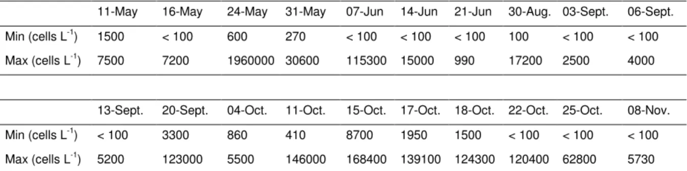
Table II. Minimal and maximal abundances (cells L-1) of Alexandrium sp. recorded during the bloom survey in 2007 at eight stations belonging to the Crique-de-l’Angle sampling grid (i.e. A00, A3, A5, A9, B3, B5, C3, C5).
Figure 1. Map of Thau lagoon located 30 km Southwest of the city of Montpellier (Hérault, South France). Shellfish farming sectors (S1, S2 and S3) represent 1/5 of total surface of the lagoon.
Figure 2. Maximum Alexandrium spp. cell densities recorded in spring and autumn in the Thau lagoon, especially into the Crique-de-l’Angle, since 1995 (data from 1995, 1998 and 1999 are available online at:
http://wwz.ifremer.fr/var/envlit/storage/documents/dossiers/toxines10ans/tableau/tab_alex.ht m).
Figure 3. Identification of Alexandrium tamarense and A. catenella resolved by specific amplicons according to their size using the semi-multiplex PCR assay. (M) 100 bp size marker; (a) 126 bp fragment length for A. tamarense (Group III-WE); (b) 240 bp fragment length for A. catenella (Group IV-TA).
Figure 4. Variation of species proportion, i.e. Alexandrium tamarense (Group III) vs.
Alexandrium catenella (Group IV), on clonal cultures established along the sampling period during the bloom of 2007 into the Crique-de-l’Angle. The specific diversity was studied in samples taken at twelve dates. The proportion of each species was represented by ‘pie charts” and reported to the number of established strains for each sampling date.
3 4 5 6 7 8 9 10 11 12 13 14 15 16 17 18 19 20 21 22 23 24 25 26 27 28 29 30 31 32 33 34 35 36 37 38 39 40 41 42 43 44 45 46 47 48 49 50 51 52 53 54 55 56 57 58 59 60
For Peer Review
Table I.
Strains (n) Sample origin Period
23 sediment winter 2004
30 seawater autumn 2004
515 seawater spring and autumn 2007
3 4 5 6 7 8 9 10 11 12 13 14 15 16 17 18 19 20 21 22 23 24 25 26 27 28 29 30 31 32 33 34 35 36 37 38 39 40 41 42 43 44 45 46 47 48 49 50 51 52 53 54 55 56 57 58 59 60
For Peer Review
Table II.
11-May 16-May 24-May 31-May 07-Jun 14-Jun 21-Jun 30-Aug. 03-Sept. 06-Sept. Min (cells L-1
) 1500 < 100 600 270 < 100 < 100 < 100 100 < 100 < 100 Max (cells L-1) 7500 7200 1960000 30600 115300 15000 990 17200 2500 4000
13-Sept. 20-Sept. 04-Oct. 11-Oct. 15-Oct. 17-Oct. 18-Oct. 22-Oct. 25-Oct. 08-Nov. Min (cells L-1 ) < 100 3300 860 410 8700 1950 1500 < 100 < 100 < 100 Max (cells L-1) 5200 123000 5500 146000 168400 139100 124300 120400 62800 5730 3 4 5 6 7 8 9 10 11 12 13 14 15 16 17 18 19 20 21 22 23 24 25 26 27 28 29 30 31 32 33 34 35 36 37 38 39 40 41 42 43 44 45 46 47 48 49 50 51 52 53 54 55 56 57 58 59 60