HAL Id: cea-02962389
https://hal-cea.archives-ouvertes.fr/cea-02962389
Submitted on 11 Mar 2021
HAL is a multi-disciplinary open access
archive for the deposit and dissemination of
sci-entific research documents, whether they are
pub-lished or not. The documents may come from
teaching and research institutions in France or
abroad, or from public or private research centers.
L’archive ouverte pluridisciplinaire HAL, est
destinée au dépôt et à la diffusion de documents
scientifiques de niveau recherche, publiés ou non,
émanant des établissements d’enseignement et de
recherche français ou étrangers, des laboratoires
publics ou privés.
Distributed under a Creative Commons Attribution| 4.0 International License
reinhardtii Two Protein Phosphatases Are Involved in
State Transitions
Federica Cariti, Marie Chazaux, Linnka Lefebvre-Legendre, Paolo Longoni,
Bart Ghysels, Xenie Johnson, Michel Goldschmidt-Clermont
To cite this version:
Federica Cariti, Marie Chazaux, Linnka Lefebvre-Legendre, Paolo Longoni, Bart Ghysels, et al..
Reg-ulation of Light Harvesting in Chlamydomonas reinhardtii Two Protein Phosphatases Are Involved in
State Transitions. Plant Physiology, American Society of Plant Biologists, 2020, 183 (4), pp.1749-1764.
�10.1104/pp.20.00384�. �cea-02962389�
reinhardtii Two Protein Phosphatases Are Involved in
State Transitions
1[OPEN]
Federica Cariti,aMarie Chazaux,b Linnka Lefebvre-Legendre,aPaolo Longoni,c,2Bart Ghysels,b,3 Xenie Johnson,band Michel Goldschmidt-Clermontc,4,5
aDepartment of Botany and Plant Biology, University of Geneva, 1211, Geneva 4, Switzerland
bAix Marseille University, Commissariat à l’Énergie Atomique et aux Énergies Alternatives, Centre National de la Recherche Scientifique, Biosciences and Biotechnologies Institute of Aix-Marseille, F–13108 Saint
Paul-Lez-Durance, France
cInstitute of Genetics and Genomics of Geneva, University of Geneva, 1205 Geneva 4, Switzerland
ORCID IDs: 0000-0002-1970-9832 (F.C.); 0000-0002-5023-2948 (M.C.); 0000-0003-0587-7621 (P.L.); 0000-0003-3424-7047 (X.J.); 0000-0003-1224-5868 (M.G.-C.).
Protein phosphorylation plays important roles in short-term regulation of photosynthetic electron transfer, and during state transitions, the kinase STATE TRANSITION7 (STT7) of Chlamydomonas reinhardtii phosphorylates components of light-harvesting antenna complex II (LHCII). This reversible phosphorylation governs the dynamic allocation of a part of LHCII to PSI or PSII, depending on light conditions and metabolic demands, but counteracting phosphatase(s) remain unknown in C. reinhardtii. Here we analyzed state transitions in C. reinhardtii mutants of two phosphatases, PROTEIN PHOSPHATASE1 and PHOTOSYSTEM II PHOSPHATASE, which are homologous to proteins that antagonize the state transition kinases (STN7 and STN8) in Arabidopsis (Arabidopsis thaliana). The transition from state 2 to state 1 was retarded in pph1, and surprisingly also in pbcp. However, both mutants eventually returned to state 1. In contrast, the double mutant pph1;pbcp appeared strongly locked in state 2. The complex phosphorylation patterns of the LHCII trimers and of the monomeric subunits were affected in the phosphatase mutants. Their analysis indicated that the two phosphatases have different yet overlapping sets of protein targets. The dual control of thylakoid protein dephosphorylation and the more complex antenna phosphorylation patterns in C. reinhardtii compared to Arabidopsis are discussed in the context of the stronger amplitude of state transitions and the more diverse LHCII isoforms in the alga.
To fulfill their energy requirements, photoautotro-phic plants and algae rely on a photosynthetic electron transfer chain embedded in the thylakoid membrane of the chloroplast. Two photosystems (PSII and PSI) and their associated light-harvesting antennae complexes (LHCII and LHCI) mediate the conversion of light en-ergy into chemical enen-ergy. In the linear mode of elec-tron flow, PSII and PSI work in series to extract electrons from water and to reduce ferredoxin (Fd) and NADPH. The two photosystems are connected through the plastoquinone (PQ) pool, the cytochrome b6f com-plex, and plastocyanin. Electron transfer along the chain is coupled to proton accumulation in the luminal compartment of the thylakoid membranes, and the resulting proton gradient is used to drive ATP synthe-sis. In the cyclic mode of electron flow (CEF), which involves PSI and the cytochrome b6f complex, ATP is produced but there is no net reduction of Fd and NADP1. The chemical energy that is stored in ATP, the condition of reduced Fd, and the presence of NADPH fuels cell metabolism, and in particular the synthesis of storage compounds such as carbohydrates. In the dark or in sink tissues, these compounds can, in
1This work was supported by the European Commission’s Marie
Curie Initial Training Network project (AccliPhot grant no. PITN– GA–2012–316427); the University of Geneva; the Institute of Genetics and Genomics of Geneva; the Swiss National Science Foundation (grant no. SNF–31003A–146300); the European Commission’s Euro-pean FP6 program (SOLAR-H grant no. STRP516510); the Agence Nationale pour la Recherche (ChloroPaths grant no. ANR–14– CE05–0041–01); and the French Ministry of Science and Education.
2
Present address: Université de Neuchâtel, 2000 Neuchâtel, Switzerland.
3Present address: Université de Liège, Laboratoire de
Bioénergé-tique, 4000 Liège, Belgium.
4Author for contact: michel.goldschmidt-clermont@unige.ch. 5Senior author.
The author responsible for distribution of materials integral to the findings presented in this article in accordance with the policy de-scribed in the Instructions for Authors (www.plantphysiol.org) is: Michel Goldschmidt-Clermont (michel.goldschmidt-clermont@ unige.ch).
M.G.-C. and X.J. conceived and coordinated the research project; F.C., M.C., L.L.-L., M.G.-C., P.L., and B.G. performed the experiments and analyzed the data; M.G.-C. and F.C. wrote the article with con-tributions of all the authors.
[OPEN]Articles can be viewed without a subscription.
www.plantphysiol.org/cgi/doi/10.1104/pp.20.00384
Ò, August 2020, Vol. 183, pp. 1749–1764, www.plantphysiol.org Ó 2020 American Society of Plant Biologists. All Rights Reserved.
turn, provide energy through glycolysis, respiration, or fermentation.
While the photosystems are highly conserved in ev-olution, the light-harvesting antennae and their orga-nization within the photosynthetic supercomplexes are more diverse. In plants, LHCI is constituted of four monomeric subunits (Lhca1–Lhca4) stably associated with PSI, while in Chlamydomonas reinhardtii, LHCI is composed of 10 subunits, consisting of two LHCA1 and one each of LHCA2–LHCA9 (Takahashi et al., 2004; Drop et al., 2011; Mazor et al., 2015; Ozawa et al., 2018; Kubota-Kawai et al., 2019). LHCII in plants is com-posed of trimers containing combinations of Lhcb1, Lhcb2, and Lhcb3, and of monomeric subunits Lhcb4 (CP29), Lhcb5 (CP26), and Lhcb6 (CP24). In C. rein-hardtii, the LHCII trimers are made of eight different subunits encoded by nine genes, LHCBM1 to LHCBM9, with LHCBM2 and LHCBM7 sharing an identical amino acid sequence, while there are only two types of monomeric subunits, LHCB4 and LHCB5 (Elrad and Grossman, 2004; Merchant et al., 2007; reviewed by Minagawa and Takahashi, 2004; Crepin and Caffarri, 2018).
Both external and internal factors induce responses that regulate the activity of the photosynthetic ma-chinery (Eberhard et al., 2008). External conditions of temperature and light vary widely, over timescales that range from months for changes linked to the seasons, to minutes or seconds for those related to the weather or the patchy shade of a canopy. Rapid variations in light are challenging for photosynthetic organisms, which have to optimize light harvesting when photonflux is limiting but avoid photodamage when light is in excess. The internal demand for ATP or reducing power can also vary widely and rapidly depending on metabolic activities, so that electron transport has to be adapted accordingly through regulatory responses of the pho-tosynthetic machinery (Yamori and Shikanai, 2016).
Under limiting light, the mechanism of state transi-tions is an important regulator for the redox balance of the electron transport chain, through the reversible al-location of a part of LHCII to either PSII or PSI (Rochaix, 2014). In state 1 (St 1), this mobile part of the antenna is connected to PSII, while in St 2 it is at least in part connected to PSI (Nagy et al., 2014; Ünlü et al., 2014; Wlodarczyk et al., 2015; Nawrocki et al., 2016; Iwai et al., 2018). State transitions represent a negative feedback regulatory loop that is important for main-taining the redox homeostasis of the PQ pool: Reduc-tion of the pool favors St 2 leading to an increase in the cross section of the antenna connected to PSI, whose activity oxidizes the pool. Conversely, oxidation of the PQ pool favors St 1 leading to an increase in the cross section of PSII, which promotes reduction of the pool. Under normal conditions, the system maintains an intermediate state between St 1 and St 2, which en-sures a balanced ratio of reduced and oxidized PQ (Goldschmidt-Clermont and Bassi, 2015). In land plants such as Arabidopsis (Arabidopsis thaliana), state transitions operate under low light in response to
changes in light quality, for example under a canopy where the spectrum is enriched in far-red light, which is more efficiently absorbed by PSI than PSII. Thus, St 1 can be experimentally favored by illumination with far-red light, while St 2 is promoted by blue light. In C. reinhardtii, the difference in spectral properties of the two photosystems is not as pronounced (Tapie et al., 1984). However, a strong transition toward St 2 involving a large part of the LHCII antenna is pro-moted by anaerobiosis in the dark (Wollman and Delepelaire, 1984; Bulte and Wollman, 1990). For lack of oxygen, production of ATP by mitochondrial res-piration is prevented, as well as PQH2 oxidation by chloroplast PLASTID TERMINAL OXIDASE, while fermentative metabolism produces an excess of re-ducing equivalents leading to a strong reduction of the PQ pool (Bulte et al., 1990; Houille-Vernes et al., 2011). CEF is tuned by redox conditions independently of state transitions (Takahashi et al., 2013). However, by favoring the activity of PSI, St 2 will enhance CEF and the production of ATP, at the expense of the linear mode of electronflow and the production of reducing power, which depend on PSII (Finazzi et al., 2002).
The transition toward St 2 is regulated through phosphorylation of LHCII by the protein kinase STATE TRANSITION7 (STN7) in land plants (Bellafiore et al., 2005), or its ortholog STT7 in C. reinhardtii (Fleischmann et al., 1999; Depège et al., 2003; Lemeille et al., 2010). The activation of this kinase requires an interaction with the stromal side of the cytochrome b6f complex and the docking of PQH2 to the Qo site of the complex (Vener et al., 1997; Zito et al., 1999; Dumas et al., 2017). The process is rapidly reversible, because the kinase is counteracted by the protein phosphatase PROTEIN PHOSPHATASE1/THYLAKOID ASSOCIATED PHOS-PHATASE38 (PPH1/TAP38) in land plants (Pribil et al., 2010; Shapiguzov et al., 2010), which favors the transition toward St 1. In Arabidopsis, the PSI–LHCI–LHCII supercomplex, which is characteristic of St 2, contains one LHCII trimer that belongs to a pool that would be loosely associated with PSII in St 1 (L trimers; Galka et al., 2012). This trimer contains both Lhcb1 and Lhcb2 subunits, but it is the phosphorylation of the Lhcb2 isoform that is crucial for state transitions by favoring docking of the LHCII trimer to PSI (Pietrzykowska et al., 2014; Crepin and Caffarri, 2015; Longoni et al., 2015; Pan et al., 2018). Recently, PSI– LHCI–LHCII complexes with a second LHCII trimer have been observed (Benson et al., 2015; Yadav et al., 2017). In C. reinhardtii, STT7 phosphorylates several subunits of LHCII trimers, and also the monomeric antenna LHCB4 (CP29; Turkina et al., 2006; Lemeille et al., 2010). The other monomeric antenna, LHCB5 (CP26) can also be phosphorylated in this alga (Bassi and Wollman, 1991). In C. reinhardtii, the PSI–LHCI– LHCII complex involves one or two LHCII trimers, and strikingly also the monomeric antennae LHCB4 (CP29) and LHCB5 (CP26; Takahashi et al., 2006). The LHCBM5 isoform of LHCII is particularly enriched in this complex, but other LHCBM subunits appear to also
participate in its formation and to be phosphorylated (Drop et al., 2014). The LHCBM2/7 subunits stabilize the trimeric LHCII and are also part of the PSI–LHCI– LHCII complex, although they may not be themselves phosphorylated (Ferrante et al., 2012; Drop et al., 2014). Supercomplexes of PSI involving the cytochrome b6f complex can be isolated from cells in St 2 or in anaerobic conditions (Iwai et al., 2010; Steinbeck et al., 2018). They are proposed to facilitate CEF and thus favor the pro-duction of ATP. However, the presence and signifi-cance of these complexes is still a matter of debate (Buchert et al., 2018).
In vascular plants, the protein kinase STN7 has a paralog called STN8 (Vainonen et al., 2005), which is involved in the phosphorylation of numerous thyla-koid proteins, including the core subunits of PSII (Reiland et al., 2011). The protein phosphatase PHO-TOSYSTEM II CORE PHOSPHATASE (PBCP) is re-quired for the efficient dephosphorylation of these subunits (Samol et al., 2012; Puthiyaveetil et al., 2014; Liu et al., 2019). While there is some overlap in their substrate specificities, the antagonistic pairs of kinases and phosphatases appear to have fairly distinct roles. STN7 and PPH1/TAP38 are mainly involved in LHCII phosphorylation and state transitions, while STN8 and PBCP influence the architecture of thylakoid mem-branes and the repair cycle of photoinhibited PSII. In monocots such as rice (Oryza sativa), a further role for STN8 and PBCP in the high light-induced phospho-rylation of the monomeric LHCII subunit Lhcb4 (CP29) has been proposed (Betterle et al., 2015, 2017; Liu et al., 2019).
While the state-transition kinase STT7 was first identified in C. reinhardtii (Depège et al. 2003), the functional homologs of the plant PPH1/TAP38 and PBCP phosphatases have not been characterized yet in the alga. Here we present evidence that in C. reinhardtii, homologs of PPH1/TAP38 and PBCP play a role in the regulation of state transitions with partially redundant functions.
RESULTS
CrPPH1 Plays a Role in State Transitions
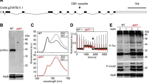
The closest homolog of the Arabidopsis gene encoding the thylakoid phosphatase PPH1/TAP38 (At4G27800) was identified in C. reinhardtii as Cre04.g218150 in recip-rocal BLASTP searches (see “Materials and Methods”). The C. reinhardtii protein, which we call CrPPH1, shares 36% sequence identity and 55% similarity with its Arabidopsis homolog. To investigate the function of CrPPH1 in C. reinhardtii, we obtained a mutant strain from the Chlamydomonas Library Project (CLiP; LMJ.RY0402.16176, https://www.chlamylibrary.org; Li et al., 2016) with a predicted insertion in intron 7 of the PPH1 gene (Fig. 1A). The site of insertion was con-firmed by PCR amplification and sequencing of the am-plified product (Supplemental Fig. S1A). This mutant line
showed an alteration of state transitions, as will be detailed below. However, according to the CLiP data-base, this line also carried a second insertion (hereafter ins2) in gene Cre24.g755597.t1.1, an insertion that was similarly confirmed by PCR analysis (Supplemental Fig. S1B). To remove the second mutation and to test whether the state transition phenotype cosegregated with the pph1 mutation, the mutant line (pph1;ins2) was backcrossed twice to the parental strain CC5155 (Supplemental Fig. S1C). From thefirst cross, 60 com-plete tetrads were analyzed for paromomycin resis-tance and for the presence of pph1 and ins2 insertions, both of which conferred paromomycin resistance. In eight of these tetrads where the two insertions segre-gated (non‐parental ditypes with four paromomycin-resistant colonies pph1/pph1/ins2/ins2), we found that altered state transitions cosegregated with the in-sertion in the pph1 gene (Supplemental Fig. S1C). In a second backcross between a pph1 single mutant deriv-ing from the first backcross and the parental line CC5155, we analyzed eight complete tetrads and ob-served cosegregation of the insertion in the pph1 gene with paromomycin resistance and the state transition phenotype (Supplemental Fig. S1D). This analysis shows that the alteration in state transitions is geneti-cally tightly linked to the pph1 mutation. To facilitate biochemical experiments, we further crossed a pph1 mutant progeny to the cell-wall–deficient strain cw15, and a double mutant, pph1;cw15, was selected for fur-ther analysis. All subsequent experiments were per-formed comparing the parental cw15 strain with the double mutant pph1;cw15; however, hereafter, for sim-plicity, these strains will be referred to as wild type and pph1, respectively. The chlorophyll content and the maximum quantum efficiency of PSII were similar in pph1 and in the wild type (Supplemental Table S1). The pph1 mutant also showed normal growth in a variety of conditions (Supplemental Fig. S2).
A rabbit antiserum was raised against recombinant CrPPH1 expressed in Escherichia coli (Supplemental Fig. S3A) to assess the presence of the protein by immuno-blotting of total protein extracts of the wild type and of the pph1 mutant (Fig. 1B). A band was observed in the wild type that was not detected in the pph1 mutant. This band migrated with an apparent molecular mass of 45 kD, somewhat slower compared to the calculated molecular mass of 40 kD for CrPPH1 after removal of a predicted chloroplast transit peptide (cTP) of 52 to 56 amino acid residues according to different algorithms (Predalgo; Tardif et al., 2012; ChloroP; Emanuelsson et al., 1999). Because of the background signal with this antibody, a low level of residual CrPPH1 expres-sion in the pph1 mutant could not be excluded (Supplemental Fig. S3).
State transitions can be monitored as a change in the fluorescence emission spectrum at 77 K (Fig. 1C). In C. reinhardtii, the transitions can be experimentally in-duced by switching between anaerobic and aerobic conditions. In the dark, anaerobiosis leads to reduction of the PQ pool and consequent establishment of St 2.
Subsequent strong aeration under the light promotes oxidation of the PQ pool and a transition to St 1. The relative sizes of the peaks at 682 nm and 712 nm qual-itatively reflect changes in the light harvesting antennae associated with PSII and PSI, respectively. In the wild type, transition from St 2 (LHCII partly connected to PSI) to St 1 (LHCII mostly allocated to PSII) caused a decrease in the PSI peak relative to the PSII peak. The spectrum was recorded after 20 min in the conditions promoting St 1, a timepoint when the transition is nearing completion in the wild type. At this time, the extent of the transition from St 2 toward St 1 was strongly diminished in the pph1 mutant compared to the wild type (Fig. 1C).
The time courses of state transitions were analyzed in more detail by measuring room-temperature chloro-phyll fluorescence using pulse-amplitude-modulation (PAM) spectroscopy (Fig. 1D). Cells that had been ac-climated to minimal medium in low light (60 mmol photons m22 s21) were transferred to the PAM fluo-rometer and the sample chamber was sealed, so that consumption of oxygen by respiration led to anaero-biosis. Steady-statefluorescence (Fs) was continuously monitored with the low intensity-measuring beam to monitor the redox state of the PQ pool, and saturating flashes were applied every 4 min to measure the max-imumfluorescence of PSII (Fm9), which is related to the cross section of its functional light-harvesting antenna.
Reduction of the PQ pool in the dark (denoted by an increase in Fs) triggered the transition to St 2 in the wild type and the pph1 mutant (decrease of Fm9). The cham-ber was then opened, and the algal sample bubbled with air, restoring an aerobic environment and allow-ing the reoxidation of the PQ pool and a transition to-ward St 1 (increase of Fm9). We observed that the transition from St 2 to St 1 was strongly delayed in pph1 (Fig. 1D), consistent with a role of CrPPH1 in dephos-phorylation of the LHCII antenna. A role in state tran-sitions was confirmed using another protocol (Hodges and Barber, 1983; Wollman and Delepelaire, 1984), whereby the transition from St 2 to St 1 was induced by actinic light in the presence of the PSII inhibitor 3-(3,4-dichlorophenyl)-1,1-dimethylurea (DCMU) under con-tinued anaerobiosis (Supplemental Fig. S4). The fact that at the onset of these experiments, pph1 is capable of a transition from St 1 toward St 2 implies that although the subsequent transition toward St 1 is delayed, pph1 can eventually reach at least a partial St 1 under the culture conditions used before the measurements (Fig. 1, C and D; Supplemental Fig. S4).
When comparing the two protocols in the wild type (Fig. 1D; Supplemental Fig. S4A), it is interesting to note that the transition from St 2 to St 1 was rapidly induced by aerobiosis (Fig. 1D), but that its onset was delayed a few minutes when it was induced in the presence of DCMU by actinic light under continued anaerobiosis
Figure 1. Characterization of the pph1 mutant. A, Schematic representation of the PPH1 gene. Exons are represented as black boxes, introns as black lines, and the 59 untranslated region and 39 untranslated region as white boxes. The arrowhead indicates the site of insertion of the CIB1 cassette in intron 7. B, Immunoblot analysis. Total protein extracts (50mg) of the wild type (WT) and of thepph1 mutant were subjected to SDS-PAGE and immunoblotting with antisera against CrPPH1 or AtpB (loading control). C, Chlorophyll fluorescence emission spectra, 77 K, under conditions that favor St 2 (anaerobiosis in the dark) and after 20 min under conditions that favor St 1 (strong aeration). The data are normalized on the PSII peak at 680 nm. D, State transitions monitored by PAM chlorophyll fluorescence spectroscopy at room temperature. Saturating light flashes were fired every 4 min and fluorescence was measured continuously (no actinic light was applied). Data were normalized on the firstFm9peak. The transition from St 1 to
St 2 was induced by sealing the sample chamber to allow respiration to deplete oxygen and cause anoxic conditions, and then the transition from St 2 to St 1 was induced by bubbling air in the sample (at the time indicated with a black arrow). E, Phospho-immunoblot analysis. Total protein extracts (10mg) of wild-type and pph1 cells in conditions favoring St 2 or St 1 (as in B) were subjected to SDS-PAGE and immunoblotting with antisera against P-Thr, P-Lhcb2, or AtpB (loading control).
(Supplemental Fig. S4A). This difference might be as-cribed to a requirement for both an oxidized PQ pool and sufficient levels of ATP to induce the transition toward St 1 (Bulte et al., 1990). While under restored aerobiosis (Fig. 1D), mitochondrial respiration could rapidly replenish the cellular ATP pool, whereas in the presence of DCMU under anaerobiosis (Supplemental Fig. S4A), CEF would replenish the ATP pool more slowly.
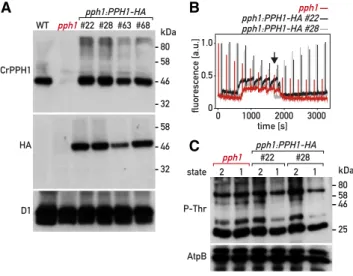
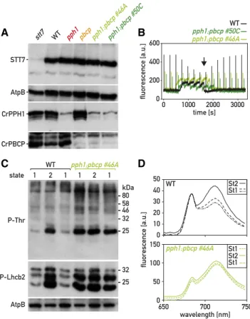
To confirm that this state-transition phenotype is due to the pph1 mutation, we transformed the mutant strain with a plasmid carrying a wild-type copy of PPH1 in which we inserted a sequence encoding a triple he-magglutinin (HA) epitope at the 39 end of the coding sequence (CDS; PPH1-HA) and a selection marker (aph799, hygromycin resistance). The transformants were screened by chlorophyll fluorescence spectros-copy, and four lines showing a restoration of state transitions were selected for further analysis. Immu-noblotting with a monoclonal HA antibody indicated that the rescued lines (pph1:PPH1-HA) expressed the tagged CrPPH1-HA protein, and immunoblotting with the anti-CrPPH1 antibodies showed that the protein was expressed at levels similar to those of the wild type or in moderate excess (Fig. 2A). PAM fluorescence spectroscopy showed that in pph1:PPH1-HA, the rate of the transition from St 2 to St 1 was comparable to that of the wild type (Fig. 2B).
To further test the role of CrPPH1 in LHCII dephos-phorylation, we examined the phosphorylation status of the major thylakoid proteins by SDS-PAGE and
immunoblotting. St 2 was established by anaerobiosis in the dark, and a subsequent transition to St 1 was promoted by strong aeration in the light, as written about above, for the analysis offluorescence at 77 K. An antibody against phospho-Thr (P-Thr) revealed a complex pattern of phosphoproteins; nevertheless, a distinct decrease in the signal of several bands was observed in the wild type upon transition from St 2 to St 1 (Fig. 1E), as well as in the complemented strains (Fig. 2C). As will be shown below, some of the same bands were clearly underphosphorylated in the mutant stt7, which is deficient for the protein kinase involved in state transitions. In the pph1 mutant, the signal of these phosphoprotein bands decreased much less after switching to the conditions that favor St 1. With an antibody against the phosphorylated form of Arabi-dopsis Lhcb2 (P-Lhcb2), several bands were observed in C. reinhardtii, corresponding to the migration of LHCII (Supplemental Fig. S5), although the sequence divergence between the peptide recognized by the an-tibody and the potential target sequences in the C. reinhardtii antenna subunits does not allow a simple assignment to specific LHCBM isoforms. In the wild type, the intensity of these bands was higher in St 2 than St 1. In the stt7 mutant (see below), these bands are detected only at low levels that are similar under St-2 or St-1 conditions. These two observations indicate that at least some of the bands revealed in C. reinhardtii by the Arabidopsis anti-P-Lhcb2 antibody are implicated in state transitions. These bands were clearly over-phosphorylated in pph1 compared to the wild type in the conditions that favor a transition to St 1. Thus, de-fects in the dephosphorylation of LHCII antenna com-ponents parallel the alteration in state transitions observed spectroscopically in the pph1 mutant.
CrPBCP Is Also Involved in State Transitions
A striking feature of the pph1 mutant was that it showed strong retardation of the transition from St 2 to St 1, but that nevertheless, under normal culture condi-tions, it was capable of approaching St 1 and undergoing a subsequent transition to St 2 (Fig. 1D; Supplemental Fig. S4). It thus appeared that at least one other protein phosphatase might be involved in state transitions and dephosphorylation of LHCII components, allowing the establishment of St 1 in pph1, albeit more slowly. This was corroborated by the identification of another phos-phatase mutant affected in state transitions.
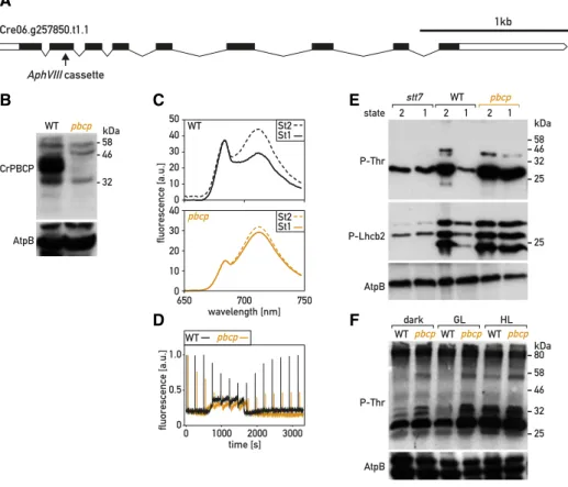
A library of mutants with random insertions of an aphVIII cassette (paromomycin resistance) was gener-ated and screened using a fluorescence imaging setup (Tolleter et al., 2011). To search for mutants in photo-protective or alternative electron transfer pathways, this library was recently screened again using a differ-ent imaging system (Johnson et al., 2009). One of the mutants (identified as pbcp, see below) showed im-paired transitions from St 2 to St 1 (Fig. 3). Using reverse-PCR techniques, the insertion was mapped to
Figure 2. Complementation of the pph1 mutant. A, Immunoblot anal-ysis. Total protein extracts (50mg) of the wild type (WT), the pph1 mutant, and four complemented lines (pph1:PPH1-HA) were subjected to SDS-PAGE and immunoblotting with antisera against CrPPH1, the HA epitope, and D1 (loading control). B, State transitions in thepph1 mutant and two complemented lines were monitored by PAM chlorophyll fluorescence spectroscopy as in Figure 1D. C, Phospho-immunoblot anal-ysis. Total protein extracts (10mg) of the pph1 mutant and of complemented lines were subjected to SDS-PAGE and immunoblotting with antisera against P-Thr or AtpB (loading control).
the gene Cre06.g257850 (Supplemental Fig. S6A), which encodes the C. reinhardtii protein most similar to PBCP from Arabidopsis, and will be called CrPBCP hereafter (Fig. 3A). To test whether the alteration of state transi-tions was genetically linked to the pbcp mutation, we backcrossed the mutant to wild-type 137C. In 40 com-plete tetrads, paromomycin resistance segregated with the insertion in the PBCP gene. In the four tetrads that we analyzed further, an overphosphorylation of thy-lakoid proteins cosegregated with the insertion in PBCP (Supplemental Fig. S6B). Moreover, pbcp and the phosphorylation phenotype also cosegregated with mating-type minus (mt2), as expected because PBCP lies in close proximity to the mating-type locus on chromosome 6. One of the pbcp mutant progeny was further crossed to cw15 to obtain a cell-wall–deficient strain (pbcp;cw15), which was used in all subsequent experiments and will be referred to as pbcp hereafter. Using a rabbit antiserum raised against recombinant CrPBCP expressed in E. coli (Supplemental Fig. S3B) and immunoblotting of total proteins separated by SDS
PAGE, the protein was detected in the wild type but not in the pbcp mutant (Fig. 3B). The chlorophyll content and the maximum quantum yield of PSII were not significantly different in pbcp and in the wild type (Supplemental Table S1). Furthermore, the pbcp mutant showed normal growth under a variety of conditions (Supplemental Fig. S2).
We analyzed state transitions in the pbcp mutant by determining itsfluorescence emission spectra at 77 K in St 2 and after a subsequent transition to conditions promoting St 1 for 20 min. Compared to the wild type, the transition to St 1 was significantly impaired in pbcp (Fig. 3C). Using PAM chlorophyll fluorescence spec-troscopy, we observed that anaerobiosis in the dark promoted a transition to St 2 in the pbcp mutant as in the wild type. However, upon aeration and reoxidation of the PQ pool, the transition from St 2 to St 1 was strongly delayed in pbcp (Fig. 3D). Similar observations were made using the protocol where this transition was in-duced by light in the presence of DCMU (Supplemental Fig. S4). Thus, CrPBCP seems to play a major role in state transitions, unlike its homolog in Arabidopsis.
Figure 3. Characterization of the pbcp mutant. A, Schematic representation of the PBCP gene. Exons are represented as black boxes, introns as black lines, and 39/59 untranslated regions as white boxes. The arrow indicates the site of insertion of the aphVIII cassette in exon 2. B, Immunoblot analysis. Total protein extracts of the wild type (WT) and of thepbcp mutant (50 mg) were subjected to SDS-PAGE and immunoblotting with antisera against PBCP or AtpB (loading control). C, Chlorophyll fluorescence emission spectra, 77 K, under condition that favor St 2 and after 20 min under conditions that favor St 1, as in Figure 1C. D, State transitions of the wild type and thepbcp mutant were monitored by PAM chlorophyll fluorescence spectroscopy as in Figure 1D. E, Phospho-immunoblot analysis of state transitions. Total protein extracts of wild type andpbcp cells in St 2 and St 1 (10 mg, treated as in Fig. 2C) were subjected to SDS-PAGE and immunoblotting with antisera against P-Thr, P-Lhcb2, or AtpB (loading control). F, Phospho-immunoblot analysis of light acclimation. The cells were grown in low light and then transferred to the dark, growth light (GL, 80mmol m22s21), or high light (HL, 300mmol m22s21) for 2 h and analyzed as in E.
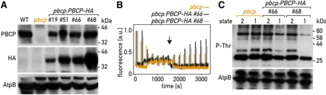
To confirm that the state-transition phenotype was due to the pbcp mutation, we transformed the pbcp mutant with a plasmid carrying a wild-type copy of PBCP tagged with a sequence encoding a triple HA epitope (PBCP-HA) and a selectable marker (aph799). Four rescued lines (pbcp;PBCP-HA) that expressed the tagged CrPBCP-HA protein were retained for further analysis. Immunoblotting with the PBCP anti-bodies (Fig. 4A) showed that the different pbcp;PBCP-HA lines expressed the protein at levels similar or somewhat reduced compared to the wild type. State transitions were restored in the pbcp;PBCP-HA lines, as monitored by PAMfluorescence spectroscopy (Fig. 4B). To investigate the alteration of thylakoid protein phosphorylation in the pbcp mutant, we used SDS-PAGE and immunoblotting (Fig. 3E). With an anti-P-Thr antibody, we observed that bands that migrate as com-ponents of LHCII (Supplemental Fig. S5) were over-phosphorylated in pbcp, but not in the complemented pbcp;PBCP-HA lines (Fig. 4C). Unfortunately, these com-mercial anti-P-Thr antibodies (now discontinued) did not clearly identify the phosphorylated forms of PSII subunits D2 (PsbD) or CP43 (PsbC; Supplemental Fig. S5). High phosphorylation of LHCII constituents was confirmed with the Arabidopsis anti-P-Lhcb2 antibodies, which showed overphosphorylation in pbcp of the bands that are underphosphorylated in the stt7 mutant. Thus, CrPBCP has a different range of targets in C. reinhardti than its homolog in Arabidopsis, where the major targets of PBCP are the subunits of the PSII core. It was striking that excess phosphorylation of thylakoid proteins in pbcp appeared not only after a transition from St 2 to St 1, but also under normal growth conditions (80mmol photons m22s21), as well as after growth under high light (300mmol photons m22s21) or in the dark (Fig. 3F).
Thepph1;pbcp Double Mutant Is Locked in State 2
Both CrPPH1 and CrPBCP are implicated in the de-phosphorylation of some components of LHCII and are essential for an efficient transition from St 2 to St 1.
Nevertheless, both individual mutants, pph1 and pbcp, are capable of eventually approaching St 1 under the culture conditions before the measurements (Figs. 1D and 3D; Supplemental Fig. S4). To further test whether the two phosphatases play partly redundant roles in the regulation of state transitions, we generated double pph1;pbcp mutants by crossing pph1 and pbcp and gen-otyping the progeny by PCR (Supplemental Fig. S7). The pph1;pbcp mutants also carry the cw15 mutation like both their parents. The double mutants were, as expected, deficient for both CrPPH1 and CrPBCP (Fig. 5A). The maximum quantum yield of PSII and the chlorophyll content of pph1;pbcp were not significantly different from the wild type (Supplemental Table S1), and the double mutant showed normal growth under a set of different conditions that were tested (Supplemental Fig. S2).
When state transitions were monitored in pph1;pbcp double mutants, it was remarkable that, after pre-acclimation under low light where the wild-type cells are in St 1, the double mutant had a comparatively low value of Fm9at the onset of the measurements, indicative of St 2 (Fig. 5B). There was also no significant further drop in Fm9when the pph1;pbcp sample became anaer-obic with the concomitant rise in Fs. The double mutant also showed no rise in Fm9when the sample was aerated again, indicating that the transition to St 1 was severely hampered. Similar results were obtained when this state transition was promoted by light in the presence of DCMU (Supplemental Fig. S4). The low level of Fm9 indicative of St 2 was matched by a high phosphoryl-ation of LHCII components (Fig. 5C). In contrast to the wild type that showed low phosphorylation in St 1, high phosphorylation after a transition to St 2, and again, low phosphorylation after a further transition to St 1, pph1;pbcp showed in all three conditions a phos-phorylation pattern similar to that of the wild type in St 2 (Fig. 5C). These observations indicated that the pph1;pbcp double mutant is essentially locked in St 2. In further support of this conclusion, we determined the fluorescence emission spectrum at 77 K of cells in all three conditions (St 1. St 2 . St 1), and observed that
Figure 4. Complementation of the pbcp mutant. A, Immunoblot analysis. Total protein extracts (50 mg) of the wild type (WT), the pbcp mutant, and four complemented lines (pbcp:PBCP-HA) were subjected to SDS-PAGE and immunoblotting with antisera against CrPBCP, the HA epitope, or AtpB (loading control). B, State transitions in thepbcp mutant and two complemented lines were monitored by PAM chlorophyll fluorescence spectroscopy at room temperature as in Figure 1D. C, Phospho-immunoblot analysis. Total protein extracts (10mg) of the pbcp mutant and of complemented lines were subjected to SDS-PAGE and im-munoblotting with antisera against P-Lhcb2, P-Thr, and AtpB (loading control).
in the pph1;pbcp double mutant thefluorescence emis-sion peak of PSI relative to PSII was always larger than in the wild type (Fig. 5D; Supplemental Fig. S4B). Our data indicate that two partially redundant protein phosphatases, CrPPH1 and CrPBCP, are involved in the transition from St 2 to St 1.
Accumulation of Thylakoid Proteins in the Mutants
In Arabidopsis, the expression of STN7 is reduced under prolonged exposure to far-red light (Willig et al., 2011), and the amount of PPH1 is downregulated at a posttranscriptional level in the psal mutant, which is deficient in the docking of LHCII to PSI and thus in-capable of completing state transitions (Rantala et al., 2016). These observations prompted us to determine
whether, in Chlamydomonas, the amounts of STT7, CrPPH1, or CrPBCP are altered in the single kinase or phosphatase mutants and also in the pph1;pbcp double mutant, using SDS PAGE and immunoblotting of pro-tein extracts from cells grown under normal conditions (Fig. 5A). However, no major differences were observed in the accumulation of the three regulatory proteins.
We also investigated the possibility that the phos-phatase mutations might be compensated in the long term by changes in the stoichiometry of the photosys-tems or other major photosynthetic complexes. Proteins extracts of the wild type, the single mutants pph1 and pbcp, and the double mutant pph1;pbcp grown under normal conditions were compared by SDS PAGE and immunoblotting (Supplemental Fig. S8). Antisera against representative subunits of the major complexes were used for this analysis: AtpB (ATP synthase), D1 (PSII), PsaA (PSI), Cytf (cytochrome b6f complex), and COXIIb (mitochondrial cytochrome oxidase). How-ever, no significant differences were apparent in the relative amounts of the photosynthetic complexes in the mutant lines.
CrPPH1 and CrPBCP Have Overlapping But Distinct Dephosphorylation Targets
In C. reinhardtii, LHCII is composed of monomeric LHCB4 (CP29) and LHCB5 (CP26) as well as trimers of isoforms LHCBM1 through LHCBM9. Based on their primary sequences, the trimer subunits belong to four types (Supplemental Fig. S9): type I in which three subgroups can be distinguished (LHCBM3; LHCBM4/ LHCBM6/LHCBM8; LHCBM9), type II (LHCBM5), type III (LHCBM2/LHCBM7, which are identical in mature sequence), and type IV (LHCBM1). While the P-Thr antiserum and the Arabidopsis P-Lhcb2 antise-rum showed clear differences in the patterns of LHCII phosphorylation in St 1 in both phosphatase mutants (Fig. 6A), the ill-defined specificity of these antisera did not allow the discrimination of the different compo-nents of the antenna, or reveal any target specificity of the respective phosphatases. To address these ques-tions, we used Phos-tag PAGE followed by immu-noblotting with specific antibodies. We obtained previously described antisera against LHCB4, LHCB5 and LHCBM5 (type II; Takahashi et al., 2006), and also generated antisera against peptides that are character-istic for the other types of LHCBM subunits. Because these isoforms share a high degree of sequence simi-larity, the antigenic peptides were selected in the N-terminal region of the proteins, which is the most divergent between LHCII types (Supplemental Fig. S9). The specificity of the affinity-purified antibodies was tested against the recombinant LHCII subunits expressed in E. coli (Supplemental Fig. S10). The anti-sera against LHCBM1 (type IV), LHCBM3 (type I), and LHCB4 and LHCB5 (minor antenna) proved to be very specific. The antiserum against LHCBM2/7 (type III) showed minor cross reactions toward type-I isoforms.
Figure 5. Characterization of the pph1;pbcp double mutant. A, Im-munoblot analysis. Total protein extracts (50mg) of stt7, wild type (WT), pph1, pbcp, and pph1;pbcp (clones 46A and 50C) were subjected to SDS-PAGE and immunoblotting with antisera against STT7, AtpB (loading control), CrPPH1, and CrPBCP. B, State transitions of the wild type and twopph1;pbcp mutants were monitored by PAM chlorophyll fluorescence spectroscopy at room temperature, as in Figure 1D. The data were not normalized to the firstFm9peak. C, Phospho-immunoblot
analysis. Total protein extracts of wild-type andpph1;pbcp cells in conditions favoring St 1, then St 2, and finally again St 1 (10mg; cells treated as in Fig. 1C) were subjected to SDS-PAGE and immunoblotting with antisera against P-Thr, P-Lhcb2, or AtpB (loading control). D, Chlorophyll fluorescence emission spectra, 77 K, under conditions se-quentially favoring St 1, St 2, and St1, obtained as in Figure 1C.
Finally, the antiserum against LHCBM4/6/8 (type I, which shares the same sequence in the N-terminal re-gion) also reacted toward LHCBM3 (also type I), but unexpectedly very strongly decorated LHCBM9 (type I). It should be noted, however, that LHCBM9 is only expressed under conditions of nutrient stress (Grewe et al., 2014).
To analyze the phosphorylation of the different pro-teins by immunoblotting, we used Phos-tag, which is a metal chelator that can be cross linked into a polyac-rylamide gel during polymerization (Kinoshita et al., 2009). This chelator binds Zn21 ions that interact with phosphate groups and thus retards the migration of phosphorylated forms of proteins during SDS-PAGE. After immunoblotting (Fig. 6B), the degree of
phosphorylation of the target protein is reflected in the ratio of the bands representing one or more slower-migrating phospho-form(s) to the band corresponding to the faster-migrating nonphosphorylated protein. To determine the migration of the nonphosphorylated form, we treated a sample of the wild type in St 2 with a nonspecific protein phosphatase (l-phosphatase) be-fore electrophoresis (lane marked l in Fig. 6B).The antipeptide antibodies are targeted to the N terminus of the LHCBM subunits, which also contains the major sites of phosphorylation. To avoid any bias due to dif-ferential recognition by these antibodies of the phos-phorylated and nonphosphos-phorylated forms of their targets, we treated the proteins after blotting onto the membrane with l-phosphatase (Longoni et al., 2015).
Figure 6. Analysis of CrPPH1 and CrPBCP targets. A, Phospho-immunoblot analysis. Total protein extracts of stt7, wild type (WT), pph1, pbcp, and pph1;pbcp (10 mg) in conditions fa-voring St 2 and then St 1 were subjected to SDS-PAGE and immunoblotting with antisera against P-Thr or AtpB (loading control). B, Phos-tag PAGE and immu-noblot analysis. Total protein extracts (10mg) of stt7, wild type, pph1, pbcp, orpph1;pbcp in St 2 and then St 1 were subjected to Phos-tag PAGE and immu-noblotting with antisera against LHCBM1, LHCBM3, LHCBM4/6/8, LHCBM2/7, LHCBM5, LHCB4, LHCB5, PsbH, or PETO. A sample of the wild type in St 2 was treated with lambda phosphatase (1l) and used as a reference for the migration of the non-phosphorylated (NP) form. The migration of the phosphor-ylated (P) forms is retarded by the Phos-tag immobilized in the polyacrylamide gel.
Because the phosphatases play a role in the transition from St 2 to St 1, we analyzed protein extracts of the wild type and the different mutants in St 2 (induced by anaerobiosis in the dark) and after a subsequent tran-sition to St 1 (triggered by aerating the sample).
The antibodies against LHCBM1 (type IV) labeled two bands in the wild type in St 2 (Fig. 6B). The lower one comigrated with the single band in the l-phosphatase–treated sample, representing the non-phosphorylated protein. The upper band, correspond-ing to a phosphorylated form, gave a strong signal relative to the lower one, indicative of a high degree of LHCBM1 phosphorylation in St 2. The ratio of the phosphorylated band to the nonphosphorylated one was much lower in the kinase mutant stt7, or after the transition to St 1. In the pph1 mutant, strong phospho-rylation of LHCBM1 was still apparent in the condi-tions promoting St 1, with a high ratio of the upper to the lower band, in contrast to the wild type or the pbcp mutant. The double mutant pph1;pbcp showed the same high degree of phosphorylation after the transition to St-1 conditions as the pph1 mutant. We infer that CrPPH1 is a major actor of LHCBM1 dephosphoryla-tion during the transidephosphoryla-tion to St 1.
The antibodies against LHCBM3 (type I) decorated two bands after Phos-tag gel electrophoresis of the l-phosphatase–treated sample (Fig. 6B), as well as after conventional gel electrophoresis of an untreated sample (Supplemental Fig. S8B), even though these antibodies were specific for LHCBM3 among the recombinant proteins expressed in E. coli (Supplemental Fig. S10). The lower band (marked with an asterisk), which was partially resolved as a doublet, is unlikely to represent processed forms of LHCBM3 lacking amino acid resi-dues at the N terminus (Stauber et al., 2003), because the LHCBM3 antibodies were raised against this region and would not recognize a truncated protein. Thus, the lower band may reflect nonspecific binding to another protein. There were two additional slower-migrating bands in the wild type in St 2, largely absent from the l-phosphatase–treated sample, suggesting that LHCBM3 may undergo phosphorylation at more than one site, or that the nonspecific protein is phosphorylated as well. One of these bands was also clearly present in stt7, suggesting the involvement of another protein kinase in LHCBM3 phosphorylation. The relative inten-sity of the top-most of the phosphorylated bands to the nonphosphorylated ones clearly decreased after transition to St 1 in the wild type. Some dephospho-rylation was still apparent in the pph1 mutant, but the overall phosphorylation level appeared to be higher. In contrast, the pbcp mutant showed a much higher ratio of phosphorylated bands in both St 2 and St 1, as did the pph1;pbcp double mutant. Thus, CrPBCP ap-pears to play the major role for dephosphorylation of LHCBM3.
With the antibodies against LHCBM4/6/8 (type I), a sin-gle major band was observed in thel-phosphatase–treated sample (Fig. 6B), as well as after conventional gel electro-phoresis (Supplemental Fig. S8B). However, these antibodies
also recognized other recombinant type-I isoforms, LHCBM3 and LHCBM9 (Supplemental Fig. S10). The latter should not be expressed under our conditions, because it is induced by nutrient stress (Grewe et al., 2014). After Phos-tag electrophoresis of the wild-type sample in St 2, the pattern of phosphorylation was complex, with most bands corresponding to those revealed with anti-LHCBM3 (but excluding the band marked with an asterisk). One of the phosphorylated forms was present in the stt7 mutant. The degree of phosphorylation of the others decreased in St 1 in both the wild type and pph1, but not in pbcp or pph1;pbcp where the most phosphorylated form was prevalent. We tentatively infer that CrPBCP is involved in de-phosphorylation of LHCBM4/6/8 and, because it also affects LHCBM3, more generally in the dephosphoryl-ation of type-I isoforms.
The antibodies against LHCBM2/7 (type III) deco-rated a strong band in the l-phosphatase–treated sample, with a second minor band above it. Likewise, after conventional gel electrophoresis, these antibodies labeled a major and a minor band (Supplemental Fig. S8B). Among the recombinant proteins expressed in E. coli, the LHCBM2/7 antibodies strongly recog-nized LHCBM2/7 but also weakly cross reacted with the type-I isoforms LHCBM3, LHCBM4/6/8, and LHCBM9 (Supplemental Fig. S10). The patterns of the phosphorylated bands, together with the upper non-phosphorylated band (marked with a triangle) but ex-cluding the strong nonphosphorylated band below (marked with a square), were similar to the pattern obtained with anti-LHCBM4/6/8, and may mostly represent the cross reaction to these isoforms (Fig. 6B). Compared to LHCBM4/6/8, the strong additional nonphosphorylated band (marked with a square) can tentatively be ascribed to LHCBM2/7, which is thus apparently not subject to phosphorylation under these conditions, or only to a small degree.
With antiserum against LHCBM5 (type II), two bands were detected in the l-phosphatase–treated sample, and at least five more in the various geno-types in St 2 (Fig. 6B). Two bands were also observed after conventional SDS-PAGE (Supplemental Fig. S8B), which could represent processed forms of LHCBM5 or cross reactions to other isoforms, so it is unclear which of thefive upper bands correspond to phosphorylated LHCBM5. The four uppermost bands were missing in stt7, suggesting that their phosphorylation relates to state-transitions. In the wild type in St 1 and in stt7, an additional band was present between the two nonphosphorylated ones, likely representing a low-phosphorylation form. Higher ratios of the upper bands to the lower ones were observed in St 1 in pph1 and pbcp, and an even higher ratio in pph1;pbcp. These observations suggest that both CrPPH1 and CrPBCP act in a partly redundant manner on the phosphoryla-tion of LHCBM5, and potentially other cross-reacting proteins decorated by the antibody.
Antiserum against LHCB4, which recognized a sin-gle band after conventional SDS-PAGE (Supplemental
Fig. S8B), decorated multiple bands in the wild type in St 2 (Fig. 6B), in accordance with the multiple phos-phorylation sites that have been observed in LHCB4 (Lemeille et al., 2010). The upper bands were missing in stt7, and the ratio of the upper ones to the lower ones decreased in the wild type in St 1 compared to St 2. In pph1, the pattern in St 1 resembled that of St 2, while in pbcp, as well as in pph1;pbcp, a very high degree of phosphorylation was observed in both states. These data suggest that CrPPH1 and CrPBCP contribute to LHCB4 dephosphorylation in St 1, and that CrPBCP may play such a role also in St 2, independently of state transitions.
The antiserum against LHCB5, which labels a single band after conventional SDS-PAGE (Supplemental Fig. S8B), decorated at least three bands in the different samples, none of which comigrated with the non-phosphorylated form in the l-phosphatase–treated sample (Fig. 6B). Two of the constitutively phosphor-ylated forms were present in stt7, while the third and slowest-migrating form was present in the wild type in St 2 but not in St 1. The latter was still present in pph1 in St 1, while it became prevalent compared to the lowest one in pbcp, and in pph1;pbcp. These observations sug-gest that CrPPH1 and CrPBCP participate in LHCB5 dephosphorylation in St 1, and that CrPBCP may also contribute in St 2.
With antiserum against the PsbH subunit of PSII, two phosphorylated forms were observed in the wild type and in stt7, in both St-2 and St-1 conditions (Fig. 6B). Phosphorylation appeared stronger in pbcp as well as pph1;pbcp, suggesting that CrPBCP is probably in-volved in the dephosphorylation of PsbH, like its ho-molog in Arabidopsis (Samol et al., 2012).
Finally, with antiserum against PETO, which is a phospho-protein implicated in the regulation of CEF (Hamel et al., 2000; Takahashi et al., 2016), a major phosphorylated form as well as one or two minor ones were observed in the wild type in St 2 that were strongly diminished in St 1 or in the stt7 mutant. In pph1 the phosphorylation was retained in St 1, in pbcp the upper band became most prevalent, and in pph1;pbcp, an even slower-migrating band was apparent. These data indicate that both CrPPH1 and CrPBCP are in-volved in the dephosphorylation of PETO in a some-what additive manner.
DISCUSSION
We have identified two protein phosphatases that are required for efficient transitions from St 2 to St 1 in C. reinhardtii. CrPPH1 is the closest homolog of Arabi-dopsis PPH1/TAP38, which is involved in state tran-sitions in plants (Pribil et al., 2010; Shapiguzov et al., 2010). Unexpectedly, we found that in C. reinhardtii, CrPBCP is also involved in state transitions. This is in contrast to PBCP from Arabidopsis, which is required for the dephosphorylation of several PSII core subunits but not of the LHCII antenna (Samol et al., 2012). In
Arabidopsis, lack of PBCP does not affect state transi-tions and it is only when this phosphatase is strongly overexpressed that it has a minor effect on the rate of state transitions. In rice, OsPBCP contributes to de-phosphorylation of LHCB4 (CP29) and is proposed to have a role in regulating the dissipation of excess en-ergy (Betterle et al., 2017). The different substrate specificities of the two phosphatases from higher plants can be explained by the different geometry of their substrate binding sites (Wei et al., 2015; Liu et al., 2019). The transition from St 2 to St 1 is strongly delayed in the pph1 and pbcp mutants of C. reinhardtii. However after incubation in conditions favoring St 1, both single mutants are nevertheless capable of approaching St 1 (Figs. 1D and 3D; Supplemental Fig. S4) and then undergo a transition toward St 2. In contrast, the double mutant pph1;pbcp remains in St 2 even in the conditions that favor St 1 in the wild type (Fig. 5; Supplemental Fig. S4). Thus, the phenotypes of the two mutants show some additivity, but the two phosphatases have partly redundant functions in state transitions.
State transitions are regulated by the phosphoryla-tion or dephosphorylaphosphoryla-tion of LHCII antenna proteins. In Arabidopsis, the Lhcb1 and Lhcb2 components of the LHCII trimers can be phosphorylated, but not Lhcb3, with phospho-Lhcb2 playing a major role in the for-mation of the PSI–LHCI–LHCII complexes in St 2 (Crepin and Caffarri, 2015; Longoni et al., 2015). To investigate the requirement of CrPPH1 and CrPBCP in antenna dephosphorylation during a transition from St 2 to St 1 in C. reinhardtii, we used Phos-tag PAGE and immunoblotting (Fig. 6B). Although some of the anti-peptide antisera that we developed displayed some cross reactions among closely related LHCBM iso-forms, it was still possible to assign many phosphory-lated forms to the different types of LHCBM. The Phos-tag PAGE technique has the advanPhos-tage of allowing an assessment of cumulative phosphorylation. Compared to the simple patterns that are obtained with Lhcb1 and Lhcb2 in Arabidopsis (Longoni et al., 2015), with each one having only a single major phosphorylated form, the patterns proved much more complex in C. rein-hardtii, raising the possibility that some LHCBM iso-forms may undergo multiple phosphorylation. The analysis revealed interesting differences and overlaps between the targets of CrPPH1 and CrPBCP. CrPPH1 was essential for dephosphorylation of the type-IV isoform LHCBM1, while mainly CrPBCP was re-quired for dephosphorylation of the type-I isoforms LHCBM3 and LHCBM4/6/8. Both CrPPH1 and CrPBCP were necessary for dephosphorylation of the type-II isoform LHCBM5 as well as the minor antennae LHCB4 and LHCB5, with CrPBCP playing a dominant role for LHCB4. An overphosphorylation phenotype was already apparent in St 2 in the pbcp mutant for LHCBM3, LHCB4, and LHCB5 and, although to a lower extent, LHCBM3 and LHCBM5 appeared over-phosphorylated in pph1 under St-2 conditions. After growth under constant condition of low light or high light, overphosphorylation was also observed in the
pbcp mutant by immunoblotting with P-Thr anti-bodies. It thus appears that CrPBCP may be active in dephosphorylation status of thylakoid proteins not only in conditions that promote St 1, but also under more balanced steady-state conditions.
In Arabidopsis, PPH1 is specific for LHCII, although its strong overexpression has an effect on the dephos-phorylation of the PSII core subunits D1 and D2 (Pribil et al., 2010; Shapiguzov et al., 2010). Conversely, PBCP is mostly required for dephosphorylation of PSII sub-units (D1, D2, and CP43), but it can also act on LHCII (Samol et al., 2012; Longoni et al., 2019). In C. reinhardtii, the major phosphorylated thylakoid proteins include, in addition to LHCII, some subunits of PSII (D2 [PsbD], CP43 [PsbC], and PsbH (Delepelaire, 1984; de Vitry et al., 1991), as well as PETO (Hamel et al., 2000). Us-ing Phos-tag gel electrophoresis, we found that both CrPPH1 and CrPBCP influence not only the dephos-phorylation of components of LHCII, but also compo-nents of the PsbH subunit of PSII. Phosphorylation of LHCII still occurred in a mutant lacking PSII (Wollman and Delepelaire, 1984), but phosphorylation of PSII did not occur in a mutant lacking LHCII proteins (de Vitry and Wollman, 1988). While the two phosphatases target partially overlapping sets of components of both PSII and LHCII, it seems likely that differences in regulation and target specificities also lie with the protein kinases STT7 and STL1. STT7 appears specific for LHCII (Turkina et al., 2006; Lemeille et al., 2010), while puta-tively STL1 might phosphorylate PSII, like its paralog STN8 in Arabidopsis (reviewed by Rochaix et al., 2012). We also obtained evidence that both phosphatases are involved in the dephosphorylation of PETO, which is implicated in the control of CEF (Takahashi et al., 2016). In summary, CrPBCP and CrPPH1 seem to have dis-tinct but overlapping sets of targets, with CrPBCP having a somewhat wider spectrum. Whether these targets represent direct substrates of the phosphatases or indirect targets through regulatory cascades cannot be determined from our genetic analysis.
In C. reinhardtii, LHCBM5, CP29, and CP26 were enriched in PSI–LHCI–LHCII complexes in St 2 (Takahashi et al., 2006, 2014), although all types of LHCBM could also be found (Drop et al., 2014). Genetic studies have shown that type-III isoform LHCBM2/7, type-I isoforms LHCBM4/6/8, and CP29 and CP26 are implicated in state transitions (Tokutsu et al., 2009; Ferrante et al., 2012; Girolomoni et al., 2017; Cazzaniga et al., 2020). We observed that, with the possible ex-ception of LHCBM2/7, all types of LHCBMs as well as LHCB4 and LHCB5 are differentially phosphorylated in St 2 compared to St 1. In particular, we found that LHCBM1 was phosphorylated in St 2, that its phos-phorylation depends on STT7, and that it was dephosphorylated in St 1, mainly reliant on CrPPH1. However mutants that lack LHCBM1 were previously found to perform state transitions (Ferrante et al., 2012). Taken together, these observations beg the question of the physiological role of LHCBM1 phosphorylation. A possible explanation might be that, for state transitions,
LHCBM1 phosphorylation acts redundantly with the phosphorylation of other antenna proteins, so that they can fulfill this role in the mutants lacking LHCBM1. Analysis of knock-down mutants indicated that LHCBM2/7 is important for state transitions (Ferrante et al., 2012). The comparison of the data we obtained with the antibodies against LHCBM2/7 and LHCBM4/6/8 suggests that the type-III isoform LHCBM2/7 may not be phosphorylated in St 2, which is consistent with the previous report that phospho-rylation of LHCBM2/7 is not detected in the PSI– LHCI–LHCII complex (Drop et al., 2014). A possible reason might be that LHCBM2/7 could be required structurally for the association of LHCII trimers to PSI–LHCI, but that this isoform would not be involved in regulation through phosphorylation. This would be analogous to the case in Arabidopsis, where Lhcb1 is not phosphorylated when it is a component of the mobile LHCII trimer that binds to PSI–LHCI, so that it is only the phosphorylation of Lhcb2 that is required for binding of LHCII to the PSI docking site (Crepin and Caffarri, 2015; Longoni et al., 2015; Pan et al., 2018).
Compared to Arabidopsis, C. reinhardtii lacks LHCB6 (CP24) but has a more complex complement of the LHCBM subunits composing the LHCII trimers (reviewed by Crepin and Caffarri, 2018). Another dif-ference is that in C. reinhardtii, some PSI–LHCI–LHCII complexes contain not only LHCII trimers but also the “minor” antennae LHCB4 and LHCB5 (Drop et al., 2014; Takahashi et al., 2014). Furthermore, the ampli-tude of state transitions is larger in C. reinhardtii than in plants (Delosme et al., 1996), and in the alga, strong state transitions are induced by changes in metabolic demands for ATP or reducing power, for example un-der anoxic conditions (Bulte and Wollman, 1990; Cardol et al., 2009). It remains a matter of speculation whether these differences are related to the more intri-cate roles of the phosphatases CrPPH1 and CrPBCP in the regulation of light harvesting that have evolved in C. reinhardtii. On the other hand, in C. reinhardtii, phosphorylation of LHCB4 is controlled by CrPBCP, reminiscent of monocots such as rice where phospho-rylation of LHCB4 is controlled by OsSTN8 and OsPBCP (Betterle et al., 2017). Further “evo-physio” investigations from a combined evolutionary and physiological perspective on the role of protein phos-phorylation in the regulation of photosynthetic accli-mation in diverse organisms promise to be a fertile approach.
MATERIALS AND METHODS Strains, Growth Conditions, and Media
The Chlamydomonas reinhardtii cw15.16 (mt1) mutant was obtained by crossing cw15 (mt2) to wild-type 137C. The C. reinhardtii CLiP strain LMJ.RY0402.16176, its parental strain CC4533 (mt2), and the corresponding strain of opposite mating type CC5155 (mt1) were obtained from the Chla-mydomonas Resource Center (https://www.chlamycollection.org/; Li et al., 2016). This CLiP strain (pph1;cao) was backcrossed twice to CC4533, and then to
cw15 to obtain the cell-wall–deficient mutant strain pph1;cw15 used in this work and referred to as pph1. The pbcp strain was isolated by screening random in-sertional mutants for aberrant chlorophyll-fluorescence induction kinetics. These mutants were generated by inserting an aphVIII cassette in wild-type 137C (mt2; Tolleter et al., 2011). The pbcp mutant was backcrossed with wild-type 137C (mt1), and further to cw15.16 (mt1) to obtain the cell-wall–deficient mutant strain pbcp;cw15 used in this work and referred to as pbcp. The gen-otype of the progeny was verified by PCR (see below) and the phengen-otype by monitoring state transitions using PAM chlorophyllfluorescence spectros-copy (see below).
Cells were grown in Tris acetate phosphate (TAP; Harris, 1989) under normal growth light (60–80 mmol m22s21) from whitefluorescent tubes. The state transitions were obtained with cells in exponential growth phase (two to three 106cells/mL) collected by centrifugation, resuspended in High Salt Minimal medium at a concentration of two to three 107cells per mL, and preacclimated in dim light (;10 mmol m22s21) for 2 h with shaking.
Mapping of the Insertion inpbcp
DNA was isolated using the cetyltrimethylammonium bromide method and quantified using a NanoDrop spectrophotometer (Thermo Fisher Scientific). The Resda-PCR protocol was used to identify the left border, and the technique was adapted from González-Ballester et al. (2005). Thefirst PCR was performed with Taq polymerase and 10% (w/v) DMSO, using DegAluI (degenerate primer) and RB1 (primer on the aphVIII cassette). The program includedfive cycles with annealing at 58°C, then 20 repetitions at 25°C, and then at 55°C for one cycle, at 58°C for two cycles, and at 40°C for one cycle. A second nested PCR was performed on the product of thefirst PCR diluted to 1/1,000 using KOD Xtreme Hot Start DNA Polymerase (Merck Millipore) and primers Qoand RB2 (González-Ballester et al., 2005). The product of the second PCR was then loaded onto an agarose gel (2% [w/v]), and fragments with a Mr. 800 bp were isolated. PCR products were extracted from the gel and purified with Nucle-oSpin Gel and a PCR Clean-up Kit (Macherey Nagel), and eluted in water before sequencing. The Genome Walker technique (Clontech) was used to identify the right border of theflanking sequence of the cassette insertion site. Genomic DNA was successively digested using the enzyme PvuII followed by the liga-tion of a specific adapter. The primary PCR uses a primer specific to the insert (AphVIII) and an adapter-specific primer. This was followed by a nested PCR whereby products .800 bp were extracted from agarose gel and cloned. Cloning of the PCR products was performed in the vector pGEM-T (Promega) containing the lactose operon and the gene coding for ampicillin resistance, and then transformed into chemo-competent Escherichia coli (DH5) by thermal shock. The bacteria were selected on luria broth Ampicillin medium in the presence of b-D-1-ThioGalactopyranoside and 5-bromo-4-chloro-3-indolyl-beta-D-galactopyranoside to allow for white/blue selection.
DNA Constructs
The protein with the highest sequence similarity to Arabidopsis (Arabidopsis thaliana) PPH1/TAP38 (At4G27800) was identified in C. reinhardtii as Cre04.g218150 in reciprocal BLAST searches performed in Phytozome 12 (E-value: 6. E-57; https://phytozome.jgi.doe.gov/pz/portal.html#!search? show5BLAST) and The Arabidopsis Information Resource (TAIR 10; E-value: 9. E-56; http://www.arabidopsis.org/Blast/index.jsp).
Vector pPL18 was obtained from pSL18 by replacing the aphVIII resistance cassette with the hygromycin resistance cassette (Sizova et al., 2001). For this, the aphVIII cassette was removed by KpnI/XhoI digestion. The hygromycin cassette was amplified from plasmid MAC1_gen3 (Douchi et al., 2016) with primers pPL 56 and pPL 57 (see Supplemental Table S2) and cloned by Gibson assembly (Gibson et al. (2009)).
The vector was further modified to allow the fusion of a triple HA epitope tag at the C terminus of the CDSs of interest. A synthetic fragment encoding the triple epitope (obtained from Biomatik) was cloned into pPL18 by BsmI/NotI diges-tion, yielding vector pFC18.
The CrPPH1 CDS was amplified from a C. reinhardtii complementary DNA library with primers pFC_216 and pFC_217 and cloned by Gibson assembly into pFC18 linearized with EcoRV/BglII, resulting in pFC18_CrPPH1_HA. The CrPBCP CDS was amplified from a complementary DNA library with primers pFC_218 and pFC_219 and cloned as above, into pFC18 resulting in pFC18_CrPBCP_HA. The CrPPH1 CDS without the predicted cTP was ampli-fied with primer pFC_265 and primer pFC_266 from pFC18_CrPPH1_HA, then cloned into vector pet28a digested with NdeI and SalI to obtain
pet28a_CrPPH1_ΔcTP. The CrPBCP CDS without the predicted cTP was am-plified with primer pFC_267 and primer pFC_268 from pFC18_CrPBCP_HA, and cloned into pet28a as above to obtain pET28a_CrPBCP_ΔcTP. All vectors used were verified by sequencing.
Transformation
Nuclear transformation by electroporation was modified from Shimogawara et al. (1998). Cells in exponential phase were collected by centrifugation and resuspended at 108cell per mL in TAP1 60 mMSuc. A volume of 300mL of cells was incubated with 1mg of plasmid DNA (linearized with XbaI) at 16°C for 20 min, and then the mix was transferred to a 4-mm–gap electroporation cu-vette and pulsed at 500 V (C5 50 mF) using a GenePulser II electroporator (Bio-Rad). The cuvette was then incubated at 16°C for 20 min. The cell suspension was diluted into 20 mL of TAP, then incubated in dim light with gentle agita-tion for 16 h and collected by centrifugaagita-tion, before plating and selecagita-tion, on TAP1 25 mg mL21hygromycin (Sigma-Aldrich).
Production of Polyclonal Antiserum of CrPPH1, CrPBCP, and LHCBMs
For production of CrPPH1 and CrPBCP, recombinant proteins were expressed from plasmids pet28a-CrPPH1_ΔcTP and pET28a-CrPBCP_ΔcTP in E. coli BL21(DE3) induced with 1 mMofb-D-1-ThioGalactopyranoside for 4 h at
37°C. Recombinant proteins purified as in Ramundo et al. (2013) were used to immunize rabbits, and antisera were purified by affinity chromatography with the corresponding protein (Eurogentec).
For production of antipeptide antisera targeting different LHCBMs isoforms (Natali and Croce, 2015), synthetic peptides (Supplemental Fig. S9) were used for rabbit immunization and affinity chromatography (Eurogentec). For assessing the specificity and cross reaction of the antibodies, pET28a plasmids carrying the CDSs of LHCBM1, LHCBM2, LHCBM3, LHCBM4, LHCBM5, LHCBM6, LHCBM9, CP26, and CP29 (Girolomoni et al., 2017) were used to express all the antenna proteins in E. coli BL21(DE3).
Fluorescence Measurements
Maximum quantum efficiency of PSII (Fv/Fm) of cells that were adapted for ;1 to 5 min in the dark was measured with a plant efficiency analyzer (Handy PEA; Hansatech Instruments) with the parameters recommended by the manu-facturer. State transitions were monitored with a PAMfluorometer (Hansatech Instruments). A 2-mL sample of cells grown and preacclimated in High Salt Minimal medium as described above was transferred to the dark in the vessel of a PAMfluorometer (Hansatech Instruments). The sample was kept in the dark with stirring, and saturating pulses (pulse width of 0.7 s; intensity 85%) were applied every 4 min. The St-1 to St-2 transition was induced by sealing the chamber, so that respiration led to anoxic conditions, and then the transition from St 2 to St 1 was induced by bubbling air in the sample. The alternate protocol for inducing state transitions in the presence of DCMU is described in Supplemental Figure S4. Chlorophyllfluorescence emission spectra at 77 K were measured with a spec-trofluorometer (Ocean Optics) with excitation from an LED light source at 435 nm. Immunoblotting
For total protein extraction, cells in exponential phase were collected by centrifugation, resuspended in lysis buffer (50 mMof Tris-HCl at pH 6.8, 2% [w/v] SDS, and 10 mMof EDTA) and 13 Protease inhibitor cocktail
(Sigma-Aldrich) and incubated at 37°C for 30 min with shaking in a thermomixer. Cell debris was removed by centrifugation at 16,000g for 10 min at 4°C, and the supernatant was used as total protein extract. For protein phosphorylation analysis, any centrifugation step of living cells was avoided by instantly dousing the cultures in 4 vol of cold acetone. After 30-min incubation on ice and centrifugation at 12,000g for 12 min in an SLA-4 rotor (Sorvall), the pellet was treated as before. Samples (10 or 50mg of total protein) were denaturated for 30 min at 37°C before SDS-PAGE (Laemmli, 1970) in 15% or 12% (w/v) acrylamide gels. After wet transfer, the nitrocellulose membranes (Bio-Rad) were blocked in TBS plus TWEEN (20 mMof Tris at pH 7.5, 150 mMof NaCl, and 0.1% [v/v] TWEEN 20) supplemented with 5% (w/v) nonfat milk (or 3% [w/v] BSA in the case of anti-P-Thr immunoblotting), for 2 h at room temperature or 16 h at 4°C. The antisera (and their sources) were as follows: monoclonal anti-HA (Promega); anti-phospho-LHCB2 (AS13-2705; Agrisera); anti-PsaA (a gift of