HAL Id: hal-02082987
https://hal.archives-ouvertes.fr/hal-02082987
Submitted on 26 May 2020
HAL is a multi-disciplinary open access
archive for the deposit and dissemination of sci-entific research documents, whether they are pub-lished or not. The documents may come from teaching and research institutions in France or abroad, or from public or private research centers.
L’archive ouverte pluridisciplinaire HAL, est destinée au dépôt et à la diffusion de documents scientifiques de niveau recherche, publiés ou non, émanant des établissements d’enseignement et de recherche français ou étrangers, des laboratoires publics ou privés.
promotes salicylic acid metabolism, and regulates
bacterial-triggered immunity
Ronny Völz, Soon-Kap Kim, Jianing Mi, Kiruthiga Mariappan, Xiujie Guo,
Jean Bigeard, Santiago Alejandro, Delphine Pflieger, Naganand Rayapuram,
Salim Al-Babili, et al.
To cite this version:
Ronny Völz, Soon-Kap Kim, Jianing Mi, Kiruthiga Mariappan, Xiujie Guo, et al.. The
Tri-helix transcription factor GT2-like 1 (GTL1) promotes salicylic acid metabolism, and regulates bacterial-triggered immunity. PLoS Genetics, Public Library of Science, 2018, 14 (10), pp.e1007708. �10.1371/journal.pgen.1007708�. �hal-02082987�
The Trihelix transcription factor GT2-like 1
(GTL1) promotes salicylic acid metabolism,
and regulates bacterial-triggered immunity
Ronny Vo¨ lzID1*, Soon-Kap Kim1, Jianing Mi1, Kiruthiga G. MariappanID1, Xiujie Guo1, Jean BigeardID2,3, Santiago Alejandro2,3, Delphine Pflieger4,5, Naganand Rayapuram1, Salim Al-BabiliID1, Heribert HirtID1,2,3*1 Desert Agriculture, Division of Biological and Environmental Sciences and Engineering, King Abdullah
University of Science and Technology, Thuwal, Saudi Arabia, 2 Institute of Plant Sciences Paris Saclay IPS2, CNRS, INRA, Universite´ Paris-Sud, Universite´ Evry, Universite´ Paris-Saclay, Orsay, France, 3 Institute of Plant Sciences Paris-Saclay IPS2, Paris Diderot, Sorbonne Paris-Cite´, Orsay, France, 4 Univ. Grenoble Alpes, CEA, Inserm, BIG-BGE, Grenoble, France, 5 CNRS, BIG-BGE, Grenoble, France
*ronny.volz@kaust.edu.sa(RV);heribert.hirt@kaust.edu.sa(HH)
Abstract
The Trihelix Transcription factor GT2-like 1 (GTL1) was previously shown to be a key regu-lator of ploidy-dependent trichome growth and drought tolerance. Here, we report that GTL1 plays an important role in coordinating plant immunity. We show that gtl1 mutants are com-promised in the regulation of basal immunity, microbial pattern-triggered immunity (PTI) and effector-triggered RIN4-mediated immunity. Transcriptome analysis revealed that GTL1 positively regulates defense genes and inhibits factors that mediate growth and develop-ment. By performing hormonal measurements and chromatin-immunoprecipitation studies, we found GTL1 to coordinate genes involved in salicylic acid metabolism, transport and response. Interaction studies and comparative transcriptomics to known data sets revealed that GTL1 is part of the MPK4 pathway and regulates oppositely the expression of differen-tially expressed genes in mpk4 plants. We introduced the gtl1 mutation in the mpk4 mutant and thereby partially suppressed its dwarfism and the high resistance against a bacterial invader. Our data show that GTL1 is part of the MPK4 pathway and acts as a positive regu-lator of bacterial-triggered immunity and SA homeostasis.
Author summary
The trihelix-transcription factor GT-2-like 1 (GTL1) belongs to the seven genes contain-ing GT-2 family of the plant-specific trihelix transcription factors. Previously, GTL1 was shown to be a key regulator of ploidy-dependent trichome growth and drought tolerance. In this report, we show that GTL1 is part of the MPK4-signaling cascade that coordinates immunity to virulent and avirulentPseudomonas syringae strains. gtl1 mutants are
promised in basal immunity, PTI and ETI. Comparative transcriptomics revealed a com-mon set of differentially regulated genes ingtl1 and mpk4. In this context, GTL1 positively
regulates defense genes and inhibits factors that mediate growth and development.
a1111111111 a1111111111 a1111111111 a1111111111 a1111111111 OPEN ACCESS
Citation: Vo¨lz R, Kim S-K, Mi J, Mariappan KG,
Guo X, Bigeard J, et al. (2018) The Trihelix transcription factor GT2-like 1 (GTL1) promotes salicylic acid metabolism, and regulates bacterial-triggered immunity. PLoS Genet 14(10): e1007708.https://doi.org/10.1371/journal. pgen.1007708
Editor: Morten Petersen, University of
Copenhagen, DENMARK
Received: March 4, 2018 Accepted: September 21, 2018 Published: October 23, 2018
Copyright:© 2018 Vo¨lz et al. This is an open access article distributed under the terms of the Creative Commons Attribution License, which permits unrestricted use, distribution, and reproduction in any medium, provided the original author and source are credited.
Data Availability Statement: All files are available
from the PLOS Genetic database, except of RNAseq data sets. The RNAseq data sets are submitted to the GEO repository (accession GSE118651).https://www.ncbi.nlm.nih.gov/geo/ query/acc.cgi?acc=GSE118651. Enter token ifwzwsqsjpixdsz into the box.
Funding: The funders had no role in study design,
data collection and analysis, decision to publish, or preparation of the manuscript. Funding for the
Salicylic acid measurements and Chromatin-Immunoprecipitation assays indicate that GTL1 directly binds and regulates genes involved in SA-biosynthesis, transport and response. Thempk4/gtl1 double mutant is compromised in the resistance to Pst AvrRPM1
and partially restored in the growth inhibition compared tompk4 single mutant. In
sum-mary, the reduced resistance of the double mutant indicates MPK4 as a negative regulator of GTL1-mediatedAvrRPM1-triggered immunity.
Introduction
Plants are faced with a constant threat of potential infections by a multiplicity of pathogenic microorganisms in their habitat. Pathogen-independent preformed physical borders like the cuticle, cell walls and wax coating represents the first line of plant defense to prevent pathogen invasion. Once the first boundary is breached, plants rely on their innate immune system to cope with different sorts of invaders and to initiate an adequate pathogen-counteracting defense response.
Plant innate immunity can be subdivided in two different recognition and response systems that relies either on the perception of pathogen-associated molecular patterns (PAMP) by plasma-membrane localized receptors (PRR) or on bacterial effectors injected in the plant cell and their recognition by intracellular receptors encoded by nucleotide binding domain leu-cine-rich repeat proteins (NLR-proteins) [1]. The perception of PAMPs, like FLAGELLIN22 (flg22), a 22 amino acid bacterial flagellum peptide, by the plasma membrane-localised recep-tor FLAGELLIN-INSENSITIV2 (FLS2) activates the mitogen-activated protein kinase (MAPK) cascades as part of the pattern-triggered immunity (PTI) [2]. Effector-triggered immunity (ETI) is activated by pathogen-derived “avirulence” (avr) effectors injected into the
plant cell by the bacterial type III-secretion system inPseudomonas syringae cv tomato (Pst).
On susceptible (r) hosts, type III effectors can contribute to virulence, by interfering with plant
immunity at the level of NPR1-dependent SA signaling [3] or the activation of the MAPKs MPK4 and 11 [4]. Some effectors are recognised by specific disease resistance (R) gene
prod-ucts leading to ETI. R and Avr proteins often co-localize within the plant cell [5]. The most common and widely distributed class of R proteins has a central nucleotide binding (NB) domain and C-terminal Leu-rich repeats (LRRs) [6]. The activation of NB-LRR proteins trig-gers a multitude of robust defense responses comprising biochemical and cellular events, like localized programmed cell death (hypersensitive response) and massive transcriptional repro-gramming to restrict pathogen propagation [7]. Resistance toPseudomonas syringae strains
expressing either AvrB and AvrRpm1 [8,9] is conferred byPseudomonas syringae pv maculi-cola 1 (RPM1), a CC-NB-LRR R protein, that is peripherally associated with the plasma
mem-brane [10]. In this context, RPM1-Interacting Protein 4 (RIN4) acts as a vital defense regulator [11] and is targeted by several pathogen effectors, such as AvrRPM1, AvrRpt2, AvrPto and AvrPtoB [12]. AvrRPM1 triggers RIN4 phosphorylation [5] by RIN4-INTERACTING RECEPTOR-LIKE PROTEIN KINASE [13] to promote the defense repression mediated by RIN4. However, plants producing RPM1 R-protein detect RIN4 phosphorylation and initiate ETI [14]. AvrRpt2 is a Cys-protease that passes through self-activation and cleavage in order to cleave RIN4 at the plasma membrane [15]. RIN4 degradation imposed by AvrRpt2 is consid-ered as a bacterial strategy to bypass AvrRpm1 induced ETI in the presence of RPM1 [16]. Recent studies have shown that SA signaling is an integral part of ETI and PTI [17,18]. Plant innate immunity activates MAP kinase cascades typically involved in early and late immune responses. MAPK cascades consist of three sequentially activated kinase modules composed of
project was provided to HH by baseline funding project BAS/1/1062 of the King Abdullah University of Science and Technology. Investissement d’Avenir Infrastructures Nationales en Biologie et Sante´ program (ProFI project, ANR-10-INBS-08) to DP (http://www.agence-nationale-recherche.fr/en/ international-cooperation/).
Competing interests: The authors have declared
a MAPK kinase kinase, a MAPK kinase and eventually a MAPK, thereby linking upstream sig-nals to downstream targets. In Arabidopsis as well as throughout the plant kingdom, the MAPK orthologues of MPK3, MPK4 and MPK6 represent the final step in the transmission of PAMP signals to respective target proteins by phosphorylation [1,19]. Although at least six PAMP-activated MAPKs have been reported to date [20,21], but so far clear evidence for a role in defense only exists for MPK3, MPK4 and MPK6, all three of which are required for complete activation of defense genes [22]. MPK3 and MPK6 are both activated by MKK4 and MKK5, but their upstream MAP3K(s) have not yet been unambiguously identified [23]. In contrast, there is clear evidence that the MAPKKK MEKK1 activates the MAPKKs MKK1 and MKK2, which converge to activate MPK4 [24–27].
MPK3 and MPK6 regulate the expression of a number of pathways, including phytoalexins, indole glucosinolate and ethylene biosynthesis [28–30]. MPK4 positively regulates basal resis-tance against pathogens [31], and about 50% of flg22-induced genes require MPK4 [22]. On the other hand,mekk1, mkk1 mkk2, and mpk4 mutant plants exhibit extreme dwarfism and
autoimmune phenotypes such as spontaneous cell death and constitutive defense gene expres-sion [24,26,31]. However, mutations in the NLR protein SUMM2 suppress these phenotypes [32], suggesting that SUMM2 monitors the integrity of the MEKK1-MKK1/2-MPK4 pathway [33,34]. ThePseudomonas syringae pathogenic effector HopAI1, targets MPK4 to block its
kinase activity and activates SUMM2-dependent defense response. In addition to SUMM2, SUMM1 is also required for activation of defense responses inmekk1, mkk1 mkk2, and mpk4
mutant plants and encodes the MAPKKK MEKK2 [35]. MEKK2 functions upstream of SUMM2 as MEKK2 overexpression results in constitutive activation of defense responses in a SUMM2-dependent manner [35]. Recently, CALMODULIN-BINDING RECEPTOR-LIKE CYTOPLASMIC KINASE 3 (CRCK3) was identified as SUMM3. CRCK3 is directly interact-ing with SUMM2 and is required for the constitutive defense responses ofmekk1, mkk1 mkk2,
andmpk4 mutant plants and suggested to function as the “guardee” or “decoy” of SUMM2
[32]. However, negative regulation of flg22-induced gene expression occurs through MPK4 phosphorylation of the transcriptional regulator ASR3 (ARABIDOPSIS SH4-RELATED3) [36] and complementation ofmpk4 mutants by a constitutively active MPK4 leads to enhanced
pathogen susceptibility [37]. ASR3 belongs to a plant-specific trihelix transcription factor fam-ily and functions as a negative regulator of PTI. ASR3 suppresses a large subset of PAMP-induced genes via MPK4-mediated phosphorylation.
The trihelix-transcription factor GT-2-like 1 (GTL1) belongs to the seven genes containing GT-2 family of the plant-specific trihelix transcription factor family [38,39]. Phylogenetic analysis of the GT-2 members shows that GT2, DF1 and GTL1 form a small clade while the other homologues are more distantly related [38–40]. A characteristic for five of the GT-2 members is the highly conserved N- and C-terminal trihelix DNA binding domain that gener-ally binds to GTcis elements (GT1 box, 5‘-GGTTAA-3‘; GT2 box, 5‘-GGTAAT-3‘; GT3 box,
5‘-GGTAAA-3’) [41–43]. Topological comparisons identified a well-conserved intervening central helix region (alpha-helical coiled-coil domain) of around 70 amino acids of unknown function. Bioinformatic analysis of GTL1 identified a putative 9-amino acid transactivation motif which fully matches to the transactivation domain previously identified for eukaryotic and viral transcription factors [44]. The loss-of-function mutantgtl1 shows large trichomes
with increased levels of endoreduplication while the overexpression ofGTL1 is sufficient to
arrest the endocycling and cell growth in trichome and other leaf epidermal cells [44,45]. GTL1 actively terminates ploidy-dependent cell growth by the transcriptional repression of
CDH1/FZR/CCS52, an activator of the anaphase-promoting complex/cyclosome (APC/C),
and is considered as a critical molecular link between developmental programming and cell-size control. In this regard,GTL1 is expressed during the post-branching stage of trichome
development, and the protein is nuclear localised. However, the expression is not restricted to leaf hairs but also found in petals, expanding roots [44], leaves [38], in the abaxial epidermis and stomata [46]. Furthermore,GTL1 is involved in the abiotic stress adaption. In this context,
GTL1 was shown to regulate water use efficiency and drought tolerance by the modulation of stomatal density via the trans-repression ofSTOMATAL DENSITY AND DISTRIBUTION1
(SDD1) expression [46].
In this report, we show that GTL1 is part of the MPK4-signaling cascade that coordinates PTI and ETI. Comparative transcriptomics revealed a common set of differentially regulated genes by GTL1 and MPK4. GTL1 positively regulates defense genes and inhibits factors that mediate growth and development. Hormone measurements and Chromatin-Immunoprecipi-tation assays indicate that GTL1 directly binds and regulates genes involved in SA biosynthesis and response. The analysis of thempk4/gtl1 double mutant suggests a genetic linkage of both
factors, indicated by its compromised resistance toPst AvrRPM1 and the increased growth
compared tompk4 single mutant, respectively.
Results
GTL1 associates with the MAPK MPK4
in vitro and in vivo
To identify unknown interaction partners of the immune MAP kinases MPK3, 4 and 6 that potentially contribute to immunity-associated processes inArabidopsis, we analysed a
collec-tion of transcripcollec-tion factors (TF) following two stringent criteria. Firstly, these TFs were shown previously to function in abiotic stress adaptation, and secondly,in silico analysis by
using the Eukaryotic Linear Motif resource [47] revealed putative MAP kinase docking sites. One of the transcription factors that emerged from this study was GTL1. The interactions of MAP kinases MPK3, 4 and 6 with GTL1 were assessed viain vitro pull-down assays by the use
of MBP-His tagged GTL1 and GST-tagged MPK3, 4 and 6 (Fig 1A). Notably, we observed the predominate interaction of GTL1 with MPK4. However, we could not detect an association of GTL1 with MPK3 and MPK6, suggesting an exclusive biological function of GTL1 in the inter-play with MPK4.
To evaluate thein vitro binding data, we applied two in vivo protein-protein interaction
studies. Firstly, bimolecular-fluorescence complementation (BiFC) inNicotiana benthaniama
was performed by the use of GTL1-YFPn and MPK4-YFPc constructs. The interaction analysis showed a nuclear signal in tobacco epidermal cells demonstrating the interaction of MPK4 with GTL1 (Fig 1B). The negative control by using MPK3 and the empty-YFPc vector did not display a fluorescence signal.
To further evaluate the binding of MPK4 with GTL1, we performed a co-immuno-precipi-tation study coupled to mass spectrometry analysis. In this experiment, 18 day-oldArabidopsis
plants were used that express anMPK4-Tandem Affinity Purification (TAP)-tagged genomic
locus. This method bears the advantage to evaluate the interaction of 2 proteins at native pro-tein levels and thereby minimising the risk to detect false-positive results imposed by the ectopic overexpression of the transgenes. We analysed three biological replicates in which GTL1 was identified and reproducibly quantified via the GTL1-specific peptide EETLALLR (amino acids 66 to 73) (Fig 1CandS1 Table), indicating that GTL1 interacts with MPK4in vivo. In addition, the binding of MPK4 to GTL1 was evaluated in three biological replicates
15min after applying flg22. GTL1 was similarly reproducibly identified and the PAMP-treat-ment did not compromise the interaction of MPK4 with GTL1 (Fig 1C). In the LC-MS/MS analysis, we detected the GTL1-specific peptide EETLALLR with a significant Mascot score of 23.9 (S1 Table). By using the Eukaryotic Linear Motif resource, we performed a protein motif analysis of GTL1 which assigned the peptide EETLALLR to a MAP kinase docking domain at
the N-terminus of the first trihelix-domain that comprises amino acids 62–71 (S1A Fig). The putative MAPkinase docking site RWPREETLAL in GTL1 follows the general MAPkinase docking pattern [KR]{0,2}[KR].{0,2}[KR].{2,4}[ILVM].[ILVF] with a valid probability of 4.324e-03[47].
Fig 1. GTL1 associates with MPK4 and allelicgtl1 mutants show higher susceptibility to various Pseudomonas syringae strains. A) In-vitro pull-down assays of
MBPHis-tagged GTL1 show interaction with MPK4, unlike to MPK3, MPK6 and single GST. Pull-down assays were performed by incubating bacterial lysates of GST (lane 1), MPK3-GST (lane 2), MPK4-GST (lane 3), and MPK6-GST (lane 4) with GST beads followed by the incubation with bacterial lysate of MBPHis-tagged GTL1 (lane 6, 10% INPUT). The pull-downs and 10% INPUT were probed with an anti-His antibody (WB: anti-His). Proteins were stained with Ponceau-S. B) Nuclear interaction of GTL1 with MPK4.Nicotiana benthamiana epidermal cells were analyzed by Bimolecular Fluorescence Complementation. MPK3 and empty
YFPn/ YFPc-vector (EV) serve as a negative control, scale bar = 25μm. C) LC-MS/MS analysis of co-immunoprecipitated MPK4-GTL1 complex. A genomic TAP-tagged MPK4 construct was generated, stably introduced inArabidopsis thaliana, and used for tandem affinity purification of MPK4 protein without treatment and
15 min after flg22 application. GTL1 was reproducibly identified via the GTL1-specific peptide EETLALLR whose relative abundance was normalized to MPK4 protein abundance. Box plots are depicted for GTL1 protein abundance associated with MPK4, boxes showing the interquartile range (IQR) 25stto 75thpercentiles, inner ellipse representing the median, whiskers show the SEM. D-F) The allelicGTL1 mutants gtl1-2 and gtl1-5, as well as two GTL1-overexpressing lines (GTL1ox1, GTL1ox) were challenged by the use of PstDC3000, PstDC3000 Δ avrPto/avrPtoB and PstDC3000 hrcC- (GTL1ox, see underS2C Fig). Plants, of three biological
replicates (n = 30), were spray-inoculated with a bacterial suspension at OD6000.2, the density of colony-forming units (cfu) was analyzed 2 and 72 hours post
inoculation (hpi). Error bars, mean± SEM, statistical significance was analyzed by Student’s test, asterisks indicate significant differences compared to treated WT,� p� 0.05,��p� 0.01,���p� 0.001.
As part of the flg22-triggered signaling cascade, MPK4 commonly regulates target pro-tein activity by phosphorylation on SP/TP sites in a PAMP-dependent manner. In several independent phosphoproteomic studies [48–52] SAAFEIAQS�PANR of GTL1 was found to be phosphorylated in a stress-dependent manner. Therefore, to discover motifs in GTL1 which are targeted for phosphorylation by MPK4,in vitro kinase assays were
car-ried out by the use of the constitutively active version of MPK4. Surprisingly, despite the availability of 5 SP and 4 TP sites in GTL1 predominantly targeted by MPK4 [53], MPK4 did not phosphorylate GTL1 at any of the sites (S1B Fig). However, the results of the pos-itive controlTarget of Myb protein 1 (TOM1) were recently published by Rayapuram,
et al. 2017 [54] which confirmed the functionality of the experimental setup. Taken together, we could not detect phosphorylation of GTL1 by MPK4 on SP or TP sites sug-gesting a regulation mechanism that relies on protein-protein interaction but not on phosphorylation.
GTL1 and innate immunity
MPK4 and the associated signaling cascades are considered as key elements inArabidopsis
innate immunity. Thus, the interaction of GTL1 with MPK4 suggests that GTL1 might play a role in the defense inArabidopsis. To test this hypothesis, pathogen assays were performed
with differentPseudomonas syringae DC3000 strains by the use of the allelic GTL1 mutants gtl1-2 (SALK_005965) [44] andgtl1-5 (Salk_044308) [46], previously described as knock-out lines. In addition, we evaluated 2 independentGTL1-GFP lines (GTL1ox1, GTL1ox2) driven
by theUBIQUITIN10 promoter. The phenotype of gtl1 is very similar to WT plants (S2A Fig) underpinned by a comparable leaf morphology and area, trichome number per leaf [44] and shoot dry weight [46]. However, the trichome and in particular the trichome-branch length is enlarged, and the stomatal density is reduced which in turn is accompanied by physiological characteristics like increased drought tolerance and increased water deficit tolerance [46]. The phenotype of theGTL1ox lines is indistinguishable from WT (S2A Fig) showing comparable biomass. For the pathogen application, we decided to apply spray inoculation of different Pseu-domonas strains because this treatment reflects most closely the natural course of infection. To
analyse the biological function of GTL1 in basal immunity, the allelicgtl1 mutants and the two GTL1ox lines were treated by the use of the virulent hemibiotrophic pathogen Pst DC3000 and Pst DC3000 ΔavrPto/avrPtoB. Two hours after spray infection, the infection levels in the
differ-ent transgenic lines corresponded to those in WT plants indicating that stomatal immunity was not affected (Fig 1D and 1E). By contrast, after 72 hours, the allelicgtl1 mutants showed a
higher proliferation level of bothPst DC3000 strains of approximately one log10value than
WT (Fig 1D and 1E). However, the bacterial titer in theGTL1ox lines was significantly
reduced afterPstDC3000 infection compared to WT. This finding shows that gtl1
mutants are compromised in basal resistance toPst infection whereas the overexpression
ofGTL1 leads to a reduced susceptibility. We evaluated these results and leaf-infiltrated Pst DC3000 in WT plants and gtl1 mutants. In accordance to the spray infection, the
pro-liferation level of the bacteria was increased in the mutant background compared to WT (S2B Fig). Based on our study, we conclude that GTL1 functions as a positive regulator of basal immunity. We also tested the suseptibility ofgtl1 mutants to spray infection by the
non-virulent PTI marker strainPst DC3000 hrcC- which is mutated in the type-III
secre-tion system and hence unable to deliver effector proteins.gtl1 mutants showed higher
proliferation levels ofPst hrcC- while the GTL1ox lines exhibit a WT-like resistance (Fig 1F,S2C Fig). These results indicate that GTL1 is a positive regulator of basal immunity and PTI.
Basal hydrogen-peroxide level is affected in
gtl1 and GTL1ox lines
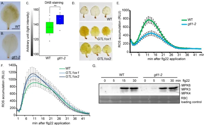
To characterise the enhanced susceptibility ofgtl1 mutants and the increased resistance of GTL1ox lines in more detail, the levels of the reactive oxygen species (ROS) H2O2was assessedby 3,3’-diaminobenzidine (DAB) staining in untreated WT plants,gtl1-2 mutant and GTL1ox
lines. We observed that the H2O2level ingtl1 is reduced in comparison to WT as depicted by a
weaker staining intensity (Fig 2A–2C). In contrast, two independentGTL1ox lines displayed
intense staining after the DAB exposure which demonstrates higher H2O2levels than WT (Fig
2D). These findings show that the basal H2O2level depends on the GTL1 function.
Further-more, ROS production and release are among the first defense reactions in response to patho-gen perception. The ROS burst after flg22 treatment was significantly reduced ingtl1 mutants
to approximately 50%, 15 min after application (Fig 2E,S2D Fig). However, theGTL1ox lines
showed elevated ROS release after flg22 treatment (Fig 2F). The differences in the ROS efflux after flg22 application ingtl1 and GTL1ox lines might be a direct consequence of the affected
basal H2O2levels. The activation of the flg22-triggered signaling cascade was evaluated by
pTpY antibody-based immunoprecipitation that targets the phosphorylated MAPK3, 4 and 6 versions. The highest activation of the three MAPK was achieved 15 min after flg22 treatment
Fig 2.gtl1 mutant shows reduced H2O2levels in leaves and is compromised in flg22-triggered ROS-burst. A-D) Evaluation ofin-situ H2O2
levels by 3,3’- diaminobenzidine staining (DAB) in untreatedgtl1-2 mutant, GTL1ox1 and GTL1ox2 compared to WT. Box plots are depicted for gtl1-2 and WT, boxes showing the interquartile range (IQR) 25stto 75thpercentiles, inner ellipse representing the median, whiskers show the SEM, outliers are depicted by dots (Min/Max range). Scale bar = 3 mm. Statistical significance was analyzed by Student’s test, asterisks indicate significant differences compared to WT,�p� 0.01. E-F) flg22-induced ROS burst assay of (E) gtl1-2 compared WT plants and (F) GTL1ox1 and GTL1ox2 compared to WT plants,1μM flg22 treatment over 45 min, the data are shown as means ± SE from 36 leaf discs (3 biological replicates)
of 5 week-old plants (negative controlS2D Fig). G) flg22-induced MAPKinase-activation assays ingtl1-2 and WT plants. 14 day-old seedlings
were treated with 1μM flg22 and samples were harvested at the indicated time points. Activation of MAPkinases 3, 4 and 6 were analyzed by immunoblot using pTpY-antibody recognizing the MAPKs in their activated form. Protein loading control was performed by Ponceau S staining for Rubisco (RBC).
in bothgtl1-2 and WT plants (Fig 2G). The comparable activation of MPK3, 4 and 6 suggests a function of GTL1 downstream of the flg22-induced MAP kinase signaling cascades.
Comparative transcriptome composition
Firstly, to identify biological processes and genes that are governed by GTL1, the transcrip-tome of 14 day-old plants ofgtl1-2 mutant and WT was analysed by performing RNA-seq. At a
stringency ofp�0.01, 1448 genes differently regulated genes (S1 Table) could be identified that show a log2fold change from -5.29 to -0.53 of negatively regulated genes and from 0.52 to
5.00 of positively regulated genes. Among these 1448 genes, 678 genes are up-regulated, and 770 genes are down-regulated (Fig 3A). The GO term analysis of down-regulated genes revealed gene functions for Innate Immune Response, Systemic Acquired Resistance and Response to biotic stimulus and suggests a reduced ability ofgtl1-2 in these processes (Fig 3B). Furthermore, we found genes being down-regulated ingtl1-2 that contribute to hydrogen
per-oxide metabolic process (S1 Table). For example,ATRBOHC/RHD2 [55] and a substantial number of peroxidases (S1 Table) that contribute to H2O2generation [56], such asPRXCB,
PER4, PRX37 and PRX25, are compromised in their expression. The GO terms in the set of
up-regulated genes emphasised gene functions in Nucleotide Biosynthesis Process, Ribosome Biogenesis and Response to Sucrose Stimulus and can be summarized in support of plant growth and development (Fig 3C,S1 Table). All in all, the GO analysis indicates GTL1 as a positive regulator of immunity-related processes and a suppressor of biological functions related to plant growth.
Secondly, we sought to investigate whether the transcriptome composition ofgtl1 and mpk4 is compromised in the same set of downstream targets. Therefore, deregulated genes in
thempk mutants [57] andgtl1 were analysed by hierarchical clustering with mutants of
MAPKs 3, 4 and 6 (Fig 3D). Interestingly,mpk3 and mpk6 mutants showed only a small
over-lap in gene expression withgtl1. However, a large number of genes in the gtl1 mutant showed
an opposite pattern of gene expression in thempk4 mutant. Among the two main clusters that
were identified in the comparison betweengtl1 and mpk4, the 123 genes in cluster I are
up-reg-ulated ingtl1-2 and down-regulated in mpk4 (Fig 3E,S2 Table). The GO term analysis high-lights gene functions in Response to Light, RNA Metabolism and Lipid Biosynthesis Process. Cluster II, comprising 319 genes, which are down-regulated ingtl1-2 and up-regulated in mpk4, displays assigned GO terms for Innate Immunity and Response to bacterium (Fig 3F,
S2 Table). The consensus matrix (Fig 3G,S2 Table) illustrates the dissimilarity of the gene sets in thegtl1 and mpk4 transcriptomes and also shows the difference to the transcriptomes of mpk3 and mpk6. These findings indicate a genetic interaction of GTL1 and MPK4 in the
regu-lation of distinct biological processes. To evaluate theRNAseq-based transcriptome
compari-son, the expression of three representative genes that contribute to SA-biosynthesis and response (S2E Fig) was analysed in thegtl1 and mpk4 mutant by qPCR. Firstly, CAM-BIND-ING PROTEIN 60-LIKE G (CBP60g) works cooperatively with SARD1 [58,59] to regulate the expression ofICS1 to induce SA-metabolism; secondly, PHYTOALEXIN DEFICIENT 3
(PAD3), that catalyses the conversion of dihydrocamalexic acid to camalexin [60,61] and lastly,ELICITOR-ACTIVATED GENE 3 (ELI3-2/CAD8) acting as alcohol:NADP+
oxidoreduc-tase [62]. The expression ofCBP60g, PAD3 and CAD8 is diminished in gtl1 when compared to
WT, but enhanced in thempk4 mutant (S3A–S3C Fig).
Salicylic acid metabolism and homeostasis are affected in
gtl1 and GTL1ox
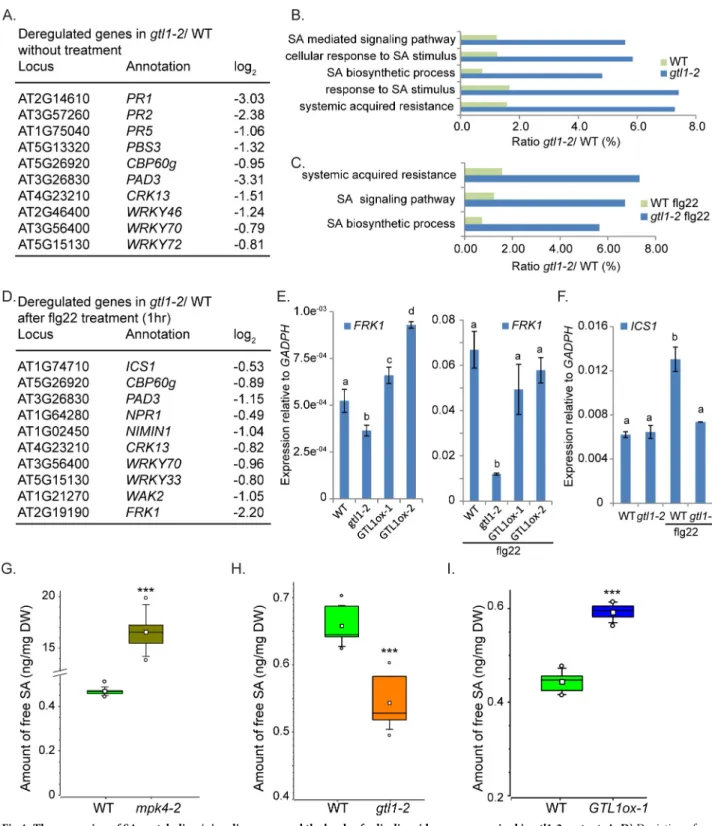
In addition toCBP60g, the RNAseq-based transcriptome analysis also revealed that a numbersignaling events (Fig 4A). Compared to WT, the transcriptome analysis ofgtl1 mutants before
and after PAMP application highlights in down-regulated genes GO categories for SA Biosyn-thesis, Systemic Acquired Resistance and Response to SA (Fig 4B and 4C). In untreated plants, the genes involved in SA biosynthesis, such asCBP60g, PBS3 and WRKY46 (Fig 4A) as well as SA signaling and PAMP-response targets, such asWRKY72, PR1 and FRK1 (Fig 4A and 4E) are down-regulated. To assess PAMP-triggered SA-metabolism and signaling ingtl1-2, the
transcriptome composition was analysed 1 hour after flg22-treatment (S3 Table). In this regard, the expression of the key-SA biosynthesis geneICS1, as well as its transcriptional
acti-vatorCBP60g, is diminished (Fig 4D and 4F). Furthermore, the expression of the genes con-sidered as central factors in SA signalingNPR1 and NIMIN1 is reduced (Fig 4D) as well as
those ofFRK1 (S3D Fig),WAK2 and PAD3 contributing to SA-mediated response (Fig 4D and 4E). If GTL1 acts as an activator of genes involved in SA metabolism and signaling, then the expression of these genes is expected to be predominantly elevated inGTL1ox lines
com-pared to thegtl1 mutant and WT, respectively. Indeed, the expression FRK1, CBP60g and PAD3 (Figs4E,5C and 5I) are significantly increased in theGTL1ox lines. These results
indi-cate that GTL1 functions as a positive regulator of SA-mediated processes.
Consequently, we determined the levels of free SA in WT,mpk4-2 and gtl1-2 mutant, and GTL1ox1 line in at least three biological replicates. Peterson et al, 2000 [31] showed that the SA accumulation inmpk4 mutants is up to 10 fold higher than WT and our measurement are in
accordance with these results (Fig 4G). After analysing 6 biological replicates, we determined a concentration of 16.56 ng SA /mg dry weight inmpk4 compared to 0.47 ng/mg in WT. The
high SA values inmpk4 indicate MPK4 as a repressor of SA accumulation. Remarkably, the
basal SA amount in thegtl1-2 mutant is consistently lower than WT levels (Fig 4H), while the basal SA concentration in theGTL1ox line is significantly increased compared to WT (Fig 4I). Taken together, these results indicate that GTL1 is a positive regulator of genes involved in SA biosynthesis and promotes basal SA accumulation.
GTL1 regulates genes involved in SA-metabolism and signaling
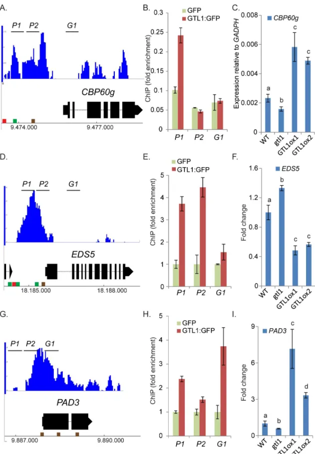
In genome-wide binding studies (ChIP-chip) [45], the association of GTL1 to regulatory sequences upstream and downstream of a large set of genes was revealed. A consensus binding motif for GTL1 was identified and described as GT3 box [5’-GGTAAA-3’]. In a previous in-vitro study, it was shown that the N-terminal DNA binding domain of GTL1 associates both to
GT1 and GT2 boxes [46]. The ChIP-chip approach was carried out by the usage of the whole aerial part of 12 day-oldgtl1-1 plants that were complemented by pGTL1::GTL1:GFP. Among
the total number of 2398 target genes, GTL1 was found to bind to the promoter regions of
CBP60g, EDS5 which codes for an SA-transporter [63] andPAD3 (Fig 5A, 5D and 5G) as indi-cated in the Integrated Genome Browser diagram. To evaluate the association of GTL1 to these direct target genes, we performed Chromatin-Immunoprecipitation (ChIP) followed by quantitative PCR (qPCR) using gene-specific primer sets (P1, P1, G1). 14 day-oldArabidopsis
seedlings expressingpUBI10::GTL1:GFP (S4A Fig) were generated and 3 independent trans-genic lines were selected and used to confirm the binding of GTL1 to selected chromatin
Fig 3. Comparative transcriptome analysis ofgtl1 and three immune MAPK mutants mpk3, -4 and -6. A) Histogram of the Log2distribution of up- and
down-regulated genes ingtl1-2 (S1 Table). B-C) Deregulated genes can be categorized in distinct Gene Ontology termsgtl1-2 (S1 Table).D) Hierarchical clustering ofgtl1-2, mpk3, mpk4 and mpk6 transcriptome highlights two main clusters showing opposite deregulated gene expression in gtl1-2 and mpk4.
log2fold (p�0.01,gtl1-2) of individual genes was used for clustering by using the average linkage method and Pearson Correlation (MeV4.0) (S2 Table).E)
Centroid graph (red) and individual expression graphs dedicated to Cluster I. GO term analyses of genes grouped in Cluster I.F) Centroid graph (red) and individual expression graphs dedicated to Cluster II. GO term analyses of genes grouped in Cluster II.G) Consensus matric (Non-Negative Matrix Factorization, Cluster samples) shows most divergent gene expression between the total set of deregulated genes ingtl1-2 and mpk4.
Fig 4. The expression of SA-metabolism/ signaling genes and the levels of salicylic acid are compromised ingtl1-2 mutant. A, D) Depiction of
down-regulated genes (A) ingtl1-2 contributing to SA-biosynthesis and signaling without treatment (S1 Table) and (D) after flg22 treatment (S3 Table) compared to the respective expression in WT. B, C) Significantly down-regulated genes (p�0.01, 769 genes) ingtl1-2 before and after flg22
treatment (p�0.0001, 715 genes) can be grouped in GO terms describing gene functions for SA-signaling and metabolism (S2andS3Tables).E-F)
Expression of SA/PTI-response geneFRK1 (E,S3D Fig) is reduced ingtl1-2 before and after flg22 treatment but shows elevated expression in the GTL1
overexpression lines in untreated conditions. The expression of the SA-biosynthesis geneICS1 (F) is diminished in gtl1-2 after flg22 treatment.
Statistical significance was analyzed by Student’s test Error bars, mean± SEM, letters above bars represent significance groups, p� 0.01.G-I) Quantitative analysis of free salicylic acid by using LC-MS/MS. Box plots are depicted for (G)mpk4-2, (H) gtl1-2 and (I) GTL1ox1 compared to WT.
regions. To discriminate against false-positive binding caused by GFP, a negative control expressing GFP under theUBI10 promoter was employed (S4A Fig). By ChIP-qPCR, the bind-ing preference of GTL1 to the promoter region ofCBP60g close to the transcriptional start
sequence (TSS) could be confirmed (Fig 5B). Intriguingly, in our ChiP-qPCR study, GTL1 binds predominantly to the region-790 bp to 707 bp upstream of the TSS that contains one
GT2 motif previously described as the binding motif of GT transcription factors [43]. Accord-ingly, the binding of GTL1 to the region upstream of the TSS ofEDS5 could be confirmed to
the 5’region P1 and P2 (Fig 5D and 5E) [45]. Upstream of the TSS ofEDS5 several GT boxes
can be found facilitating the binding of GTL1. Moreover, GTL1 binds to the promoter region ofPAD3 upstream of the TSS and as well in the 5’-ORF (Fig 5G and 5H). Notably, several
GT-boxes (GT1) dedicated to GTL1 binding can be found indicating specific binding of GTL1 to
thePAD3 genomic region. Taken together, in accordance with the genome-wide binding
stud-ies of Breuer et al, 2012 and the presented ChIP evaluations,CBP60g, EDS5 and PAD3 could
be confirmed as direct downstream targets of GTL1. To find out whether GTL1 exerts tran-scriptional control onCBP60g, EDS5 and PAD3, we analysed their expression in the gtl1
mutant and theGTL1ox lines. On the one hand, the expression of CBP60g and PAD3 is
reduced in thegtl1 mutant and elevated in the GTL1ox lines (Fig 5C and 5I) under untreated conditions which suggest GTL1 as a transcriptional activator of these genes under non-stress conditions. After flg22-treatment, the expression ofCBP60g and PAD3 is also reduced in the gtl1 mutant, but in the GTL1ox lines, the expression is ambiguous for CBP60g and WT-like in
the case ofPAD3 (S4B and S4D Fig). On the other hand, the expression ofEDS5 is elevated in gtl1 and reduced in the GTL1ox lines (Fig 5F) which implies GTL1 as a repressor ofEDS5
expression. After flg22-treatment, the expression ofEDS5 is not broadly perturbed from WT
(S4C Fig). Our results suggest GTL1 as a transcriptional regulator of these genes involved in SA-biosynthesis, transport and response.
GTL1 regulates immunity against the bacterial effectors AvrRpm1 and
AvrRpt2
Since MPK4 is also involved in effector-triggered immunity [37], we tested the bacterial strains
Pst DC3000_AvrRpm1 and PstDC3000_AvrRpt2 which upon injection of the bacterial effectors
trigger RIN4-dependent ETI [5]. Two hours after spray infection, the growth level of the bacte-ria in the transgenic lines was indistinguishable from WT (Fig 6A and 6B). However 72 hours after infection, we observed enhanced bacterial growth of about one fold change in eitherPst DC3000 strains in the allelic gtl1 mutants (Fig 6A and 6B), while theGTL1ox lines are not
broadly perturbed in their immunity compared to WT, respectively (S4E and S4F Fig). These results show thatgtl1 mutants are compromised in RIN4-AvrRpm1/AvrRpt2 induced
immu-nity which indicates a function of GTL1 in the effector-triggered immuimmu-nity. To pinpoint whether GTL1 contribute to SA accumulation after bacterial infection, we determined the free SA levels by LC-MS/MS analysis 24 hours afterPst DC3000_AvrRpm1 infection in the gtl1
mutant. We found that the SA accumulation is significantly reduced to 3.5 ng/mg compared to WT showing an SA level of about 4.7 ng/mg in the average of 4 biological replicates, respec-tively (Fig 6C). Our results show that GTL1 is necessary for the SA accumulation as part of the ETI.
depicted by dots (Min/Max range). Statistical significance was analyzed by Student’s test. Asterisks indicate significant differences compared to WT,��� p� 0.001.
Fig 5. The SA-biosynthesis activatorCBP60g, the SA-transporter EDS5 and the SA-responsive gene PAD3 are bound and
gtl1 partially restores susceptibility of mpk4 and growth defects
Previously, it was reported that the strong autoimmune phenotype inmpk4-3 largely depends
on SUMM2 [34]. However, the accumulation of H2O2andPR gene expression are only to
some extent diminished in thempk4/summ2 double mutant and show still significant
enhancement compared to WT [34]. Furthermore, the severe dwarfism of thempk4 mutant is
not fully restored by the introduction of different allelicsumm2 mutations [34]. Eventually, these previous results indicate that MPK4 is involved in immune and growth regulation inde-pendently of SUMM2 [34]. To test whether defense response to bacterial attack in GTL1 depends on MPK4, we generated and analysed thempk4-2/gtl1-2 double mutant. As depicted
inFig 7A and 7B, the fresh weight of the double mutant is increased by about 14% of 4 week-old plants and 34% of 7 week-week-old plants (S4G Fig) compared tompk4-2 single mutants,
respectively. Furthermore, the trichome branch length is extended in the double mutant com-pared tompk4 single mutant suggesting a partial suppression of developmental defects in mpk4 plants by the gtl1 mutation (Fig 7C and 7D). To evaluate the genetic interaction of
the 5’-region ofCBP60g (A), EDS5 (D) and PAD3 (G). Brown cube refer to the GT1-box, green cube indicates GT2-box and red cube
shows GT3-box. B, E, F) ChIP-qPCR by using three biological replicates ofpUBI10::GTL1:GFP expressing plants. GTL1 binding to
genomic regions close toCBP60g (B), EDS5 (E) and PAD3 (H) were tested with sets of three primer pairs (P1, P2, G1) for each locus.
Y-axis shows the fold enrichment in thepUBI10::GTL1:GFP lines normalized to GFP immunoprecipitation, driven by the pUBI10
promoter (0.64 kb). C, F, I) The expression ofCBP60g (C) and PAD3 (I) is diminished in gtl1-2 and elevated in GTL1ox1 and GTL1ox2. The expression of EDS5 is elevated in gtl1-2 and reduced in GTL1ox1 and GTL1ox2. Asterisks indicate significant
differences compared to WT,�p� 0.05.��p� 0.01. Statistical significance was analyzed by Student’s test. Letters above bars represent
significance groups,p� 0.05.
https://doi.org/10.1371/journal.pgen.1007708.g005
Fig 6.gtl1 mutants are compromised in resistance to avirulent PstDC3000 strains and accumulate less SA after
PstDC3000_avrRPM1 infection. A-B) The allelic GTL1 mutants gtl1-2 and gtl1-5 were challenged by the use of PstDC3000_avrRPM1 and PstDC3000_avrRPT2. Results of two GTL1-overexpressing lines (GTL1ox1, GTL1ox2) are shown in SFig 4E and 4F. Plants, of three biological replicates (n = 30), were spray-inoculated with a bacterial suspension at OD6000.2, the density of colony-forming units
(cfu) was analyzed 2 and 72 hours post inoculation (hpi). Error bars, mean± SEM, statistical significance was analyzed by Student’s test, asterisks indicate significant differences compared to treated WT,�p� 0.05��p� 0.01,���p� 0.001. C) Quantitative analysis of
free salicylic acid by using LC-MS/MS. Box plots are depicted forgtl1-2 and WT, 24 hrs after PstDC3000_avrRPM1 spray-inoculation,
untreated conditions are shown inFig 4H. Boxes showing the interquartile range (IQR) 25stto 75thpercentiles, inner square
representing the median, whiskers show the SEM, outliers are depicted by dots (Min/Max range). Statistical significance was analyzed by Student’s test. Asterisks indicate significant differences compared to WT,��p� 0.01.
MPK4 with GTL1 in the effector-triggered immunity, the double mutant was treated byPst DC3000 AvrRPM1. At two hours after infection, the bacterial titer was indistinguishable from
WT ingtl1, mpk4 single mutants, and mpk4/gtl1 double mutant thereby suggesting that
stoma-tal immunity is not perturbed (Fig 7E). However, after 72 hours, the proliferation level in the
mpk4 single mutant was significantly reduced compared to WT, while the bacterial titer was
elevated inmpk4/gtl1 compared to mpk4 single mutants (Fig 7E). Based on our findings, we postulate that MPK4 functions as a negative regulator of GTL1 inAvrRPM1-triggered
RIN4-mediated immunity.
Discussion
Role of GTL1 in basal resistance and PTI
In this study, we identified the trihelix transcription factor GTL1 as a regulator of immunity. Using pathogen assays with virulentPst DC3000, Pst DC3000 ΔavrPto/avrPtoB and
non-viru-lentPst DC3000 hrcC- strains, we showed that GTL1 is a positive regulator of basal defense and
PTI, respectively. Transcriptome analysis suggested that GTL1 functions on similar targets as the MPK4 pathway. However, whereas MPK4 negatively regulates the overlapping set of tar-gets contributing to defense and immunity, GTL1 regulates them in a positive manner. In this context, thempk4 mutant exhibits enhanced resistance to Pst DC3000 and elevated
expres-sion of defense markers [31]. Since PAMP-triggered MAPK activation is not affected ingtl1
mutants, it is likely that GTL1 functions downstream of the MEKK1-MKK1/2-MPK4 cascade. This hypothesis is supported by the fact that GTL1 forms part of the MPK4 protein complex. We also found that GTL1 can directly interact with MPK4 and that this interaction is specific as no interaction was detected with the related immune MAPKs MPK3 and MPK6. Interest-ingly, we could not detect phosphorylation of GTL1 by MPK4, suggesting a regulation mecha-nism that relies on protein-protein interaction rather than phosphorylation.
Fig 7. Mutation inGTL1 partially restores the mpk4 growth and resistance phenotype. A-B) Overview of the phenotype (A) and
shoot fresh weight (B) of WT,gtl1-2, mpk4-2 and mpk4/gtl1 double mutants. The shoot fresh weight was analyzed of 4 week-old
plants in 5 biological replicates and 7 week-old plants (S4G Fig). Error bars, mean± SEM, statistical significance was analyzed by Student’s test. Letters above bars represent significance groups,p� 0.005. Scale bar = 1cm. C-D) Bright-field microscopy of WT, gtl1, mpk4 and mpk4/gtl1 leaves/plants. Scale bar = 500 μm. Error bars, mean ± SEM, statistical significance was analyzed by Student’s test.
Letters above bars represent significance groups,p� 0.01. D) Quantitative analysis of trichome branch length of WT, gtl1, mpk4 and mpk4/gtl1 plants. Trichomes of 4 week-old plants were measured by the use of ZEN lite 2012 software. Boxes showing the
interquartile range (IQR) 25stto 75thpercentiles, inner square representing the median, whiskers show the SEM, Statistical significance was analyzed by Student’s test. Letters above boxes represent significance groups,p� 0.01. E) WT, gtl1-2, mpk4-2 and mpk4/gtl1 mutant were treated with PstDC3000 avrRPM1 Plants, of three biological replicates (n = 30), were spray-inoculated with a
bacterial suspension at OD6000.2, the density of colony-forming units (cfu) was analyzed 2 and 72 hours post inoculation (hpi). Error
bars, mean± SEM, statistical significance was analyzed by Student’s test. Letters above bars represent significance groups, p� 0.01. https://doi.org/10.1371/journal.pgen.1007708.g007
GTL1 is a positive regulator of SA biosynthesis and signaling but negatively
regulates growth
The transcriptome pattern ofgtl1 mutants revealed that GTL1 is a positive factor for defense
gene expression but a negative regulator of genes involved in growth, supporting the concept that growth and defense are inversely coupled. In agreement with a role of GTL1 in suppress-ing growth,gtl1 mutants are also slightly bigger than WT plants under water-deficiency [46], but as shown here, this feature comes with the caveat of being more susceptible to pathogen attack. Conversely,mpk4 mutant plants are dwarfed but are incredibly pathogen resistant. A
characteristic feature ofmpk4 mutants is the increased SA level that correlates with its
enhanced resistance to the virulent strains ofPseudomonas syringae [31]. Interestingly, SA amounts ingtl1-2 mutant and lines expressing a constitutively active MPK4 version [37] were consistently lower than WT suggesting an opposing regulation of SA homeostasis. Sincegtl1
has reduced whereasmpk4 massively, enhanced SA levels, one might be tempted to conclude
that these different sensitivities could be solely due to SA amounts. However, this assumption is probably too simple as suppression of SA levels inmpk4 mutants could only relieve the
dwarf phenotype to some extent [31].
Transcriptome analysis confirmed a role of GTL1 as a positive regulator of SA and defense as well, by showing reduced levels of the SA/PAMP-marker genes, such asPR1 and FRK1. This
effect seems to be mediated both at the level of SA signaling genes, exemplified byNPR1 and NIMIN1, as well as at the level of the biosynthesis gene ICS1/SID2 and its regulator CBP60g.
The analysis of available ChIP-chip data [45] indicated that GTL1 binds to a number of its tar-get genes via interaction of the GT boxes. By ChIP-qPCR, we could verify that GT elements are involved in the regulation of theCBP60g, EDS5 and PAD3 genes by GTL1. CBP60g binds
to and promotes the expression of the SA-biosynthesis geneICS1 and the SA-signaling NPR1
[59]. Furthermore, WRKY33 and MKS1 are two downstream target proteins of flg22-activated MPK4 that mutually regulate the expression ofPAD3 encoding an enzyme required for
syn-thesis of antimicrobial camalexin [61,64]. Recently, it was shown that the MEKK1-MKK1/ 2-MPK4 cascade is guarded by the NB-LRR gene SUMM2 and that the guardee of this system is CRCK3 which directly interacts with SUMM2 [32]. The double mutant ofmpk4/summ2 is
to some extent suppressed in thempk4 autoimmune phenotype and partially restored in
growth. Eventually, these data explain the severempk4 mutant phenotypes and suggest that
MPK4 acts actually as a positive regulator of defense. However, the only partial suppression indicates that MPK4 is involved in immune and growth regulation independently of SUMM2. The up-regulation ofPAD3 is unaffected in the summ2 mutant after flg22-application [34]. Unlike thegtl1-2 mutant, the compromised up-regulation of PAD3 after flg22-treatment refers
directly to the cooperation of MPK4 and GTL1 in a SUMM2 independent manner. However, MPK4 also negatively regulates defense genes as evidenced by the fact that expression of a constitutively active version of MPK4 results in pathogen hypersensitivity [37]. Moreover, a negative role of MPK4 in defense gene expression is also provided by the work on the transcriptional repressor ASR3, whereby PAMP-induced MPK4 phosphorylation of ASR3 was shown to enhance its DNA binding and repression of a considerable number of defense target genes [36]. ASR3 acts as a transcriptional repressor through its EAR motif and displays oppositeFRK1 regulation as GTL1. Interestingly, ASR3 is also a member of the plant-specific
trihelix transcription factor family but belongs to an SH4 clade. Theasr3 mutant shows, unlike gtl1, an enhanced resistance against virulent bacterial strains. By contrast, the susceptibility of asr3 to infection by the avirulent strain PstDC3000 avrRpt2 matches WT plants. Pathogen
resistance toPstDC3000 AvrRpt2 and PstDC3000 AvrRpm1 is triggered upon perception by
PstDC3000 strains demonstrates that both trihelix TF family members do not act redundantly
and exert distinct and opposite biological functions in immunity.
Interestingly, GTL1 was recently also shown to alter drought tolerance ofArabidopsis [46], and the underlying mechanism was suggested to be due to the altered, reduced number of sto-mates ingtl1 plants making them more robust under drought conditions. GTL1 is assumed to
monitor the water status in plants to determine the most appropriate number of stomates dur-ing plant development. This effect was shown to be exerted through the repression of the
SDD1 gene as a direct target of GTL1.
In summary, the current data suggest that fine-tuning of GTL1 activity plays an important role in defining the balance between growth, defense and developmental adaptations to biotic and abiotic stress conditions. Given its involvement and role in these processes, further studies are warranted into the regulation of GTL1 at the post-translational level.
GTL1 contribute to RIN4-mediated Effector-triggered immunity
RPM1-INTERACTING PROTEIN 4 (RIN4) interacts with AvrRpm1 andPseudomonas syrin-gae pv maculicola 1 (RPM1) [5,65], whereby the association of AvrRpm1 provokes the phos-phorylation of RIN4 by RIN4-interacting receptor-like protein kinase (RIPK) [13] enhancing its activity as a negative regulator of plant defense. However, phosphorylated RIN4 induces the activation of the R-protein RPM1 triggering the RPM1-dependent defense response [13]. MPK4 is a crucial regulator of defense against virulent pathogens and PTI, but the protein kinase is also implicated in ETI regulation [37]. Consistent with a role ofGTL1 in ETI,
resis-tance ofgtl1 plants infected with the avirulent Pst AvrRpm1 and Pst AvrRpt2 strain was
com-promised in thegtl1 background, indicating that GTL1 is a positive regulator of RPM1/
RPT2-mediated ETI. Interestingly,mpk4 mutants complemented by the constitutively active
MPK4 (CA-MPK4) exhibit distinct responses to different avirulentPseudomonas strains.
CA-MPK4 lines are affected in pathogen resistance mediated by TIR-NB-LRR, but not CC-NB-LRR, receptors. In this regard, CA-MPK4 lines retained WT-like resistance toPst DC3000 AvrRpm1 recognised by CC-NB-LRR receptors, whereas we showed that the mpk4
mutant is more resistance, while thempk4/gtl1 mutant partially restored susceptibility.
There-fore, we postulate that MPK4 functions as a negative regulator of GTL1 inAvrRpm1 -triggered
RIN4-mediated immunity. In summary, we reason that GTL1 is embedded in the MPK4 path-way and coordinates SA-metabolism and homeostasis which directly impacts basal immunity, PAMP- and effector-triggered immunity.
Materials and methods
Plant material and growth conditions
Experiments were performed by the usage ofArabidopsis thaliana of the Columbia accession
grown on soil in plant growth chambers (Percival Scientific) under short-day conditions (8h light/ 16 h dark) at 22˚C.Nicotiana benthamiana were grown under long-day conditions (16 h
light + 8 h darkness) at 28˚C.gtl1-2 (Salk_005965), gtl1-5 (Salk_044308) and mpk4-2 (Salk_056245) seeds were obtained from NASC.
Accession numbers
GTL1 (AT1G33240), MPK4 (AT4G01370).
Additional Materials and Methods
Supporting information
S1 Fig. Domain map of GTL1 andin-vitro kinase assay. A) Schematic representation of
GTL1; NTH-Myb-like, N-terminal trihelix domain; CC, Coiled coil domain; CTH-Myb-like, C-terminal trihelix domain; red bar, putative MAPK interaction domain.
B) Inin-vitro kinase assays followed by LC/MS-MS, MPK4 does not phosphorylate GTL1 at
the previously reported phosphopeptide nor at another site. (TIF)
S2 Fig. Phenotypical and functional characterization ofgtl1 and GTL1ox lines. A) Overview
of the phenotype and shoot-fresh weight of WT,gtl1-2, gtl1-5, GTL1ox1 and GTL1ox2 plants.
The shoot fresh weight was analyzed of 2 week-old plants in 3 biological replicates. Error bars, mean± SEM, statistical significance was analyzed by Student’s test; n.s, non-significant against WT. Scale bar = 1cm.
B) The allelicGTL1 mutants gtl1-2 and gtl1-5 were challenged by leaf infiltration with PstDC3000. Plants, of three biological replicates, were leaf-infiltrated with a bacterial
suspen-sion at OD6000.005, the density of colony-forming units (cfu) was analyzed 2 and 72 hours
post inoculation (hpi). Error bars, mean± SEM, statistical significance was analyzed by Stu-dent’s test, asterisks indicate significant differences compared to treated WT,�p � 0.05,��
p � 0.01,���p � 0.001.
C) Pathogen-treatment ofGTL1ox1 and GTL1ox2 lines refers toFig 1F. D) Negative control for ROS-burst assay (Fig 2E)
E) Expression ofPAD3 and CAD8 after SA application. 14 day-old WT seedlings were treated
with 1μM SA for 6 hours. Error bars, mean ± SEM, Asterisks indicate significant differences compared to untreated WT,�p � 0.05,���p � 0.001.
(TIF)
S3 Fig. Expression ofCBP60g, CAD8, PAD3 and FRK1. A-B) Expression of CBP60g (A),
PAD3 (B) and CAD8 (C) in gtl1-2 and mpk4.
D) Expression ofFRK1 after SA-treatment. 14 day-old WT seedlings were treated with 1μM
SA for 6 hours. Error bars, mean± SEM, statistical significance was analyzed by Student’s test, letters above bars represent significance groups,p � 0.01.
(TIF)
S4 Fig. Expression ofCBP60g, EDS5 and PAD3 and further pathogen-treatments of GTL1ox lines. A) GTL1_GFP fusion protein and single GFP in Arabidopsis root of
indepen-dent transgenic lines used for ChIP.
B-D) Expression ofCBP60g (B) and EDS5 (C) and PAD3 (D) in gtl1-2, GTL1ox1 and GTL1ox2
after flg22 application (1μM, flg22 for 1hr). Statistical significance was analyzed by Student’s test. Letters above bars represent significance groups,p � 0.05; n.s, non-significant.
E-F) Pathogen-treatment ofGTL1ox1 and GTL1ox2, refers toFig 6A and 6B.
G) WT,gtl1-2, mpk4-2 and mpk4/gtl1 double mutant. The shoot fresh weight was analyzed of
7 week-old plants in 3 biological replicates. Error bars, mean± SEM, statistical significance was analyzed by Student’s test. Letters above bars represent significance groups,p� 0.001.
(TIF)
S1 Table. Transcriptome composition ofgtl1-2 /WT of 14 day-old seedlings. GO terms
of up- and down-regulated genes. Isolation of GTL1 in the in vivo MPK4-Tandem Affinity Purification approach combined with an LC-MS/MS analysis before and after flg22 applica-tion.
S2 Table. Genes dedicated to cluster I and II, GO term analysis, Matrix-cluster.
(XLSX)
S3 Table. Transcriptome composition ofgtl1-2 /WT of 14 day-old seedlings, 1 hr after
flg22-treatment,p�0.0001, Go term analysis of down-regulated genes.
(XLSX)
S1 Materials and Methods.
(DOCX)
Acknowledgments
BiFC-MAPK constructs were kindly provided by J. Colcombet. We thank Stefan Arold for making GST-tagged MAPK constructs available. We are grateful to Yunhe Jiang and Arsheed Sheikh for technical support.
Author Contributions
Conceptualization: Ronny Vo¨lz, Heribert Hirt. Data curation: Ronny Vo¨lz.
Formal analysis: Ronny Vo¨lz, Soon-Kap Kim, Kiruthiga G. Mariappan, Xiujie Guo, Jean
Bigeard, Naganand Rayapuram.
Funding acquisition: Heribert Hirt.
Investigation: Jianing Mi, Santiago Alejandro, Delphine Pflieger.
Methodology: Ronny Vo¨lz, Jianing Mi, Santiago Alejandro, Delphine Pflieger.
Validation: Salim Al-Babili, Heribert Hirt. Writing – original draft: Ronny Vo¨lz.
Writing – review & editing: Ronny Vo¨lz, Heribert Hirt.
References
1. Tsuda K. and Katagiri F., Comparing signaling mechanisms engaged in pattern-triggered and effector-triggered immunity. Curr Opin Plant Biol, 2010. 13(4): p. 459–65.https://doi.org/10.1016/j.pbi.2010.04. 006PMID:20471306
2. Nakagami H., Pitzschke A., and Hirt H., Emerging MAP kinase pathways in plant stress signalling. Trends Plant Sci, 2005. 10(7): p. 339–46.https://doi.org/10.1016/j.tplants.2005.05.009PMID: 15953753
3. Chen H., et al., A Bacterial Type III Effector Targets the Master Regulator of Salicylic Acid Signaling, NPR1, to Subvert Plant Immunity. Cell Host Microbe, 2017. 22(6): p. 777–788 e7.https://doi.org/10. 1016/j.chom.2017.10.019PMID:29174403
4. Eschen-Lippold L., et al., Bacterial AvrRpt2-Like Cysteine Proteases Block Activation of the Arabidopsis Mitogen-Activated Protein Kinases, MPK4 and MPK11. Plant Physiol, 2016. 171(3): p. 2223–38. https://doi.org/10.1104/pp.16.00336PMID:27208280
5. Mackey D., et al., RIN4 interacts with Pseudomonas syringae type III effector molecules and is required for RPM1-mediated resistance in Arabidopsis. Cell, 2002. 108(6): p. 743–54. PMID:11955429
6. Belkhadir Y., et al., Arabidopsis RIN4 negatively regulates disease resistance mediated by RPS2 and RPM1 downstream or independent of the NDR1 signal modulator and is not required for the virulence functions of bacterial type III effectors AvrRpt2 or AvrRpm1. Plant Cell, 2004. 16(10): p. 2822–35. https://doi.org/10.1105/tpc.104.024117PMID:15361584
7. Wu L., et al., Go in for the kill: How plants deploy effector-triggered immunity to combat pathogens.
8. Bisgrove S.R., et al., A disease resistance gene in Arabidopsis with specificity for two different pathogen avirulence genes. Plant Cell, 1994. 6(7): p. 927–33.https://doi.org/10.1105/tpc.6.7.927PMID: 8069104
9. Grant M.R., et al., Structure of the Arabidopsis RPM1 gene enabling dual specificity disease resistance. Science, 1995. 269(5225): p. 843–6. PMID:7638602
10. Boyes D.C., Nam J., and Dangl J.L., The Arabidopsis thaliana RPM1 disease resistance gene product is a peripheral plasma membrane protein that is degraded coincident with the hypersensitive response. Proc Natl Acad Sci U S A, 1998. 95(26): p. 15849–54. PMID:9861059
11. Kim M.G., et al., Two Pseudomonas syringae type III effectors inhibit RIN4-regulated basal defense in Arabidopsis. Cell, 2005. 121(5): p. 749–59.https://doi.org/10.1016/j.cell.2005.03.025PMID:15935761
12. Deslandes L. and Rivas S., Catch me if you can: bacterial effectors and plant targets. Trends Plant Sci, 2012. 17(11): p. 644–55.https://doi.org/10.1016/j.tplants.2012.06.011PMID:22796464
13. Liu J., et al., A receptor-like cytoplasmic kinase phosphorylates the host target RIN4, leading to the acti-vation of a plant innate immune receptor. Cell Host Microbe, 2011. 9(2): p. 137–46.https://doi.org/10. 1016/j.chom.2011.01.010PMID:21320696
14. Chung E.H., et al., Specific threonine phosphorylation of a host target by two unrelated type III effectors activates a host innate immune receptor in plants. Cell Host Microbe, 2011. 9(2): p. 125–36.https://doi. org/10.1016/j.chom.2011.01.009PMID:21320695
15. Mackey D., et al., Arabidopsis RIN4 is a target of the type III virulence effector AvrRpt2 and modulates RPS2-mediated resistance. Cell, 2003. 112(3): p. 379–89. PMID:12581527
16. Kim H.S., et al., The Pseudomonas syringae effector AvrRpt2 cleaves its C-terminally acylated target, RIN4, from Arabidopsis membranes to block RPM1 activation. Proc Natl Acad Sci U S A, 2005. 102 (18): p. 6496–501.https://doi.org/10.1073/pnas.0500792102PMID:15845764
17. Dempsey D.A. and Klessig D.F., Salicylic acid, active oxygen species and systemic acquired resistance in plants. Trends Cell Biol, 1994. 4(9): p. 334–8. PMID:14731471
18. Durrant W.E. and Dong X., Systemic acquired resistance. Annu Rev Phytopathol, 2004. 42: p. 185– 209.https://doi.org/10.1146/annurev.phyto.42.040803.140421PMID:15283665
19. Bigeard J., Colcombet J., and Hirt H., Signaling mechanisms in pattern-triggered immunity (PTI). Mol Plant, 2015. 8(4): p. 521–39.https://doi.org/10.1016/j.molp.2014.12.022PMID:25744358
20. Meng X. and Zhang S., MAPK cascades in plant disease resistance signaling. Annu Rev Phytopathol, 2013. 51: p. 245–66.https://doi.org/10.1146/annurev-phyto-082712-102314PMID:23663002
21. Nitta Y., Ding P., and Zhang Y., Identification of additional MAP kinases activated upon PAMP treat-ment. Plant Signal Behav, 2014. 9(11): p. e976155.https://doi.org/10.4161/15592324.2014.976155 PMID:25482788
22. Frei dit Frey N., et al., Functional analysis of Arabidopsis immune-related MAPKs uncovers a role for MPK3 as negative regulator of inducible defences. Genome Biol, 2014. 15(6): p. R87.https://doi.org/ 10.1186/gb-2014-15-6-r87PMID:24980080
23. Asai T., et al., MAP kinase signalling cascade in Arabidopsis innate immunity. Nature, 2002. 415 (6875): p. 977–83.https://doi.org/10.1038/415977aPMID:11875555
24. Ichimura K., et al., MEKK1 is required for MPK4 activation and regulates tissue-specific and tempera-ture-dependent cell death in Arabidopsis. J Biol Chem, 2006. 281(48): p. 36969–76.https://doi.org/10. 1074/jbc.M605319200PMID:17023433
25. Nakagami H., et al., A Mitogen-activated protein kinase kinase kinase mediates reactive oxygen spe-cies homeostasis in Arabidopsis. J Biol Chem, 2006. 281(50): p. 38697–704.https://doi.org/10.1074/ jbc.M605293200PMID:17043356
26. Gao M., et al., MEKK1, MKK1/MKK2 and MPK4 function together in a mitogen-activated protein kinase cascade to regulate innate immunity in plants. Cell Res, 2008. 18(12): p. 1190–8.https://doi.org/10. 1038/cr.2008.300PMID:18982020
27. Qiu J.L., et al., Arabidopsis mitogen-activated protein kinase kinases MKK1 and MKK2 have overlap-ping functions in defense signaling mediated by MEKK1, MPK4, and MKS1. Plant Physiol, 2008. 148 (1): p. 212–22.https://doi.org/10.1104/pp.108.120006PMID:18599650
28. Liu Y. and Zhang S., Phosphorylation of 1-aminocyclopropane-1-carboxylic acid synthase by MPK6, a stress-responsive mitogen-activated protein kinase, induces ethylene biosynthesis in Arabidopsis. Plant Cell, 2004. 16(12): p. 3386–99.https://doi.org/10.1105/tpc.104.026609PMID:15539472
29. Ren D., et al., A fungal-responsive MAPK cascade regulates phytoalexin biosynthesis in Arabidopsis. Proc Natl Acad Sci U S A, 2008. 105(14): p. 5638–43.https://doi.org/10.1073/pnas.0711301105PMID: 18378893
30. Xu J., et al., Pathogen-Responsive MPK3 and MPK6 Reprogram the Biosynthesis of Indole Glucosino-lates and Their Derivatives in Arabidopsis Immunity. Plant Cell, 2016. 28(5): p. 1144–62.https://doi. org/10.1105/tpc.15.00871PMID:27081184
31. Petersen M., et al., Arabidopsis map kinase 4 negatively regulates systemic acquired resistance. Cell, 2000. 103(7): p. 1111–20. PMID:11163186
32. Zhang Z., et al., The NLR protein SUMM2 senses the disruption of an immune signaling MAP kinase cascade via CRCK3. EMBO Rep, 2017. 18(2): p. 292–302.https://doi.org/10.15252/embr.201642704 PMID:27986791
33. Roux M.E., et al., The mRNA decay factor PAT1 functions in a pathway including MAP kinase 4 and immune receptor SUMM2. EMBO J, 2015. 34(5): p. 593–608.https://doi.org/10.15252/embj. 201488645PMID:25603932
34. Zhang Z., et al., Disruption of PAMP-induced MAP kinase cascade by a Pseudomonas syringae effec-tor activates plant immunity mediated by the NB-LRR protein SUMM2. Cell Host Microbe, 2012. 11(3): p. 253–63.https://doi.org/10.1016/j.chom.2012.01.015PMID:22423965
35. Kong Q., et al., The MEKK1-MKK1/MKK2-MPK4 kinase cascade negatively regulates immunity medi-ated by a mitogen-activmedi-ated protein kinase kinase kinase in Arabidopsis. Plant Cell, 2012. 24(5): p. 2225–36.https://doi.org/10.1105/tpc.112.097253PMID:22643122
36. Li B., et al., Phosphorylation of trihelix transcriptional repressor ASR3 by MAP KINASE4 negatively reg-ulates Arabidopsis immunity. Plant Cell, 2015. 27(3): p. 839–56.https://doi.org/10.1105/tpc.114. 134809PMID:25770109
37. Berriri S., et al., Constitutively active mitogen-activated protein kinase versions reveal functions of Ara-bidopsis MPK4 in pathogen defense signaling. Plant Cell, 2012. 24(10): p. 4281–93.https://doi.org/10. 1105/tpc.112.101253PMID:23115249
38. Smalle J., et al., The trihelix DNA-binding motif in higher plants is not restricted to the transcription fac-tors GT-1 and GT-2. Proc Natl Acad Sci U S A, 1998. 95(6): p. 3318–22. PMID:9501260
39. Kaplan-Levy R.N., et al., The trihelix family of transcription factors—light, stress and development. Trends Plant Sci, 2012. 17(3): p. 163–71.https://doi.org/10.1016/j.tplants.2011.12.002PMID: 22236699
40. Nagano Y., et al., Trihelix DNA-binding protein with specificities for two distinct cis-elements: both important for light down-regulated and dark-inducible gene expression in higher plants. J Biol Chem, 2001. 276(25): p. 22238–43.https://doi.org/10.1074/jbc.M102474200PMID:11301338
41. Dehesh K., et al., GT-2: a transcription factor with twin autonomous DNA-binding domains of closely related but different target sequence specificity. EMBO J, 1992. 11(11): p. 4131–44. PMID:1396594
42. Zhou D.X., Regulatory mechanism of plant gene transcription by GT-elements and GT-factors. Trends Plant Sci, 1999. 4(6): p. 210–214. PMID:10366876
43. Ni M., et al., GT-2: in vivo transcriptional activation activity and definition of novel twin DNA binding domains with reciprocal target sequence selectivity. Plant Cell, 1996. 8(6): p. 1041–59.https://doi.org/ 10.1105/tpc.8.6.1041PMID:8672890
44. Breuer C., et al., The trihelix transcription factor GTL1 regulates ploidy-dependent cell growth in the Ara-bidopsis trichome. Plant Cell, 2009. 21(8): p. 2307–22.https://doi.org/10.1105/tpc.109.068387PMID: 19717615
45. Breuer C., et al., Transcriptional repression of the APC/C activator CCS52A1 promotes active termina-tion of cell growth. EMBO J, 2012. 31(24): p. 4488–501.https://doi.org/10.1038/emboj.2012.294PMID: 23143274
46. Yoo C.Y., et al., The Arabidopsis GTL1 transcription factor regulates water use efficiency and drought tolerance by modulating stomatal density via transrepression of SDD1. Plant Cell, 2010. 22(12): p. 4128–41.https://doi.org/10.1105/tpc.110.078691PMID:21169508
47. Dinkel H., et al., ELM 2016—data update and new functionality of the eukaryotic linear motif resource. Nucleic Acids Res, 2016. 44(D1): p. D294–300.https://doi.org/10.1093/nar/gkv1291PMID:26615199
48. Reiland S., et al., Large-scale Arabidopsis phosphoproteome profiling reveals novel chloroplast kinase substrates and phosphorylation networks. Plant Physiol, 2009. 150(2): p. 889–903.https://doi.org/10. 1104/pp.109.138677PMID:19376835
49. Reiland S., et al., Comparative phosphoproteome profiling reveals a function of the STN8 kinase in fine-tuning of cyclic electron flow (CEF). Proc Natl Acad Sci U S A, 2011. 108(31): p. 12955–60.https://doi. org/10.1073/pnas.1104734108PMID:21768351
50. Wang P., et al., Quantitative phosphoproteomics identifies SnRK2 protein kinase substrates and reveals the effectors of abscisic acid action. Proc Natl Acad Sci U S A, 2013. 110(27): p. 11205–10. https://doi.org/10.1073/pnas.1308974110PMID:23776212