HAL Id: hal-01818486
https://hal.archives-ouvertes.fr/hal-01818486
Submitted on 27 Jan 2020
HAL is a multi-disciplinary open access
archive for the deposit and dissemination of
sci-entific research documents, whether they are
pub-lished or not. The documents may come from
teaching and research institutions in France or
abroad, or from public or private research centers.
L’archive ouverte pluridisciplinaire HAL, est
destinée au dépôt et à la diffusion de documents
scientifiques de niveau recherche, publiés ou non,
émanant des établissements d’enseignement et de
recherche français ou étrangers, des laboratoires
publics ou privés.
Distributed under a Creative Commons Attribution - NonCommercial - NoDerivatives| 4.0
International License
Muscle involvement in limb-girdle muscular dystrophy
with GMPPB deficiency (LGMD2T)
S.T. Oestergaard, T. Stojkovic, J. Dahlqvist, C. Bouchet-Seraphin, J.
Nectoux, F. Leturcq, M. Cossée, G. Solé, C. Thomsen, T.O. Krag, et al.
To cite this version:
S.T. Oestergaard, T. Stojkovic, J. Dahlqvist, C. Bouchet-Seraphin, J. Nectoux, et al.. Muscle
involve-ment in limb-girdle muscular dystrophy with GMPPB deficiency (LGMD2T). Neurology Genetics,
American Academy of Neurology, 2016, 2, pp.e112. �10.1212/NXG.0000000000000112�. �hal-01818486�
S.T. Oestergaard, BSc T. Stojkovic, PhD J.R. Dahlqvist, MD C. Bouchet-Seraphin, PhD J. Nectoux, PhD F. Leturcq, PharmD M. Cossée, PhD G. Solé, MD C. Thomsen, DMSc T.O. Krag, PhD J. Vissing, DMSc Correspondence to Sofie T. Oestergaard: sofie.thuroe.oestergaard.02 @regionh.dk Supplemental data at Neurology.org/ng
Muscle involvement in limb-girdle
muscular dystrophy with GMPPB
deficiency (LGMD2T)
ABSTRACT
Objective:In this study, muscle involvement assessed by MRI and levels of GMPPB and glycosyl-ation ofa-dystroglycan expression in muscle were examined in patients with limb-girdle muscular dystrophy (LGMD) type 2T.
Methods: Six new patients with genetically verified mutations in GMPPB were studied. T1-weighted magnetic resonance images were obtained in 4 participants. Muscle strength and potential involvement of extramuscular organs were examined. Glycosylation ofa-dystroglycan in muscle was studied, and GMPPB anda-dystroglycan expression was analyzed by Western blotting. Prevalence of LGMD2T was calculated from the total LGMD population in Denmark. GMPPB was sequenced in all unclassified cases.
Results:Two patients carried 3 new mutations inGMPPB. The other 4 patients carried previously described pathogenic mutations inGMPPB. MRI showed that the paraspinal muscles were the most affected, followed by involvement of hamstrings. Our results showed a loss of glycosylation of a-dystroglycan as well as secondary loss of merosin expression on Western blotting. The prevalence of LGMD2T in the Danish cohort of patients with LGMD is 1.5%.
Conclusions:The new findings of this study are (1) the consistent finding of a preferential affec-tion of paraspinal and hamstring muscles in LGMD2T, (2) 3 new mutaaffec-tions in GMPPB, (3) vari-able loss of glycosylation tested with IIH6 and VIA4 antibodies, and (4) a prevalence of LGMD2T of 1.5% in a well-characterized Danish LGMD cohort. Neurol Genet 2016;2:e112; doi: 10.1212/NXG.0000000000000112
GLOSSARY
CK5 creatine kinase; CMD 5 congenital muscular dystrophy; GDP 5 guanosine diphosphate; LGMD 5 limb-girdle muscular dystrophy.
Limb-girdle muscular dystrophy (LGMD) designates a heterogeneous group of more than 31
muscle disorders characterized by weakness and atrophy of the proximal muscles of the shoulder
and pelvic girdles.
1Recently, a new gene (GMPPB), responsible for causing both LGMD type 2T and congenital
muscular dystrophy (CMD), was identified.
2GMPPB codes for the protein, guanosine
diphos-phate (GDP)-mannose pyrophosphorylase B (GMPPB), which catalyzes the formation of
GDP-mannose, required for glycosylation of proteins and lipids, including
a-dystroglycan.
a-dystroglycan is part of the dystroglycan protein complex, which forms a critical link between
the contractile elements and extracellular matrix in muscle cells. It is important for cell stability
and membrane integrity.
3Mutations in
GMPPB lead to hypoglycosylation of a-dystroglycan.
2Approximately
40 patients with
GMPPB mutations and muscular dystrophy have been reported worldwide
From the Copenhagen Neuromuscular Center (S.T.O., J.R.D., T.O.K., J.V.), Department of Neurology, Department of Diagnostic Radiology (C.T.), Rigshospitalet, University of Copenhagen, Denmark; AP-HP, Institute of Myology (T.S.), Centre de reference des maladies neuromusculaires Paris Est, G-H Pitié-Salpêtrière; AP-HP, Hôpital Bichat (C.B.-S.), Département de Biochimie et de Génétique, Paris; Service de génétique et biologie moléculaires (J.N., F.L.), HUPC Hôpital Cochin, Paris; Laboratoire de Génétique Moléculaire and UE 7402 (M.C.), CHRU Montpellier, Université Montpellier; and Center of Reference for Neuromuscular Disorders (G.S.), CHU Bordeaux, France. Funding information and disclosures are provided at the end of the article. Go to Neurology.org/ng for full disclosure forms. The Article Processing Charge was paid by the authors.
This is an open access article distributed under the terms of the Creative Commons Attribution-NonCommercial-NoDerivatives License 4.0 (CC BY-NC-ND), which permits downloading and sharing the work provided it is properly cited. The work cannot be changed in any way or used commercially.
with phenotypes equally distributed between
LGMD and CMD.
2The majority of the
pa-tients with the LGMD phenotype are able to
walk short distances. Patients with GMPPB
deficiency and CMD have been described
with hypotonia, epilepsy, intellectual
disabil-ity, cataracts, cardiomyopathy, cerebellar/
pontine hypoplasia, and neuromuscular
junc-tion dysfuncjunc-tion.
4–6However, phenotypic
characterization in patients with the LGMD
phenotype, especially knowledge about the
pattern of muscle involvement, is lacking.
We therefore studied 6 new cases of
LGMD2T clinically, by MRI, with
immuno-histology and Western blotting, and
esti-mated the prevalence of LGMD2T among
LGMDs in Denmark.
METHODS Standard protocol approvals, registrations, and patient consents.The study was approved by the Danish National Committee on Health Research Ethics (H-3-2012-163 with amendment #41665, #43449, and #50556). All patients consented to participate.
Participants.Six patients from nonconsanguineous parents and with genetically verified LGMD2T were included. For demo-graphic data, see table. Two of them (cases 1 and 5) were brothers.
Clinical evaluation. Standard clinical and laboratory assess-ments are evident from the table. Limb muscle strength was assessed by manual muscle testing (Medical Research Council scale).
MRI.The MRI protocol included T1-weighted brain and whole-body examination. Four cross-sectional slices were chosen for the evaluation of muscle involvement (figure 1). Replacement of muscle by fat was scored using the Mercuri scale.7
Muscle biopsy and molecular examinations.Muscle biopsies were procured from the tibialis anterior or deltoid muscles and were analyzed immunohistochemically for glycosylateda-dystroglycan and merosin and by Western blotting for GMPPB anda-dystroglycan, using standard protocols.8
The Copenhagen Neuromuscular Center is a national referral center for people affected by muscle diseases, and 201 patients are registered with LGMD, including 23 unclas-sified cases. The 23 unclasunclas-sified Danish patients and the 3 French and 3 Danish patients reported with LGMD2T were tested for aberrations inGMPPB using exome sequencing tech-nology. A prevalence of LGMD2T in the Danish LGMD cohort was calculated as follows: number of Danish LGMD2T patients/total number of patients with LGMD in the cohort.
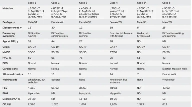
Table Baseline characteristics of 6 patients with LGMD2T
Case 1 Case 2 Case 3 Case 4 Case 5a Case 6
Mutation c.859C.T (p.Arg287Trp), c.79G.C (p.Asp27His) c.859C.T (p.Arg287Trp), c.79G.C (p.Asp27His) c.464G.A (p.Arg155His), c.1039_1043dup (p.Ile349Serfsa) c.79G.C (p.Asp27His), c.760G.A (p.Val254Met) c.859C.T (p.Arg287Trp), c.79G.C (p.Asp27His) c.902C.G (p.Ser301Cys), c.1069G.A (p.Val357Ile) Sex/age, y Male/51 Female/44 Female/32 Female/33 Male/53 Male/59
Disease onset, y 15 30 12 18 5 25 Presenting symptoms Difficulties running Difficulties climbing stairs Difficulties running Exercise intolerance with fatigue Walked at 5 years old Difficulties walking and running Age at MRI, y 51 44 27 28 ND ND
Origin CA, DK CA, DK CA, Fr CA, Fr CA, DK CA, Fr
MMSE 30/30 30/30 30/30 27/30 ND 28/30
FVC, % 59 66 76 95 61 43
ECG Normal Normal Normal Normal Normal Normal
Cardiac echo Normal Normal Normal Normal Normal Ejection fraction 48% 10-m walk test, s 13 11 6 14 7 Cannot walk Walking aids Wheelchair, but
ambulant
Scooter None Wheelchair, but ambulant
None Wheelchair
FSS 48/63 61/63 35/63 58/63 ND 43/63
EMG Myopathic ND Myopathic Myopathic ND ND Decrement,b% 26–29 ND 11–13 10–23 ND 13
CK, U/L 2,390 1,520 1,604 1,200 1,327 619
Abbreviations: CK5 creatine kinase; CA 5 Caucasian; DK 5 Danish; Fr 5 French; FSS 5 Fatigue Severity Scale; FVC 5 forced vital capacity; MMSE 5 Mini-Mental State Examination; ND5 not done.
aData obtained at the latest visit 12 years ago, at age 41 years. This patient is the brother of case 1 and has intellectual disability due to prolonged oxygen
deficiency at birth and could not cooperate to perform MRI.
bAnalyses of decrement, using 3-Hz stimulation frequency, were obtained in different muscles (recording electrode on trapezius muscle, anconeus muscle,
and the tibialis anterior muscle, respectively).
Mutations inGMPPB in the Danish participants were con-firmed by whole-exome sequencing on DNA samples at the Broad Institute’s Genomics Platform, using Illumina exome capture, 38-Mb baited target, and the Broad’s in-solution
hybrid selection process, and confirmed by Sanger sequencing. Sanger sequencing with bidirectional sequencing of amplicons using a Big Dye terminator was used in the French participants.
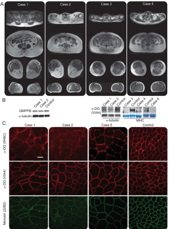
Figure 1 MRI and muscle biopsy findings
(A) T1-weighted, cross-sectional magnetic resonance images of muscles in cases 1–4. Images were acquired at C6 and L4 of the spine, at the middle of the thighs, and at the thickest part of the calves. (B) Expression of muscle proteins on Western blotting and immunofluorescence in patients with LGMD2T. Two patients express normal levels of GMPPB protein. Same analysis was not available for the rest of the participants. Expression ofa-dystroglycan (a-DG) is reduced in all patients. (C) Merosin (clone 22B2) expression appeared near normal on immunofluorescence-stained muscle sections. IIH6C-specific glycosylation ofa-DG is significantly reduced in patients, while VIA4-specific glycosylation appears normal. Bar is 50 mm. MHC5 major histocompatibility complex.
RESULTS Clinical characteristics. Onset of disease varied from 5 to 30 years of age (table). All partici-pants presented with first symptoms related to diffi-culties in running and climbing stairs. Two women (cases 3 and 4) complained of fatigability and wors-ening of motor deficit after exercise. Five were ambu-latory, but 3 of them used wheelchair outside. Strength testing showed hamstrings and hip flexor muscles to be most affected, with some asymmetry
(figure 2). Four had hypertrophy of calves. None had symptoms from eyes or dysphagia, and only one had mildly affected left ventricular ejection frac-tion on echo.
MRI.Brain MRI was normal in all participants. The lumbar erector spinae muscle was the most severely involved muscle in all participants (figures 1 and 2). The second most affected muscles were hamstrings, which were more involved than anterior thigh muscles in all patients. We found an age-dependent gradient of muscle involvement, but no correlation between disease duration and level of muscle involvement.
Muscle biopsy.All muscle biopsies showed dystrophic changes with ring fibers, increased fibrosis, necrosis, and multiple regenerating fibers. Surprisingly, GMPPB protein was not downregulated on Western blotting (figure 1) from 2 participants, while merosin was absent (data not shown).
Immunohistology demonstrated that glycosyla-tion ofa-dystroglycan was reduced to a spotty pres-ence in myofibers using the IIH6C antibody, but not with the VIA4 antibody, except for 1 case (figures 1 and e-1 at Neurology.org/ng), while merosin expres-sion appeared near normal.
Molecular findings.The mutations inGMPPB found in 4 of the participants have previously been described as pathogenic variants.4–6 One
partici-pant (case 3) was compound heterozygous for 2 new variants in GMPPB, and case 6 had 1 new mutation not present in the databases, such as Exome Variant Server and 1000 Genomes Project. The c.1039_1043dup (p.Ile349Serfs*) mutation is inherently pathogenic due to a frameshift, and the mutations, c.464G.A (p.Arg155His) and c.902C.G (p.Ser301Cys), affect highly con-served sites and are predicted to be pathogenic by 3 databases (Single Nucleotide Polymor-phism/GO, SIFT, and PANTHER). The preva-lence of LGMD2T in the Danish cohort was estimated to be 1.5%. Creatine kinase (CK) level was elevated in all patients (table).
Neurophysiology.EMG showed a myopathic pattern mostly in proximal muscles, and repetitive stimu-lation disclosed a decremental response primarily in the proximal muscles at 3-Hz stimulation of the radial, peroneral, and spinal nerves with re-cordings from the anconeus, tibialis anterior, and trapezius muscles (11%–29%). However, treat-ment with acetylcholine esterase inhibitors had on-ly substantial effect in one of the 3 participants (case 3) in whom this was tested. In this latter patient, the decrement also decreased on repetitive stimulation.
Figure 2 Muscle involvement and muscle strength evaluation
(A) Levels of muscle involvement evaluated by T1-weighted MRI using the Mercuri scale.7(B)
Muscle strength evaluation using the Medical Research Council (MRC) scale. Values 0–5, including plus and minus for 4 and 5 (41 equals 4.33 and 52 equals 4.66).
DISCUSSION The principal new findings of this study are (1) the consistent finding of a preferential affection of paraspinal and hamstring muscles in LGMD2T, (2) 3 new mutations inGMPPB, (3) vari-able loss of glycosylation tested with IIH6 and VIA4 antibodies, and (4) a prevalence of LGMD2T of 1.5% in Denmark. Muscle MRI and pattern of weak-ness have not been investigated in the approximately 14 other known ambulatory patients with LGMD2T reported so far. The pattern of preferential affection of lumbar erectors and hamstrings resembles the pat-tern observed in LGMD2A and is supported by find-ings reported in one other patient with LGMD2T.6
Calf hypertrophy in 4 of the participants is in agree-ment with previous findings in LGMD2T.4 The
muscle involvement evaluated on MRI was not cor-related with the duration of disease (table). This may be due to the patients’ different understandings of disease onset as this was self-reported.
Approximately 90% of patients with LGMD fol-lowed at the Copenhagen Neuromuscular Center have a molecularly defined diagnosis. The remaining undiagnosed 10% of cases were tested for aberrations inGMPPB. Thus, in a genetically well-characterized group of 201 LGMDs, LGMD2T constitutes 1.5%. Future studies must show whether there are geo-graphic differences in frequency of this disorder.
We found that the level of GMPPB protein was the same in patients and healthy controls. This can be attributed to the allele carrying the c.79G.C mutation because the c.859C.T mutation has been shown to yield no protein, whereas the c.79G.C does.6 Patients had decreaseda-dystroglycan
glyco-sylation. This presumably affects the binding of a-dystroglycan to merosin, explaining the loss of mer-osin seen on Western blots, and thus linkage to the extracellular matrix. Presumably because of differen-ces in sensitivity between Western blotting and immunofluorescence, merosin appeared near normal and uniformly distributed, not mosaic as seen in the VIA4 and IIH6C stains. Three of the participants, car-rying the same mutations, had reduceda-dystroglycan glycosylation assessed with the IIH6C antibody, but not VIA4, but another case with different mutations had reduced glycosylation with the VIA4 antibody (figure e-1). Patients with LGMD2I and 2M always show reduced glycosylation ofa-dystroglycan with the VIA4 antibody.9,10 This demonstrates that both the
VIA4 and IIH6C antibodies, commonly used for the diagnosis of dystroglycanopathies, are required for diagnosing patients suspected to have a-dystroglycan glycosylation defects.
It has been suggested that the c.79G.C mutation leads to a mild phenotype of LGMD2T5 and the
c.859C.T to a severe one.6Three cases were
com-pound heterozygous for these 2 mutations inGMPPB,
and MRI from these patients showed severe muscle involvement in comparison with case 4, who carried 2 mutations associated with the mild MRI involve-ment, thus indicating a genotype/phenotype relation-ship. This fits the pattern seen in the present 17 known a-dystroglycanopathies, where phenotype/genotype relationships are generally recognized.
In accordance with previous findings in LGMD2T,6
repetitive EMG stimulation elicited a decrement of the muscle action potential, while attempts to treat the abnormality of neurotransmission have been sluggish at best. In line with this, only one patient had improved endurance on treatment with pyridostigmine. These findings suggest abnormalities of the neuromuscular junction related to abnormal glycosylation, which can-not be completely corrected by conventional antimyas-thenic treatment. Thus, in a context of LGMD with high CK levels, a decremental response may be an important diagnostic clue toward genetic analysis of GMPPB.
AUTHOR CONTRIBUTIONS
S.T. Oestergaard: design of study, analysis, acquisition and interpreta-tion of data, and drafting the manuscript. T. Stojkovic: acquisiinterpreta-tion of data and revision of manuscript. J.R. Dahlqvist: design of study, analy-sis, acquisition and interpretation of data, and drafting the manuscript. C. Bouchet-Seraphin, J. Nectoux, F. Leturcq, M. Cossée, G. Solé, and C. Thomsen: acquisition of data and revision of manuscript. T.O. Krag: acquisition and interpretation of data and revision of manuscript. J. Vissing: design of study, acquisition and interpretation of data, and revision of manuscript.
ACKNOWLEDGMENT
The authors thank Dr. Norma Beatriz Romero, Dr. Pascale Marcorelles, and Emmanuelle Lacéne for providing immunohistochemistry pictures from the French participants. They also thank Dr. Fanny Duval from the center of reference for neuromuscular disorders, CHU Bordeaux, France, for the opportunity to include an additional participant to the article.
STUDY FUNDING No targeted funding reported.
DISCLOSURE
S.T. Oestergaard has received travel funding from the Lundbeck Foundation. Dr. Stojkovic has received speaker honoraria from the Laboratory LFB (Lab-oratoire français du fractionnement et des biotechnologies). Dr. Dahlqvist, Dr. Bouchet-Seraphin, Dr. Nectoux, Dr. Leturcq, and Dr. Cossée report no disclosures. Dr. Solé has served on the scientific advisory board of CSL Behring; has received travel funding from Laboratoire Français de biotech-nologie, CSL Behring, and Sanofi Genzyme; and has received research sup-port from CSL Behring and AFM-Téléthon. Dr. Thomsen holds a patent for Optical motion tracking of an object (WO 2011/113441 A2). Dr. Krag has received research support from the AP Møller Foundation and Augustinus Foundation. Dr. Vissing has served on the scientific advisory board of Gen-zyme; has received travel and speaker honoraria from GenGen-zyme; has served on the editorial board ofNeuromuscular Disorders and the Journal of Neuro-muscular Diseases; has been a consultant for NOVO Nordic Industries (Denmark); and has received research support from the Lundbeck Founda-tion, the NOVO FoundaFounda-tion, the Danish Medical Research Council, Uni-versity of Copenhagen, and the research Committee of the National Hospital. Go to Neurology.org/ng for full disclosure forms.
REFERENCES
1. Nigro V, Savarese M. Genetic basis of limb-girdle muscular dystrophies: the 2014 update. Acta Myol 2014;33:1–12. 2. Carss KJ, Stevens E, Foley AR, et al. Mutations in
GDP-mannose pyrophosphorylase B cause congenital and limb-girdle muscular dystrophies associated with hypoglycosylation ofa-dystroglycan. Am J Hum Genet 2013;93:29–41. 3. Adams JC, Brancaccio A. The evolution of the
dystrogly-can complex, a major mediator of muscle integrity. Biol Open 2015;4:1163–1179.
4. Cabrera-Serrano M, Ghaoui R, Ravenscroft G, et al. Expand-ing the phenotype of GMPPB mutations. Brain 2015;138: 836–844.
5. Jensen BS, Willer T, Saade DN, et al. GMPPB-associated dystroglycanopathy: emerging common variants with phe-notype correlation. Hum Mutat 2015;36:1159–1163. 6. Belaya K, Rodríguez Cruz PM, Liu WW, et al. Mutations
in GMPPB cause congenital myasthenic syndrome and
bridge myasthenic disorders with dystroglycanopathies. Brain 2015;138:2493–2504.
7. Mercuri E, Talim B, Moghadaszadeh B, et al. Clinical and imaging findings in six cases of congenital muscu-lar dystrophy with rigid spine syndrome linked to chro-mosome 1p (RSMD1). Neuromuscul Disord 2002;12: 631–638.
8. Krag TO, Vissing J. A new mouse model of limb-girdle muscular dystrophy type 2I homozygous for the common L276I mutation mimicking the mild phenotype in hu-mans. J Neuropathol Exp Neurol 2015;74:1137–1146. 9. Krag TO, Hauerslev S, Sveen ML, et al. Level of muscle
regeneration in limb-girdle muscular dystrophy type 2I relates to genotype and clinical severity. Skelet Muscle 2011;1:31.
10. Riisager M, Duno M, Hansen FJ, et al. A new mutation of the fukutin gene causing late-onset limb girdle muscular dystrophy. Neuromuscul Disord 2013;23:562–567.