HAL Id: tel-01403855
https://tel.archives-ouvertes.fr/tel-01403855
Submitted on 28 Nov 2016HAL is a multi-disciplinary open access archive for the deposit and dissemination of sci-entific research documents, whether they are pub-lished or not. The documents may come from teaching and research institutions in France or abroad, or from public or private research centers.
L’archive ouverte pluridisciplinaire HAL, est destinée au dépôt et à la diffusion de documents scientifiques de niveau recherche, publiés ou non, émanant des établissements d’enseignement et de recherche français ou étrangers, des laboratoires publics ou privés.
ribosome
Iskander Khusainov
To cite this version:
Iskander Khusainov. Structural studies of the Staphylococcus aureus ribosome. Biophysics. Université de Strasbourg, 2015. English. �NNT : 2015STRAJ071�. �tel-01403855�
UNIVERSITÉ DE STRASBOURG
ÉCOLE DOCTORALE des Sciences de la Vie et de la Santé
IGBMC - CNRS UMR 7104 - Inserm U 964
THÈSE
présentée par :Iskander KHUSAINOV
soutenue le : 27 Novembre 2015pour obtenir le grade de :
!"#$%&'($')*%+,-$&.,#/'($'0#&1.2!%&3
Discipline/ Spécialité: Biophysique et biologie structurale
(Biophysics and Structural biology)
Structural studies of the
Staphylococcus aureus ribosome
Etudes structurales du ribosome de
Staphylococcus aureus
THÈSE dirigée par :
M. YUSUPOV Marat Directeur de recherche, Université de Strasbourg
Mme. YUSUPOVA Gulnara Directeur de recherche, Université de Strasbourg
RAPPORTEURS :
M. JENNER Lasse B. Associate Professor, Aarhus University (Denmark)
M. MECHULAM Yves Directeur de recherche, École Polytechnique (Paris)
AUTRES MEMBRES DU JURY :
M. GEISSMANN Thomas Chargé de recherches, ENS de Lyon (Lyon)
2
ACKNOWLEDGEMENTS
First of all a would like to thank all jury members, Dr. Yves Mechulam, Dr. Lasse Bohl Jenner, Dr. Thomas Geissmann and Dr. Stefano Marzi who agreed to evaluate my PhD work. I am extremely thankful to meet them in Strasbourg and to discuss.
I would like to express my sincere gratitude to my advisors, Gula and Marat for their constant support and patience, for their motivation, advice and confidence. I am thankful to them for giving me the opportunity to learn many different of methods of biochemistry and structural analysis in the laboratory. Every single day I learn something new here. Working in Marat's and Gula's laboratories is priceless experience.
This work would not be complete without electron microscopy involvement. I express my gratitude to our collaborator Yaser Hashem with whom we obtained the first structure of Staph ribosome; and to Alex Myasnikov for his significant contribution in cryo-EM data collection and helpful discussions. Special regards to Dr. Pascale Romby. It is always a pleasure to discuss with her and to get a fresh view on the project.
I am thankful to the structural biology platform of IGBMC: Catherine, Peirre and Alastair, who taught me a lot of techniques of biophysics and crystallization. In addition, I thank Prof. Richard Beckett and Dr. Farida Minibayeva for patiently correcting my thesis and bringing it to its presentable form.
Now it is a good time to thank all of my colleagues who made me feel comfortable here. First, I want to cordially thank two people: Sergey, whose endless enthusiasm and striking erudition inspired me to move forward from day to day; and Natasha, who helped me to not get lost in the ribosome world during my first year. Gracias/Grazie to my eEF2 team Eduardo and Simone. These two people present an unbelievable combination of experience, knowledge, curiosity, diligence, belief and reasonable skepticism that make them extremely nice companions. And particular credits to Alexey of course. Always strict, always straightforward, sometimes sarcastic but never disingenuous, he made our synchrotron trips very special. Many thanks to our French teammates Justine, Mélanie, David. Thanks to them, I knew more about French culture, cuisine, and most importantly, they were always helpful with the French language and administration duties. And many thanks to all the present and former members of our lab, Irina, Yuzuru, Marie, Daniya, Muminjon, Jean-Paul, Raphaël, Nico, Sylvie, Lasse, Liudmila, for their support and friendly atmosphere in the laboratory.
3 Besides work in the laboratory, we had a great time outside IGBMC. First, I would like to thank our Eastern Union team Anna and Kate. We met here during the interview and kept the friendship over the whole PhD. I appreciate the hospitality of Ben and Damien, who showed us Strasbourg and helped with integration into the French life during first year. I am grateful to Nico and Claire, Firas and Irene, Marie and Gérald, Irina, Anna, Kate, Karima, Heena, Shilpy, Alexey, Vova, Grigory, Valerio, Xavi, Ismail, Xieyang and others for the unforgettable moments and adventures that we had together: via ferrata in Jura, opera in Dijon, ski in the Alps, board games at Alexey's place, crazy night bike trip along the Rhine and many others. I would like to thank all the members of SPB and personally Michel who always gave me a hand when I asked. Many thanks to my friends in Kazan - Aidar, Igor, Sergey and Olga. It is always a pleasure to come to the hometown and meet them there. Most importantly, I would like to thank my family. I am very grateful to my parents Rezeda and Shamil who accepted my wish to come to France with understanding. To my aunt Farida, who showed me the beauty of science and biology when I was child; and to my grandmother Jasmina, who patiently listens to all my stories about synchrotrons, diffraction and gives a good advice when I need one. I am also grateful to my brother Farid and my niece Rushaniya with whom we spent a great time in Saint-Petersburg this year. I am very happy to have such an adorable family!
My PhD thesis has been possible only because of constant support and participation of all the people mentioned above. I've never walked alone. Thank you very much!
4
TABLE OF CONTENTS
ACKNOWLEDGEMENTS ... 2
TABLE OF CONTENTS ... 4
TABLE OF FIGURES AND TABLES ... 6
SUMMARY ... 8
RÉSUMÉ DE THÈSE ... 12
ABBREVIATIONS ... 22
INTRODUCTION ... 22
THE RIBOSOME ... 25
Core of the ribosome ... 25
Specifications of bacterial and eukaryotic ribosome ... 26
Protein synthesis ... 27
Structural studies of the ribosome ... 29
STAPHYLOCOCCUS AUREUS ... 34
Main characteristics of S. aureus ... 34
Virulence determinants ... 35
S. aureus - superbug ... 38
Antibiotic resistance of S. aureus ... 39
Mechanisms of antibiotics action and bacterial resistance. ... 40
Current drugs used in treatment of S. aureus diseases ... 44
S. aureus ribosome as a target for new antibiotics ... 49
TRANSLATION ELONGATION FACTORS ... 50
Translation elongation cycle ... 50
eEF2 is a potential target for drug development ... 54
PROJECT OUTLINE ... 55
RESEARCH PROJECT 1. STRUCTURE DETERMINATION OF S. AUREUS RIBOSOME... 51
S. AUREUS CELL GROWTH ... 58
S. aureus strain ... 58
Cells preparation ... 58
RIBOSOMES PURIFICATION ... 59
Buffers ... 60
Lysis ... 60
PEG precipitation and sucrose cushion ... 61
5
Ribosome concentration and storage ... 62
RIBOSOMES CHARACTERIZATION ... 63
One- and two-dimensional polyacrylamide gel electrophoresis (PAGE)... 63
Agarose gel electrophoresis ... 64
Analytical sucrose density gradient centrifugation ... 64
Analytical ultracentrifugation ... 67
Mass spectrometry ... 67
CRYSTALLOGRAPHIC STUDIES OF S. AUREUS RIBOSOME... 70
Why x-rays and why crystals?... 70
Macromolecular crystallization in theory ... 71
Macromolecular crystallization in practice ... 73
Crystallization of S. aureus ribosome ... 76
Post-crystallization treatment ... 82
Post-crystallization treatment of S. aureus ribosome crystals ... 83
X-ray data collection ... 86
CRYO-EM STRUCTURE DETERMINATION OF S. AUREUS RIBOSOME ... 89
Latest improvements in cryo-EM ... 89
Electron microscope organization ... 90
Sample preparation and data collection ... 92
Image processing for single particle cryo-EM ... 94
RESEARCH PROJECT 1: SUMMARY ... 105
RESEARCH PROJECT 2. PURIFICATION AND CRYSTALLIZATION OF HUMAN eEF2 ... 100
PURIFICATION AND CRYSTALLIZATION OF HUMAN eEF2 PROTEIN ... 107
HeLa cells preparation ... 107
Purification of native eEF2... 107
Characterization of human eEF2. ... 110
Crystallization of human eEF2. ... 112
Diffraction experiment ... 113
Cloning of human eEF2 ... 114
RESEARCH PROJECT 2: SUMMARY ... 119
DISCUSSION ... 121
PERSPECTIVES ... 125
6
TABLE OF FIGURES AND TABLES
FIGURES:
Figure 1. Structural organization and functional sites of the ribosome ... 25
Figure 2. Composition of bacterial and eukaryotic ribosomes and the common core, based on x-ray studies... 27
Figure 3. Schematic diagram of bacterial protein synthesis ... 28
Figure 4. Regulation network of production of virulence factors in s. aureus ... 36
Figure 5. RnaIII secondary structure ... 38
Figure 6. Antibacterial resistance mechanisms ... 41
Figure 7. Antibiotics targeting sites during protein synthesis ... 43
Figure 8. Antibiotics susceptibility profiles of S. aureus.. ... 51
Figure 10. The structures of apo eEF2, ef-g in complex with GDP, sordarin, enterotoxin a ... 53
Figure 11. Sucrose gradient profile of S. aureus ribosome sample ... 62
Figure 12. One-dimensional and two-dimensional SDS-PAGE, agarose gel of S. aureus ribosome sample.. ... 64
Figure 13. Characterization of S. aureus ribosomes sample by sucrose density gradient centrifugation ... 66
Figure 14. Sedimentation profile of S. aureus ribosome sample. ... 67
Figure 15. Schematic representation of diffraction experiment ... 71
Figure 16. Schematic phase diagram of protein crystallization. ... 72
Figure 17. Idealized phase diagrams showing the trajectories of three different crystallization methods, vapour-diffusion, batch and liquid-diffusion ... 75
Figure 18. Crystals of S. aureus 70S ribosome. ... 81
Figure 19. Scheme of post-crystallization treatment ... 85
Figure 20. Crystals cracked during post crystallization treatment. ... 85
Figure 21. Diffraction patterns of different crystals ... 88
Figure 22. Schematic representation of basic composition of an electron microscope. ... 91
Figure 23. Scattering of the electrons by atom. ... 91
Figure 24. Representation of the cryo-EM grid at different scales ... 92
Figure 25. Electron micrograph of the vitrified S. aureus 70S ribosome sample taken on the F-30 polara microscope.. ... 93
Figure 26. Processing of S. aureus ribosome cryo-EM images taken at different defocus ... 96
Figure 27. Schematic and idealized representation of particle extraction and 3D reconstruction .. 97
Figure 28. Schematic representation of 3D classification of S. aureus ribosome dataset ... 98
Figure 29. Resolution curve of cryo-em structure reconstruction after 3D auto-refinement and post-processing ... 99
Figure 30. Local resolution map of S. aureus ribosome ... 100
Figure 31. Cryo-em structure of S. aureus ribosome. ... 101
Figure 32. Comparison of cryo-EM structures of B. subtilis ribosome and S. aureus ribosome ... 102
Figure 33. Examples of the comparative analysis of secondary structure elements of the rRNAs from different organisms. ... 103
Figure 34. X-ray structure of t. thermophilus ribosome fitted into S. aureus ribosome structure . 104 Figure 35. Purification of human eEF2 protein ... 109
Figure 36. MALLS profiles of two independent experiments ... 111
Figure 37. Crystals of eEF2 protein obtained by robotic screening... 113
7
Figure 39. Two strategies of eEF2 cloning experiment. ... 115 Figure 40. Agarose gel of amplified fragments of eEF2 DNA ... 116 Figure 41. Secondary structures of S. aureus and S. cerevisiae rRNA ... 122
TABLES:
Table 1. Mechanisms of action and resistance mechanisms of selected ribosome-targeting
antibiotics 44
Table 2. Several antimicrobial agents and their therapeutic utilization 47
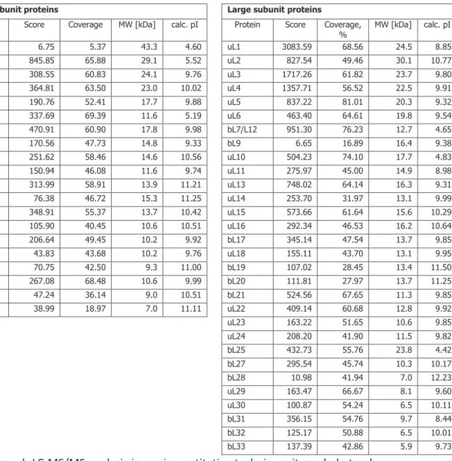
Table 3. List of ribosomal proteins found in S. aureus 70S ribosome sample 68
Table 4. List of non-ribosomal proteins found in S. aureus ribosome sample 69
Table 5. Published crystal conditions for 70S ribosomes 77
Table 6. Mass spectrometry results of eEF2 protein sample 112
8
SUMMARY
The high-resolution structures of ribosomes and ribosomal subunits obtained by crystallography and electron microscopy have revolutionized the field of protein translation. Knowledge of the precise positions of residues in the ribosome in various states has led to a deeper understanding of the complex mechanisms of protein synthesis. Crystal structures of ribosomal complexes with antibiotics and antifungal compounds have provided unparalleled insight into their mechanisms of action, and they also facilitate the design of more effective drugs (Garreau de Loubresse et al., 2014; see for review Wilson, 2014). It was discovered that most of translation inhibitors bind to the conserved regions of the ribosomes (PTC, tRNA-binding sites, protein exit tunnel, etc). However, the effects of the same drug on different organism can vary from species to species. An excellent example is provided by the drug-resistant bacteria, which are tolerant to many of commonly used antibiotics. Among them is Staphylococcus aureus (S. aureus), an aggressive pathogen that causes numerous infections in humans. This bacterium is rather unique and thus, requires individual approach for treatment.
S. aureus is responsible for severe nosocomial and community-acquired infections. It can
cause a variety of life-threatening diseases in humans. Deeply penetrating infections of S. aureus include endocarditis, peritonitis, necrotizing pneumonia, bacteremia, meningitis, osteomyelitis, septic arthritis, and infections of bones, joints and organs. Some skin conditions caused by staphylococcal exfoliative toxins include blisters, skin loss, pimples, furuncles, impetigo, folliculitis, abscesses, poor temperature control, fluid loss, and secondary infection. Certain strains of S. aureus produce the superantigen TSST-1, which is responsible for toxic shock syndrome (TSS) (Murray et al., 2002), from which mortality is very high; death can occur within two hours (Chen et al., 2007).
Another focus area in S. aureus research is its increasing resistance to antibiotics. A 2014 report by World Health Organization (WHO) reveals that antimicrobial resistance, including antibiotic resistance is no longer a prediction for the future, it is occurring now in every region of the world and has the potential to affect anyone, of any age, in any country. Antibiotic resistance is now a major threat to public health. According to the report, as many as 60% of S. aureus infections are determined to be methicillin-resistant (MRSA) in Europe. This value was 90% in the WHO Region of Americas, 80% in African Region, 80% in Western Pacific Region. In the Eastern Mediterranean Region 50% of S. aureus infections were reported to be methicillin-resistant, while this value was more than 25% in some parts of South-East Asia.
9 The resistance to the ribosome binding antibiotics is provided by the small modifications (mainly methylation) of the rRNA in the antibiotic binding sites. These modifications do not affect the process of protein synthesis, but prevent the binding of the drug. Solving the structure of the ribosome from S. aureus will be the first step in understanding precise mechanism of its resistance and will facilitate the design of new antistaphylococcal compounds.
Research project 1.
The project dedicated to solving the structure of 70S ribosome of S. aureus was initiated in 2011. By that time the structures of Thermus thermophilus and Escherichia coli ribosomes had already been solved by X-ray crystallography. However, the structure of any Gram-positive or any pathogenic bacteria ribosome was still unidentified. It is known that some aspects of protein synthesis differ between Gram-negative and Gram-positive bacteria. Additionally, pathogenic bacteria have evolved complex mechanisms of translation regulation, that provide high degree of pathogenesis and facilitate survival in stressful conditions. Moreover, many pathogenic bacteria (including S. aureus) show extremely high resistance to ribosome-targeting antibiotics. The structure of the ribosome from the Gram-positive multi-resistant pathogenic bacterium
Staphylococcus aureus would enable us to understand the peculiar structural features of its
translation machinery, leading to a better understanding of the specific features of how this bacterium regulates protein synthesis, and how it can survive stress. Most importantly, the structure of the ribosome will serve as a model system for developing new anti-staphylococcal drugs.
We first focused on the crystallographic studies of the S. aureus 70S ribosome. This involved: preparing the cells: growing, harvesting, breaking;
developing a purification protocol for obtaining pure and stable ribosomes that are suitable for crystallization;
crystallization and post-crystallization treatment: searching for crystallization conditions, growing big crystals and preparation for diffraction analysis;
developing the strategy of data collection at the synchrotron; solving the structure: processing the data and building the model.
A protocol for the efficient purification of S. aureus 70S ribosomes using a minimal amount of cells was developed. It is based on the protocols used for the bacterium Thermus thermophilus (Gogia et al., 1986, Yusupov et al., 2001) and the yeast Saccharomyces cerevisiae (Ben-Shem et al., 2011). Lysis procedures, ionic conditions, magnesium and polyethylene glycol (PEG) concentrations, sucrose gradients, etc. were optimized during the development of the purification protocol.
10 Ribosomes were shown to be pure, stable and capable of being crystallized. The size of the crystals of S. aureus 70S ribosome was sufficient for X-ray analysis (200 × 80 × 20 µm); however the diffraction of these crystals after post-crystallization treatment reached a maximum resolution of 17.5 Å.
As we experienced specific difficulties with the diffraction of the crystals and because of time constraints, we used a cryo-EM technique to determine the structure of the S. aureus ribosome. The purity of the sample and recent advances in cryo-EM field gave us a hope that we would be able to obtain a high-resolution structure. The data were collected using the latest generation microscope (Titan Krios, installed at the Centre de Biologie Intégrative (CBI, Illkirch, France)). After image processing, the first structure of S. aureus 70S ribosome was obtained at 3.9 Å resolution. The structure at this resolution allows visualization of the side chains of the ribosomal proteins, base-paring of the nucleotides of S. aureus rRNA, and other fine details of the structure, however, does not gives us the ability to analyse the mechanisms of binding of the antibiotics. Therefore, the resolution will be further improved by performing several additional steps of image processing. At the same time my colleagues continue crystallographic studies of 70S ribosome from S. aureus.
The results I have obtained during my PhD will be discussed in details in the next chapters.
Research project 2.
One of the scientific goals of our laboratory is the crystal structure determination of the human 80S ribosome. During our development of the protocol for human 80S ribosome purification we discovered that most of the ribosomes co-purify with translation elongation factor 2 (eEF2) protein. The same observation was reported in the recent cryo-EM study of human 80S ribosome: the sample of vacant 80S ribosome contained some fraction of 80S ribosome complex with eEF2 (Anger, et al., 2013). Thus, the part of my work was devoted to the determination of the crystal structure of human eEF2.
It is well known that eEF2 is one of the major regulators of translation in eukaryotes, and it is often involved in cell stress response and cancer. Determination of the structure of human eEF2 can clarify some aspects of regulation and repression of protein synthesis in humans. The eEF2 protein is known to be a target of specific protein synthesis inhibitors, and therefore the knowledge of the structure of human eEF2 could also help to discover potential anti-cancer compounds.
The project on eEF2 started by developing a protocol to purify the native eEF2 protein using the ribosome-free cytosolic fraction of HeLa cells. The protocol of purification was developed and first crystals of native eEF2 protein were obtained. However, purification of native protein gave a
11 very low yield, not suitable for large scale crystallization experiments. Therefore, we switched to the recombinant production of the protein in bacteria. The gene of human eEF2 protein was amplified from the cDNA library of HeLa cells and first cloning experiments were performed.
12
RÉSUMÉ DE THÈSE
La biosynthèse protéique est un processus fondamental, retrouvé au sein de toutes les cellules, qui permet d'assurer le décodage de l'information génétique. Ce mécanisme de traduction implique de nombreux partenaires, protéines et acides nucléiques, et nécessite une parfaite coordination de leurs actions afin d'assurer la fidélité du transfert de l'information génétique. Le !"#$#%&'&$(')*+,(&- ',&.( +)'/&',&'0 #,&$$-$'/&'( +/-,(!#.1'2"#./+.('/+.$')&$',&))-)&$'&(',#.$& 34' au cours de l'évolution, celu!5,!' &$(' ,#%0#$4' /&' /&-6' $#-$5-.!(4$7' -.&' 0&(!(&' &(' -.&' 8 +./&7' combinant des protéines et des acides nucléiques qui s'associent au cours de la traduction pour former un ribosome fonctionnel. Dans cet état, le ribosome est capable de catalyser l'addition séquentielle des acides aminés de la chaîne peptidique en cours de synthèse en utilisant l'ARN %&$$+8& '92:;%<',#%%&'%+( !,&'&(')&$'2:;'/&'( +.$=& ('+%!.#+,>)4$'9++52:;(<',#%%&'$-"$( +($1' Les deux sous-unités présentent des fonctions bien distinctes. Le déc !"#$% !$% &'()* +,"-( )% #.).-(/0$%1 )-$)0%!")2%&'345,%$2-%"220+.%6"+%&"%6$-(-$%2 0270)(-.%-")!(2%/0$%&"%#+")!$%2 0270)(-.% 1"-"&82$% &"% +."1-( )% !$% -+")26$6-(!"-( )% 6$+,$--")-% &"% * +,"-( )% !'0)$% &("(2 )% 6$6-(!(/0$% $)-+$% chaque acide aminé. Une dizaine de facteurs protéiques se lient successivement au ribosome afin de catalyser les différentes étapes de la traduction, à savoir: l'initiation, l'élongation, la terminaison et le recyclage. Bien que la synthèse des protéines conservée parmi tout les domaines du vivant, tous les organismes ont des spécificités. Soit, les bactéries Gram positives et Gram négatives ont leurs propres spécificités concernant la régulation et l'organisation de la machinerie traductionnelle.
Les structures à haute résolution des ribosomes et sous-unités ribosomales obtenues par cristallographie aux rayons X et microscopie électronique ont révolutionnées le domaine de la traduction des protéines. La connaissance des positions précises des résidus dans le ribosome dans divers états a conduit à une meilleure compréhension des mécanismes complexes de la synthèse des protéines. Les structures de complexes ribosomaux avec des antibiotiques et des composés antifongiques ont fourni un aperçu sans précédent de leurs mécanismes d'action et facilitent également la conception de médicaments plus efficaces (Garreau de Loubresse et al, 2014;. revue Wilson, 2014). Il a été découvert que la plupart des inhibiteurs de la traduction se lient aux régions conservées des ribosomes (centre de peptidyltransférase, les sites de liaison d'ARNt, tunnel de 2 +-($% !$% &"% 6+ -.()$9% $-1:;:% <$6$)!")-9% &$2% $**$-2% !'0)% ,=,$% ,.!(1",$)-% 20+% !$2% $26>1$2% différentes peuvent varier. Un excellent exemple est fourni par les bactéries résistantes à de
13 nombreux antibiotiques couramment utilisés. Parmi celles-ci, Staphylococcus aureus (S. aureus), un "#$)-%6"-? #>)$%/0(%6+ @ /0$%!$%#+"@$2%$-%) ,A+$02$2%()*$1-( )2%1?$B%&'? ,,$:%<$--$%A"1-.+($% est assez unique et donc, requiert une approche individuelle pour le traitement.
S. aureus est responsable des infections nosocomiales et infections sévères et peuvent causer
0)$% @"+(.-.% !$% ,"&"!($2% 6 -$)-($&&$,$)-% , +-$&&$2% 1?$B% &'? ,,$:% <$2% ()*$1-( )2% 1 ,6+$))$)-% l'endocardite, la péritonite, pneumonie nécrosante, la bactériémie, la méningite, l'ostéomyélite, l'arthrite septique, et les infections des os, des articulations et des organes (Fridkin et al, 2005).
S. aureus est un pathogène majeur d'une importance croissante en raison de sa haute
résistance aux antibiotiques (Lowy, 1998). Un rapport par l'Organisation mondiale de la Santé (OMS) révèle que la résistance aux antimicrobiens, y compris la résistance aux antibiotiques n'est plus une prédiction pour l'avenir, elle se passe maintenant dans toutes les régions du monde et a le potentiel de toucher tout le monde, de tout âge, dans tous les pays. La résistance aux antibiotiques est maintenant une menace majeure pour la santé publique. Selon ce rapport, pas moins de 60% des infections à S. aureus sont déterminées comme résistantes à la méthicilline (SARM) en Europe (World Health Organization, 2015).
Les gènes responsables de la résistance sont souvent transmis par des éléments génétiques mobiles tels que les plasmides et, par conséquent, populations bactériennes peuvent acquérir une résistance très rapidement. Divers mécanismes de résistance aux antibiotiques sont indiqués sur la Fig I. Les bactéries peuvent empêcher la pénétration de l'antibiotique dans la cellule (a), le retirer de la cellule en utilisant une pompe d'efflux (b), de dégrader (h) ou modifier l'antibiotique (g). Alternativement les bactéries peuvent muter (c), modifier (d) ou protéger (f) les cibles des antibiotiques. La conception de nouveaux agents antimicrobiens est fortement requise pour le traitement des maladies causées par S. aureus.
14
Fig I. Mécanismes de résistance antibactérienne (adapté de Wilson, 2014).
Pour comprendre les aspects structurels de la résistance aux antibiotiques nous avons besoin de connaître la structure de leur cible. Par conséquent nous nous sommes concentrés sur la structure du ribosome, qui est la principale cible des antibiotiques les plus couramment utilisés. La résolution de la structure du ribosome de S. aureus sera la première étape vers la compréhension du mécanisme précis de sa résistance aux antibiotiques et facilitera la conception de nouveaux composés antistaphylococciques.
Projet de recherche 1.
Le projet dédié à la résolution de la structure du ribosome de S. aureus a été initié en 2011. À cette époque, les structures des ribosomes de Thermus thermophilus et Escherichia coli avaient déjà .-.% +.2 &0$2% 6"+% 1+(2-"&& #+"6?($% "0C% +"8 )2% D:% <$6$)!")-9% &"% 2-+01-0+$% !'0)% +(A 2 ,$% !$% - 0-$% bactérie Gram positif ou tout ribosome pathogène restait manquante. Hors, il est connu que certains aspects de la synthèse des protéines diffèrent entre les bactéries à Gram négatif et à Gram positif. En outre, les bactéries pathogènes ont développé des mécanismes complexes de régulation de la traduction, qui offre un rendement élevé de la pathogenèse et facilite la survie de la bactérie dans des conditions stressantes. En outre, de nombreuses bactéries pathogènes (y compris
15 ribosomes. Cette résistance est souvent due à des modifications au sein du ribosome lui-même. La structure du ribosome de la bactérie pathogène multi-résistante à Gram positif Staphylococcus
aureus révélerait les caractéristiques structurelles particulières de machinerie de traduction nous
permettant ainsi de mieux comprendre les spécificités de la régulation de la synthèse des protéines, et de la survie de cet agent pathogène. Plus important encore, elle servira comme un système modèle pour le développement de nouveaux médicaments contre Staphylococcus.
Nous avons d'abord mis l'accent sur les études cristallographiques du ribosome de S. aureus. Cela a impliqué:
E%&"%6+.6"+"-( )%!$2%1$&&0&$2F%10&-0+$9%&"%+.1 &-$9%&82$ ;
E% &G.&"A +"-( )% !G0)% 6+ - 1 &$% !$% 60+(*(1"-( )% /0(% * 0+)(+"% !$2% +(A 2 ,$2% 60+$2% $-% 2-"bles, propices à la cristallisation ;
E%&"%,(2$%"0%6 ()-%!$%1 )!(-( )2%!$%1+(2-"&&(2"-( )% 6-(,"&$2%6$+,$--")-%!' A-$)(+%!$2%1+(2-"0C% dans grande taille et en quantité suffisante
E% &"% ,(2$% "0% 6 ()-% !$% -+"(-$,$)-2% !$% 6 2--cristallisation nécessaires pour l'analyse par diffraction aux rayons X;
E%&'.&"A +"-( )%!$%&"%2-+"-.#($%!$%1 &&$1-$%!$%! )).$2%"0%28)1?+ -+ )H
E%&"%+.2 &0-( )%!$%&"%2-+01-0+$F%&$%-+"(-$,$)-%!$2%! )).$2%$-%!$%&"%1 )2-+01-( )%!0%, !>&$: I'"(% !.@$& 66.% &$% 6+ - 1 &$% !$% 60+(*(1"-( )% !$2% +(A 2 ,es S. aureus efficace utilisant une quantité minimale de cellules. Il est basé sur les protocoles utilisés pour les bactéries Thermus
thermophilus (Gogia et al., 1986, Yusupov et al., 2001) et les levures Saccharomyces cerevisiae
(Ben-Shem et al., 2011). La procédure de lyse, les conditions ioniques, des concentrations de magnésium et de polyethylene glycol (PEG), des gradients de saccharose, etc. ont été optimisés au cours du développement du protocole de purification. J'.chantillon purifié a été caractérisé en utilisant plusieurs méthodes biophysiques et biochimiques. Des gels SDS ont confirmé la pureté de l'échantillon (Fig II, A et B); gel d'agarose a montré le caractère intact de l'ARNr (Fig II, C) ultracentrifugation analytique a démontré que l'échantillon contenait principalement des particules 70S (Fig II, D). J'"nalyse par spectrométrie de masse a révélé l'absence de protéines ribosomiques S1 et L9. La stabilité du ribosome sous différentes concentrations Mg2+ a été analysée par centrifugation en gradient de saccharose. La concentration minimale de Mg2+ qui permet de garder les sous-unités associées a été déterminée et les conditions pour la dissociation des 70S dans en sous-unités 50S et 30S ont été trouvées.
16
Fig II. SDS-PAGE (électrophorèse sur gel de polyacrylamide en présence de dodécylsulfate de
sodium) unidimensionnel (A) et bidimensions (B) del'échantillon du ribosome de S. aureus. Électrophorèse sur gel d'agarose des ribosomes (C): 1 - T. thermophilus 70S (5 Kg), 2 et 3 - 70S de S. aureus (15 Kg). Les bandes supérieure et inférieure correspondent à des ARNr 23S et 16S, respectivement. Profil de sédimentation de l'échantillon du ribosome de S. aureus (D).
Particules cristallines initiales de ribosome 70S de S. aureus ont été obtenus en utilisant une recherche robotique. Après plusieurs étapes d'optimisation de cristallisation, de grands cristaux tridimensionnels ont été obtenus. Typiquement, les cristaux sont apparus de façon reproductible dans les 7 - 10 jours et ont atteint leur taille moyenne (200 × 80 × 20 Km) après deux semaines supplémentaires (Fig III). La taille des cristaux de ribosome de S. aureus est suffisante pour l'analyse par diffraction aux rayons X. Les conditions de déshydratation et de cryo-refroidissement ont été optimisées pour les cristaux obtenus. Les premières données de diffraction de cristaux du ribosome 70S de S. aureus ont été recueillies. La diffraction de ces cristaux atteint au maximum 17.5 Å, le groupe de l'espace a été déterminée comme P42212 et la taille de l'unité asymétrique était: 450 × 450 × 280 Å.
17
Fig III. Les cristaux du ribosome 70S de S. aureus.
L)%+"(2 )%!$%!(**(10&-.2%"@$1%/0"&(-.%!$%&"%!(**+"1-( )%!$2%1+(2-"0C9%) 02%"@ )2%1? (2(%!'0-(&(2.%&"% technique de cryo-microscopie electronique (cryo-ME) pour déterminer la structure du ribosome de S. aureus. La pureté de l'échantillon et de sérieux progrès dans le domaine de la cryo-ME au cours des dernières années a donné un espoir d'obtenir la structure à haute résolution grâce à cette technique. Les données ont été recueillies en utilisant la dernière génération de microscope (Titan Krios, installé au Centre de Biologie Intégrative (CBI, Illkirch, France)).
Les conditions appropriées pour la préparation de la grille cryo-ME et de la collecte de données ont été trouvées pour l'échantillon du ribosome 70S de S. aureus (Fig IV, A). Le traitement des images cryo-ME a été réalisé avec Scipion workflow et les algorithmes intégrés (Fig IV, B). Après le traitement des images, la structure à une résolution de 3.9 Å a été obtenu. La structure à cette résolution permet de voir les chaînes latérales des protéines ribosomales, la base-azotée des )01&. -(!$2%!$%&'345+%!$%S. aureus, etc (Fig IV, C). Cette résolution donne également la possiblilité d'analyser la liaison des antibiotiques et, en combinaison avec les connaissances biochimiques, de développer des composés antimicrobiens efficacent contre S. aureus.
18
Fig IV. A. Micrographie électronique du ribosome 70S de S. aureus. L'échantillon a été vitrifiés
en éthane liquide et prélevé sur le Polara F-30 microscope. Agrandissement 59000X, tension 300 kV, détecteur CCD eagle 4Kx4K. La structure Cryo-ME de S. aureus ribosome. B. Paramètres de collection de données sur Titan Krios microscope et de traitement des image.
Nous avons effectué une analyse comparative du ribosome de S. aureus et des objets de modèle structures cristallines du ribosomes 70S de T. thermophilus (Jenner et al, 2010) et de E. coli (Schuwirth et al., 2005) et la structure cryo-ME du ribosome 70S de B. subtilis (Sohmen et al., 2015). Le ribosome de T. thermophilus est le ribosome plus structurellement étudiée, le ribosome de E. coli est le plus étudié biochimiquement et la bactérie Bacillus est phylogénétiquement l'organisme le plus proche du genre
Staphylococcus. Plusieurs hélices d'ARN ont été trouvées pour être étendues dans la structure de S. aureus (Fig V). Certainses des extensions sont conservées par E. coli (H26, H44, H27, H68, H79), ou T. thermophilus (h9, H15, H63), tandis que d'autres sont spécifiques des bactéries à Gram-positif S. aureus, ou B. subtilis (H6, H10, H17, H25, H54).
19
Fig V. La structure cristalline du ribosome de T. thermophilus (Code PDB 4V4F) monté dans la
structure du ribosome de S. aureus comme corps rigide: unité 30S est colorée en bleu, sous-unité 50S est colorée en or. Les extensions de certaines hélices d'ARNr et les protéines ribosomiques sont colorées en rouge.
Un autre détail important observé dans la structure de S. aureus est l'absence de protéines ribosomiques S1 et L9. Protéine S1 est la protéine ribosomique la plus grande et la plus acide, et son rôle est de faciliter l'adaptation de l'ARNm de la sous-unité 30S lors de l'initiation de la traduction dans les bactéries. La protéine S1 !'E. coli ou de T. thermophilus se compose d'environ 550 acides aminés se pliées en six domaines, et il se lie au ribosome relativement faiblement. La protéine S1 de S. aureus est constituée de 391 acides aminés se pliées en quatre domaines et ne se lie pas au ribosome. Le ,.1")(2,$%!'interaction de la protéine S1 avec le ribosome de S. aureus reste difficile à déterminer. La protéine L9 est également une spécificité bactérienne. Elle située sur la périphérie très proche de tige L1 dans le domaine V de l'ARNr et se trouve sur le côté du ribosome bactérien. La position de la protéine L9 près de la voisine ribosome 5'-amont dans polysomes top-to-top suggère qu'il pourrait contribuer à
20 la régulation inter-ribosome. (Dunkle et Cate, 2011). D'autre par-9%&"%6+ -.()$%+(A 2 ,"&$%JM%)'$2-%pas essentiel dans E. coli ou T. thermophilus, mais elle est conservé$%!")2%!'"0-+$2 bactéries.
N 02% &$2% +.20&-"-2% A-$)02% !")2% 1$2% -+"@"0C9% *"1(&(-$+ )-% &"% !$21+(6-( )% O% &'.1?$&&$% "- ,(/0$% !0% ribosome de S. aureus $-% 2$2% 1 ,6&$C$2% * )1-( ))$&2'% '!")2% 0)% *0-0+% 6+ 1?$:% J"% 1 ,A()"(2 )% !$2% méthodes de cristallographie aux rayons X et de cryo-ME aidera à atteindre cet objectif. Les résultats obtenus serviront de base pour le développement de nouveaux composés contre la bactérie pathogène $-%$C-+=,$,$)-%+.2(2-")-$%/0'$2-%S. aureus.
Projet de recherche 2.
Une autre partie de mon travail a été consacrée à la détermination de la structure tridimensionnelle par radio-cristallographie du facteur d'élongation humain de type 2 (eEF2). Cette protéine a été jugée plutôt étroitement lié aux ribosomes 80S humains lors de la purification effectuée au sein de notre laboratoire. Il est bien connu que eEF2 est le principal régulateur de la traduction chez les eucaryotes et est souvent impliqué dans la réponse au stress cellulaire et dans le développement du cancer (revue White-Gilbertson, 2009). La détermination de la structure de eEF2 humaine permettrait de clarifier certains aspects de la réglulation et de la répression de la 28)-?>2$%!$2%6+ -.()$2%1?$B%&'? ,,$:%J"%6+ -.()$%$LPQ%$2-%1 ))0$%6 0+%=-+$%0)$%1(A&$%!G()?(A(-$0rs spécifiques de la synthèse des protéines, de sorte que la structure de eEF2 humaine pourrait aussi aider à découvrir des composés anticancéreux potentiels.
Le travail a commencé avec l'élaboration du protocole de purification de la protéine native eEF2 utilisant comme source la fraction cytosolique libre des ribosomes des cellules HeLa. La pureté, l'homogénéité et la stabilité de la protéine purifiée a été analysée par SDS-PAGE, diffusion dynamique de la lumière, diffusion de la lumière multiangle, essai de décalage thermique et la spectrométrie de masse. Les premiers cristaux ont été obtenus par recherche robotique (Fig VI). L'analyse par diffraction de ces cristaux au synchrotron 2'$2- avéré être des cristaux de protéine. Cependant, la qualité de la diffraction ne suffit pas à collecter les données.
21
Fig VI. Les cristaux de protéine eEF2 obtenus par recherche robotique.
En outre l'optimisation de &"%1+(2-"&&(2"-( )%!'eEF2 était impossible en raison du manque de matériel. La purification de la protéine native requise de nombreuses cellules humaines, qui sont coûteux. Nous avons donc décidé de cloner et exprimer la protéine hétérologue. Initialement, car elle offre la plus facile, la moins chère et le plus rapide système d'expression, E. coli a été sélectionnée.
Cependant, nous avons choisi de poursuivre par la purification de la protéine eEF2 recombinante dans des bactéries. Pour le moment, le travail est à l'étape de clonage de la région codante de la protéine eEF2 dans un vecteur d'expression bactérien pET-15b (His-tag, la thrombine site de clivage, résistance à l'ampicilline, IPTG inductible). La gène complet d'eEF2 humain ou ses fragments ont été amplifiés en utilisant la bibliothèque HeLa ADNc incorporée dans un bactériophage. Le travail est toujours en cours.
22
ABBREVIATIONS
Ribosome:
aa-tRNA aminoacyl-tRNA
A-site aminoacyl site, (A-tRNA R A-site tRNA) EF elongation factor (eEF R eukaryotic EF) E-site exit site (E-tRNA R E-site tRNA)
IF initiation factor (eIF R eukaryotic IF) mRNA messenger ribonucleic acid
PTC peptidyl transferase centre
P-site peptidyl site, (P-tRNA R P-site tRNA) RF release factor (eRF R eukaryotic RF) RP ribosomal proteins
RRF ribosome recycling factor rRNA ribosomal ribonucleic acid SD Shine-Dalgarno
tRNA transfer ribonucleic acid
Methods:
NMR nuclear magnetic resonance Cryo-EM cryo-electron microscopy
LC-MS/MS liquid chromatography coupled mass spectrometry DLS dynamic light scattering
MALLS multiangle laser light scattering
MALDI matrix assisted laser desorption/ionization PCR polymerase chain reaction
PAGE polyacrylamide gel electrophoresis
S. aureus:
B. subtilis Bacillus subtilis E. coli Escherichia coli
MRSA methicillin-resistant S. aureus mrMRSA multi resistant MRSA
MSSA methicillin-susceptible S. aureus nmrMRSA non-multi resistant MRSA PBPs penicillin binding proteins
S. aureus Staphylococcus aureus T. thermophilus Thermus thermophilus
TSS toxic shock syndrome
VISA vancomycin-intermediate S. aureus VRSA vancomycin-resistant S. aureus WHO World Health Organization SA sigma factor A
SB sigma factor B
Cryo-electron microscopy:
23 CTF contrast transfer function
DDD direct detector device
DQE detective quantum efficiency FEG field emission gun
FSC Fourier shell correlation SNR signal-to-noise ratio
TEM transmission electron microscopy
Reagents:
DMSO dimethyl solfoxide DTT dithiothreitol
EDTA ethylenediaminetetraacetic acid EG ethyloene glycol GDP guanosine diphosphate GTP guanosine triphosphate LB lysogeny broth MPD 3-methyl,1, 5-pentanediol PBS phosphate-buffered saline PEG polyethylene glycol
PMSF phenylmethylsulfonyl fluoride SDS sodium dodecyl sulfate
25
THE RIBOSOME
Protein synthesis is performed by the ribosome in all living organisms. It is one of the most conserved biological processes. The central dogma of molecular biology states that DNA is transcribed into RNA and RNA is subsequently translated into proteins. Ribosome is macromolecular machine which translates the nucleotides-based language of the mRNA into amino acid-based language of proteins. The ribosome is a major link between genes and proteins and it is made of nucleic acid (ribosomal RNA, or rRNA) and protein (ribosomal proteins, or RP) components.
Core of the ribosome
Structurally ribosomes in all living cells are composed of two subunits, called small ribosomal subunit (SSU) and large ribosomal subunit (LSU). Both are composed of rRNA(s) and proteins with average ratio of 2:1 RNA to protein (the exceptions are mitochondrial and chloroplast ribosomes which have ratios 1:2 and 3:2 respectively (see for review Sharma and Agrawal, 2012). The ribosome is an asymmetric macromolecular complex and each subunit has particular structural and functional organization, thus carries out different functions in translation (Figure 1).
Figure 1. Structural organization and functional sites of the ribosome. The two ribosomal
subunits left and right) assemble together to form the full ribosome (center). Main functional sites and natural ligands (mRNA, tRNA) are annotated (adapted from Melnikov et al., 2012).
N?$%2,"&&%20A0)(-%(2%+$26 )2(A&$%* +%-?$%!$1 !()#%6+ 1$22%T?$+$%",() "18&7-453%U""-tRNA) is selected according to the mRNA sequence. Its major functional sites are the mRNA path used to conduct mRNA during translation, the decoding center responsible for decoding, and the tRNA
26 A()!()#%2(-$2%U39%V%")!%L;:%N?$%372(-$%2$+@$2%- %A()!%",() "18&7-453%"2%it enters into the ribosome !0+()#% 6+ -$()% 28)-?$2(29% -?$%V72(-$% ? &!2%-453% 1"++8()#% -?$% )"21$)-%6 &86$6-(!$% 1?"()% U6$6-(!8&7 tRNA), and the E-site (exit) is where tRNA dissociates from the ribosome. During translation, tRNAs are translocated from the A to -?$%V72(-$%")!%*+ ,%-?$%V%- %-?$%L72(-$:%N?$%&"+#$%20A0)(-%1"-"&8B$2% peptide bond formation. Its major functional sites are the tRNA binding sites (A, P and E), the peptide exit tunnel that extends through the body of the large subunit, and the peptidyl transferase centre (PTC). The PTC is responsible for peptide bond formation and is located at the entrance to the peptide tunnel in a conserved region on the interface that is mainly composed of rRNA. As a result of peptide bond formation in the PTC, the nascent polypeptide chain is transferred from the 6$6-(!8&7-453%()%-?$%V7%2(-$%- %-?$%""7-453%()%-?$%372(-$9%-?02%$C-$)!()#%-?$%)"21$)-%1?"()%A8% )$% amino acid.
Specifications of bacterial and eukaryotic ribosome
Despite the universal conservation of the core, ribosome composition varies between domains of life, taxonomic subgroups, organelles and even within a single individual, although to a smaller extent. Ribosomes may contain their own set of specific moieties: specific proteins, insertions and extensions of conserved proteins and expansion segments of rRNAs.
Major differences in ribosome composition are observed between domains of life. Apart from the core, the bacterial ribosome (E. coli or T. thermophilus) contains 21 A"1-$+("726$1(*(1%6+ -$()29%"% few extensions of the conserved proteins and of ribosomal RNA. The eukaryotic ribosome (S. cerevisiae;%1 )-"()2%WX%$0Y"+8 -$726$1(*(1%6+ -$()2%UZ[[%Y\";%")!%$C-$)2( )2%")!%()2$+-( )2%()% most of the proteins of the core (200 kDa), and the rRNA harbors several extensions in the conserved rRNA chains (about 800 nucleotides -?"-%"11 0)-%* +%]^[%Y\";%U_$)7`?$,%$-%"&:9%Q[aaH%b$&)(Yov et al., 2012). Sedimentation coefficients are used to characterize and to name isolated or associated subunits. Hence, association of the small subunit (30S in bacteria, 40S in eukaryotes) with the large subunit (50S in bacteria, 60S in eukaryotes) results in the full ribosome R 70S in bacteria and 80S in eukaryotes.
27
Figure 2. Composition of bacterial and eukaryotic ribosomes and the common core, based on
X-Ray studies. Bacterial and eukaryotic ribosomes share a conserved core composed of RNA (light blue) and proteins (light red). In addition to the core, ribosomes in each domain of life contain their own set of proteins, extensions and insertions in conserved proteins (both in red), and extension segments in ribosomal RNA (blue). 5.8S and 25S rRNA are both homologous to 23S rRNA in bacteria. Dashed lines around the core indicate positions of flexible stalks of the ribosomes that are usually disordered in X-ray structures. For simplicity, these lines are not shown in the other structures. The figure is based on X-ray and Cryo-EM structures from Armache et al., 2010, Ben-Shem et al., 2011, Jenner et al., 2010, Jarasch et al., 2012 (adapted from Melnikov et al., 2012).
Protein synthesis
Protein synthesis in bacteria can be divided into four main steps: initiation, elongation of the polypeptide chain, termination and recycling of ribosomes (Figure 3). Each of these steps is assisted by protein factors termed initiation factors (IF), elongation factors (EF), release factors (RF) and recycling factors (RRF). Initiation in bacteria involves the accurate positioning of the mRNA start codon (usually AUG), together with the initiator tRNA (fMet-tRNA) at the ribosomal P-site of small (30S) subunit and following binding of large (50S) subunit resulting in formation of 70S ribosome. The efficiency and fidelity of this process is controlled by three specialized translation initiation
28 factors: IF1, IF2 and IF3. The elongation phase involves a cycle of four major steps. First, an aminoacylated tRNA (aa-tRNA) is delivered to the A-site of the ribosome by elongation factor Tu (EF-Tu) in complex with GTP.
Figure 3. Schematic diagram of bacterial protein synthesis. The main steps comprising the
translation process are shown. Details regarding each step are provided in the main text. For simplicity, some intermediate stages are omitted. The mRNA is depicted as a strand running horizontally along the small (30S) subunit, with alternating white and black segments, each representing one codon. The tRNAs bind at A-, P- and E-sites. The nascent polypeptide is shown as a string of spheres. The individual structures and cartoons are not drawn to scale (adapted from Agirrezabala and Frank, 2010).
During the process of decoding, the ribosome monitors the base-pairing interaction between the mRNA codon and the tRNA anticodon, ensuring that only tRNAs bearing the correct amino acid are accommodated. Peptide-bond formation occurs between the amino acids attached to the tRNAs in the A- and P-sites, resulting in transfer of the amino acid (or the polypeptide chain in later elongation cycles) from the P-site tRNA to the aa-tRNA in the A-site. In order to accommodate the next incoming aa-tRNA, the tRNAs are moved from the A-and P-sites to the P-and E-sites in a process known as translocation, which is facilitated by EF-G. As the polypeptide chain elongates, it passes through an exit tunnel in the 50S subunit before entering the cytoplasm, where protein folding
29 occurs. The elongation cycle continues until a stop codon is encountered. The stop codon (UAG, UGA or UAA) recruits release factor instead of tRNA which allows the release of the newly synthesized nascent peptide chain. Finally, the two subunits are dissociated in the recycling step making them available for the next translation cycle. This basic mechanism of the protein synthesis is conserved across species, but there are differences in regulation and certain steps, such as initiation, associated with more complex organisms.
Structural studies of the ribosome
At present the methods of structural biology are at the forefront in the understanding the molecular mechanisms of protein synthesis. Modern structural approaches are bringing fundamental biochemical knowledge about the ribosome accumulated during last century to an atomic level (when visualization of interactions between atoms is available). Pioneering works on structural investigation of biological material (mainly single proteins and nucleic acids) were made in the middle of 20th century using X-ray crystallography: structures of vitamin B12 (Hodgkin et al., 1956), insulin (Blundell et al., 1971), penicillin (Crowfoot et al., 1949) by D. Hodgkin and coworkers; helical structure of the DNA (Watson and Crick, 1953) by R. Franklin, J. Watson, F. Crick, M. Wilkins; structure of protein haemoglobin (Perutz et al., 1960); and many others. Later, in 1984 the laboratory of K. Wüthrich produced the first protein structure determined by NMR (nuclear magnetic resonance), a spectroscopy technique (Williamson et al., 1985). The same year can be considered as an origin of cryo-electron microscopy (cryo-EM) of biological materials, when the first images of adenovirus embedded in a vitrified layer of water were shown (Adrian et al., 1984). The importance of all this discoveries and their colossal impact in biology, chemistry and medicine cannot be overestimated.
Although solving structures of proteins and nucleic acids became possible, structures of big macromolecular complexes such as ribosome remained enigmatic. Massive, asymmetric, highly flexible macromolecular complex composed of two types of molecules (RNA and proteins), the ribosome was always extremely difficult object for structural analysis.
Use of NMR technique is commonly limited by the size of the molecule to analyse, typically 25 100 kDa, although sometimes NMR can be used to obtain structural information in large but symmetric systems as big as 900 kDa (Fiaux et al., 2002). At the same time, electron microscopy is limited for working with small objects but suitable for resolving the big ones. Hence, the first 3D reconstructions of the ribosome were obtained using electron microscopy. In 1983 A. Spirin with colleagues modelled the 70S ribosome from E. coli showing the main structural features of both
30 subunits (Vasiliev et al., 1983; see also Vasiliev et al., 1974; Lake 1976 and refs therein; Kastner et al., 1981). During next decades, J. Frank and colleagues worked on ribosome structure determination using cryo-electron microscopy technique. They have achieved significant progress in that area, by reaching 11.5 Å resolution of 70S ribosome in 2000 and contributed to the development of cryo-EM method in general (Wagenknecht et al, 1989; Stark et al., 1995; Gabashvili et al., 2000).
In the middle of 1980s two groups started to work in the direction of X-ray crystallography, the most developed, the most accurate and the most reliable method. However, this method is one of the most challenging one, because the researcher need to produce highly ordered radiation tolerant solid crystals, which requires a lot of stable homogeneous material and specific conditions. Obviously, this becomes excessively difficult task working with the ribosome which is large, asymmetric, flexible, and very dynamic RNA-protein complex. However, years of work and the sharing of experience have enabled researchers to achieve this aim.
X-ray crystallography of the ribo some
X-ray crystallography is the presently the only one technique that conceivably provide the high-resolution structural information of the ribosome.
The first crystals of the large ribosomal subunit were obtained from the thermophilic bacterium Bacillus stearothermophilus and extreme halophilic archaeons Haloarcula marismortui. in the Max Plank Institute (Berlin) by H. Wittmann and A. Yonath in 1982. Their pioneering work provided the crystals of the large bacterial and archaeal 50S subunits diffracting to 18 Å resolution (Yonath et al., 1984; Yonath et al., 1986; Shoham et al., 1986).
In 1983 new extreme thermophile Thermus thermophilus was introduced in the field of ribosome crystallography in the group of Dr. M. Yusupov from the Institute of Protein Research, Academy of Sciences of USSR (Puschino). In this group purification procedures of the ribosomes were developed and reported for the first time crystallization procedures of small ribosomal 30S subunit and full 70S ribosome (Trakhanov et al., 1987; Yusupov et al., 1987). From 1995 to 2000, V. Ramakrishnan from Cambridge University (UK) and A. Yonath groups published the
T. thermophilus 30S structure at atomic resolution (Schluenzen et al. 2000, Wimberly et al. 2000).
In 2000, T. Steitz (Yale University, USA) with colleagues produced a 2.4 Å electron density map of the H. marismortui 50S subunit (Ban et al., 2000).
The first crystal structure of full 70S ribosome from T. thermophilus containing bound functional ligands such as a messenger RNA and three transfer RNAs was solved at 7.8 Å resolution
31 in 1999 in collaboration between M. Yusupov, G. Yusupova and H. Noller (University of California, Santa Cruz) (Cate et al., 1999). In 2001 the resolution of this crystal form was extended to 5.5 Å, providing for the first time detailed analysis of the interactions between the subunits and their interactions with messenger RNA and tRNAs in the A-, P- and E-sites (Yusupov et al., 2001, Yusupova et al, Cell, 2001). Within the next few years, a similar crystal form pushed their resolution to 3.7 Å (Korostelev et al., 2006). The group of J. Cate (University of California, Berkeley, USA) developed experimental protocol for E. coli 70S ribosome and reached 3.5 Å resolution (Schuwirth et al., 2005; Zhang et al., 2009). The resolution limits were soon surpassed again with discovery of a new crystal form of T. thermophilus which diffracted to 2.8 Å (Selmer et al., 2006). Once the procedures of ribosome purification and crystallization became well established many of functional complexes have been determined and provided unprecedented insights into process of protein synthesis in bacteria. In 2009 V. Ramakrishnan, T. Steitz and A. Yonath received the Nobel Prize in chemistry for the studies of the structure and function of the ribosome.
However, many questions remained unanswered. Genome analysis, biochemical and biophysical characterization, electron microscopy studies of eukaryotic ribosomes have shown the differences in protein translation between eukaryotes and bacteria, but the atomic structure of eukaryotic ribosome remained elusive. But recently in 2010, the first structure of the full eukaryotic ribosome from Saccharomyces. cerevisiae was solved at 4.2 Å (Ben-Shem et al., 2010) in the laboratory of M. Yusupov (IGBMC, Strasbourg, France). In 2011 the same group reported the crystal structure of the 80S ribosome from S. cerevisiae!including nearly all ribosomal RNA bases and protein side chains as well as an additional protein, Stm1!at a resolution of 3.0 angstroms (Ben-Shem et al., 2011). Structures of the large 60S and small 40S ribosomal subunits of Tetrahymena
thermophila at 3.5 Å and 3.9 Å respectively were obtained by N. Ban (ETH, Zurich, Switzerland) and
his colleagues (Klinge et al., 2011; Rabl et al., 2011). The only crystal structure of mammalian ribosome obtained so far is rabbit 43S initiation complex solved at 11 Å resolution in the laboratory of T. Steitz (Lomakin and Steitz, 2013). Significant advances in deciphering detailed mechanisms, fidelity and stalling of protein synthesis using X-ray analysis were made by the laboratories of H. Noller, M. Yusupov, J. Cate, T. Steitz, V. Ramakrishnan, who carried out many unique studies on crystal structures of different intermediate states of translating or stalled ribosome (references marked by *).
While X-ray crystallography has been highly informative, one of its limitations is that the result is a static model. The translation process, however, passes through a cycle where major interactions physically change with time. A consequence of the nature of the technique is that only interactions
32 that minimally alter the ribosome's structure and do not interfere with crystal packing can be observed by modifying any known crystal form. To overcome this problem, the cryo-electron microscopy is employed by researchers.
Cryo-electron microscopy of the ribosome
The relatively "young" but extremely ambitious technique of cryo-electron microscopy (cryo-EM)) is currently receiving a lot of attention from structural biologists. Some would even call it "the new era of cryo-EM" where structures at near-atomic resolution are no longer the prerogative of X-ray crystallography or NMR spectroscopy (W. Kühlbrandt, 2014a; W. Kühlbrandt, 2014b). Indeed, cryo-electron microscopy made a huge progress during last several years and due to technological and computational developments is able to reach near atomic and even atomic resolution (Fisher et al., 2015; Khatter et al., 2015; Bartesaghi et al., 2015). Notably, the ribosome played an important role in this development. There are several major reasons why ribosomes are a convenient objects for cryo-EM:
Size (2.5 4.3 MDa): big size and globular shape make it readily visible on cryo-EM micrographs;
Stability: due to high RNA content it is more stable during the exposure to electrons; Availability of crystal structures: fitting already known crystal structure into cryo-EM
density facilitates modelling of similar structures.
Cryo-electron microscopy delivers the number of strong methodological advantages to the area of structural investigation of macromolecules. First, molecules in solution can be studied, thus obviating the demanding and limiting step of crystallization. Another advantage is that the amount of material and the concentration required for cryo-EM is considerably lower than that required for crystallography. Third, it provides an opportunity to analyse different populations (even minor ones) of the same sample allowing the trapping of intermediate states of dynamic biological processes.
The advantages outlined above overcame the limitations of X-ray crystallography and have shed light on high resolution structures of the ribosomes from Trypanosoma brucei (Hashem et al., 2013), Plasmodium falciparum (Wong et al., 2014), humans (Anger et al., 2013, Khatter et al., 2015). Furthermore, it revealed the structures of mitochondrial ribosomal subunits and the full mito-ribosome (Greber et al., 2014 and 2015; Kaushal et al., 2014; Brown et al., 2014; Amunts et al., 2014 and 2015), the chloroplast ribosome (Manuell et al., 2007), the stalled Bacillus subtilis ribosome (Sohmen et al., 2015), the membrane bound mammalian ribosomes (Voorhees et al., 2014) and many functional complexes that were difficult to get by X-ray crystallography (see references
33 marked by **).
Cryo-EM does however suffer from some strong disadvantages. First, the flexibility of the particles on cryo-EM grid is much higher than flexibility of the particles inside the crystal. For example, a distinctive feature of the human ribosome is the presence of human-specific long rRNA expansion segments, which contribute to ~25% of molecular weight of the ribosome. Despite solving structures of human ribosome at 5.0 and 3.6 Å resolution, the structure of these 1 MDa expansion segments remains enigmatic. Another weak point is the problem of resolution. Researchers use different methods to estimate the resolution of their structures due to the absence of a unified approach to calculate it. Moreover, the resolution of the structure is not homogeneous. Due to the movements of the specimen during the exposure with electrons, the resolution on the periphery can be significantly lower than in the centre of the particle. And finally, the field of cryo-EM still lacks the unified tools for validation of the structures. This can cause misinterpretations and errors in the final model.
Summarizing, methods of structure determination are too different to be compared between each other. Every technique has its limitations that must be respected. Especially working with very big asymmetric molecular assemblies like ribosome, we need to be particularly careful with data interpretation, since the resolution is still far below that of the real atomic resolution, which is 1.5 Å and higher (Blow, 2002).
34
STAPHYLOCOCCUS AUREUS
The high-resolution structures of ribosomes and ribosomal subunits obtained by crystallography and electron microscopy have revolutionized the field of protein translation. Knowledge of the precise positions of residues in the ribosome in various states has led to a deeper understanding of the complex mechanisms of protein synthesis. Crystal structures of ribosomal complexes with antibiotics and antifungal compounds and other ribosome inhibitors have provided unparalleled insight into their mechanisms of action, and they also facilitate the design of more effective drugs (Garreau de Loubresse et al., 2014; see for review Wilson, 2014). It was discovered that most of translation inhibitors bind to the conserved regions of the ribosomes (PTC, tRNA-binding sites, protein exit tunnel, etc). However, the effects of the same drug on different organism can vary from species to species. An excellent example is provided by the drug-resistant bacteria which are tolerant to many of commonly used antibiotics. Among them is Staphylococcus aureus
(S. aureus), a severe pathogen that causes numerous infections in humans. This bacterium is rather
unique and thus, requires individual approach for treatment. Therefore, solving the structure of the ribosome from S. aureus will be the first step in understanding precise mechanism of its resistance to antibiotics and will facilitate the design of new antistaphylococcal compounds.
Main characteristics of S. aureus
Staphylococci are Gram-positive bacteria, with diameters of 0.5 "#$%&'%()*%+,(-(+./-01/*%
by individual cocci, which divide in more than one plane to form grape-like clusters (Harris et al., 2002). To date, there are at least 40 species and 11 sub-species in the genus Staphylococcus, many of which preferentially colonize the human body. Staphylococcus aureus and Staphylococcus
epidermidis are the two most characterized and studied strains due to their medical importance. Staphylococci are non-motile, non-spore forming facultative anaerobes that grow by aerobic
respiration or by fermentation. Pathogenic species are commonly identified by their ability to produce coagulase, and thus clot blood. Members of Staphylococcus genus are catalase-positive and oxidase-negative, distinguishing them from the genus streptococci, which are catalase-negative, and have a different cell wall composition to staphylococci.
S. aureus is a major pathogen of increasing importance due to high antibiotic resistance (Lowy,
1998). The species name aureus refers to the fact that colonies often have a golden colour when grown on solid media. The cell wall of S. aureus is a tough protective coat, about 20 40 nm thick
35 (Shockman and Barrett,1983). About 50% of the cell wall mass is made of peptidoglycan (Waldvogel, 1990). Another cell wall constituent is a group of phosphate-containing polymers called teichoic acids, which contribute about 40% of cell wall mass (Knox and Wicken, 1973). Teichoic acids contribute a negative charge to the staphylococcal cell surface and play a role in the acquisition and localization of metal ions, particularly divalent cations, and the activities of autolytic enzymes (Wilkinson, 1997). Peptidoglycan and teichoic acid together only account for about 90% of the weight of the cell wall, the rest is composed of surface proteins, exoproteins and peptidoglycan hydrolases (autolysins). Some of these components are involved in attaching the bacteria to surfaces and are virulence determinants.
S. aureus is responsible for severe nosocomial and community-acquired infections. It can
cause a variety of self-limiting to life-threatening diseases in humans. Some skin conditions caused by Staphylococcal exfoliative toxins include blisters, skin loss, pimples, furuncles, impetigo, folliculitis, abscesses, poor temperature control, fluid loss, and secondary infection (Murray et al., 2002; Le Loir et al., 2003; Einstein, 2008; Fridkin et al., 2005). Certain strains of S. aureus produce the superantigen TSST-1, which is responsible for toxic shock syndrome (TSS) (Murray et al., 2002), from which mortality is very high; death can occur within 2 hours (Chen et al., 2007). Deeply penetrating S. aureus infections can be severe. Such infections include endocarditis, peritonitis, necrotizing pneumonia, bacteremia, meningitis, osteomyelitis, septic arthritis, and infections of bones, joints and organs (Murray et al., 2002; Einstein, 2008; Fridkin et al., 2005).
S. aureus is a successful pathogen due to a combination of nasal carriage and bacterial
immune-evasive strategies (Kluytmans et al., 1997; Cole et al., 2001). S. aureus can grow in a pH range from 4.2 to 9.3 and in salt concentrations of up to 15% (Le Loir et al., 2003). The bacteria can survive for up to 42 days on carcasses and organs, for up to 7 days on floors, for up to 46 hours on glass, after exposure to 17 or 7 hours of sunlight or UV respectively, for 60 days on meat products, for up to 7 days on coins, or from 30 min to 38 days on skin (Cimolai, 2008). It has evolved a number of regulatory mechanisms to control the synthesis of its multiple virulence factors in response to the host, stresses and environmental changes (Lowy, 1998).
Virulence determinants
During bacterial infections, various genes must be expressed in a temporally coordinated manner to allow the bacterium to adapt to the environment in the host and to cause infection. Therefore, the multiplicity of these virulence regulatory factors and the interconnected networks that they form may be required to adapt to most environments (Figure 4). The S. aureus genome