HAL Id: hal-01345999
https://hal.archives-ouvertes.fr/hal-01345999
Submitted on 18 Jul 2016
HAL is a multi-disciplinary open access
archive for the deposit and dissemination of
sci-entific research documents, whether they are
pub-lished or not. The documents may come from
teaching and research institutions in France or
abroad, or from public or private research centers.
L’archive ouverte pluridisciplinaire HAL, est
destinée au dépôt et à la diffusion de documents
scientifiques de niveau recherche, publiés ou non,
émanant des établissements d’enseignement et de
recherche français ou étrangers, des laboratoires
publics ou privés.
Facile and selective covalent grafting of an RGD-peptide
to electrospun scaffolds improves HUVEC adhesion
Monica Dettin, Annj Zamuner, Martina Roso, Giovanna Iucci, Valérie
Samouillan, Roberta Danesin, Michele Modesti, Maria Teresa Conconi
To cite this version:
Monica Dettin, Annj Zamuner, Martina Roso, Giovanna Iucci, Valérie Samouillan, et al.. Facile and
selective covalent grafting of an RGD-peptide to electrospun scaffolds improves HUVEC adhesion.
Journal of Peptide Science, Wiley, 2015, vol. 21 (n° 10), pp. 786-795. �10.1002/psc.2808�.
�hal-01345999�
O
pen
A
rchive
T
OULOUSE
A
rchive
O
uverte (
OATAO
)
OATAO is an open access repository that collects the work of Toulouse researchers and
makes it freely available over the web where possible.
This is an author-deposited version published in :
http://oatao.univ-toulouse.fr/
Eprints ID : 15718
To link to this article : dx.doi.org/10.1002/psc.2808
URL :
http://dx.doi.org/10.1002/psc.2808
To cite this version :
Dettin, Monica and Zamuner, Annj and Roso,
Martina and Iucci, Giovanna and Samouillan, Valérie and Danesin,
Roberta and Modesti, Michele and Conconi, Maria Teresa Facile
and selective covalent grafting of an RGD-peptide to electrospun
scaffolds improves HUVEC adhesion. (2015) Journal of Peptide
Science, vol. 21 (n° 10). pp. 786-795. ISSN 1075-2617
Any correspondence concerning this service should be sent to the repository
administrator:
staff-oatao@listes-diff.inp-toulouse.fr
Facile and selective covalent grafting of an
RGD-peptide to electrospun scaffolds improves
HUVEC adhesion
Monica Dettin,
a
* Annj Zamuner,
a
Martina Roso,
a
Giovanna Iucci,
b
Valerie Samouillan,
c
Roberta Danesin,
a
Michele Modesti
a
and Maria Teresa Conconi
d
The development of a biomimetic surface able to promote endothelialization is fundamental in the search for blood vessel substitutes that prevent the formation of thrombi or hyperplasia. This study aims at investigating the effect of functionalization of poly-ε-caprolactone or poly(L-lactic acid-co-ɛ-caprolactone) electrospun scaffolds with a photoreactive adhesive peptide. The designed peptide sequence contains four Gly-Arg-Gly-Asp-Ser-Pro motifs per chain and a p-azido-Phe residue at each terminus. Different peptide densities on the scaffold surface were obtained by simply modifying the peptide concentration used in pretreatment of the scaffold before UV irradiation.
Scaffolds of poly-ε-caprolactone embedded with adhesive peptides were produced to assess the importance of peptide covalent grafting.
Our results show that the scaffolds functionalized with photoreactive peptides enhance adhesion at 24 h with a dose-dependent effect and control the proliferation of human umbilical vein endothelial cells, whereas the inclusion of adhesive pep-tide in the electrospun matrices by embedding does not give satisfactory results.
Keywords:biomaterials; poly-ε-caprolactone; poly(L-lactic acid-co-ɛ-caprolactone); peptides; covalent grafting; HUVEC
Introduction
Cardiovascular diseases (CVDs) are the leading cause of death in Western countries, and the need for vascular substitutes continues to grow [1]. Although autologous vessels, such as the internal mam-mary artery, radial artery, or saphenous vein, are considered the golden standard for the replacement of malfunctioning or diseased blood vessels, their availability is limited especially in elderly pa-tients and because of concomitant diseases [2]. Synthetic materials, such as Dacron® and expanded polytetrafluoroethylene (ePTFE), can be used successfully to substitute large diameter vessels [3]. However, they are not as suitable for small diameter (<6 mm) arte-rial grafts because of their thrombogenicity and increased rate of infections and inflammation [4]. Three main types of tissue-engineered blood vessels (TEBVs) have been designed for vascular regeneration: (i) biodegradable synthetic polymer-based con-structs, (ii) cell self-assembly blood vessels, and (iii) decellularized tissue grafts [5].
Several polymers have been used to obtain TEBVs: poly (dimethylsiloxane), poly-ε-caprolactone (PCL), poly(methyl methac-rylate), poly-L-lactic acid (PLLA), polyglycolic acid, poly(glycerol
sebacate), and polyvinyl alcohol [6,7]. Several studies have shown that the behavior of vascular cells (fibroblasts, endothelial, and mus-cle cells) depends on the topography of biomaterial: presence of fi-bers or pores, pore size of the fifi-bers, spacing between these elements, roughness, orientation of the fibers, and microscale or nanoscale [8]. These features can be produced in a wide range of
biomaterials through various techniques, such as photolithography, etching, molding, particulate leaching, and electrospinning [9]. The latter can be used to create fibers with diameters down to the nanoscale range, possessing high porosity and spatial interconnec-tivity and developing a large specific surface area for loading of bio-active molecules [10].
To guarantee an in vivo long-term patency of TEBVs, intimal hy-perplasia and graft occlusion must be avoided, and a continuous lining of endothelial cells (ECs) on the luminal surface of TEBVs seems to be essential because it represents a physical barrier that is able to prevent platelet adhesion and the activation of the coag-ulation cascade [11]. Indeed, in vivo studies have shown that in vitro re-endothelialized conduits lack thrombogenic complications [12].
* Correspondence to: Monica Dettin, Department of Industrial Engineering, University of Padova, Via Marzolo, 9 35131 Padova, Italy.
E-mail: monica.dettin@unipd.it
a Department of Industrial Engineering, University of Padova, Padova, Italy b Department of Physics, University Roma Tre, Rome, Italy
c Inter-university Centre for Materials Research and Engineering, University
Toulouse–Paul Sabatier, Toulouse, France
d Department of Pharmaceutical and Pharmacological Sciences, University of
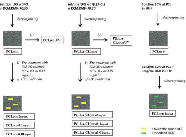
Although in vitro re-endothelization of the grafts remains time-consuming, the capability of the biomaterial to induce the in vivo adhesion of ECs derived from both the neighboring tissues or circu-lating progenitors seems to be a desirable effect. Thus, polymer sur-faces have been modified with cell adhesion sequences derived from extracellular matrix (ECM) macromolecules, such as fibronec-tin, laminin, and type-1 collagen [13]. Among these, the RGD pep-tide is the main adhesion motif used in vascular tissue engineering [14]. Covalent linking and simple coating were carried out to modify the surface. The former approach allows peptides to be uniformly distributed throughout the biomaterial, whereas the latter is less effective in terms of quantity of molecules adsorbed. Starting from these considerations, this study aims at developing a functionalized polymeric surface able to promote endothelializa-tion and consequently mimic the lumen of a blood vessel. Electrospun matrices of two bioresorbable polymers – poly-ε-caprolactone and the 70 : 30 copolymer poly-ε- poly-ε-caprolactone-co-polylactic acid (P(LLA-CL) – were taken into consideration. These surfaces became biomimetic through enrichment with a linear adhesive peptide carrying four Gly-Arg-Gly-Asp-Ser-Pro (GRGDSP) motifs per chain, designed in our laboratory, that showed higher capacity to promote osteoblast adhesion when compared with branched peptides or sequences with a single motif (RGD or GRGDSPK) [15,16]. Later in this article, it will be referred as RGD-peptide for the sake of simplification. Two different strategies were used to create the biomimetic surfaces: (i) covalent bond for photo-activation of specific surface azido groups inserted at the end of the peptide sequence (Figures 1 and 2) and (ii) inclusion of adhesive peptides in the polymeric solutions before electrospinning (only in the case of PCL scaffolds – Figure 2).
In vitro assays with endothelial cells (human umbilical vein
en-dothelial cells (HUVEC)) were performed to test the potential of biomimetic surfaces to promote adhesion and control proliferation.
Experimental Section
Materials
The solid support resins Sasrin and Amide MBHA were from Novabiochem (Merck KGaA, Darmstadt, Germany). The Fmoc protected amino acids were from Novabiochem (Merck KGaA, Darmstadt, Germany). The coupling reagents 2-(1H-benzotriazole-1-yl)-1,1,3,3-tetramethyluronium hexafluorophosphate (HBTU) and 1-hydroxybenzotriazole (HOBt) were from Advanced Biotech (Seveso, MI, Italy). N,N-Diisopropylethylamine (DIEA) and piperidine were from Biosolve (Leenderweg, Valkenswaard, Netherlands). Triethoxysilane (TES) was from Sigma-Aldrich (Steinheim, Germany). Solvents such as N,N-dimethylformamide (DMF), trifluoroaceticacid (TFA), N-methyl-2-pyrrolidone (NMP), and dichloromethane (DCM) were from Biosolve (Leenderweg, Valkenswaard, Netherlands). Poly-ɛ-caprolactone (Mn = 60 KDa), acetonitrile, and 1,1,1-3,3,3-hexafluoro-2-propanol (HFIP) were purchased from Sigma-Aldrich (Steinheim, Germany). The copolymer poly(L-lactic acid-co-ɛ-caprolactone) (70 : 30) was
purchased by PURAC biochem (Gorinchen, Holland).
Phosphate-buffered saline (PBS) tablets were purchased from Gibco Invitrogen Corp. (Paisley, UK). The Endothelial Cell Growth Medium MV2 was purchased from PromoCell GmbH (Heidelberg, Germany). Cell strainer, tissue culture-treated dishes, and fibronec-tin were from BD Biosciences (San Jose, CA, USA), whereas
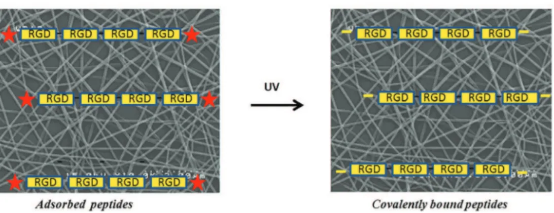
3-(4,5-Figure 1. Schematic representation of covalent grafting of photoreactive peptide N3RGD to electrospun scaffold surfaces. The photoreactive peptide is
physisorbed on the surface of the scaffold, and then the UV irradiation induces the formation of nitrenes from azido groups inserted at the ends of the peptide chain causing the formation of covalent bonds between the peptide and the polymer.
dimethylthiazol-2-yl)-2,5-dimethyltetrazolium bromide-tetrazolium dye (MTT) was provided by Sigma-Aldrich (St. Louis, MO, USA).
Methods
Peptide design and synthesis
The peptide named N3RGD (sequence: H-Phe(p-N3
)-Gly-Arg-Gly- Asp-Ser-Pro-Gly-Arg-Gly-Asp-Ser-Pro-Gly-Arg-Gly-Asp-Ser-Pro-Gly-Arg-Gly-Asp-Ser-Pro-Phe(p-N3)-NH2) is characterized by the
introduction of two aryl azides at the termini of a linear peptide composed by four GRGDSP motifs. The introduction of two aryl azides per chain was realized to increase successful grafting. The sites of introduction of photoreactive groups, used to anchor the adhesive sequence, preserves the pattern of adhesive motifs that are functional to biological activity observed. The peptide N3RGD
was synthesized as a p-amino analog with Fmoc chemistry using a Syro-I synthesizer (Multisyntech Gmbh, Germany) on Rink amide MBHA resin (0.66 mmol/g substitution, 0.13 mmol scale). The con-densation was carried out via HBTU/HOBt using double couplings (5 eq. for each amino acid insertion). The side-chain protective groups were as follows: OtBu for Asp, Pmc for Arg, and tBu for Ser Boc for Phe(p-NH21). The peptide was cleaved from the resin and
side-chain deprotected with 9.5 ml trifluoroacetic acid, 0.25 ml TES, and 0.25 ml H2O for 2 h 15 min in the dark. After the conversion
of amino groups into azido groups by reaction in solution with so-dium nitrite and soso-dium azide [17], the N-terminal Fmoc-protected crude product was Fmoc-deprotected and then purified by reverse phase-high-performance liquid chromatography (RP-HPLC) with-out use of UV-detector. The purified peptide had a 92% homogene-ity as resulted by the integration of chromatography pattern (conditions: column Vydac C18 218TP54 protein and peptide
(5 μm, 300 Å, 4.6 × 250 mm); flow rate = 1.0 ml/min; eluent A = 0.05% TFA in H2O MilliQ; eluent B = 0.05% TFA in CH3CN;
gradi-ent = 13–23% B over 20 min; and detector 214 nm).
The peptide identity was established by electrospray ionization-time of flight (ESI-TOF) mass analysis (theoretical mass: 2671.70 Da; experimental mass: 2671.18 Da).
The peptide named RGD (sequence: H-Ser-Pro-Gly-Arg-Gly-Asp- Gly-Arg-Gly-Asp-Ser-Pro-Gly-Arg-Gly-Asp-Ser-Pro-Gly-Arg-Gly-Asp-Ser-Pro-Lys-OH) was synthesized and purified as previously de-scribed [15]. The peptide homogeneity grade was 97.4% (condi-tions: column Vydac C18 218TP54 protein and peptide; flow
rate = 1.0 ml/min; eluent A = 0.05% TFA in H2O MilliQ; eluent
B = 0.05% TFA in CH3CN; gradient = 4–14% B over 20 min; and
de-tector 214 nm). ESI-TOF mass analysis confirmed the identity of the product (theoretical mass: 2424.51 Da; experimental mass: 2424.15 Da).
Electrospinning
Three different set of electrospun samples (Figure 2) were obtained by processing the following polymeric solutions:
• 10% wt/wt P(LLA-CL) (70–30) solution in DCM:DMF = 70 : 30 wt/wt;
• 25% PCL wt/wt solution in DCM:DMF = 70 : 30 wt/wt; • 10% wt PCL with or without 1 mg/ml RGD in HFIP.
The process conditions in terms of flow rate, distance be-tween electrodes, and applied voltage, are summarized in Table 1 for each polymer/solvent system. All the samples were collected for 1-h time.
The aluminum foil (15 × 15 cm) used as the collector was cut in circular samples of 1.2-cm diameter.
Scaffold sampling
The samples of copolymer were not cut directly on polymer-covered aluminum foil because the copolymer shrinks itself once placed in water. Consequently, the scaffold was immersed in water, detached from the aluminum support by peeling, and successively dried and cut. The copolymer resulted difficult to manipulate for its tendency to curl. The samples of PLC did not present these problems.
Surface crosslinking through photoreactive adhesive peptides
The peptide pre-conditioning was obtained through the incubation of samples with a solution of 1 mg N3RGD in 1 ml MilliQ water,
0.1 mg/ml, or 0.01 mg/ml for 1 h at room temperature (Figure 2). The samples were dried under vacuum in the presence of P2O5.
Each sample was irradiated for 30 min at 254 nm (distance from the lamp, 1 cm – model UVSL-15 mineralight lamp 220 V, 50 Hz, 0.12 AMPS, Ultraviolet Products INC, San Gabriel, CA, USA) [18]. The samples were extensively washed with MilliQ water and dried under vacuum.
Scanning electron microscopy
Electrospun scaffolds were sputter coated with carbon (EMITECH K950x Turbo Evaporator, EBSciences, East Granby, CT) and observed under SEM (Cambridge Stereoscan 440 SEM, Cambridge, UK). Im-ages were taken at magnifications of 30 000× with an accelerating voltage of 15 kV. The diameter range of the fabricated nanofibers was measured using image analysis software (ImageJ, National In-stitutes of Health, Bethesda, MD,USA). For each sample, three im-ages at different magnification and in three different zones of the sample were taken.
X-ray photoelectron spectroscopy measurements
X-ray photoelectron spectroscopy (XPS) investigations were per-formed in an instrument of our own construction and design equipped with a 150-mm mean radius hemispherical electron ana-lyzer and a 16-channel detector. Mg Ka non-monochromatized X-ray radiation (hν = 1253.6 eV) was used to record peptide (C1s, N1s, and O1s) and polymer (C1s and O1s) core-level spectra on the respective samples. XPS spectra of N3RGD were also recorded
as reference. In all the investigated samples, the Al2p signal from the Al foil substrate was too low to be detected; this indicated the presence of a thick mat of polymer nanofibers. The spectra were en-ergy referenced to the C1 signal of aliphatic carbons located at a binding energy (BE) of 285.0 eV [19]. The standard deviation on the measured BE values was 0.1 eV. Atomic ratios (±10%) were cal-culated from the peak intensities with Scofield’s cross sections and experimentally determined sensitivity factors. A curve-fitting analy-sis of the C1s, N1s, and O1s spectra was performed with Gaussian curves as fitting functions.
Contact angle measurements
Contact angles (CA) were measured using a SURTENS angulometer (OEG, GmbH, Frankfurt, Germany) at 25 °C ± 0.5 °C and for a relative humidity of 30% ± 2%. One sample for each kind of scaffold was set onto a silica slide, and droplets of PBS (3 μl) were dispensed onto the scaffold for contact angle measurement.
Differential scanning calorimetry
Differential scanning calorimetry apparatus (DSC from Pyris Elmer) was calibrated using Hg and In as standards, resulting in tempera-ture accuracy ±0.1 °C and enthalpy accuracy ± 0.2 J/g.
Poly(L-lactide-co-ε-caprolactone) samples in the solid state, 5 mg
in weight, were set into aluminum pans and submitted to the fol-lowing temperature program under nitrogen flow: cooling at !20 °C/min until !100 °C and successive heating from !100 to 200 °C at 10 °C/min resulting in a first DSC scan. After quenching at!100 °C, this temperature program is repeated resulting in a sec-ond DSC scan.
Chromatography determination of RGD release from the scaffold PCLHFIP 1mg/ml
The assay was carried out on three different scaffolds (PCLHFIP1mg/ml)
prepared with 1 mg/ml of RGD. Each sample was cut and its surface area determined. The samples were separated from the aluminum foil and weighed. Each sample was left in a glass tube with 1 ml of PBS at 37 °C: at each time point, a sample (0.115 ml) of solution was picked up and injected in HPLC to determine the peak area. The same volume was replaced by PBS. The peak area was correlated to RGD nmoles through a weighted titration chromatographic curve obtained injecting in HPLC known quantities of RGD. The chromatographyc conditions used were as follows: column, Vydac-C18 (5 μm, 300 Å, 4.6 × 250 mm Grace Vydac, Hesperia, CA); eluent A (H2O MilliQ with 0.05% TFA), eluent B (CH3CN, 0.05%
TFA); gradient, from 0% to 80% of B in 25 min; flow rate, 1 ml/min.; and detector, 214 nm.
Biological Assays
Cell cultures
Primary cultures of human umbilical vein endothelial cells were ob-tained by enzymatic digestion of umbilical vein endothelial layer with a 0.1% collagenase IV solution. The cells were seeded on Petri dishes previously coated with 1 μg/ml fibronectin and cultured with Endothelial Cell Growth Medium MV2supplemented with 5% fetal
calf serum (FCS), 1 μg/ml ascorbic acid, 10 ng/mL hFGF-2, 5 ng/mL hEGF, hydrocortisone 0.2 μg/ml, 20 ng/ml R3-IGF-1, 0.5 ng/ml VEGF (endothelial MV2medium kit), and 1% antibiotic solution,
contain-ing 10 ng/ml streptomycin sulfate, 250 ng/ml amphotericin-B, and 100 U/ml penicillin. This medium was referred as complete me-dium. Cultures were incubated at 37 °C in a humidified atmosphere. HUVECs were used until the fourth passage and harvested at 80% confluence.
MTT assay
The 3-(4,5-dimethylthiazol-2-yl)-2,5-dimethyltetrazolium bromide tetrazolium dye assay was used to evaluate cell adhesion at 24 h and cell proliferation at 3 and 7 days. HUVECs (3 × 10⁵cells/cm2) were seeded on PCLHFIP, PCLDCM, and P(LLA-CL)DCMlacking
adhe-sive peptides or containing them in various concentrations (1, 0.1, and 0.01 mg/ml). The peptide named RGD was incorporated in
Table 1. Process parameters for electrospinning Polymer/solvent
system Flow rate Distance Applied voltage Needle i.d. (ml/h) (cm) (kV) (mm)
P(LLA-CL)/DMC-DMF 1 10 8 0.4
PCL/DMC-DMF 1 15 8 0.4
PCLHFIP, whereas PCLDCMand P(LLA-CL)DCMcarried the photoactive
peptide N3RGD. Furthermore, some cultures grew on PCLDCM, and P
(LLA-CL)DCM without peptides and treated with UV light. Before
seeding, all scaffolds were put onto wells of 12-well plates, and cells were cultured by using Basal MV2 Medium supplemented with 1% FCS and lacking growth factors. Briefly, after 24 h, 3 and 7 days from seeding, cells were treated with MTT (0.50 mg/ml; Sigma) for 4 h. Formazane precipitates were dissolved in acidic 2-propanol (0.04 M HCl in 2-propanol; Sigma) and optical density was mea-sured at 570 nm, using a Microplate autoreader EL 13 (BIO-TEK in-struments Inc., Winooski, Vermont, USA). The linearity of absorbance of formazan over a range of 3 × 10³
–20 × 104 cells was established by determining the linear coefficient (0.9858). Re-sults were expressed as either percent of control cultures grown on polymers without peptides (taken as 100) or cell number. Their statistical comparison was performed by analysis of variance, followed by Student’s t-test.
Results
Peptide Synthesis
Initially, the synthesis of the N3RGD peptide was carried out using
Fmoc-Phe(p-N3)-OH, but this strategy did not produce the target
peptide. Instead, the introduction of Phe(p-NH2) residues and the
following conversion of amino groups into azido groups produced a yield of the target peptide (30 mg (11 μmoles) with a purity grade >95% with respect to a synthetic scale of 0.25 mmoles).
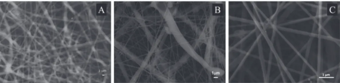
Morphological Analysis by SEM
In poly(L-lactide-co-ε-caprolactone) scaffolds, the fibers were
ran-domly oriented, the diameter varied between 0.6 and 2 μm, and the mesh width varied between 3.5 and 20 μM(Figure 3A).
The electrospun PCL, obtained with the same solvent and with the same parameters applied for P(LLA-CL), resulted in a nonwoven mat with many defects or beads. To produce the scaffold used for the biological assays, the SEM image of which was reported in Figure 3B, the concentration of the polymer solution, and the dis-tance between the needle and the collector were increased (con-centration from 10% to 25% wt/wt and distance from 10 to 15 cm). The scaffold showed a bimodal distribution of the fibers with a diameter between 1 and 3 μm in the first case, and of a few hun-dred nanometers in the second case. The formation of fiber bun-dles produced a rough surface visible to the naked eye in which meshes of tens of μm alternate with meshes of hundreds of μm. In the scaffold electrospun from a solution of PCL in HFIP, the fiber diameter varied from 0.1 to 0.2 μm and presented rather dense meshes (4 μm) (Fig. 3, C).
No morphological changes (data not shown) occurred through the enrichment of the polymer solution with peptides before electrospinning or surface treatment with photoactive peptide so-lution followed by irradiation.
X-ray Photoelectron Spectroscopy Analysis
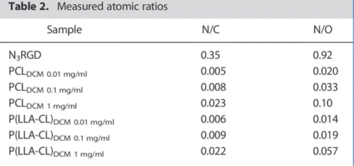
The poly-ε-caprolactone and P(LLA-CL) scaffolds and their biomi-metic analogs obtained by adding three different concentration peptide solutions and irradiation were characterized by XPS. A de-tailed description of the data is reported in reference [20]. The study demonstrated the immobilization of the RGD peptide on the sur-face for both materials, as evidenced by the appearance of an N1s peak at BE = 400.0 eV, in agreement with the expected value for amide nitrogens [19]; the N1s spectrum of pristine N3RGD shows
a single peak located at 400.0 eV, exactly. The N1s spectra recorded for both PCL and P(LLA-CL) after incubation with peptide solution at 1.0 mg/ml are shown in Figure 4 (right). Table 2 shows the mea-sured N/C and N/O atomic ratios for the two polymer films after peptide immobilization and for the reference N3RGD peptide. The
N1s signal can be detected even for the lowest peptide concentra-tion (0.01 mg/ml). The amount of immobilized peptide, measured by the N/C and N/O ratios, increases with peptide concentration of the mother solution. A plot of the measured N/C and N/O ratios as a function of peptide concentration is also shown in Figure 4 (left); the trend is very similar for the two polymer films. However, the efficiency of peptide immobilization was low: the reaction yield was, even for the samples prepared with the highest peptide con-centration solutions, less than 10% of the values calculated for the initial peptide quantity [20].
Release of the Peptide RGD from PCLHFIP 1mg/mlScaffold
A study of peptide release in solution (10 mMsodium phosphate
and 150 mMNaCl buffer at pH 7.4) at 37 °C was performed for the
PCLHFIP 1mg/mlscaffold (Figure 2) in which the peptide is not
cova-lently linked to PCL.
The kinetics of peptide release showed that the concentration of the peptide solution reached a constant value after 15 h: the quan-tity of peptide released is about one third of the initial peptide quantity (Figure 5).
Characterization of P(LLA-PC)DCM; P(LLA-PC)DCMUV; P(LLA-PC) DCM 0.01 mg/ml; P(LLA-PC)DCM 0.1 mg/ml, and P(LLA-PC)DCM 1 mg/ml
The hydrophobicity of all P(LLA-CL) samples detailed in Figure 2 was evaluated, and their thermal transitions were determined in or-der to evaluate the effect of UV irradiation and peptide functionalization on these physical characteristics.
Figure 3. SEM images of electrospun scaffolds produced using (A) 25% (wt/wt) PCL in DCM/DMF (70 : 30 wt/wt); (B) 10% (wt/wt) PCL in HFIP; and (C) 10% (wt/wt) P(LLA-CL) (70 : 30) in DCM/DMF (70 : 30 wt/wt). PCL, poly-ε-caprolactone; P(LLA-CL), poly(L-lactic acid-co-ɛ-caprolactone).
Contact angle measurements
The results of PBS CA measurements are reported in Table 3. The measurements are quite reproducible and all the sam-ples showed a high level of hydrophobicity, with CA values of about 120°.
Differential scanning calorimetry
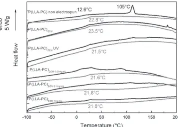
The DSC thermograms of P(LLA-CL) samples are reported in Figure 6. The first DSC thermogram of initial P(LLA-CL) samples
was characterized by a glass transition at Tg = 12.6 °C associated with the amorphous phase and a first-order transition attributed to the melting of the crystalline phase at Tm = 105 °C. On the sec-ond DSC scan performed after quenching, the only evidenced was the glass transition phenomenon at 22.8 °C. On the electrospun P(LLA-CL) thermograms, the glass transition on the first and second heating were detectable, but there was no evidence for a crystalline phase. The values of the glass transition determined from the sec-ond scan are pointed on the figure.
Biological Assays
At 24 h from seeding, cell adhesion on PCL significantly (p < 0.05) increased in all cultural conditions compared with that verified in control cultures without peptide (Figure 7). However, the cova-lent binding of the N3RGD peptide on electrospun PCL (samples
PCLDCM 0.01 mg/ml; PCLDCM 0.1 mg/ml; PCLDCM 1 mg/ml) significantly
enhanced the adhesion of endothelial cells compared with the UV irradiated surface (PCLDCMUV) and the scaffold with embedded
peptide (PCLHFIP 1 mg/ml). Furthermore, the effect appeared to be
dose-dependent and reached its maximum when pre-treated with a 1 mg/ml peptide solution (PCLDCM 1 mg/ml). Although cell
prolifer-ation was significantly higher in PCL containing peptide (samples PCLDCM 0.01 mg/ml; PCLDCM 0.1 mg/ml; and PCLDCM 1 mg/ml) compared
with the proliferation observed on the polymer alone, after 7 days, no statistical differences were detected among scaffolds with em-bedded and covalently linked peptides. Thus, the effects of the bio-mimetic surfaces on cell proliferation were not as visible as those on cell adhesion. Overall, our data suggests that the covalent binding
Figure 4. N1s spectra of PCL and P(LLA-CL) films after incubation with 1.0 mg/ml peptide solution; markers represent experimental points and the line represent the fitting component (right). Plot of the measured atomic ratios (N/C and N/O) as a function of peptide concentration in the mother solution; both abscissa and ordinate are in log scale (left). Error bars (±10%) are also shown in the figure. PCL, poly-ε-caprolactone; P(LLA-CL), poly(L-lactic
acid-co-ɛ-caprolactone).
Table 2. Measured atomic ratios
Sample N/C N/O N3RGD 0.35 0.92 PCLDCM 0.01 mg/ml 0.005 0.020 PCLDCM 0.1 mg/ml 0.008 0.033 PCLDCM 1 mg/ml 0.023 0.10 P(LLA-CL)DCM 0.01 mg/ml 0.006 0.014 P(LLA-CL)DCM 0.1 mg/ml 0.009 0.019 P(LLA-CL)DCM 1 mg/ml 0.022 0.057
Figure 5. Release of RGD from PCLHFIP 1 mg/ml scaffold. PCL,
poly-ε-caprolactone.
Table 3. Contact angle measurements of P(LLA-CL) scaffolds
Sample N Mean CA (°) CA SD(°) Control P(LLA-CL)DCM 3 118.9 0.52 P(LLA-CL)DCMUV 3 122.9 1.06 P(LLA-CL)DCM 0.01mg/ml 3 118.3 1.19 P(LLA-CL)DCM 0.1mg/ml 2 120.4 0.99 P(LLA-CL)DCM 1mg/ml 3 122.3 0.53
approach is more valuable than simple embedding to induce endo-thelial cell adhesion.
Similar to that observed on PCL, at 24 h from seeding, the cova-lent binding of the N3RGD peptide on electrospun P(LLA-CL)
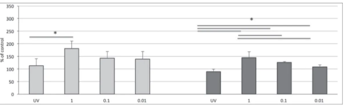
(sam-ples P(LLA-CL)DCM 0.01 mg/ml; P(LLA-CL)DCM 0.1 mg/ml; and P(LLA-CL) DCM 1 mg/ml) caused a significant (p < 0.05) increase in cell adhesion
compared with the control cultures without peptide (Figure 8). Pre-treatment with a 1 mg/ml peptide solution seemed to be the most effective concentration (P(LLA-CL)DCM 1 mg/ml), because the
en-hancement of cell adhesion was significantly higher than that veri-fied on the UV-irradiated polymer (P(LLA-CL)DCMUV). At 7 days, cell
proliferation was increased significantly with 1 and 0.1 mg/ml linked peptide compared with that verified on control and UV-irradiated scaffolds.
Discussion
X-ray photoelectron spectroscopy data confirmed the possibility to anchor bioactive peptides on the surface of an electrospun bioma-terial through the introduction in the peptide sequence of an azido group, which is converted by UV irradiation into a nitrene. The
proposed strategy allows grafting the bioactive sequence to every type of surface in a one-step procedure. This method did not re-quire the use of particular complementary functional groups on the surface. In addition, the introduction of the azido group in the side chain of each terminal residue, not involved in the bioactive se-quence, ensures the specifically oriented grafting of the peptide. This method allows producing different peptide surface densities by simply modifying the peptide solution concentration used to treat the scaffold before UV-irradiation, while the efficiency of im-mobilization is low: the reaction yield is, even for the samples pre-pared with the highest peptide concentration solutions, less than 10% of the values calculated for the initial peptide quantity [20].
Our data showed that UV irradiation and peptide grafting on electrospun scaffolds did not produce important modifications in wettability and global thermal properties.
The effect of UV irradiation on polymeric surface wettability is controversial. Yeh et al. showed that even with a long time irradia-tion of PCL with UV at 254 nm, no change is detected on the hydro-philicity of PCL, i.e. the surface remains hydrophobic [21]. Furthermore, EC adhesion and proliferation may be improved by UVC irradiation (in the 172 nm range) or plasma treatment that in-creases the hydrophilicity of some polymer surfaces [22].
Surface wettability is an important property of biomaterials and can affect the attachment, proliferation, migration, and viability of cells. The water contact angle performed on dry surfaces can be used to evaluate wettability and surface modification and it can be compared with values from literature on different scaffolds. We chose to perform CA measurements with PBS instead of pure deionized (DI) water because PBS is a medium of interest for biolog-ical simulations, and PBS drops are already used in literature data [23]. Because surface tension of water only increases of about 0.4% in PBS, the errors that would be caused by the use of PBS are of the same order of magnitude as the statistical errors that usu-ally occur in normal CA. So CA measurements in PBS are analogous to CA measurements in DI water [24].
All the samples showed a high level of hydrophobicity, with CA values of about 120°, with variations that do not exceed 4% and that cannot be associated with significant changes. Water CA values of about 70–74° have been reported for flat PCL films [21,25] and of about 77° for flat PLLA films [26]. In the case of electrospun P(LLA-CL) scaffolds, the values of about 120° certainly reflect a super hydrophobic behavior because of the specific roughness of the surface [27] as observed by SEM. It was already shown that post-electrospinning nanofibers of PCL could exhibit enhanced hydrophobicity [28]. Initial P(LLA-CL) are semi-crystalline. It has been previously proven by X-ray diffraction in P(LLA-CL) 70 : 30 that only lactide units were able to crystallize [29]. It was also reported that copolymerization of PLLA with CL had an important effect on the thermal properties; both the glass transition of the co-polymer and the melting temperature of PLLA crystalline sequences decrease with the increase of the amount of CL, because the intro-duction of CL units, which have five methylene groups, increases the chain flexibility and mobility [30]. By comparison, with previous DSC studies on different P(LLA-CL) 70 : 30 copolymers, the commer-cial P(LLA-CL) 70 : 30 copolymer studied here is a moderately block polymer with a number average sequence length for LLA units between 4 and 3 [31]. At 37 °C, P(LLA-CL) 70 : 30 is in a visco-elastic state in contrast with pure PLLA (Tg ~ 60 °C), which is glassy.
As evidenced on the first DSC thermogram of electrospun P (LLA-CL) with the vanishing of the melting phenomenon and the persistence of the glass transition, electrospinning induces amorphization of the copolymer. The crystalline microstructure in
Figure 7. MTT assay at 24 h and 7 days from seeding on electrospun PCL. Results, expressed as percent of control cultures grown on polymers without peptides (taken as 100), are means ± SD of three independent experiments. Bars: light gray 24 h; dark gray 7 days. *p < 0.05, Student’s t-test.
Figure 6. DSC thermograms (black line: 1st heating scan; gray line: 2nd heating scan) of P(LLA-CL) samples. DSC, differential scanning calorimetry; P(LLA-CL), poly(L-lactic acid-co-ɛ-caprolactone).
electrospun fibers has often been reported as being not well-developed or totally inhibited [32].
As shown in Figure 6, UV irradiation induces a slight decrease (!2 °C) of the glass transition temperature, whereas peptide grafting does not result in further modifications of this parameter, meaning that the global thermal properties of the biomaterial are preserved.
While the functionalization process did not modify wettability and global thermal properties, it is important to underline that the different matrices used (PCLDCM, PCLHFIP, and P(LLA-CL)DCM)
were characterized by different initial fiber diameter and mesh dimension.
Biological assays demonstrated that in all cases, the peptide en-richment of the scaffolds increases HUVEC adhesion. The best re-sults are obtained at the higher peptide solution concentration (1 mg/ml). This peptide incorporated to PCL did not give the same results. The different performance may be due to the random pep-tide presentation toward the cells, to the partial peppep-tide presenta-tion on the surface, and to de-adsorppresenta-tion of the embedded and non-covalently linked peptide. As demonstrated by the peptide re-lease curve (Figure 5) registered for PCLHFIP 1 mg/mlsample, the
con-centration of the peptide in solution reached a constant value after 15 h: the quantity of peptide released was about one third of the initial peptide quantity. It is important to note that RGD peptides in solution could promote the inhibition of cell adhesion, an effect opposite to that induced by grafted RGD sequences. Arginine-Gly-cine-Aspartic Acid peptides acts as adhesion factors when chemi-cally bound to the surface of biomaterials, or represents pro-apoptotic agents when used as soluble factors [33]. Indeed, they compete with ECM components for the binding to cell surface, thus inducing a lack of anchorage. In our previous work [34], we have demonstrated that (GRGDSP)4K dissolved in culture medium
inhibited the adhesion and proliferation of HUVECs. Consequently, this study provides further evidence on the superiority of covalent binding versus physical adsorption in the formulation of bioactive surfaces for tissue engineering or regenerative medicine applications.
The comparison between the two scaffolds functionalized with photoreactive peptides (Figure 9) showed no difference when treated with a higher concentration of peptide solution, but a bet-ter performance of the copolymer at the lower concentration of peptide solution or in the control surface, whether or not treated with UV irradiation. The difference can be due to the structure of the electrospun copolymer whose mesh dimensions are ideal for
epithelial cells. In fact, endothelial cells prefer surfaces with porosity in the range of 18–60 μm [9]. Also worth noting is the capacity of the biomimetic coverage to eliminate this difference at optimized peptide concentrations on the surface.
The scaffolds functionalized with photoreactive peptides seem to promote HUVEC adhesion more than HUVEC proliferation. In view to blood vessel replacement, a key factor is a quick re-endothelization of biomaterials, because the coverage with endo-thelial cells avoid platelet adhesion and, in turn, thrombus forma-tion. On the other hand, cell growth is an event that takes place after adhesion but it is not particularly desired for our aims: prolifer-ation of endothelial cells, seeded on the biomimetic scaffold, could cause intimal hyperplasia and finally blood vessel obstruction.
In the past, the functionalization of biomaterials with adhesive peptides has raised some skepticism. The perplexity concerns the efficacy of such strategy in in vivo contexts due to serum proteins adsorption on the functionalized surface. Recently, Battista E. et al. [35], engrafting an RGD peptide on PCL surfaces, demonstrated that the presence of serum proteins effectively creates a layer that covers peptide covalently conjugated on the surface but, after the first adhesion phase, cells dig into the physisorbed protein layer and reach the submerged RGD peptide for establishing a more sta-ble adhesion. The driving force of the process is the preference by cells for adhering on firmly bound RGD on which to build more ro-bust focal adhesions and a mechanically stable cytoskeleton via mechanosensing mechanism. The data reported [35] are in
Figure 9. Comparison between electrospun PCL (light gray) and P(LLA-CL) (dark gray) at 24 h from seeding. Results, expressed as cell number, are means ± SD of three independent experiments. *p < 0.05, Student’s t-test. PCL, poly-ε-caprolactone; P(LLA-CL), poly(L-lactic acid-co-ɛ-caprolactone).
Figure 8. MTT assay at 24 h and 7 days from seeding on electrospun P(LLA-CL). Results, expressed as percent of control cultures grown on polymers without peptides (taken as 100), are means ± SD of three independent experiments. Bars: light gray 24 h; dark gray 7 days; 1, 0.1, and 0.01 refer to the concentration in mg/ml of N3RGD in the solution used for pretreatment before irradiation. *p < 0.05, Student’s t-test. The statistical comparison between cultures grown on
electrospun PCL and P(LLA-CL) (Figure 7) revealed that at 24 h from seeding, cell adhesion was higher on P(LLA-CL) in UV-irradiated cultures and in those containing the peptide, covalently linked with 0.01 mg/ml solution. On the contrary, no differences were detected when the (GRGDSP)4K peptide was
agreement with our results [36] demonstrating that the covalent functionalization of biomaterials with adhesive peptides increases cell adhesion but also adhesion strength. In addition, the prefer-ence by cells for firmly bound RGD could explain the different per-formance of covalent grafted RGD versus embedded RGD.
These new experimental evidences shed light on the mecha-nisms that give rise to the behavior of the cells seeded on function-alized surfaces; support the efficacy of the covalent conjugation of adhesive motifs also in the presence of serum and open new per-spectives in the development of biomaterials decorated with finely tuned ligands able to support the assembly of the cell cytoskeleton in a controlled manner.
Conclusions
The use of azido-tagged peptides allows the surface modification of electrospun matrices. The covalent anchoring by activation of the azido groups with ultraviolet radiation is preferable to the simple in-clusion of the adhesive peptides in the electrospun scaffold as regards the promotion of cell adhesion. While not being particularly efficient, the proposed method is simple, versatile, and able to pro-vide matrices with different degree of functionalization (peptide density) and to assure the selective grafting of the adhesive pep-tides. Although the matrices used in this study showed similar ther-mal and surface properties, they have different morphological characteristics. Nevertheless, the covalent functionalization with adhesive peptides seems to be the dominant factor in the outcome of the bioassay.
Acknowledgements
The authors would like to thank Dr Paolo Pontini for his help in the preparation of the scaffolds.
References
1 Hirsch AT, Allison MA, Gomes AS, Corriere MA, Duval S, Ershow AG, Hiatt WR, Karas RH, Lovell MB, McDermott MM, Mendes DM, Nussmeier NA, Treat-Jacobson D. A call to action: women and peripheral artery disease: a scientific statement from the American Heart Association. Circulation 2012; 125: 1449–1472. DOI: 10.1161/ CIR.0b013e31824c39ba.
2 Piccone V. Alternative techniques in coronary artery reconstruction.In
Modern Vascular Grafts, McGraw-Hill Book Company: New York, NY,
1987; 253–260.
3 Neff LP, Tillman BW, Yazdani SK, Machingal MA, Yoo JJ, Soker S, Bernish BW, Geary RL, Christ GJ. Vascular smooth muscle enhances functionality of tissue-engineered blood vessels in vivo. J. Vasc. Surg. 2011; 53: 426–434. DOI: 10.1016/j.jvs.2010.07.054.
4 Thomas AC, Campbell GR, Campbell JH. Advances in vascular tissue engineering. Cardiovasc. Pathol. 2003; 12: 271–276. DOI: 10.1016/ S1054-8807(03)00086-3.
5 Nemeno-Guanzon JG, Lee S, Berg JR, Jo YH, Yeo JE, Nam BM, Koh YG, Lee JI. Trends in tissue engineering for blood vessels. J. Biomed.
Biotechnol. 2012; 2012: 956345. DOI: 10.1155/2012/956345.
6 Hung HS, Chen HC, Tsai CH, Lin SZ. Novel approach by nanobiomaterials in vascular tissue engineering. Cell Transplant. 2011; 20: 63–70. DOI: 10.3727/096368910X532864.
7 Conconi MT, Borgio L, Di Liddo R, Sartore L, Dal ZD, Amistà P, Lora S, Parnigotto PP, Grandi C. Evaluation of vascular graft based on polyvinyl alcohol cryogels. Mol. Med. Rep. 2014; 10: 1329–1334. DOI: 10.3892/mmr.2014.2348.
8 Dettin M. Bioactive surfaces using peptide grafting in tissue engineering. In Cellular Response to Biomaterials, Woodhead Publishing Limited CRC Press: Cambridge, UK, 2009; 479–502.
9 Andrews KD, Hunt JA. Developing smaller-diameter biocompatible vascular grafts.In Cellular Response to Biomaterials, Woodhead Publishing Limited, CRC Press: Cambridge, UK, 2009; 212–233.
10 Liu W, Thomopoulos S, Xia Y. Electrospun nanofibers for regenerative medicine. Adv. Healthc. Mater. 2012; 1: 10–25. DOI: 10.1002/ adhm.201100021.
11 Pankajakshan D, Agrawal DK. Scaffolds in tissue engineering of blood vessels. Can. J. Physiol. Pharmacol. 2010; 88: 855–867. DOI: 10.1139/ y10-073.
12 Zhao Y, Zhang S, Zhou J, Wang J, Zhen M, Liu Y, Chen J, Qi Z. The development of a tissue-engineered artery using decellularized scaffold and autologous ovine mesenchymal stem cells. Biomaterials 2010; 31: 296–307. DOI: 10.1016/j.biomaterials.2009.09.049.
13 Monchaux E, Vermette P. Effects of surface properties and bioactivation of biomaterials on endothelial cells. Front. Biosci. 2010; 2: 239–255. 14 Li J, Ding M, Fu Q, Tan H, Xie X, Zhong Y. A novel strategy to graft RGD
peptide on biomaterials surfaces for endothelization of small-diameter vascular grafts and tissue engineering blood vessel. J. Mater. Sci. Mater.
Med. 2008; 19: 2595–2603. DOI: 10.1007/s10856-007-3354-5.
15 Dettin M, Conconi MT, Gambaretto R, Pasquato A, Folin M, Di Bello C, Parnigotto PP. Novel osteoblast-adhesive peptides for dental/orthopedic biomaterials. J. Biomed. Mat. Res. 2002; 60: 466–471. 16 Dettin M, Conconi MT, Gambaretto R, Bagno A, Di Bello C, Menti AM,
Grandi C, Parnigotto PP. Effect of synthetic peptides on osteoblast adhesion. Biomaterials 2005; 26: 4507–4515.
17 Grant GA. Synthetic Peptides: A User’s Guide, Oxford University Press: New York, 2002; 328–329.
18 Woulters W, Van Dun J, Laduron PM. Photoaffinity labelling of dopamine receptors. Synthesis and binding characteristics of azapride. Eur. J.
Biochem. 1984; 145: 273–278.
19 Moulder JF, Stickle WF, Sobol PE, Bomben KD. Handbook of X-ray
Photoelectron Spectroscopy, Physical Electronics Inc. eds: Eden Prairie,
MN, 1996.
20 Iucci G, Ghezzo F, Danesin R, Modesti M, Dettin M. Biomimetic peptide-enriched electrospun polymers: a photoelectron and infrared spectroscopy study. J. Appl. Pol. Sci. 2011; 122: 3574–3582.
21 Yeh C-C, Chen C-N, Li Y-T, Chang C-W, Cheng M-Y, Chang H-I. The effect of polymer molecular weight and UV radiation on physical properties and bioactivities of PCL films. Cellular Polymers 2011; 30: 227–242. 22 De Mel A, Jell G, Stevens MM, Seifalian AM. Biofunctionalization of
biomaterials for accelerated in situ endothelialization: a review.
Biomacromolecules 2008; 9: 2969–2979. DOI: 10.1021/bm800681k.
23 Kim J, Shen AQ, Lee KH, Cangelosi GA, Chung JH. Contact angle changes induced by immunocomplex formation. Analyst 2014; 139: 1340–1344. DOI: 10.1039/c3an02189k.
24 Van Oss CJ. The Properties of Water and Their Role in Colloidal and Biological
Systems, Academic Press, Elsevier: Amsterdam, Holland, 2008; 160.
25 Chung T-W, Yang M-G, Liu D-Z, Chen W-P, Pan C-I, Wang S-S. Enhancing growth human endothelial cells on Arg-Gly-Asp (RGD) embedded poly (ε-caprolactone) (PCL) surface with nanometer scale of surface disturbance. J. Biomed. Mater. Res. 2005; 72A: 213–219.
26 Cui YL, Di Qi A, Liu WG, Wang XH, Wang H, Ma DM, De Yao K. Biomimetic surface modification of poly(l-lactic acid) with chitosan and its effects on articular chondrocytes in vitro. Biomaterials 2003; 24: 3859–3868. 27 Yoshimitsu Z, Nakajima A, Watanabe T. Effects of surface structure on the
hydrophobicity and sliding behavior of water droplets. Langmuir 2002; 18: 5818–5822.
28 Bosworth L, Downes S. Biocompatible Three-Dimensional Scaffolds for
Tendon Tissue Engineering using Electrospinning, in Cellular Response to Biomaterials, Elsevier: Amsterdam, Holland, 2008; 1–27.
29 Ugartemendia JM, Sarasua JR, Proceedings of the 69th Annual Technical Conference of the Society of Plastic Engineering ANTEC 2011; 1: 230. 30 Lu XL, Sun ZJ, Cai W, Gao ZY. Study on the shape memory effects of poly
(L-lactide-co-epsilon-caprolactone) biodegradable polymers. J. Mater.
Sci. Mater. Med. 2008; 19: 395–399.
31 Fernández J, Etxeberria A, Ugartemendia JM, Petisco S, Sarasua J-R. Effects of chain microstructures on mechanical behavior and aging of a poly(L-lactide-co-ε-caprolactone) biomedical thermoplastic-elastomer. J. Mech. Behav. Biomed. Mater. 2012; 12: 29–38. DOI: 10.1016/j.jmbbm.2012.03.008.
32 Zong X, Ran S, Fang D, Hsiao BS, Chu B. Control of structure, morphology and property in electrospun poly(glycolide-co-lactide) non-woven membranes via post-draw treatments. Polymer 2003; 44: 4959–4967. 33 Swenson S, Ramu S, Markland FS. Antiangiogenesis and RGD
-containing snake venom disintegrins. Curr. Pharm. Des. 2007; 13: 2860–2871.
34 Conconi MT, Ghezzo F, Dettin M, Urbani L, Grandi C, Guidolin D, Nico B, Di Bello C, Ribatti D, Parnigotto PP. Effects on in vitro and in vivo
angiogenesis induced by small peptides carrying adhesion sequences.
J. Pept. Sci. 2010; 16: 349–357. DOI: 10.1002/psc.1251.
35 Battista E, Causa F, Lettera V, Panzetta V, Guarnieri D, Fusco S, Gentile F, Netti PA. Ligand engagement on material surfaces is discriminated by cell mechanosensoring. Biomaterials 2015; 45: 72–80. DOI: 10.1016/j. biomaterials.2014.12.012.
36 Bagno A, Piovan A, Dettin M, Brun P, Gambaretto R, Palù G, Di Bello C, Castagliuolo I. Improvement of Anselme’s adhesion model for evaluating of human osteoblast response to peptide-grafted titanium surfaces. Bone 2007; 41: 704–712.