HAL Id: inserm-02529958
https://www.hal.inserm.fr/inserm-02529958
Submitted on 7 Apr 2020
HAL is a multi-disciplinary open access
archive for the deposit and dissemination of
sci-entific research documents, whether they are
pub-lished or not. The documents may come from
teaching and research institutions in France or
abroad, or from public or private research centers.
L’archive ouverte pluridisciplinaire HAL, est
destinée au dépôt et à la diffusion de documents
scientifiques de niveau recherche, publiés ou non,
émanant des établissements d’enseignement et de
recherche français ou étrangers, des laboratoires
publics ou privés.
Corine Bertolotto
To cite this version:
Charlotte Pandiani, Guillaume Beranger, Justine Leclerc, Robert Ballotti, Corine Bertolotto. Focus on
cutaneous and uveal melanoma specificities. Genes and Development, Cold Spring Harbor Laboratory
Press, 2017, 31 (8), pp.724-743. �10.1101/gad.296962.117�. �inserm-02529958�
REVIEW
Focus on cutaneous and uveal melanoma
specificities
Charlotte Pandiani, Guillaume E. Béranger, Justine Leclerc, Robert Ballotti, and Corine Bertolotto
U1065, Institut National de la Santé et de la Recherche Médicale Centre Méditerranéen de Médecine Moléculaire, Université Côte d’Azur, 06200 Nice, France
Cutaneous melanoma (CM) and uveal melanoma (UM) derive from cutaneous and uveal melanocytes that share the same embryonic origin and display the same cellular function. However, the etiopathogenesis and biological behaviors of these melanomas are very different. CM and UM display distinct landscapes of genetic alterations and show different metastatic routes and tropisms. Hence, therapeutic improvements achieved in the last few years for the treatment of CM have failed to ameliorate the clin-ical outcomes of patients with UM. The scope of this review is to discuss the differences in tumorigenic pro-cesses (etiologic factors and genetic alterations) and tumor biology (gene expression and signaling pathways) between CM and UM. We develop hypotheses to explain these differences, which might provide important clues for research avenues and the identification of actionable vul-nerabilities suitable for the development of new therapeu-tic strategies for metastatherapeu-tic UM.
Physiological function of melanocytes
Melanocytes are cells responsible for the synthesis of melanin pigments within organelles called melanosomes through an enzymatic cascade involving tyrosinase, tyrosinase-related protein-1 (TYRP1), and TYRP2/DCT (dopachrome tautomerase). Two types of pigment are pro-duced: the brown/black pigment eumelanin and the or-ange/yellow pigment pheomelanin; the latter is formed in the presence of cysteine or glutathione. The proportion of these two types of melanin defines the variation in skin and iris color. The ratio of eumelanin/pheomelanin is sig-nificantly greater in both dark brown skin and eyes than in pale skin and eyes with light-colored irises (hazel, green, yellow-brown, and blue in color) (Rees 2004; Wakamatsu et al. 2008).
Melanocytes derive from neural crest cells. These un-differentiated cells, called melanoblasts, migrate to their
final location, where they synthesize melanin. They are found in various parts of the human body, such as the skin, eyes, meninges, heart, and cochlea. The role and function of melanocytes are well established in skin but not in other anatomical locations.
In the epidermis, melanocytes transfer melanin-con-taining melanosomes to neighboring keratinocytes to en-sure homogeneous pigmentation and provide efficient skin protection against the harmful effects of ultraviolet radiation (UVR) from solar light (Brenner and Hearing 2008).
In the eyes, melanocytes can be found in (1) the conjunc-tiva, a nonkeratinized epithelium that covers the anterior part of the sclera and the internal surface of the eyelids, and (2) all areas of the uvea: the iris, ciliary body, and cho-roid. The role of melanocytes in the conjunctiva remains unknown. The quantity and quality of melanin pigment in the iris determine eye color. However, in contrast to the skin, the iris color remains stable after exposure to sunlight. Furthermore, the presence of melanin in uveal melanocytes is thought to contribute to eye protection against ocular diseases that can cause blindness, including age-related macular degeneration and uveal melanoma (UM) (Sarna 1992). However, how melanin mediates this protection remains mostly unknown.
The presence of melanocytes in organs that are not ex-posed to UVR indicates that these cells might have func-tions other than those solely related to photoprotection. Melanocytes in the stria vascularis of the cochlea are in-volved in the generation of endolymph-mediated action potentials necessary for normal hearing (Barrenas and Lindgren 1990; Tachibana 1999) and in equilibrium func-tion (Takeda et al. 2007). Brain melanocytes are associated with neuroendocrine functions and may also protect against oxidative damage (Zecca et al. 2008). Heart mela-nocytes play a role in the mechanical properties of the valves (Carneiro et al. 2015) and have been shown to be in-volved in atrial arrhythmia (Levin et al. 2009).
[Keywords: cancer; melanoma; skin] Corresponding author: [email protected]
Article is online at http://www.genesdev.org/cgi/doi/10.1101/gad.296962. 117.
© 2017 Pandiani et al. This article is distributed exclusively by Cold Spring Harbor Laboratory Press for the first six months after the full-issue publication date (see http://genesdev.cshlp.org/site/misc/terms.xhtml). After six months, it is available under a Creative Commons License (At-tribution-NonCommercial 4.0 International), as described at http:// creativecommons.org/licenses/by-nc/4.0/.
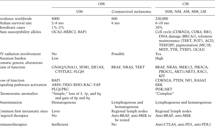
In this review, we provide several hypotheses to explain why cells sharing the same embryonic origin and cellular functions (i.e., melanin synthesis) are subjected to differ-ent tumor transformation processes. We discuss the bio-logical and genetic differences between skin and eye melanomas and, based on these differences, how treat-ment and clinical outcomes are affected (Table 1).
Classification and prognosis of cutaneous melanoma (CM) and ocular melanoma (OM)
Both CM and OM arise from melanocyte transformation and represent deadly forms of cancer. Their rate is higher among Caucasians compared with African Americans (McLaughlin et al. 2005; Jovanovic et al. 2013). In most cases, they both occur de novo but can also develop from pre-existing melanocytic lesions such as nevi or pri-mary acquired melanocytosis (Tsao et al. 2003; Jovanovic et al. 2013).
The incidence of CM, which develops from cutaneous melanocytes, has increased dramatically in white
popula-tions over the past several decades to reach 230,000 new cases worldwide each year (World Health Organization) and accounts for 1.6% of all diagnosed cancers.
A clinico-anatomical classification (Clark’s classifica-tion) based on the site of cancer occurrence and histological morphology distinguished the following five types of CM: superficial spreading melanoma, nodular melanoma, len-tigo maligna melanoma, mucosal melanoma, and acral melanoma (Clark et al. 1986). Superficial spreading mela-noma presents as an enlarging patch during the radial growth phase, which subsequently extends downward through the skin in the vertical growth phase. Nodular mel-anoma, which presents as a nodule, has a propensity to grow vertically and display aggressive behaviors. Lentigo maligna melanoma grows in diameter slowly over many years. It is associated with cumulative sun exposure and thus is found most often in the elderly. Acral melanoma in-volves the nonpigmented palmoplantar and subungual ar-eas, and mucosal melanoma can occur in all mucosal surfaces. These lesions have been termed acral lentiginous melanoma because they share several features and often present a lentiginous component (Arrington et al. 1977).
Table 1. Comparison of cutaneous melanoma (CM) and UM features
OM CM
UM Conjunctival melanoma SSM, NM, AM, MM, LM
Incidence worldwide 8000 800 230,000
Median survival rate 2–8 mo 8 mo 6–10 mo
Hereditary cases 1%–2% 10%
Main susceptibility alleles OCA2–HERC2, BAP1 Cell cycle (CDKN2A, CDK4, RB1),
DNA damage (BRCA1), telomere maintenance (TERT, POT1, ACD, TERF2IP), pigmentation (MC1R, MITF, TYR, TYRP1, OCA2)
UV radiation involvement No Possible Yes
Mutation burden Low High
Somatic genetic alterations
Gain of function GNAQ/GNA11, SF3B1, EIF1AX, CYSTLR2,ΡLCβ4
BRAF, NRAS, TERT BRAF, NRAS, MEK1/2, PIK3CA, PIK3CG, AKT1/AKT3, RAC1, KIT
Loss of function BAP1 CDKN2A, PTEN, NF1, RASA2
Signaling pathways activation ARF6–TRIO–RHO–RAC–YAP ERK
PLCβ/PKC PI3K/AKT
Chromosome anomalies “Simple,” loss of 3, 1p, and 6q and gain of 6p and 8q
“Complex”
Dissemination Hematogenous Lymphogenous and
hematogenous
Lymphogenous and hematogenous Common first metastatic sites Liver Regional lymph nodes Regional lymph nodes
Targeted therapies No Anti-BRAF, anti-MEK to
be tested
Anti-BRAF, anti-MEK
Immunotherapies Inefficient No Anti-CTLA4, anti-PD1, anti-PDL1
(OM) Ocular melanoma; (SSM) superficial spreading melanoma; (NM) nodular melanoma; (AM) acral melanoma; (MM) mucosal mela-noma; (LM) lentigo maligna melamela-noma; (BAP1) BRCA1-associated protein; (CDKN2A) cyclin-dependent kinase (CDK) inhibitor 2A; (BRAF) brain rapidly accelerated fibrosarcoma; (TERT) telomerase reverse transcriptase; (MC1R) melanocortin-1 receptor; (MITF) microphthalmia-associated transcription factor; (GNAQ) guanine nucleotide-binding protein G(q) subunitα; (GNA11) guanine nucleo-tide-binding protein subunitα-11; (SF3B1) splicing factor 3B, subunit 1; (EIF1AX) eukaryotic translation initiation factor 1A, X-linked; (PLCβ) phospholipase C β; (PI3K) phosphatidylinositol-3-kinase; (PKC) protein kinase C; (AKT) protein kinase B; (RAC1) Ras-related C3 botulinum toxin substrate 1; (PTEN) phosphatase and tensin homolog; (NF1) neurofibromin 1; (RASA2) RAS (Rous sarcoma) p21 protein activator 2; (ARF6) ADP-ribosylation factor 6; (YAP) yes-associated protein; (ERK) extracellular regulated kinase; (CTLA4) cy-totoxic T-lymphocyte-associated protein 4; (PD1) programmed cell death 1; (PDL1) PD ligand 1.
Very early stage skin-localized melanoma (Breslow <1 mm) can be cured by wide surgical excision and has a 5-yr survival rate of >98%. In contrast, when diagnosis is de-layed, CM becomes increasingly more devastating, and in-dividuals display an increased risk of developing lymph node and visceral metastases. CM is believed to spread mainly via the lymphatic route, although hematogenous diffusion has also been reported (Zbytek et al. 2008). Al-most all organs can be involved, but the Al-most common sites for distant CM metastases are the lungs, liver, bones, and brain. Until 2012, studies have shown that patients with distant metastatic CM had a median survival rate typically ranging from 6 to 10 mo and a 5-yr survival rate of∼15%–20% (Tas 2012).
OM, which originates from eye melanocytes, is the most common primary malignancy in the adult popula-tion. OM is classified based on the anatomic site of origin as conjunctival melanoma or UM. The large majority of OM originates from the uvea (95%), comprising the poste-rior uvea (choroid 90% and ciliary body 5%) and the ante-rior uvea (iris 5%). The UM staging system is based on the largest basal tumor diameter, ciliary body involvement, and extraocular involvement (Kujala and Kivela 2005). Approximately 8000 new cases of UM and 800 new cases of conjunctival melanoma are diagnosed worldwide each year. CM is 20–30 times more common than UM and 360–900 times more common than conjunctival melano-ma (Singh and Topham 2003; Wong et al. 2014). In con-trast to the incidence of UM, which has remained stable over the last three decades, the incidence of conjunctival melanoma is increasing (Triay et al. 2009). In the early stages, UM usually presents as a pigmented choroidal nodular mass in the eye fundus, growing toward the vitre-ous space with a typical mushroom shape. It can extend through sclera or the optic nerve in advanced stages. Symptoms of UM include blurred vision and seeing flash-ing lights and shadows, but most UM tumors are initially completely asymptomatic and are diagnosed by an oph-thalmologist during a routine sight test, accounting for their frequent late stage diagnosis. Despite successful treatment of the primary tumor, only 1%–3% of patients have detectable extraocular lesions at diagnosis; up to 50% of patients develop metastases. Consequently, micrometastases appear to be established several years be-fore the diagnosis of UM. UM spreads mainly via the bloodstream (i.e., hematogenously) (Dithmar et al. 2000). In 80%–90% of UM cases, the liver is the most common metastatic site, with the second most common site being the lung (Rietschel et al. 2005; Singh et al. 2005). Impor-tantly, a nonliver first metastasis has been correlated with improved survival. At the metastatic stage, long-term survival is rare. Patients with liver metastases have a median survival time of 2–8 mo, and 80% of patients die within 1 yr (Diener-West et al. 2005). Conjunctival melanoma is a completely different entity and generally presents as a pigmented nodular lesion that is usually on the bulbar conjunctiva and often involves the limbus (Shields et al. 2011b). Most conjunctival tumors do not cause symptoms and are diagnosed during a routine eye examination by an ophthalmologist. Approximately
20%–30% of people will develop metastatic disease. Con-junctival melanoma disseminates via the lymphatics and the bloodstream to invade the lungs, brain, liver, skin, bones, and gastrointestinal tract but can undergo direct extension to the eyeball and orbit (Kenawy et al. 2013). The melanoma-specific survival rate is 86% at 5 yr and 71% at 10 yr (Missotten et al. 2005).
Risk factors and genetic predisposition
The etiology of melanoma is complex and heterogeneous because it involves environmental, phenotypic, and ge-netic risk factors.
The major risk factors for CM include a personal and fa-milial history of CM, a large number of nevi/dysplasic nevi, sun exposure, and skin reactions to sun exposure ac-cording to the phototype. Approximately 10% of CM is es-timated to exhibit familial inheritance. Mutations in cyclin-dependent kinase (CDK) inhibitor 2A (CDKN2A) are found in up to 40% of cases of familial melanoma (Hus-sussian et al. 1994). CDKN2A encodes completely distinct proteins from two alternatively spliced transcripts, p16INK4a (inhibitor of kinase a) and p14ARF (alternative reading frame). p16INK4ainhibits CDK4 and CDK6, thus preventing phosphorylation of the retinoblastoma tumor suppressor RB1 and blocking E2F transcriptional activa-tion. p14ARFinhibits human double minute 2 (HDM2), leading to p53 stabilization and increased expression of its target gene, CDKN1A.
The second high-risk CM susceptibility gene, for which only a few families have been reported to carry mutations, is CDK4. Germline mutations in CDK4 contain arginine at position 24 instead of cysteine (p.R24C) or histidine (p.R24H) and prevent its interaction with p16INK4A(Zuo et al. 1996).
Additionally, germline inactivation of RB1 predisposes carriers to CM, at least those who survive their retinoblas-toma, a rare cancer of the eye (Fletcher et al. 2004). Hence, multiple mechanisms operate in CM to overcome the RB-dependent G1 arrest, thereby favoring improper progres-sion from G1 to S phase and allowing uncontrolled cell proliferation. Furthermore, RB plays a pivotal role in the induction and maintenance of senescence. Therefore, all of the above-described alterations in the RB pathway favor senescence bypass, which is a mandatory step toward mel-anoma progression (Sherr and McCormick 2002).
In recent years, other high-risk genes have been discov-ered and may explain∼1%–2% of familial CM. Although not discussed in detail here, these candidates are associated with genes implicated in DNA repair, such as the gene en-coding BRCA-1-associated protein (BAP1) (Wiesner et al. 2011), and in telomere maintenance, including POT1, ACD, TERF2IP, and TERT (telomerase reverse transcrip-tase) (for review, see Aoude et al. 2015). Thus, the process of senescence appears to be central to the development of melanoma because the melanoma susceptibility genes mentioned above are also linked to cellular senescence.
In addition to these rare but highly penetrant muta-tions, which confer a high risk of CM, more common
single-nucleotide polymorphisms (SNPs) represent low to intermediate CM susceptibility alleles. Two susceptibil-ity genes with medium penetrance, melanocortin-1 recep-tor(MC1R) and microphthalmia-associated transcription factor(MITF), have also been implicated in the risk of CM. MC1R encodes the melanocyte-stimulating hormone receptor that acts by activating the cyclic adenosine mo-nophosphate/protein kinase A (cAMP/PKA) pathway to control MITF expression and the pigmentation process (Bertolotto et al. 1998a, b). MC1R variants reduce the ability to stimulate eumelanin production, causing melanocytes to favor pheomelanin synthesis, and are re-sponsible for the red hair color (RHC) phenotype (Schioth et al. 1999). Furthermore, MC1R interacts with PTEN (phosphatase and tensin homolog) and protects it from degradation, allowing moderation of the downstream phosphatidylinositol-3-kinase (PI3K) signaling pathway. Interestingly, the MC1R RHC variants do not interact with PTEN (Cao et al. 2013) and therefore might favor sus-tained activation of the PI3K pathway, which supports senescence bypass in the context of BRAFV600E(brain rap-idly accelerated fibrosarcoma V600E) melanoma cells (Dankort et al. 2009; Vredeveld et al. 2012). Moreover, MC1R is also linked to DNA repair mechanisms (for re-view, see Herraiz et al. 2017). Therefore, alterations of MC1R functions in photoprotective melanin synthesis, DNA repair, and senescence bypass by RHC variants might explain the increased risk of melanoma in carriers. MITFis a master regulator gene of melanocyte develop-ment and differentiation (Steingrimsson et al. 2004) and has also been associated with melanoma development and progression (Garraway et al. 2005). Recently, we and others identified a recurrent germline mutation in MITF (p.E318K) that predisposes carriers to melanoma (Bres-sac-de-Paillerets et al. 2002; Bertolotto et al. 2011; Yokoyama et al. 2011; Ghiorzo et al. 2013). Additionally, variants of pigmentation genes (MC1R, ASIP, MATP, TYRP1, SLC45A2, and OCA2) or nonpigmentation genes (MTAP, PARP1, and CASP8) represent low penetrance mutations (for review, see Aoude et al. 2015). Although both medium to low penetrance genes per se have a weak impact on melanoma predisposition, they can act as modifiers of high-risk genes and somatic mutations and can dramatically impact melanoma development.
UM also occurs in a familial setting in 1%–2% of cases (Krygier et al. 2001). Major risk factors include fair skin and light eye color, a large number of dysplasic nevi, the presence of oculodermal melanocytosis or nevus of Ota, variation in the HERC2/OCA2 region that influences the human pigmentation phenotype (Sturm and Larsson 2009; Ferguson et al. 2016), and infrequent mutations in the tumor predisposition syndrome gene BAP1 (Harbour et al. 2010; Gupta et al. 2015). UM patients have a signifi-cantly increased risk of CM (Scelo et al. 2004), but the mechanisms underlying this risk remain unexplained. Variants in MC1R that were shown to influence the qual-ity and quantqual-ity of melanin production do not play a role in the susceptibility to developing UM (Metzelaar-Blok et al. 2001; Hearle et al. 2003a,b; Vajdic et al. 2003). Like-wise, current data argue against an important role of the
CDKN2Agene in UM susceptibility. However, methyla-tion of the p16INK4Agene and inhibition of its expression or cyclin D overexpression have been reported (van der Velden et al. 2001). Moreover, although RB and p53 are in-frequently mutated in UM, their respective pathways may be functionally inactivated (Brantley and Harbour 2000a, b). Tumor rarity and the few population-based studies re-strict robust conclusions regarding potential risk factors for conjunctival melanoma.
Acquired risk factors: genetic alterations CM
Recently, Bastian (2014) updated the classification of mel-anoma by integrating the huge amount of data revealing the genetic alterations in melanoma in association with specific clinical or histopathological characteristics and different environmental factors such as UVR, thereby pro-viding an integrated taxonomy of melanocytic neoplasia. These acquired genetic alterations are depicted below (Fig. 1). In this review, we mainly refer to driver mutations, which, by definition, confer a selective growth advantage to the cells in which they occur (Vogelstein et al. 2013).
Whole-genome and whole-exome sequencing of large CM series confirmed the presence of BRAF (50%) and NRAS(20%–25%) mutations that had been identified pre-viously using candidate gene approaches and also revealed a panel of novel frequent somatic genetic alterations that activate oncogenes or inactivate tumor suppressor genes (Berger et al. 2012; Hodis et al. 2012; Krauthammer et al. 2012; Network 2015).
The Ras family consists of the three isoforms HRAS, NRAS, and KRAS, each encoding a membrane-localized small GTPase that triggers the activation of RAF family serine/threonine kinases (ARAF, RAF1, and BRAF) and the downstream ERK (extracellular regulated kinase) pathway. The ERK pathway plays a very important role in tumor development, particularly in melanoma devel-opment, because it is involved in the control of several key cellular processes, including migration, survival, and proliferation (Dhillon et al. 2007).
Somatic NRAS mutations are concentrated within two hot spots that occur most frequently in exon 1 (leading to the substitution of glycine at position 12 [p.G12V]) or exon 2 (leading to the substitution of glutamine at posi-tion 61 [Q61K/L/R]) (Platz et al. 2008). The mutaposi-tions pre-vent GTP (guanosine triphosphate) hydrolysis and lock NRAS in a permanently active state that continually acti-vates downstream effectors such as BRAF.
Ninety percent of the hot spot somatic mutations in the serine/threonine protein kinase BRAF cause the amino acid substitution p.V600E in exon 15 (Davies et al. 2002). This mutation disrupts the normal intramolecular interaction that holds BRAF in an inactive conformation, thereby constitutively activating BRAF (Garnett and Mar-ais 2004). Mutations in BRAF and NRAS occur in a mutu-ally exclusive pattern.
Remarkably, among the new recurrent driver muta-tions that have been identified, two genes, neurofibromin
1 (NF1) and RAS (Rous sarcoma) p21 protein activator 2 (RASA2), which are mutated in∼15% and 5% of CM, re-spectively, function as RAS-GAP. NF1 and RASA2 under-go loss-of-function mutations that increase the level of active RAS-GTP and the activation of downstream ERK and PI3K signaling pathways (Hodis et al. 2012; Kraut-hammer et al. 2012, 2015; Arafeh et al. 2015).
Mutations in MAP2K1 (mitogen-activated protein ki-nase kiki-nase 1; also called MEK1) and MAP2K2 (MEK2), which function downstream from BRAF, have also been identified in 8% of cases and confer resistance to MEK and BRAF inhibitors (Emery et al. 2009; Nikolaev et al. 2011; Villanueva et al. 2013).
Altogether, mutations in the genes discussed above are found in >80% of CM patients and result in constitutive activation of the ERK signaling pathway.
It is noteworthy that these driver mutations do not nec-essarily translate into tumor induction, since NRAS and BRAF mutations are frequently found in congenital (Bauer et al. 2007) and common acquired (Pollock et al. 2003)
nevi, respectively. The nevus is thought to be a pretu-moral lesion that displays blunted progression toward melanoma by senescence (Michaloglou et al. 2005; Denoyelle et al. 2006; Zhuang et al. 2008). Additional epi-genetic or epi-genetic alterations of CDKN2A or in the PI3K pathway are required to allow melanoma development (Ackermann et al. 2005; Dankort et al. 2009; Dhomen et al. 2009; Vredeveld et al. 2012). Consistent with these findings, recurrent somatic mutations in BRAF are fre-quently associated with a deletion in PTEN (7%) and/or CDKN2A, which also occurs at a somatic level in a large proportion of melanomas (30%). A recent study from our laboratory supports this idea, demonstrating that the MITFE318Kvariant functions by inhibiting cell cycle in-hibitors, including p16INK4A, to delay the implementation of BRAFV600E-mediated oncogene-induced senescence (OIS) (Bonet et al. 2017).
Hence, the PI3K pathway and CDKN2A represent im-portant modulators of ERK-dependent melanoma tumor progression (Tsao et al. 2004; Janku et al. 2011; Shull et al. 2012) by favoring OIS bypass and CM development. PI3K also plays an instrumental role in melanoma. PI3K activation stimulates the downstream kinase AKT (pro-tein kinase B) and engages pleiotropic cellular responses, including the regulation of cell motility, survival, and pro-liferation. In CM, the PI3K pathway can be activated as a consequence of constitutive NRAS activation and activat-ing mutations or amplification in the catalytic subunit of PI3K(PIK3CA 4% and PIK3CG 3%) (http://cbioportal.org; Janku et al. 2011; Shull et al. 2012) as well as AKT (1%– 4%) (Davies et al. 2008). However, the inactivation of PTENthat occurs in 20%–30% of CM cases due to muta-tions (8%), deletion (6%), or epigenetic silencing is the main cause of PI3K pathway activation (Wu et al. 2003; Mirmohammadsadegh et al. 2006). Supporting the key role of the PI3K pathway in melanoma, mutations in other effectors of the PI3K pathway have also been reported. The AKT family can activateβ-catenin, another factor that is important for melanoma formation, which can also be al-tered by mutations (7% in The Cancer Genome Atlas [TCGA] cohort) (for reviews, see Larue and Delmas 2006; Bennett 2008). β-Catenin activation can impair OIS (Damsky et al. 2011) and, through p16INK4A inhibi-tion, promote senescence evasion and immortalization in mouse melanocytes (Delmas et al. 2007). PTEN loss can also act through a PI3K-independent and caveolin-de-pendent pathway to trigger nuclear β-catenin shuttling, p16INK4Arepression, and senescence bypass (Conde-Perez et al. 2015).
Also operating downstream from AKT, the mTOR sig-naling pathway is commonly affected (>15% of melanoma cases) by mutations in MTOR, TSC1, TSC2, RICTOR, and RPTOR, as observed in the TCGA melanoma cohort (http ://cbioportal.org; Cerami et al. 2012; Gao et al. 2013). However, further studies are required to determine their potential contribution to CM development.
Mutations in RAC1 (Ras-related C3 botulinum toxin substrate 1), one of the key targets/effectors of PI3K, occur at a frequency of∼4%–7%. RAC1 belongs to the Rho fam-ily of genes that includes >20 members and encodes GTP
Figure 1. Main genetic and signaling pathway alterations in UMs and CMs. Signaling modules implicated in CMs (blue box), UMs (orange boxes), and both lesions (gray box). Gain-of-function (red) and loss-of-Gain-of-function (green) mutations are indicated as well as genes mutated in both UM and CM (yellow star). In CM, growth factor binding to receptor tyrosine kinase or muta-tions trigger the activation of the RAS (Rous sarcoma)–BRAF– MEK–ERK (extracellular regulated kinase) and/or PI3K–AKT (protein kinase B)–mTOR signaling cascades. In UM, ligand bind-ing to G-protein-coupled receptors (GPCRs), among which is CYSLTR2 (cysteinyl leukotriene receptor 2), enables GNAQ/11 [guanine nucleotide-binding protein G(q) subunitα/guanine nu-cleotide-binding protein subunitα-11] to signal to downstream ef-fectors such as ARF6 (ADP-ribosylation factor 6), TRIO, and PLCβ (phospholipase C β) and triggers activation of multiple sig-naling pathways. These pathways include the TRIO–RHO–RAC (Ras-related C3 botulinum toxin)–YAP (yes-associated protein) cascade, which is involved in actin cytoskeleton reorganization and PKC (protein kinase C)–ERK, which controls cell prolifera-tion. Moreover, BAP1, which is lost in a large proportion of UM, controls DNA integrity, proliferation, and survival of mela-noma cells, the dysregulation of which is involved in transforma-tion. Note that GNAQ/11 and BAP1 are also affected to a lesser extent in CMs.
hydrolases, which are known to affect the cell cytoskele-ton and motility. Additionally, aberrant activation of PREX2 (phosphatidylinositol-3,4,5-trisphosphate-depen-dent RAC exchange factor 2), a member of the DBL family of guanine nucleotide exchange factors (GEFs) specific for RAC1, has been identified in 14% of cases. PREX2 has also been reported as a PTEN-interacting protein and neg-ative regulator of its phosphatase activity (Fine et al. 2009; Berger et al. 2012). However, whether PREX2 is a true mel-anoma driver gene has been questioned recently and thus remains to be formally determined (Horrigan et al. 2017). Of note, 50% of CMs harbor genetic alterations in genes encoding PTEN, catalytic subunits of PI3K, AKT, and RAC1, which function in the PI3K signaling pathway.
CM can also display mutations or amplification in the transmembrane receptor tyrosine kinase KIT (5%–8% of cases). These alterations appear to be more frequent in acral, mucosal, or lentigo maligna melanomas (Curtin et al. 2006; Beadling et al. 2008). They lead to stem cell fac-tor (SCF/KIT ligand)-independent activation of KIT and its associated downstream signaling cascade, including MAPK/ERK, PI3K, and phospholipase C (Carvajal et al. 2011; Allegra et al. 2014).
Most importantly, whole-genome/exome studies of CM have also led to the identification of frequent muta-tions in other genes, including the tumor suppressor gene TP53(19%), the subunit of the PBAF chromatin remodel-ing complex ARID2 (7%), or the serine/threonine phos-phatase PPP6C (12%), which is involved in the control of the cell cycle. The presence of recurrent somatic muta-tions in the TERT promoter (∼75% of metastases and 33% of primary lesions) has been reported in CM (Patel et al. 2016). All of these mutations might also function to inhibit or delay OIS. A large panel of other (not dis-cussed in this review), less frequently mutated genes has been revealed, all of which might represent CM driv-ers or modifying genes and are potential targets for new therapeutic approaches (for reviews, see Bertolotto 2013; Shtivelman et al. 2014; Zhang et al. 2016; Weyden et al. 2017).
Thus, this mutational landscape emphasizes the impor-tance of the ERK and PI3K signaling pathways as well as the bypass of senescence in CM development, progres-sion, and resistance to therapies.
UM
Similar large-scale whole-genome and whole-exome se-quencing studies performed in UM validated previously occurring mutations mainly in GNAQ [guanine nucleo-tide-binding protein G(q) subunitα] or GNA11 (guanine nucleotide-binding protein subunit α-11) (83%), BAP1 (40%), SF3B1 (splicing factor 3B, subunit 1) (20%), and EIF1AX (eukaryotic translation initiation factor 1A, X-linked) (8%) (Decatur et al. 2016; Johansson et al. 2016; Moore et al. 2016). These studies also pinpointed other driver genes that provide a basis for identifying molecular frameworks for the design of therapeutic strategies.
The Gα subunit of the heterotrimeric G proteins (GNAQ) or its paralog (GNA11) share 90% sequence
homology and represent the most frequently mutated genes in UM. Ninety-seven percent of the hot spot somatic mutations cause the amino acid substitution p.Q209L (the most common) or p.Q209P in exon 5. The other 3% of mutations cause a p.R183C amino acid chan-ge in exon 4. The Q209 mutation trigchan-gers a complete loss of intrinsic GTPase activity and renders GNAQ/GNA11 constitutively active to prolong its downstream signaling. In contrast, the R183 mutation is weakly activating because it causes only a partial loss of intrinsic GTPase ac-tivity. The Q209 and R183 mutations occur in a mutually exclusive pattern in UM. Mutations in GNAQ or GNA11 are found in 45% and 32% of primary UM, respectively, and 22% and 57% of metastatic UM, respectively (Onken et al. 2008; Van Raamsdonk et al. 2009, 2010). Although differences between GNAQ and GNA11 signaling have not been elucidated, these findings suggest that GNA11 mutant tumors have a greater tendency to metastasize. GTP-GNAQ/11 and β–γ subunits transfer the signal from the receptor to downstream effectors that stimulate diverse signaling pathways, including the MAPK path-way, possibly via DAG (diacylglycerol)-mediated activa-tion of protein kinase C (PKC) isoforms, the ADP-ribosylation factor 6 (ARF6)–TRIO–RHO/RAC implicated in cytoskeletal organization, and yes-associated protein 65 (YAP), a key component of the HIPPO signaling path-way (Feng et al. 2014; Yu et al. 2014; Yoo et al. 2016). ARF6 acts as a proximal node of oncogenic GNAQ signal-ing and contributes to activation of the downstream path-ways (Yoo et al. 2016). Of note, no ARF6 mutations have been discovered to date in UM or other cancers.
The importance of GNAQ in UM is highlighted by the observation that mutations in upstream regulators and downstream effectors were identified recently. Cysteinyl leukotriene receptor 2 (CYSLTR2) is a seven-transmem-brane G protein-coupled receptor (GPCR) and a member of the rhodopsin-like family that responds to purinergic or pyrimidinergic nucleotides (P2Ys). As a member of the GPCR family, CYSLTRs signal through activation of GNAQ/11 (Mong et al. 1988). Recurrent p.L129Q as well as p.R136H mutations in CYSLTR2 have been discovered in UM patients (Moore et al. 2016). Only p.L129Q CYSLTR2, which favors the transition toward an active conformation, is oncogenic. CYSLTRs are acti-vated by cysteine-containing leukotrienes (LTC4, LTD4 [leukotriene D4], and LTE4), which are lipid mediators and robust inflammatory mediators. LTC4 and LTD4 are potent mitogens of normal human epidermal melano-cytes (Morelli et al. 1989). Leukotrienes also have roles in multiple diseases, including cancer. LTD4, a CYSLTR2 agonist, facilitates cell survival and proliferation of in-testinal epithelial cells through β-catenin activation (Mezhybovska et al. 2005). LTD4 also regulates the survival and migration of human colon cancer cells by regulation of an anti-apoptotic member of the BCL2 family and activating integrin, respectively (Massoumi et al. 2003; Wikstrom et al. 2003a,b). Collectively, these data are consistent with a role for LTD4 in cancer, includ-ing UM. Additionally, in response to a pathobiological event, CYSLTR2 can mediate an increase in vascular
permeability in some tissues (Beller et al. 2004), a process that might contribute to UM blood dissemination.
Among the downstream effectors of GNAQ/11 are members of the phospholipase C (PLC) family (mainly PLCβ), which hydrolyzes PIP2 (phosphatidylinositol-2 phosphate) to generate IP3 (inositol trisphosphate) and DAG. IP3 triggers the release of calcium ions from the en-doplasmic reticulum, whereas DAG activates the PKC signaling cascade. A recurrent mutation in PLCB4, which encode p.D630Y, was identified recently in UM patients (Johansson et al. 2016; Moore et al. 2016). This mutation is located in the Y domain of the highly conserved catalyt-ic core of PLCB4, whcatalyt-ich is activated by direct interaction with GNAQ (Lyon and Tesmer 2013). A novel mutation in PLCB3, encoding p.K898N, has also been discovered (Johansson et al. 2016). Again, this mutation lies in a domain, the CTD linker, that is linked to GNAQ activa-tion (Lyon and Tesmer 2013). However, the role of PLCB3as a UM driver gene remains to be demonstrated. Mutations in CYSLTR2, GNAQ, GNA11, and PLCB4 are mutually exclusive, suggesting that they operate in the same pathway.
UM metastases are also associated with inactivating somatic mutations in BAP1 in ∼80% of cases, which generally cause protein truncations and are associated with a poor prognosis (Harbour et al. 2010). BAP1 is a chromatin-associated deubiquitinase that induces poly (ADP-ribose)-dependent recruitment of the polycomb deubiquitylase complex PR-DUB (polycomb-repressive deubiquitylase) to sites of DNA damage and is required for efficient assembly of the homologous recombination (HR) factors BRCA1 and RAD51 (An et al. 2014). Conse-quently, its mutation impairs its function in DNA dou-ble-strand break repair (Ismail et al. 2014). Moreover, BAP1 impacts histone H2A ubiquitination, regulates transcriptional programs that support the maintenance of melanocytic cell identity, and blocks their transition toward a stem-like phenotype (Landreville et al. 2012; Matatall et al. 2013).
Dysregulation of the activity of two other genes, SF3B1 and EIF1AX, has prognostic value in UM.
EIF1AX is a component of the 43S preinitiation com-plex (PIC), which mediates the recruitment of the small 40S ribosomal subunit to the 5′cap of messenger RNAs. It is not clear how these mutations might promote cancer, but the dysregulation of mRNA translation is a frequent feature of neoplasia (Bhat et al. 2015). Recurrent muta-tions in EIF1AX are found mainly in low metastatic risk tumors with no ciliary body involvement (Furney et al. 2013; Martin et al. 2013; Johansson et al. 2016), meaning they are associated with a good prognosis.
SF3B1encodes a core component of the U2 small nucle-ar ribonucleoprotein (snRNP) complex of the spliceosome involved in 3′splice site recognition during RNA splicing (Furney et al. 2013; Zhang and Manley 2013; Alsafadi et al. 2016). Alternative splicing contributes to structural tran-script variation and proteome diversity, a process involved in disease progression (Sveen et al. 2016). The SF3B1 mis-sense hot spot mutations (p.R625C, p.R625H, p.K666T, and p.K700E) are associated with low risk for
metasta-sis (Furney et al. 2013; Harbour et al. 2013; Martin et al. 2013; Alsafadi et al. 2016). Recently, Harbour’s group (Field et al. 2016a,b) has shown that SF3B1 mutations of-ten occur in tumors expressing the oncogene preferential-ly expressed antigen in melanoma(PRAME), which is an independent biomarker for metastasis. PRAME expres-sion appears to be inversely associated with EIF1AX mutations.
BAP1, SF3B1, and EIF1AX mutations are almost mutu-ally exclusive with each other (Martin et al. 2013; Decatur et al. 2016; Royer-Bertrand et al. 2016; Yavuzyigitoglu et al. 2016).
Finally, whole-genome/exome studies have revealed somatic missense or truncating mutations in a panel of other genes, but their roles in UM remain to be elucidated (Johansson et al. 2016; Royer-Bertrand et al. 2016).
Interestingly, GNAQ/11 mutations have also been iden-tified in ∼5% of skin melanomas, but oncogenic driver mutations similar to those identified in UM (Q209P or Q209L) are found in only ∼2% of CM cases (http ://cbioportal.org). One Q209 mutation was found in a CM from chronically sun-damaged skin among the 74 samples analyzed (Van Raamsdonk et al. 2010). Further-more, PLCB4 is recurrently mutated with a high frequency (21%–28%) in CM (Wei et al. 2011; Hodis et al. 2012; The Cancer Genome Atlas Network 2015; Krauthammer et al. 2015). However, none of these mutations is identical to those found in UM, and no hot spot mutations can be found, indicating that they are unlikely to function as driver mutations. It should be noted that TRIO is also af-fected in 10% of CM. In 5% of the cases, these alterations seem to be passenger mutations, but, in the remaining 5%, TRIOis amplified (http://cbioportal.org) and might partic-ipate in CM development. Nevertheless, elucidation of the role of GNAQ/11 and its downstream effectors, PLCB4 and TRIO, in CM requires additional investigation.
Finally, BAP1 is affected in 40% of UM cases, and the majority of the alterations is truncating mutations. In con-trast, BAP1 is affected in 3% of CM cases, but truncation mutations are a rare event (http://cbioportal.org).
Little is known about the genetic perturbations in con-junctival melanomas. Nevertheless, primary and meta-static conjunctival melanomas harbor BRAF, mainly p. V600E (27%–50%) and NRAS p.Q61K/R/L (18%) muta-tions (Griewank et al. 2013). Mutamuta-tions in the promoter of TERT are also detected in conjunctival melanomas (32%) but not in UM (Griewank and Murali 2013). They harbor a UV signature identical to those found in CM that mediates increased expression of TERT by gener-ating new binding motifs for Ets transcription factors (Horn et al. 2013). Therefore, from a genetic perspective, conjunctival melanoma seems to have more similarities to CM than to UM.
Cytogenetic alterations
Before the use of deep sequencing approaches, the genetic modifications in CM and UM were determined by cytoge-netic studies. Comparative genomic hybridization was
used to map copy number abnormalities. The frequent chromosomal aberrations are reported below.
CM exhibits complex cytogenetic alterations (for re-view, see van den Bosch et al. 2010). They are character-ized by frequent losses involving chromosomes 4, 5, 6q, 8p, 9p, 10q, 11q, 12q, 14, 15, 16, 21, and 22, whereas gains most often occurred at 1q, 6p, 7, 8q, 18, and 20q (Bastian et al. 1998; Pirker et al. 2003). It is worth mentioning the rearrangement in chromosome 1, where NRAS (1p13 region) and AKT3 (1q44 region) are located; of some 7, harboring the BRAF gene (7q34); and of chromo-some 9 with CDKN2A (9p21).
In UM, chromosomal aberrations include mainly monosomy 3 (50%) as well as 6p and 8q gain. UM tumors with monosomy 3 and polysomy 8q correlate with high metastatic risk and poor prognosis (de Lange et al. 2015; Versluis et al. 2015). Monosomy 3 occurs in 50% of the an-alyzed cases, is rather specific for UM because this chro-mosomal aberration is rarely encountered in other cancer types, and is the most widely used predictor of UM metastatic disease (van den Bosch et al. 2012). Chro-mosome 3 likely hosts tumor suppressor genes. One of the most studied is BAP1. Conversely, genes that can contrib-ute to tumor progression are located in the 8q region, such as MYC (8q24) (Muller et al. 2010) or ASAP1 (DDEF1; 8q24) (Meyer and Penn 2008). Interestingly, ArfGAP with the SH3 domain, ankyrin repeat, and PH domain (ASAP1) is a GTPase-activating protein for ARF1 as well as ARF6 (Furman et al. 2002). Although the gain of chro-mosome 8q is also found in 25% of CM, co-occurrence of both monosomy 3 and the gain of 8q is rare in CM. UM tumors with such a 6p gain are less likely to show chromosome 3 loss and are associated with better sur-vival. Moreover, chromosome 10, which contains PTEN, is also altered in UM (27%) but to a lower frequency com-pared with CM (60%). PTEN down-regulation seems to occur in UM lesions with high genomic instability, sup-porting a role late in tumor progression (Ehlers et al. 2008). Several other DNA copy number alterations, including the gain of 1q or loss of 8p, 1p, and 6q, also characterize UM (Aalto et al. 2001; Trolet et al. 2009; Damato et al. 2010; Royer-Bertrand et al. 2016).
Collectively, CM tumors display very complex karyo-types that cannot be used to provide valuable prognostic information. In contrast, UM presents a relatively “sim-ple” karyotype, with recurrent chromosomal anomalies, which has valuable prognostic impact for patients. Conse-quently, high-risk patients may benefit from accurate sur-veillance (including of the liver, which is the most common metastatic site) or enter clinical trials investigat-ing adjuvant therapy.
The gene expression program
Cytological and histochemical methods have long been recognized as useful for analyzing the intertumor and intratumor heterogeneity in CM. More recently, high-throughput approaches (DNA arrays and RNA sequenc-ing) have confirmed that CM displays a high degree of
intertumor heterogeneity even in the same individual (Kemper et al. 2015). Intratumoral heterogeneity was val-idated at the single-cell level (Ennen et al. 2015; Tirosh et al. 2016). Tumor phenotypic heterogeneity may be caused by genetic heterogeneity but also may be generat-ed by the impact of the tumor microenvironment without any requirement for new or additional genetic events. Un-derstanding and deciphering tumor heterogeneity remain a challenge to cancer therapy.
CM cells can be classified into at least two major states; i.e., proliferative and invasive (Hoek et al. 2008). A model derived from these findings, the“phenotype-switching” model, predicts that melanoma cells are plastic and may switch between these two states to generate intratumoral heterogeneity (Hoek and Goding 2010). This model postu-lates that high MITF activity triggers a differentiation phe-notype, whereas low MITF activity is associated with mesenchymal transition and an invasive phenotype (Car-reira et al. 2006; Cheli et al. 2011; Ohanna et al. 2011). More recently, a larger gene repertoire linked to activation of the HIPPO–YAP pathway (Muller et al. 2014; Verfaillie et al. 2015) and metabolic stress responses (Falletta et al. 2017) has been established as an indicator of this plastici-ty. Previous reports have demonstrated that phenotype switching toward a more mesenchymal cell state is asso-ciated with intrinsic and acquired resistance to targeted therapies (Johannessen et al. 2013; Konieczkowski et al. 2014; Muller et al. 2014; Van Allen et al. 2014). Melanoma phenotype switching also negatively impacts immune checkpoint blockade and impairs the efficiency of immu-notherapy (Landsberg et al. 2012; Riesenberg et al. 2015; Falletta et al. 2017).
Intertumor heterogeneity has also been described in UM. Molecular analyses classify UM into two prognosti-cally significant molecular classes (Onken et al. 2004, 2006, 2010a). Class 1 UM tumors, which have been fur-ther divided into well-defined class 1A and class 1B, retain a differentiated melanocytic phenotype and have a low to intermediate metastatic risk, respectively. EIF1AX and SF3B1are associated with class 1, with SF3B1 showing a particular association with class 1B (Harbour et al. 2013). Class 2 UM tumors exhibit a dedifferentiated stem cell-like and epithelioid phenotype that is associated with monosomy of chromosome 3 and a high metastatic risk (Field and Harbour 2014). Interestingly, the class 2 ex-pression program is mainly a consequence of monosomy 3 and loss of function of BAP1. Indeed, depletion of BAP1 in cultured class 1 UM cells induced a loss of the melanocyte differentiation markers and the acquisition of a class 2 gene expression profile (Landreville et al. 2012; Matatall et al. 2013).
The gene expression profile capable of distinguishing class 1 and class 2 primary UM has been further restricted to a set of 12 genes (Table 2). This set has been shown to be more accurate than all other clinical and pathologic fac-tors, such as chromosome 3 status (monosomy 3), cytopa-thology, and tumor size, in predicting the development of metastases (Onken et al. 2010b).
Importantly, coexistence of the spindle and epithelioid cells in some UM tumors revealed by histopathological
analysis suggests the existence of intratumor heterogene-ity in addition to intertumor heterogeneheterogene-ity.
Furthermore, UM metastasis and poor patient outcome are associated with monosomy 3 and the loss of differen-tiation markers. Remarkably, MITF, which controls mela-nocyte differentiation (Bertolotto et al. 1998a; Cheli et al. 2010) is located in 3p13. Moreover, in skin melanocyte cells, MITF controls the expression of a repertoire of genes involved in DNA repair and replication (Giuliano et al. 2010; Strub et al. 2011). Consequently, one might hypoth-esize that, in UM, monosomy 3 triggers a reduction
of both BAP1 and MITF levels and dampens accurate DNA repair, favoring chromosomal instability and UM progression.
In support of this idea, tumors with disomy 3 contain fewer chromosomal abnormalities, rarely metastasize, and thus are associated with a better survival rate (Onken et al. 2010a; Shields et al. 2011a), which might be ex-plained by the maintenance of MITF. Moreover, MITF haploinsufficiency might favor UM cell switching from a differentiated to undifferentiated metastatic-prone phe-notype as observed for CM and therefore might contribute
Table 2. Gene expression signature in UM Gene
symbol Gene name
Genomic location Direction in class 2 primary UM Function Correlation with survival in CM; P-value
CDH1∗ E-cadherin 16q22.1 Up Cell–cell adhesion,
mobility and
proliferation of epithelial cells
No
ECM1∗ Extracellular matrix protein 1 1q21.2 Up Cell adhesion, cell-to-cell communication and differentiation
No
RAB31∗ RAB31, member RAS oncogene family 18p11.22 Up Regulator of vesicle trafficking No HTR2B∗ 5-hydroxytryptamine (serotonin) receptor 2B 2q37.1 Up Receptor Positive (P = 0.044)
ID2∗† Inhibitor of DNA binding 2 2p25.1 Down Transcriptional regulator Positive (P = 0.008) EIF1B∗ Eukaryotic translation initiation
factor 1B
3p22.1 Down Poly(A) RNA binding and translation initiation factor activity
No
FXR1∗ Fragile X mental retardation autosomal homolog 1
3q26.33 Down Nucleic acid binding and RNA binding
No LMCD1∗ LIM and cysteine-rich domains 1 3p25.3 Down Protein–protein
interactions,
Transcriptional regulator No
ROBO1∗† Roundabout, axon guidance receptor 1
3p12.3 Down Receptor No
SATB1∗ SATB homeobox 1 3p24.3 Down Transcriptional regulator Positive (P = 0.046) MTUS1∗† Microtubule-associated tumor
suppressor 1
8p22 Down Signaling Positive (P = 0.019)
LTA4H∗ Leukotriene A4 hydrolase 12q23.1 Down Poly(A) RNA binding and peptidase activity
No
AZGP1† α-2-glycoprotein 1, zinc 7q22.1 Down Metabolism No
ENPP2† Ectonucleotide pyrophosphatase/ phosphodiesterase 2 (autotaxin)
8q24.12 Down Metabolism No
EDNRB† Endothelin receptor typeΒ 13q22.3 Down Receptor Positive (P = 0.004)
GPR37† G protein-coupled receptor 37 (endothelin receptor type B-like)
7q31.33 Down Receptor No
IL12RB2† Interleukin 12 receptor,β 2 1 p31.3 Down Receptor No
SPP1† Secreted phosphoprotein 1 (osteopontin, bone sialoprotein I, early T-lymphocyte activation 1)
4q22.1 Down Cell matrix interaction No
VAMP8† Vesicle-associated membrane protein 8 (endobrevin)
2p11.2 Down Regulator of vesicle trafficking
Positive (P = 0.006) List of genes that distinguishes low metastatic risk (class 1 signature) and high metastatic risk (class 2 signature) primary UMs (Onken et al. 2004, 2010b). This list contains the high stringency genes that discriminate class 1 and class 2 genes (†) (fold change >5 and false discovery rate <0.001) (Onken et al. 2006) and the set of 12 genes (∗) that has been proposed using a clinically practical
PCR-based prognosis assay to identify high-risk patients (Onken et al. 2010b). The direction of the gene expression change in class 2 versus class 1 primary UMs is shown (fourth column). Kaplan-Meier survival analysis for TCGA CM data between the lower and upper quar-tiles was computed (sixth column). A positive correlation indicates that high gene expression is associated with a good prognosis (P-value is given).“No” indicates no correlation. Thus, some genes may have important functions in both UM and CM.
to metastasis development. Collectively, these observa-tions suggest that MITF, which is a nexus in CM patholo-gy, might also play a critical role in UM.
Activation of the transcriptional coactivator YAP, a critical downstream effector of the HIPPO signaling path-way, has been reported in both CM and UM (Feng et al. 2014; Nallet-Staub et al. 2014; Yu et al. 2014). YAP is ac-tivated in UM cells downstream from the oncogenic mu-tation of GNAQ/11 and is required for GNAQ/11-induced tumorigenicity in UM. The YAP–TEAD cascade seems to be implemented in CM cells that have lost MITF and en-gages in an invasive gene expression program (Verfaillie et al. 2015). These observations suggest that these sig-naling molecules (MITF, BAP1, and YAP) may represent suitable pharmacological intervention strategies in both tumor types.
Role of UV
It has long been suspected that UVR exposure was the main environmental risk factor for melanoma. However, the link with melanoma development was not fully un-derstood until recently. Large-scale genomic studies have revealed a higher rate of somatic mutations in CM tumors than in any other tumor types (Lawrence et al. 2013), with a median of 16.8 mutations per megabase (Berger et al. 2012; Hodis et al. 2012; Krauthammer et al. 2012; The Cancer Genome Atlas Network 2015). The highest average mutation rate was observed in chronically sun-damaged skin melanomas (21 per megabase), whereas acral and mucosal melanomas displayed a very low muta-tional load (The Cancer Genome Atlas Network 2015). These studies have unequivocally provided genomic evi-dence for a direct mutagenic role of UV light in melanoma pathogenesis. The mutations are predominantly C-to-T or tandem CC-to-TT transitions at specific dipyrimidine se-quences, which is the mutational signature of UVB light (Harris 2013), or G-to-T substitutions that might reflect a transversion following oxidative DNA damage (Cheng et al. 1992). Specifically, it was found that 46% and 9% of melanoma driver mutations can be attributed to C > T or G > T mutations, respectively (Hodis et al. 2012). BRAFV600variants, particularly BRAFV600E, which is the main driver gene in melanoma, do not bear the traditional UVB signature mutations. However, sunlight UV is also composed of UVA, which is thought to promote mutagen-ic lesions through oxidative damage (Besaratinia et al. 2004). Accordingly, DNA lesions induced by UVA expo-sure resemble the BRAFV600Evariant mutation (Thomas 2006; Besaratinia and Pfeifer 2008).
From the genomic analyses, it appears that UM tumors have a low mutation burden. Johansson et al. (2016) iden-tified a mean of 10.6 protein-changing mutations per sam-ple, which is consistent with other studies reporting 17 variants per tumor on average (Royer-Bertrand et al. 2016). The mean mutation rate across the UM patient genomes ranges between 0.24 and 0.50 per megabase, which is lower than that of metastatic CM, and the muta-tion spectrum is not consistent with an UVR signature
(Johansson et al. 2016; Royer-Bertrand et al. 2016), strengthening the lack of UV involvement in UM etiolo-gy. However, a C-to-T transition has been described in few cases of UM with a GNAQ/11 R183 mutation, sug-gesting a possible role of UV in UM.
Comparative biology of UM and CM
Why are driver mutations different in UM and CM? As mentioned above, despite the fact that both CM and UM are derived from melanocytes that originate from neural crest cells, the driver mutation landscape is completely different in these two neoplasms. This differ-ence might be ascribed to the lack of UVR involvement in UM etiology, while UVR is a proven risk factor for CM. However, the risk of intraocular melanoma is much higher in Caucasians than in African Americans and peo-ple with light-colored eyes (Seddon et al. 1990; Vajdic et al. 2002; Tsai et al. 2005), suggesting a possible link to sun-light exposure. Further studies have also proposed a role for short-wavelength blue-light exposure in the etiology of UM (de Lange et al. 2015). Blue light, which is part of the visible light spectrum, reaches deeper into the eye and causes damage to the back of the eye. Although only a part of blue light is harmful, our exposure to it is in-creasing due to the use of digital devices and modern light-ing, which emit blue light. Furthermore, as has been shown for CM, the pheomelanin pigment pathway might contribute to uveal melanomagenesis by a UVR-indepen-dent carcinogenic mechanism that depends on oxidative damage (Mitra et al. 2012). Again, this is consistent with the fact that both CM and OM are more common in indi-viduals with pale skin and eyes with light-colored irises in comparison with those with dark-brown skin and eyes (Tsai et al. 2005).
Of note, the main driver mutations—GNAQ/11Q209L (c.626A > T) and CYSLTR2L129Q(c.386T > A) in UM and BRAFV600E (c.1799T > A) in CM—display homologous base substitutions, which are not related to UVR, suggest-ing a convergsuggest-ing mutational mechanism.
In addition to understanding the cause of the muta-tions, it is also important to consider why causative driver mutations are not identical in UM and CM. It should be noted that BRAF and NRAS mutations dominate in le-sions arising in an epithelial context such as conjunctival melanoma and CM, excluding blue nevi and blue-nevus-like melanoma. GNAQ/11 mutations are found in mela-nocytic lesions with an extraepithelial location.
Indeed, GNAQ/11 mutations are not restricted solely to UM but are also found in melanocytic lesions located in leptomeninges (diffuse melanocytosis and meningeal mel-anomatosis) and dermis (nevus of Ota, blue nevi, and blue nevus-like melanomas) (Van Raamsdonk et al. 2009, 2010; Murali et al. 2012). Most of these lesions arise in the cra-niofacial region, albeit blue nevi can be found anywhere on the body. A plausible explanation for GNAQ/11 muta-tion predominance in nonepithelial melanocytes involves subtle geographic variances in the embryonic origin of ep-ithelial and nonepep-ithelial melanocytes. Along the
embryonic axis, several distinct neural crest populations differ in both their migratory pathways and range of deriv-atives (Bronner-Fraser 1994). In this context, it has been suggested that nonepithelial melanocytes derive from the cranial rather than the truncal neural crest (Francis et al. 2016), which indicates that although the melano-cytes have the same embryonic origin (i.e., the neural crest), they might behave differently as a function of their relative anterior–posterior position (Mayor and Theve-neau 2013). However, blue nevi located on the legs, for in-stance, are unlikely to derive from the cranial neural crest. Furthermore, it should be noted that epidermal melano-cytes interact with epithelial cells, whereas noncutane-ous melanocytes interact with mesodermal stroma. The growth and differentiation of epidermal melanocytes appear to be dependent on KIT signaling, whereas noncu-taneous melanocytes seem more dependent on the endo-thelin and hepatocyte growth factor (HGF) signaling pathways (Wilson et al. 2004; Aoki et al. 2009). Therefore, it could be argued that CM and UM acquire the same mu-tations, but direct cell contact or the paracrine signal pro-duced by the tissue-specific environment might allow proliferation of cells only with specific mutational events and therefore favor selection of specific driver mutations. Finally, Adameyko et al. (2009) showed that Schwann cells, which also originate from the neural crest and differ-entiate to form myelin sheaths that surround the mature nerve, constitute another source of melanocytes. A muta-tion in GNA11 has been reported in a melanotic Schwan-noma, a soft tissue benign neoplasm of Schwann cells, which shares histologic features with melanocytic tumors and Schwannomas (Tatsi et al. 2016). Because the muta-tional spectra of these lesions overlap, it has been hypoth-esized previously that both cell types could derive from the same developmental mechanism (Van Raamsdonk et al. 2010; Bastian 2014).
Why are gene expression signatures different in UM and CM?
In both CM and UM, extensive efforts have been under-taken to identify genes or sets of genes that can predict the clinical outcome of the patients, including metastasis development and survival. As mentioned earlier, the study of numerous primary UM patients has led to a well-defined molecular classification (Table 2). Class 2 UM has the poorest survival prognosis and is character-ized by an increase in epithelial markers such as E-cad-herin (CDH1) and a loss of differentiation markers such as tyrosinase (TYR) or DCT. For CM, the gene expression signature has been obtained by analysis of metastatic mel-anoma cell lines or short-term cultures and defines a high-MITF/differentiated group endowed with fast prolifera-tion capacity and a low-MITF/poorly differentiated group with highly invasive behavior. The low-MITF CM gener-ally lost epithelial markers such as E-cadherin and gained a mesenchymal phenotype. In these analyses, the use of tumor tissue for UM versus cultured metastatic cells for CM might explain this apparent discrepancy. Of note, analysis of CM tumor tissue also identified a“keratin
sub-class” of CM with high expression levels of keratin and ep-ithelial markers (CDH1), indicating a worse prognosis (The Cancer Genome Atlas Network 2015). However, this subclass also displays a high level of differentiation markers, in contrast to reports for primary UM samples. The natures of analyzed UM and CM samples are not sim-ilar. Primary lesions are predominant in UM cohorts, while CM tissues are mainly from metastatic samples (80%), of which∼50% are lymph node metastases (The Cancer Genome Atlas Network 2015).
The natural history of metastatic tumor development requires, first, an in situ proliferation phase followed by an invasion phase, allowing melanoma cells to reach met-astatic sites. Once at the metmet-astatic site, cells with inva-sive properties can migrate and colonize other organs or proliferate to promote an increased tumor size, functional failure of the affected organ, and, ultimately, patient death. Therefore, survival will be affected by the ability of tumor cells to implement both the invasive and subse-quent proliferative programs. In primary skin melanoma lesions, cells must enable the invasive program, while metastatic cells require only the proliferative program. Accordingly, the gene expression signature associated with poor prognosis might be different if it stems from pri-mary UM lesions or CM metastatic samples.
Despite the invaluable basic information provided by gene expression analyses of melanoma lesions, their translation into clinical advances are currently minimal for CM. Furthermore, primary CM shows a very high level of contamination by keratinocytes that may hinder accu-rate gene expression profiling. For UM, gene expression profiling has a real clinical impact, perhaps because the analyzed samples are more homogenous.
Why does UM display prominent liver tropism?
Metastasizing UM displays a tropism to the liver in 90% of cases, while CM does not demonstrate such a preferential tropism. Indeed, CM cells disseminate to skin or lymph nodes and almost equivalently to the lungs, liver, brain, and bones (Balch et al. 2003). Several factors might be im-portant for favoring UM liver metastasis progression, such as the invasion route used by tumoral cells. It is accepted that CM cells prefer the lymphatic system, while UM cells spread almost exclusively hematogenously. The hematog-enous routes might favor dissemination of tumor cells from the eye to the liver rather than to others organs. It is also possible that the specific loose structure of the liver endothelial vasculature, presenting large fenestrae (Wisse et al. 2008), might favor liver colonization by UM cells. Another mechanism could be related to the metabolic sta-tus, which has been shown in breast cancer cells to dictate the site of metastasis (Dupuy et al. 2015).
Finally, recent data demonstrated that UM-derived exo-somes expressing integrinα V/integrin β5 are taken up by liver-specific cells to prepare the premetastatic niches and steer the liver tropism of UM cells (Hoshino et al. 2015). In addition to favoring the nesting of UM cells, the liver might also provide a favorable environment for sustaining the growth of UM. This phenomenon could be achieved
through liver production of HGF, which stimulates the proliferation and survival of UM cells expressing the sur-face receptor c-Met (Bakalian et al. 2008). A complete identification of the mechanisms mediating this tropism will undoubtedly help to improve patient surveillance and outcome.
From bench to bedside: the therapeutic options
The identification of specific biological, molecular, and ge-netic tumor features have led to the development of “per-sonalized therapy”; i.e., therapies tailored to patient-specific molecular aberrations. More precisely, the discov-ery of the BRAFV600Emutation led to the development of BRAF (vemurafenib and dabrafenib) and MEK (trametinib and cobimetinib) inhibitors (Sullivan and Flaherty 2015b). Bitherapy, combining both BRAF and MEK inhib-itors, is the reference treatment for patients with metastat-ic CM harboring a mutation at codon 600 of BRAF; however, in some centers, it is superseded by immuno-therapy (described below). The targeted therapies have al-lowed, for the first time, a >12-mo increase in the median overall survival of the patients (Long et al. 2014; Ugurel et al. 2016). However, despite the success of these treat-ments, most patients eventually develop secondary resis-tance and relapse.
The genetic studies suggest that conjunctival melanoma behaves more similarly to CM than UM. Although there is no standard recommendation for the treatment of patients with conjunctival melanoma, there is ample evidence to test patients for mutations at codon 600 of BRAF and assess the efficacy of the BRAF and MEK inhibitors.
Currently, there is no systemic treatment for UM once it has spread. UM metastases are remarkably refractory to conventional chemotherapy and nonsensitive to external radiotherapy. Mortality rates have not changed in the last decades. In patients with UM metastases, BRAF and/ or MEK inhibitors are generally ineffective due to the ab-sence of the BRAF mutation (for review, see Oliva et al. 2016). However, because a phase 2 trial showed promising results with the MEK inhibitor selumetinib (Carvajal et al. 2014), there are several ongoing trials for UM patients as-sessing the efficacy of selumetinib in combination with chemotherapies or with PKC and AKT inhibitors (Chatto-padhyay et al. 2016).
Nevertheless, the discovery of GNAQ, GNA11, CYSLTR2, and PLCB4 mutations has paved the road to-ward specific targeted therapies for UM. In this context, FR900359, a selective inhibitor of GNAQ/11/14, has been identified recently and shown to blunt the signaling downstream from GNAQR183Cand GNAQQ209L(Schrage et al. 2015). Moreover, compounds with anti-CYSLTR2 ac-tivity are currently being tested in phase 2 clinical trials (Wunder et al. 2010). As CYSLTR2 functions upstream of GNAQ/11, CYSLTR2 inhibitors are expected to be effi-cient only in UM without GNAQ/11 or PLCB4 mutations. Inhibitors of TRIO (Blangy et al. 2006; Schmidt and Debant 2013) or ARF6 (Yoo et al. 2016) have been de-scribed and would be of wider use because they will block
oncogenic signaling downstream from mutated GNAQ/ 11 and CYSLTR2, overall representing∼95% of the UM cases. These inhibitors might not be efficient in UM with PLCB4 mutations.
Finally, CM and UM might be sensitive to the same in-hibitors. For example, UM with mutations in EIF1AX might benefit from inhibitors of the formation of the EIF4F complex, which is required downstream from EIF1AX for the regulation of cap-dependent translation. Such inhibitors have been described and have shown some efficacy in a preclinical model of CM (Boussemart et al. 2014). The potential targets and drugs are summa-rized in Table 3.
Additionally, studies have elucidated how CTLA4 (cy-totoxic T-lymphocyte-associated protein 4) and PD-1 (pro-grammed cell death 1) decrease activation of the immune system, thereby leading to the development of monoclo-nal antibodies against CTLA4 (ipilimumab) and PD-1 (nivolumab and pembrolizumab). These antibodies have demonstrated clear clinical benefits in patients with met-astatic CM. Indeed, objective response rates are obtained at 15%–30% and demonstrate durable responses, reaching an 18-mo increase in median survival (Hodi et al. 2010; Topalian et al. 2014; Carlino and Long 2016). The combi-nation of ipilimumab and nivolumab showed even greater improvements in patient overall survival compared with anti-CTLA4 or anti-PD1 monotherapy (Kaufman et al. 2013; Sullivan and Flaherty 2015a). Clinical trials investi-gating antibodies against PDL1 (programmed cell death li-gand 1), a PD1 lili-gand, have demonstrated promising activity in advanced CM, albeit with generally lower re-sponse rates than PD1 antibodies (Tsai et al. 2014; Maho-ney et al. 2015).
Until now, immunotherapeutic approaches targeting immune checkpoints have revealed limited efficacy in metastatic UM (Danielli et al. 2012; Algazi et al. 2016; van der Kooij et al. 2017). Ipilimumab has failed to demon-strate a clear objective clinical response (for review, see Oliva et al. 2016). Similarly, anti-PD1 (either pembrolizu-mab or nivolupembrolizu-mab) or anti-PDL1 (Algazi et al. 2016) has demonstrated poor clinical activity (Algazi et al. 2016; van der Kooij et al. 2017).
The weak response of metastatic UM to immunothera-pies might be ascribed to the maintenance of ocular immune privilege, which has been involved in the sup-pression of both adaptive and innate immune effector mechanisms (McKenna and Chen 2010). Moreover, the CM response to immunotherapies seems to be correlated to the mutational burden, which is thought to generate neoantigens (Johansson et al. 2016; Royer-Bertrand et al. 2016). The low mutational burden of UM might also ex-plain the moderate response to immunotherapies.
Clinical trials evaluating the efficacy of pembrolizumab in monotherapy, which nevertheless has shown a more favorable response in one study (Kottschade et al. 2016), and the combination of ipilimumab and nivolumab are currently under way (Chattopadhyay et al. 2016). In the absence of rational treatment options for metastatic UM, immunotherapy should be considered, and addition-al clinicaddition-al triaddition-als should be scheduled. In support of this
idea, combination therapies of checkpoint inhibitors with local, targeted, and immunotherapy for metastatic UM must be explored to determine whether they could im-prove patient prognosis.
Moreover, strategies using genetically modified T-cell-based adoptive immunotherapy approaches, including chimeric antigen receptor (CAR) and engineered T-cell re-ceptor (TCR) T-cell therapies, have yielded encouraging clinical responses by overcoming immune evasion and re-directing the specificity of cytotoxic T lymphocytes to tu-mor cells (Sharpe and Mount 2015). Due to advances in sequencing technology, somatically mutated UM anti-gens (neoantianti-gens) have been identified and have become compelling targets for immunotherapy. Adoptive immu-notherapies could therefore represent therapeutic options for low mutation burden cancers such as UM.
Furthermore, PRAME is associated with class 1b meta-static UM (Field et al. 2016a) and is immunogenic, in-creasing the attractiveness of the development of novel immune therapies for PRAME. Phase 1 clinical trials are currently evaluating the safety and immunogenicity of a PRAME vaccine (Gutzmer et al. 2016). This new strategy might soon be offered to patients with UM and improve their outcome.
Conclusion
Cutaneous and uveal melanocytes share the same embry-onic origin and the same cellular function; however, they are subjected to different oncogenic transformation pro-cesses. In recent years, immense progress has been achieved in the cellular and molecular characterization of UM and skin melanoma. Large-scale genomic studies have demonstrated the direct mutagenic role of UVR in CM pathogenesis, whereas there is no conclusive proof
linking UV exposure to UM etiology. The majority of CM (80%) carries a mutation in BRAF, NRAS, or NF1, leading to the deregulation of the ERK pathway. In UM, activating mutations in GNAQ/11 dominate (83%) and engage specific signaling pathways, including ARF6/ TRIO/RHO/RAC/YAP and PLCβ/PKC/ERK cascades. Moreover, recurrent genetic alterations in BAP1, which functions in the cell cycle, cell identity, and genome in-tegrity, are found in UM (40%) and are associated with the development of metastasis. UM metastases display a very strong liver tropism, while CM metastases involve, almost equivalently, the lungs, liver, bones, and brain. Because of these cellular and molecular differences, the re-cently developed therapies (targeted and immunothera-pies) that show clinical activity for metastatic CM are still ineffective in patients with metastatic UM.
Nevertheless, recent discoveries have delineated the contours of UM disease and identified targets for rational therapies. The concerted effort of talented researchers and clinicians working in the field will undoubtedly replicate in UM the extraordinary clinical advances recently achieved in CM.
Acknowledgments
We thank Dr. L. Larue (Institut Curie, Paris, France) and R. Kelsh (University of Bath, Bath, UK) for helpful discussions. This work was funded by Institut National du Cancer grant INCa_10573 to C.B. The team is“Equipe Labellisée Fondation ARC” to R.B. C.P. is a recipient from the La Ligue Nationale Contre le Cancer.
References
Aalto Y, Eriksson L, Seregard S, Larsson O, Knuutila S. 2001. Con-comitant loss of chromosome 3 and whole arm losses and Table 3. Clinically relevant targets in UM
Drugs Targets Indication Clinical trials in UM Comments
ONO-6950 CYSLTR2 Asthma Asthma: NCT01551147
HAMI3379 CYSLTR2 Asthma Preclinical development
FR900359 GNAQ/11 Asthma Preclinical development
NAV-2729 ARF6 Inflammatory
conditions
Preclinical development
ITX3 TRIO Preclinical development
TRIP TRIO Preclinical development
FL3 EIF1AX Preclinical development
Crizotinib MET Lung cancer NCT02223819 FDA-approved
Cabozantinib MET Kidney cancer NCT01835145 FDA-approved
Verteporfin YAP Retinopathy FDA-approved
Sotrastaurin PKC DLBCL, UM NCT01801358 NCT01430416
NCT02273219
Trials in combination with MEK of PI3K inhibitors
LXS-196 PKC UM NCT02601378
Trametinib MEK CM NCT01979523 FDA-approved in combination with
BRAF inhibitor Selumetinib
(Adjuvant)
MEK Thyroid cancer NCT02768766 NCT01143402
Novel potential treatment strategies (first column) against essential genetic pathway alterations (second column), clinical indications (third column), clinical trials that are under way (fourth column), and the stages of drug development (fifth column) are indicated. (DLBCL) Diffuse large B-cell lymphoma.
![[70,l+] baryons in large Nc QCD revisited: The effect on Regge trajectories](data:image/gif;base64,R0lGODlhAQABAIAAAP///wAAACH5BAEAAAAALAAAAAABAAEAAAICRAEAOw==)