HAL Id: hal-01878493
https://hal.umontpellier.fr/hal-01878493
Submitted on 4 Jun 2021
HAL is a multi-disciplinary open access
archive for the deposit and dissemination of
sci-entific research documents, whether they are
pub-lished or not. The documents may come from
teaching and research institutions in France or
abroad, or from public or private research centers.
L’archive ouverte pluridisciplinaire HAL, est
destinée au dépôt et à la diffusion de documents
scientifiques de niveau recherche, publiés ou non,
émanant des établissements d’enseignement et de
recherche français ou étrangers, des laboratoires
publics ou privés.
Cell death and renewal during prey capture and
digestion in the carnivorous sponge Asbestopluma
hypogea (Porifera: Poecilosclerida)
C. Martinand-Mari, J. Vacelet, M. Nickel, G. Worheide, P. Mangeat, S.
Baghdiguian
To cite this version:
C. Martinand-Mari, J. Vacelet, M. Nickel, G. Worheide, P. Mangeat, et al.. Cell death and renewal
during prey capture and digestion in the carnivorous sponge Asbestopluma hypogea (Porifera:
Poe-cilosclerida). Journal of Experimental Biology, Cambridge University Press, 2012, 215 (22),
pp.3937-3943. �10.1242/jeb.072371�. �hal-01878493�
INTRODUCTION
Sponges (Porifera), the oldest still extant metazoan phylum, are organized in a unique ground pattern based on filter feeding by means of a prevalent canal system. They are devoid of any organ, specialized tissue or nervous system, and all their functions rely on the individual behavior of a limited number of cell types (Bergquist, 1978; Simpson, 1984). Despite this simplified organization, the destiny of the cells is governed through the three fundamental mechanisms, i.e. proliferation, differentiation and programmed cell death (PCD), which are landmarks of metazoan homeostasis.
Because sponges do not show a clear distinction between germ-and somatic-cell lineages, it has been postulated germ-and subsequently proven that sponge cells have an unlimited capacity for cell proliferation. This high proliferation capacity correlates with high levels of telomerase activity (Koziol et al., 1998). Because all the sponge species show a characteristic body plan (Simpson, 1984), it was compelling to postulate a developmental mechanism that is based on a tuned balance between an (almost) unlimited production of immortal cells on one side and a controlled elimination of cells, by either PCD or local repression of proliferation (Wagner et al., 1998; Müller et al., 1999), on the other.
A surprising point is that some sponges, generally localized in the deep sea, have been able to drastically modify their strategy and
organization as filter feeders by becoming carnivorous, discarding the unique aquiferous system of the Porifera but keeping the mobile and plastic comportment of the cells (Vacelet and Boury-Esnault, 1995). Carnivorous sponges are a unique example of metazoans digesting macroprey without any digestive cavity. This implies that cells, likely responding to a presently unknown signal, migrate toward the prey in order to degrade and digest it (Vacelet and Duport, 2004). These sponges represent a fascinating evolution of the body plan among poriferans.
The feeding mechanism of carnivorous sponges implies a significant reorganization of the tissues involving alternate cycles of extension/retraction of prey-capturing filaments. In this paper we asked to what extent PCD, responsible for the demise of cells during many physiological processes, is implied in this reorganization. Two main pathways have been described for PCD: (1) apoptosis, of which caspase proteins are the major effectors, and (2) autophagy, first studied for its role in cell survival by intracellular protein and organelle clearance and involving autophagy-related (ATG) proteins (Godefroy et al., 2009). We therefore studied how cell migration, PCD and cell renewal are acting during the feeding cycle of
Asbestopluma hypogea. This rare species is accessible due to its
cavernicolous habitat and can easily be permanently maintained under laboratory conditions, unlike its deep-sea relatives.
SUMMARY
The sponge Asbestopluma hypogea is unusual among sponges due to its peculiar carnivorous feeding habit. During various stages of its nutrition cycle, the sponge is subjected to spectacular morphological modifications. Starved animals are characterized by many elongated filaments, which are crucial for the capture of prey. After capture, and during the digestion process, these filaments actively regress before being regenerated during a subsequent period of starvation. Here, we demonstrate that these morphological events rely on a highly dynamic cellular turnover, implying a coordinated sequence of programmed cell death (apoptosis and autophagy), cell proliferation and cell migration. A candidate niche for cell renewal by stem cell proliferation and differentiation was identified at the base of the sponge peduncle, characterized by higher levels of BrdU/EdU incorporation. Therefore, BrdU/EdU-positive cells of the peduncle base are candidate motile cells responsible for the regeneration of the prey-capturing main sponge body, i.e. the dynamic filaments. Altogether, our results demonstrate that dynamics of cell renewal in sponge appear to be regulated by cellular mechanisms as multiple and complex as those already identified in bilaterian metazoans.
Supplementary material available online at http://jeb.biologists.org/cgi/content/full/215/22/3937/DC1 Key words: cell differentiation, cell motility, regeneration.
Received 7 March 2012; Accepted 7 August 2012 The Journal of Experimental Biology 215, 3937-3943
© 2012. Published by The Company of Biologists Ltd doi:10.1242/jeb.072371
RESEARCH ARTICLE
Cell death and renewal during prey capture and digestion in the carnivorous sponge
Asbestopluma hypogea (Porifera: Poecilosclerida)
Camille Martinand-Mari
1,6, Jean Vacelet
2, Michael Nickel
3, Gert Wörheide
4,5, Paul Mangeat
1,7and
Stephen Baghdiguian
1,6,*
1Université Montpellier 2, Place Eugène Bataillon, 34095 Montpellier Cedex 5, France, 2Aix-Marseille Université, CNRS, IMBE
UMR7263, 13007 Marseille, France, 3Institut für Spezielle Zoologie und Evolutionsbiologie mit Phyletischem Museum,
Friedrich-Schiller-Universität Jena, Erbertstrasse 1, 07743 Jena, Germany, 4Ludwig-Maximilians-Universität München,
Department of Earth and Environmental Sciences, Palaeontology & Geobiology, 80333 Munich, Germany, 5Bayerische
Staatssammlung für Paläontologie und Geologie, 80333 Munich, Germany, 6Institut des Sciences de lʼEvolution, UMR5554, CNRS,
34095 Montpellier, France and 7Centre de Recherche de Biochimie Macromoléculaire (CRBM), CNRS, 34293 Montpellier, France *Author for correspondence (stephen.baghdiguian@univ-montp2.fr)
MATERIALS AND METHODS Animal husbandry
Specimens of A. hypogea (Vacelet and Boury-Esnault, 1996) were collected in the 3PP cave near La Ciotat on the Mediterranean coast of France (Vacelet et al., 1994). The animals were transferred to small seawater tanks, 0.5–2l, in the laboratory where they were kept in a dark chamber at a temperature of 13°C. Under these conditions, the sponge can thrive for years, with a monthly change of water and feeding with living Artemia nauplii.
Histological and cytological study
The sponges were fixed after more than 15days of starvation or 4days after feeding by Artemia nauplii. The fixation was performed at room temperature in 2.5% glutaraldehyde in a buffer composed of 0.4moll–1sodium cacodylate/seawater (1/1) for 24h. The sponges were then post-fixed in 2% osmium tetroxide in seawater for 2h and desilicified in 5% hydrofluoric acid for 30min. They were then embedded in Araldite for light and transmission electron microscopy (TEM). Semi-thin sections were stained with Toluidine Blue. Sections, contrasted with uranyl acetate and lead citrate, were observed under a Zeiss EM 912 electron microscope (IBDML Microscopy Platform, Marseille, France).
TUNEL staining, BrdU/EdU incorporation and indirect immunofluorescence analysis
One month after starvation or 4days after feeding, the sponges were fixed for 20min with 3.7% paraformaldehyde in filtered seawater at room temperature and permeabilized for 10min with 0.2% Triton X-100 in Tris-buffered (pH7.4) saline. Indirect immunofluorescence and TUNEL staining (Roche Molecular Biochemicals, Indianapolis, IN, USA) were performed on whole-mount, undesilicified specimens as described previously (Chambon et al., 2002). Positive and negative controls were performed on DNAse (3000Uml–1)-treated specimens, with or without terminal deoxynucleotidyl transferase (TdT) present in the dUTP staining solution, respectively. For double labeling studies, ATG8 was visualized with a rabbit polyclonal antibody raised against amino acids 1–50 mapping at the N terminus of MAP 1A LC3 of human origin (sc-28266). ATG8 was found to be conserved (supplementary material Fig.S1A,B) and expressed in A. hypogea (supplementary material Fig.S1C). Blocking steps were performed with Tris-buffered (pH7.4) saline containing 0.2% gelatin. Appropriate secondary antibody was FITC-conjugated donkey-anti-rabbit IgG (Jackson Laboratories, West Grove, PA, USA). Specimens were analyzed with a Leica TCS-SPE laser confocal microscope (Montpellier RIO Imaging platform, Montpellier, France).
The sponges were incubated for 24h at 13°C in seawater containing 50mmoll–1bromodeoxyuridine (BrdU) (Sigma B5002, St Louis, MO, USA), and then fixed with 1% paraformaldehyde overnight at 4°C and permeabilized as above. After 1h of DNase treatment (50Uml–1) at 37°C, whole-mount, undesilicified specimens were incubated with monoclonal anti-BrdU (GE Healthcare RPN 202, Pittsburgh, PA, USA) for 2h at 37°C. Blocking steps were performed with Tris-buffered (pH7.4) saline containing 0.2% gelatin. The appropriate secondary antibody was TRITC-conjugated donkey-anti-mouse IgG (Jackson Laboratories). Negative controls were performed by omitting the primary anti-BrdU antibody. The Click-IT EdU imaging kit (Invitrogen, Life Technologies, Carlsbad, CA, USA) was used as an alternative to BrdU labeling. EdU (5-ethynyl-2⬘-deoxyuridine) is a nucleoside analog of thymidine and is incorporated into DNA during active DNA synthesis (Salic and Mitchison, 2008). Detection is based on
a click copper-catalyzed covalent reaction between an azide and an alkyne. Sponges were incubated for 24h with 50mmoll–1 EdU followed by paraformaldehyde fixation, permeabilization and DAPI staining. Specimens were analyzed with a Leica TCS-SPE laser confocal microscope (Montpellier RIO Imaging platform).
Database analysis, ATG8 mRNA isolation and qPCR
Protein sequences from Homo sapiens, Caenorhabditis elegans and
Drosophila melanogaster were found in the NCBI database
(http://www.ncbi.nlm.nih.gov/) and those from Ciona intestinalis on the ANISEED web site (http://www.aniseed.cnrs.fr/) (Godefroy et al., 2009). Each selected sequence was then used to conduct TBLASTN searches against the A. hypogea expressed sequence tag database (T. Nosenko and G.W., unpublished). Among hit sequences, we selectively kept those that were indifferently found with the four reference species. This step gave clusters of sequences (i.e. AS15547755, AS15550259, AS15546683 and AS15544626). Sponges were lysed in Trizol (Invitrogen) and total RNA was isolated according to the supplier’s instructions. Reverse transcription was performed with Superscript II reverse transcriptase (Invitrogen) with an oligodT primer. PCR was realized on a Light Cycler 480 with the SYBR Green I Master kit (Roche; qPHD UM2/GenomiX Platform). For each gene identified on the database, a set of forward (F) and reverse (R) primers was designed to amplify specific fragments (supplementary material TableS1). To obtain complete coding sequences, we used the Gene Racer Core kit (Invitrogen) (supplementary material Fig.S1). The sequencing of isolated fragments was produced using the molecular genetic analysis technical facilities of the Centre Méditerranéen ‘Environnement Biodiversité’.
Digital time-lapse imaging and image analysis
Digital images of a whole A. hypogea specimen were taken for 70.5h at a resolution of 2048⫻1536pixels at regular intervals of 1min, resulting in an image-data accumulation rate of 35MBh–1. During imaging, a 4h pre-feeding sequence was followed by an
Artemia nauplii feeding pulse of 30min and 66h of post-feeding monitoring.
A Nikon Coolpix 990E digital camera (Nikon, Sendai, Japan) in manual macro focus and exposure mode was used to acquire grayscale images. The camera was connected to a Nikon SB 24 flash unit, set to manual mode (24mm, output 1/16). The camera was controlled by a PC, using a USB connection cable and the software DC_RemoteShutter V. 2.3.0 in conjunction with DC_TimeTrigger V. 1.0 (Madson, 2003-2010). Images were downloaded, saved on the PC and erased on the CF-card of the camera instantly after being taken. A reference image including a scale bar placed next to the sponge was taken for each experimental series to allow scaling. In all cases, a black background was used to optimize contrast.
Image analysis was performed using ImageJ 1.42 (National Institutes of Health, Bethesda, MD, USA), based on built-in functions. Time-lapse movies were created from the total individual at reduced resolution as well as from the stalk region at full resolution. To visualize cell movement, pseudo-color LUT (lookup table ‘fire’) was applied. Cell velocities were determined by following cells manually over time and measuring the movement distance.
RESULTS
The sponge is composed of an ovoid body supported by a thin peduncle attached by an enlarged base to rocky substrate. The body
bears a variable number of long, thin filaments, up to 10mm-long in sponges starved for a long time in an aquarium (Fig.1A). As described by Vacelet and Duport (Vacelet and Duport, 2004), the capture of prey induces a drastic shortening and regression of the filaments (Fig.2A, Fig.3A, Fig.4B). The digestion lasts a variable number of days according to the size and number of prey, after which the filaments regenerate and extend again (Fig.4A).
The occurrence of the two main pathways of PCD, apoptosis and autophagy, were evidenced during the cycle of feeding in A.
hypogea using standard procedures. In unfed animals (Fig.1A), both apoptosis (Fig.1D,F) and autophagy (Fig.1F) were observed. TUNEL-positive cells (negative and positive controls are shown in Fig.1B,C, respectively) were observed in the base of the filaments (Fig.1F) and with the highest frequency in the peduncle (Fig.1D). Autophagy detected by a human MAPLC3 antibody was found to be strictly located at the base of the dynamic filaments that help the sponge to capture prey (Fig.1F). It is noteworthy that at the filament base, both types of cell death markers were recorded either in different cells or within the same cells (Fig.1F). In sharp contrast, no labeling was detected in the filament extremity (Fig.1E) or in the central body region (Fig.1G).
In fed animals undergoing regression of the filaments (Fig.2A), TUNEL-positive cells were still detected in the peduncle (Fig.2B). Apoptosis was confirmed to take place at the ultrastructural level. Chromatin condensation, a hallmark of apoptosis, was unambiguously identified in T-shaped pinacocytes (Fig.2C) and archeocytes (Fig.2D). In addition, numerous TUNEL-positive cells, up to 10% of total cells (TUNEL-positive cells versus DAPI-labeled nuclei), were also observed in the regression area of the filaments (Fig.2E). No apoptosis was found to occur in the central body region (Fig.2F). These results show that apoptosis, together with cell migration, contributes to the regression of filaments after feeding.
The new cycle of filament extension after digestion (Fig.4A) appears intuitively connected to cell renewal. We therefore followed the extent of cell proliferation taking place at this stage of the A.
hypogea life cycle. Remarkably, BrdU incorporation was mainly
observed in the peduncle (Fig.3F), with a few proliferative cells also present at the base of the filaments (Fig.3D) and almost none at the extremity of those structures (Fig.3C). All stages of the S phase were indeed observed during cell renewal (Fig.3G–J). The above observations led us to pinpoint the peduncle and the enlarged attachment base as the potential niches harboring cell renewal. To confirm this hypothesis, we performed a comparative study of the peduncle cellular organization between starved (Fig.4A) and fed animals (Fig.4B). Indeed, we observed a higher number of proliferative cells in the peduncle of fed (Fig.4D) than unfed animals (Fig.4C). In addition, stained semi-thin tissue sections revealed the differential presence of numerous vesicle-containing cells in fed (Fig.4F) versus unfed animals (Fig.4E). These cells were identified as phagosome-containing cells at the ultrastructural level by TEM (Fig.4G).
BrdU incorporation was difficult to achieve in this type of non-filtering sponge, living at low temperature. We tested a range of 50mmoll–1 to 50mmoll–1 BrdU. Reproducible incorporation was attained with very unusual concentrations of BrdU that could induce potential drawbacks [DNA synthesis could be inhibited by an imbalance of the nucleotide pool (Gutierrez et al., 1983)], causing cell arrest at the G1/S border (Harper, 2005). To verify the results obtained with this high concentration of BrdU, we used 50mmoll–1 EdU as an alternative to BrdU labeling (Fig.4H-I). Convincingly, BrdU- and EdU-positive cells were found in the same proportions in similar sponge areas and with similar differential extents of labeling between fed (Fig.4D,I) and unfed animals (Fig.4C,H). In starving sponges, 3% of cells were found to incorporate BrdU and 2% EdU (percentage of positive nuclei versus DAPI-positive nuclei), whereas in fed animals the percentages increased to 16 and 12%, respectively. Preliminary observations in time-lapse imaging showed a bi-directional movement of cells in the peduncle, which was present at all times (see supplementary material Movie1). In starving sponges and in the first stage of prey capture, a slightly stronger upward migration was observed. Relatively small cells, most likely
E F G B C F G
F
A
DB
D
E
C
G
Fig.1. Occurrence of programmed cell death in
Asbestopluma hypogea. Animals (A) were
collected, whole-mount fixed and processed for TUNEL (B–G), MAPLC3 (F) and DAPI (B–E,G) labeling. TUNEL-positive cells are indicated in red (B–E, G) and green (F); autophagic cells are indicated in red (F); and DAPI is indicated in blue. Insets in F and G represent the characteristic spicule organization present at the filament base (F) and in the central body region (G). Boxes in A refer to the analyzed areas shown in B–G: (B–D) foot peduncle; (E) filament extremity; (F) filament base; (G) central body region. Scale bars, (A) 0.4mm, (B,C) 20mm, (D,F) 10mm, (G) 20mm.
of the type observed in the peduncle of starving sponges (Fig.4E), migrated upwards from the attachment base to the body. Migrating cells were evenly distributed along the peduncle, resulting in a continuously flowing granular pattern in time-lapse imaging (see supplementary material Movie1). When a prey item was captured, cell masses accumulated at the attachment spot, where they remained for an initial period, which was longer than 24h in the observed cases, before they started to move away from the prey. Subsequently, larger cells and cell clusters, most likely of the type observed in the peduncle of fed sponges, with numerous phagosomes and vitelline granules (Fig.4F,G), migrated from the body towards the attachment base. Migration speeds were measured at 205mmh–1(±36mmh–1,
N7) with no obvious difference between the two directions of migration (statistical analysis was not possible due to the small size of the data set).
DISCUSSION
Here, we show that two mechanisms of PCD coexist in A. hypogea. The detection of autophagy was limited to unfed animals, this observation being consistent with the well-known function of autophagy during starvation. Indeed, for starving animals this represents an economic way to renew the pools of nucleotides, amino acids and ATP using metabolic compounds that are regenerated from autophagic cells. In contrast, apoptosis was observed both in unfed and fed animals. Remarkably, in unfed sponges, some cells were found to be subjected to both events, as previously shown in C.
intestinalis (Baghdiguian et al., 2007). The coexistence of the two
processes in the same cell, an observation well established in higher metazoans where autophagy and apoptosis are clearly connected (Pattingre et al., 2005), might therefore be of ancient evolutionary origin. Indeed, our present observations indicate that they might be connected as well in A. hypogea. These results confirm previous PCD studies where some of the apoptotic genes or encoded proteins have been identified in the sponges Geodia cydonium and Suberites
domuncula, namely, the MA-3 gene (Wagner et al., 1998), the
potential anti-apoptotic Bcl-2 homologous proteins and the potential
pro-apoptotic molecule DD2 (Wiens et al., 2000a; Wiens et al., 2000b; Aubry et al., 2002; Wiens et al., 2003; Wiens et al., 2004; Wiens et al., 2006).
A striking conclusion of our study is how strongly regionalized apoptotic events were located within the entire sponge. The central body region of A. hypogea appeared mostly PCD-free, whereas areas of intense cell reorganization were subjected to localized PCD. In the sponge peduncle, a constant level of PCD was recorded, whereas PCD was induced by feeding at the ends of filaments, where up to a maximum of 10% cells was found to being eliminated through this mechanism at the time point of the regression process. Similarly, regionalized PCD was also observed during the morphogenesis of the Lubomirskia baicalensis (Wiens et al., 2006), whereas in Halisarca caerulea cell shedding is the major process involved in cell turnover (De Goeij et al, 2009). With respect to regionalization, PCD in A. hypogea appears as an important component in the control of the naturally occurring periodic cell regeneration, which follows the sponge life cycle with repetitive alternate filament extension and regression triggered by food capturing and ingestion. The data demonstrate that apoptosis contributes to the regression of filaments, in conjunction with cell migration towards the sponge body. During regression, some cells die, especially spiculocytes and spongocytes, due to the elimination of megasclere spicules and anisochele microscleres of the filament skeleton. Other cells migrate towards the prey and differentiate as digestive cells (Vacelet and Duport, 2004).
Our data also demonstrate the unexpected central role of the peduncle and the attachment base, which behave as a dynamic tissue controlling the A. hypogea life cycle. This region of the sponge is subjected to intense cellular turnover, combining apoptotic events (indeed it is the main place of PCD, with the exception of filament extremity in fed animals) and cell proliferation, as indicated by the numerous BrdU- or EdU-positive cells (five times more numerous in recently fed animals compared with starved ones). We have also shown that these proliferative cells correlate with the presence of many highly motile phagosome-containing cells. Cell movements
B E F C D
D
B
E
A
C
C
F
F
Fig.2. Apoptosis is induced by feeding at the filament extremity in Asbestopluma hypogea. Fed animals (A) were collected and treated for TUNEL (B,E,F; red) and DAPI (B,E,F; blue) staining. (C,D)Apoptotic T-shaped pinacocyte and archeocyte observed by TEM. The nuclear membrane disappears when apoptosis progresses to result in a rounded condensed chromatin (arrows). Boxes in A refer to the analyzed areas in B–F: (B–D) peduncle; (E) filament in regression; (F) central body region. In F, the left picture refers to the phase contrast image showing the spicule organization in the central body region. Note the numerous apoptotic cells at the extremity of the regressing filament (E). Scale bars, (A) 2mm, (B) 6mm, (C) 4mm, (D) 2mm, (E) 250mm, (F) 300mm.
in the peduncle are bilateral, with a tendency towards upstream movement during starvation and in the initial prey digestion phase, and a tendency towards downstream movement in the later digestion phase. Similar cell migration behaviors have been demonstrated in other demosponges (Bond and Harris, 1988; Bond, 1992; Nickel and Brümmer, 2004). For example, in the body expansions of Tethya
wilhelma, actinocytes move at speeds up to 400mmh–1(Nickel and Brümmer, 2004), which is significantly faster than in A. hypogaea. These T. wilhelma filaments are structurally almost identical to the
A. hypogea peduncle, with central styles and collagen as the main
axis and dense cell masses around the core, covered by pinacocytes. The average movement speed of 205mmh–1in A. hypogea might
be biased by the fact that only larger cell packages could be traced under the given experimental setup. This also explains the lack of deeper statistical analysis of movement in the present study, which would require more clearly traceable cell packages than we could use. Thus a deeper microscopic study will be necessary to evaluate single cell speeds in the peduncle. However, such an experiment requires a differential interference contrast microscopy setup, which allows researchers to image peduncles of whole animals, applying objectives of at least 60⫻. Such a setup will have to be developed in the future, as the only alternative available at the moment would be the use of dissected peduncles, in which cells might not migrate in a natural way. C D E B,F
C
A
B
D
E
G
H
F
I J
Fig.3. Cell proliferation is predominantly present in the peduncle in Asbestopluma
hypogea. Fed animals (A) were incubated with
BrdU for 24h, then collected, fixed and processed with anti-BrdU antibody (B–J; red) and DAPI (B–J; blue). (B)Negative control performed without anti-BrdU antibody. Boxes in A refer to the respective analyzed areas shown in B–F: (C) filament extremity; (D) filament base; (E) central body region; (F) peduncle. Enlargements in G–J refer to characteristic stages of the S phase [from early (G) to late (J)] of BrdU-positive cells of the foot peduncle (F). Scale bars, (A) 3.2mm, (B) 7mm, (C–F) 14mm, (G–J) 2.5mm.
These highly motile phagosome-containing cells are candidates for the digestion of prey and filament regeneration, because they migrate from the body towards the attachment base during prey digestion. Therefore, the peduncle and the attachment base appear to be the niche of cell renewal and differentiation required after
each cycle of feeding. A maximal range of 12–16% proliferative cells was recorded in the peduncle and one should ask whether this proliferative rate is sufficient to regenerate all cells lost during regression of the filaments. An actual answer to this question will be possible only when the duration of the cell cycle is determined.
B
A
C
D
E
F
G
n
phH
I
Fig.4. Cell proliferation and appearance of spherulous cells are induced by feeding in the foot peduncle in Asbestopluma hypogea. Starving (A,C,E,H) and fed (B,D,F,I) animals were incubated with BrdU (C,D) or EdU (H,I), fixed and processed with anti-BrdU antibody (C,D) and DAPI (C,D,H,I) as in Fig.3. Boxes in A and B refer to the analyzed area of the foot peduncle shown in C,D and H,I, respectively. Semi-thin sections of foot peduncle from starved (E) and fed (F) animals were stained with Toluidine Blue. Note the drastic difference of cell organization between the two states, with numerous phagosome-containing cells being differentiated in the fed condition (F). (G)Phagosome-containing cell observed by TEM in fed animals (n, nucleus; ph, phagosome). Scale bars, (A,B) 4mm, (C,D) 6mm, (E,F) 400mm, (G) 2mm, (H,I) 10mm.
In tropical H. caerulea, this duration is as short as 5.4h with a 0.5h S phase (De Goeij et al., 2009). If this is also the case in A. hypogea, a much higher number of proliferative cells should have been recorded. It is possible that the duration of the cell cycle in A.
hypogea living in a cooler environment is much longer. This appears
appropriate with the long period (days and even weeks) observed for filament regression and regeneration.
The presence of a stem-cell-like niche at this specific location is consistent with its protection. In the case of A. hypogea, this is best done near the substrate because this part of the sponge is most likely not affected by mechanical damage or loss. In addition, these structures might also act as a cell reserve during starvation. Indeed, a form of energy storage is extremely valuable for animals living in unstable or nutrient-deprived conditions. One should note that A.
hypogea might represent a unique model in which potential stem
cells (the phagosome-containing cells) directly control the digestive homeostasis in the absence of a specific differentiated organ. Therefore, it appears that the base of the peduncle might be more important for the physiology of the sponge than was previously thought. We propose that the base of the peduncle could represent the ‘main’ body of the sponge, a role previously attributed to the central body region, a more exposed area, which rather would appear to act solely as a supporting structure from which filaments extend and retract, as well as the location of sexual reproduction. Overall, our main results show a temporal and spatial localization of apoptosis and cell renewal during the feeding cycle, and a previously unsuspected role of the sponge peduncle and attachment base acting as a cell reserve. Finally, one major question that remains to be answered is how the cycles of PCD and cell renewal are spatially and temporally coordinated at the molecular level.
ACKNOWLEDGEMENTS
Data used in this study were (partly) produced using the molecular genetic analysis technical facilities of the SFR Montpellier Environnement Biodiversité. We thank Chantal Bézac (Centre dʼOcéanologie de Marseille, France) for technical assistance in electron microscopy, Jacques Lubrano for logistic support, Christian Pétron for his invaluable help in collecting the sponge specimens, Tetyana Nosenko for processing the transcriptomic data and Alain Sahuquet for
Asbestopluma hypogea photography. This is publication no. 2012-122 of the
institut des Sciences de lʼEvolution de Montpellier (UMR5554-CNRS).
FUNDING
The sequencing of the transcriptome data was possible due to funding by the German Science Foundation (DFG) through a grant to G.W. within the DFG Priority Programme SPP1174 Deep Metazoan Phylogeny, Project Wo896/6.
REFERENCES
Aubry, L., Mattei, S., Blot, B., Sadoul, R., Satre, M. and Klein, G. (2002).
Biochemical characterization of two analogues of the apoptosis-linked gene 2 protein in Dictyostelium discoideum and interaction with a physiological partner in mammals, murine Alix. J. Biol. Chem. 277, 21947-21954.
Baghdiguian, S., Martinand-Mari, C. and Mangeat, P. (2007). Using Ciona to study
developmental programmed cell death. Semin. Cancer Biol. 17, 147-153.
Bergquist, P. (1978). Sponges. Berkeley, CA: University of California Press. Bond, C. (1992). Continuous cell movements rearrange anatomical structures in intact
sponges. J. Exp. Zool. 263, 284-302.
Bond, C. and Harris, A. K. (1988). Locomotion of sponges and its physical
mechanism. J. Exp. Zool. 246, 271-284.
Chambon, J. P., Soule, J., Pomies, P., Fort, P., Sahuquet, A., Alexandre, D., Mangeat, P. H. and Baghdiguian, S. (2002). Tail regression in Ciona intestinalis
(Prochordate) involves a caspase-dependent apoptosis event associated with ERK activation. Development 129, 3105-3114.
De Goeij, J. M., De Kluijver, A., Van Duyl, F. C., Vacelet, J., Wijffels, R. H., De Goeij, A. F. P. M., Cleutjens, J. P. M. and Schutte, B. (2009). Cell kinetics of the
marine sponge Halisarca caerulea reveal rapid turnover and shedding. J. Exp. Biol.
212, 3892-3900.
Godefroy, N., Hoa, C., Tsokanos, F., Le Goff, E., Douzery, E. J., Baghdiguian, S. and Martinand-Mari, C. (2009). Identification of autophagy genes in Ciona
intestinalis: a new experimental model to study autophagy mechanism. Autophagy 5,
805-815.
Gutierrez, C., Gonzalez-Gil, G. and Hernandez, P. (1983). Analysis of baseline and
BrdU-dependent SCEs at different BrdU concentrations. Exp. Cell Res. 149, 461-469.
Harper, J. V. (2005). Synchronization of cell populations in G1/S and G2/M phases of
the cell cycle. Methods Mol. Biol. 296, 157-166.
Koziol, C., Borojevic, R., Steffen, R. and Müller, W. E. (1998). Sponges (Porifera)
model systems to study the shift from immortal to senescent somatic cells: the telomerase activity in somatic cells. Mech. Ageing Dev. 100, 107-120.
Madson, P. (2003-2010). DC_RemoteShutter and DC_TimeTrigger. Available at
http://www.digital-camera.dk/.
Müller, W. E. G., Wiens, M., Batel, R., Steffen, R., Schroder, H. C., Borojevic, R. and Custodio, M. R. (1999). Establishment of a primary cell culture from a sponge:
primmorphs from Suberites domuncula. Mar. Ecol. Prog. Ser. 178, 205-219.
Nickel, M. and Brümmer, F. (2004). Body extension types of Tethya wilhelma: cellular
organisation and their locomotory function. Boll. Mus. Ist. Biol. Univ. Genova 68, 483-489.
Pattingre, S., Tassa, A., Qu, X., Garuti, R., Liang, X. H., Mizushima, N., Packer, M., Schneider, M. D. and Levine, B. (2005). Bcl-2 antiapoptotic proteins inhibit
beclin1-dependent autophagy. Cell 122, 927-939.
Salic, A. and Mitchison, T. J. (2008). A chemical method for fast and sensitive
detection of DNA synthesis in vivo. Proc. Natl. Acad. Sci. USA 105, 2415-2420.
Simpson, T. L. (1984). The Cell Biology of Sponges. New York: Springer. Vacelet, J. and Boury-Esnault, N. (1995). Carnivorous sponges. Nature 373,
333-335.
Vacelet, J. and Boury-Esnault, N. (1996). A new species of carnivorous sponge
(Demospongiae: Cladorhizidae) from a Mediterranean cave. Bull. Inst. R. Sci. Nat.
Belg. Biol. 66, 109-115.
Vacelet, J. and Duport, E. (2004). Prey capture and digestion in the carnivorous
sponge Asbestopluma hypogea (Porifera: Demospongiae). Zoomorphology 123, 179-190.
Vacelet, J., Boury-Esnault, N. and Harmelin, J. G. (1994). Hexactinellid cave, a
unique deep-sea habitat in the scuba zone. Deep Sea Res. 41, 965-973.
Wagner, C., Steffen, R., Koziol, C., Batel, R., Lacorn, M., Steinhart, H., Simat, T. and Müller, W. E. G. (1998). Apoptosis in marine sponges: a biomarker for
environmental stress (cadmium and bacteria). Mar. Biol. 131, 411-421.
Wiens, M., Krasko, A., Müller, C. I. and Müller, W. E. (2000a). Molecular evolution of
apoptotic pathways: cloning of key domains from sponges (Bcl-2 homology domains and death domains) and their phylogenetic relationships. J. Mol. Evol. 50, 520-531.
Wiens, M., Krasko, A., Blumbach, B., Müller, I. M. and Müller, W. E. (2000b).
Increased expression of the potential proapoptotic molecule DD2 and increased synthesis of leukotriene B4 during allograft rejection in a marine sponge. Cell Death
Differ. 7, 461-469.
Wiens, M., Krasko, A., Perovic, S. and Müller, W. E. (2003). Caspase-mediated
apoptosis in sponges: cloning and function of the phylogenetic oldest apoptotic proteases from Metazoa. Biochim. Biophys. Acta 1593, 179-189.
Wiens, M., Perović-Ottstadt, S., Müller, I. M. and Müller, W. E. (2004). Allograft
rejection in the mixed cell reaction system of the demosponge Suberites domuncula is controlled by differential expression of apoptotic genes. Immunogenetics 56, 597-610.
Wiens, M., Belikov, S. I., Kaluzhnaya, O. V., Schröder, H. C., Hamer, B., Perovic-Ottstadt, S., Borejko, A., Luthringer, B., Müller, I. M. and Müller, W. E. (2006).
Axial (apical-basal) expression of pro-apoptotic and pro-survival genes in the lake baikal demosponge Lubomirskia baicalensis. DNA Cell Biol. 25, 152-164.
A
B
C
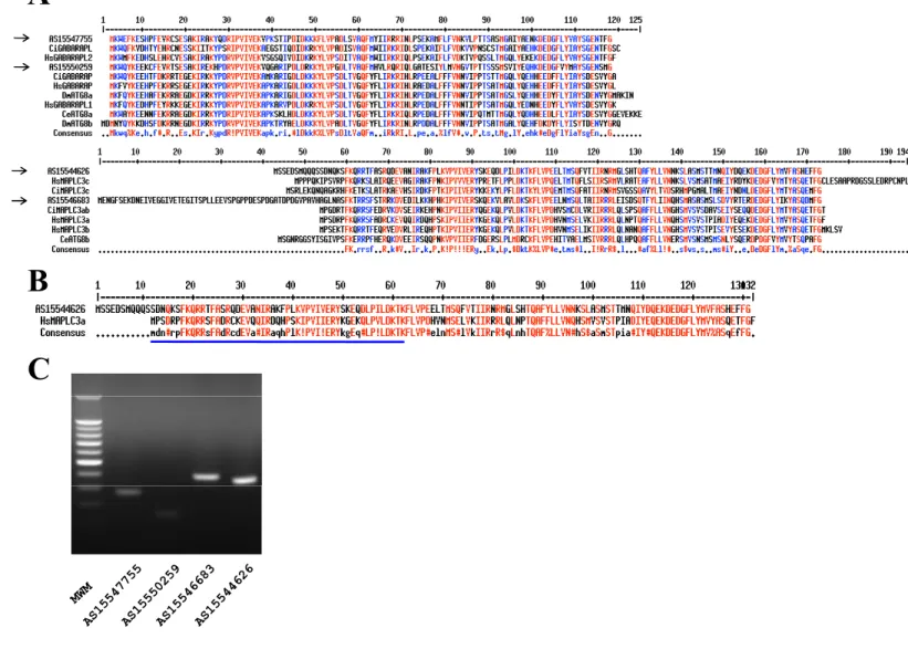
Fig. S1. Key autophagy encoding gene is present in the genome of Abestopluma hypogea. (A) Amino acid
sequences of A. hypogea ATG8 isolated clones as deduced from cDNA sequencing and compared with human,
Ciona, Drosophila and nematode ATG8 sequences (Godefroy et al., 2009). Two main clusters (framed) were
obtained: AS15547755 and AS15550259, related to the GABARAP family; and AS15544626 and AS15546683,
related to the MAPLC3 family. Residues shared by at least 90 and 50% sequences are indicated in red and blue,
respectively. A consensus sequence is indicated below each frame. The arrows point out sponge ATG8 clones.
(B) Alignment of AS15544626 with human MAPLC3a. The underlined sequence corresponds to the peptide
used as antigen to produce the anti-human MAPLC3 antibody used in this study. Residues shared by at least
90% sequences are indicated in red. (C) mRNA expression of A. hypogea ATG8.
Table S1. Set of forward and reverse primers designed to amplify the ATG8 gene
Name Forward primer Reverse primer PCR fragment size (bp)
AS15547755 GCA-AAA-ATC-AGG-GCC-AAA-TA CCA-AAT-GTG-TTC-TCC-CCA-CT 407
For Race PCR GTCCCCAAGTCCACCATCCCAGATA CCATGGACGCACTTGTTGTGGGTAG
AS15550259 CAT-GTG-CTG-CGA-CAG-AGA-AT
CGG-AGT-CTG-CGA-AGA-TAA-GG
TCC-CCA-CTG-TAT-GCC-ATG-TA 152
289
For Race PCR TGGTCCCTGGAGACTTGACAGTTGC GTTGGAGTGACCCCATTGACCATGA
AS15546683 AGG-TGG-GAT-TGT-CGA-GAC-TG
AGA-TGA-GTC-ACC-GGA-TGG-AG
TAT-CCT-GGG-AGG-CGT-ATT-TG 470
404
For Race PCR AGCCAGCTCACTGCCATCATCAGAC GAACGCTCCACGATCACAGGAATCT
AS15546683 AGC-AAT-CTT-CCG-ACA-ACC-AG CAG-CCA-AAG-AAC-TCG-TGT-GA 370