HAL Id: hal-00786577
https://hal.archives-ouvertes.fr/hal-00786577
Submitted on 1 Sep 2013
HAL is a multi-disciplinary open access
archive for the deposit and dissemination of
sci-entific research documents, whether they are
pub-lished or not. The documents may come from
teaching and research institutions in France or
abroad, or from public or private research centers.
L’archive ouverte pluridisciplinaire HAL, est
destinée au dépôt et à la diffusion de documents
scientifiques de niveau recherche, publiés ou non,
émanant des établissements d’enseignement et de
recherche français ou étrangers, des laboratoires
publics ou privés.
Dielectrophoretic separation of blood pathogens ou
Bacterial extraction from biological samples using DEP
forces
Emilie Bisceglia, Myriam Cubizolles, Frédéric Mallard, Olivier Français,
Bruno Le Pioufle
To cite this version:
Emilie Bisceglia, Myriam Cubizolles, Frédéric Mallard, Olivier Français, Bruno Le Pioufle.
Dielec-trophoretic separation of blood pathogens ou Bacterial extraction from biological samples using DEP
forces. lab on a chip world congress, Sep 2012, San Diego, United States. pp.1. �hal-00786577�
Dielectrophoretic separation of blood pathogens
ou
Bacterial extraction from biological samples using DEP forces
Emilie Bisceglia,
aMyriam Cubizolles,
aFrederic Mallard,
bOlivier Francais
cand Bruno Le Pioufle
cReceived Xth XXXXXXXXXX 20XX, Accepted Xth XXXXXXXXX 20XX First published on the web Xth XXXXXXXXXX 200X
DOI : 10.1039/b000000x
ABSTRACT
The abstract should be a single paragraph which summarises the content of the article. Any references in the abstract should be written out in full e.g. [Surname et al., Journal Title, 2000, 35, 3523].
1
Introduction
Rapid detection and identification of pathogens from biolo-gical matrices is a major issue in microbiolobiolo-gical diagnosis. Reference techniques are time-consuming methods based on culture, increasing time significantly between the collection and the analysis of pathogens. For diagnosis of sepsis, traditio-nal techniques detection provide a measuring result after 48h to 7 days, because pre-enrichment of the low micro-organism concentration in blood (below 100 cfu/mL) is usually required to acknowledge its presence. Faster sample preparation would provide great benefits, and would mainly alow medical staff to prescribe broad spectrum antibiotics and thus would reduce the risk of new multi-resistance pathogens emergence. In addi-tion, more accurate therapies could be set up for patient well-ness.
Dramatic advances in microfluidic and microtechnology research provide numerous emerging methods to cope with the lack of simple sample preparation methods.
– surface-based approaches (antibody-antigene interac-tion,...) limited by diffusion
– bulk based approaches (MACS, FACS,...) rapid expan-sion because of micro-system emergence
– focus on DEP
– DEP applications in other fields than cell sorting (high throughput drug assay1/ concentration of biothreat
agents from food matrices2)
essor de la DEP depuis la maitrise des micro-technologies
aDepartment of Technology for Biology and Health, CEA LETI-Minatec, 17
rue des Martyrs, 38054 Grenoble Cedex 9, France.
bbioMerieux, 5 rue des Berges, 38000 Grenoble, France.
cInstitut d’Alembert, ENS Cachan, 61 av President Wilson, 94235 Cachan
Cedex, France.
citation : trapping based method = target cells suspended in the sample stream are selectively trapped and concentrated onto the designated area inside a microfluidic channel while other cells are continuously drawn out by the flow3
We report here a method based on DEP separation to concentrate pathogens out of a biological sample by combi-ning positive and negative DEP to separate pathogens from the sample matrix. In this approach, we take advantage from the large tolerance of micro-organisms towards osmotic shock to perform dielectrophoretic separation in a low electric conductivity medium. This condition enables to collect micro-organisms by positive DEP, while lysed blood cells are repelled from the electrodes by negative DEP.
Our microfluidic device is designed based on numerical simulation.
This approach is validated on a large range of micro-organisms, since it has been tested with different species of bacteria (S. epidermidis (Gram +) and E. coli (Gram -)) and yeasts (C. albicans).
2
Methods and materials
2.1 Device design
The device used for the DEP sorting experiments is made up of an electrode array topped with a microfluidic chamber. The electrode array is made of 5 pairs of interdigited electrodes : the electrode width and gap are 90µm and 10µm, respectively. An overview of the device is presented in figure 1. (faire aussi un schema des electrodes ?)
Fig. 1 Schematic of the microfluidic device for cell separation using DEP
2.2 Device fabrication
The device was fabricated using standard photolithography techniques.
Si + SiO2+ Al + SiO2
+ de details sur la fabrication : mesure d’epaisseur par ellipsometrie la semaine du 19/03
The electrodes are covered with a SiO2 passivation layer to minimize electrochemical reactions with the processed sample.
double-sided tape over the electrodes (Notto Denko MC-2033) to design the sorting chamber with an accurate height of 30µm. Then the chamber is closed with a coverglass to enable microscopic observation.
2.3 Theory and electrid field simulation
2.3.1 DEP theory. When a dielectric particule is suspen-ded in a conductive medium and an electric field is applied, the particule will become polarised. The interaction between the induced dipole and the applied non-uniform field will ge-nerate a force. When the applied electric field is non-uniform, the strength of the electric field on one side of the dipole is greater than on the other side. Thus a force is induced, called dielectrophoretic force (DEP force) because this force will set the particule in motion4.
The time average DEP force in the dipole approximation for a spherical cell is given by equation 1.
~
FDEP= 2πεmr3Re[ fCM]~∇|E|2 (1)
where r is the particule radius, εmis the permittivity of the
sus-pending medium, Re[ fCM] is the real part of Clausius-Mossoti
factor and E is the root-mean-square electric field.
The Clausis-Mossoti factor, given by equation 2 for an ho-mogenous particule, stands for the relative polarizability of the
particule in the suspending medium. It depends on the applied field frequency and can take values from -0.5 to 1.
fCM=
ε∗p− εm∗ εp∗+ 2εm∗
(2)
where εp∗= εp− jσp/ω and εm∗= εm− jσm/ω are the complex
permittivities of the particule and the medium, respectively. εp, εmand σp, σmare the permittivities and the conductivities
of the particule and the medium, respectively.
Depending on the sign of Re[ fCM], particules will move in
opposite direction : if the particule is more polarisable than the suspending medium (Re[ fCM] > 0), particule motion will
be induced towards the local minima of the applied electric field (positive dielectrophoresis or DEP+), and conversly if the particle is less polarisable than the medium (Re[ fCM] < 0),
particule motion will be induced towards the local maxima of the applied electric field (negative dielectrophoresis or DEP-). tri en fonction de l’amplitude de la force ou de la direction Thus depending on the frequency of the applied electric field, DEP can be used to separate two different populations of cells by binary sorting (ref).
2.3.2 Electric field simulation. We solved numerically the electric field produced by interdigited electrodes by using finite element analysis software (Comsol Multiphysics 4.1). It enables to create the geometry and the mesh and then to solve the associated differential equations. The software solved the electric potential using the Laplace equation (or Electrostatic Poisson Equation ?). Then the computed electric field (~E= −~∇V ) is used to determine ~Fdep, which is
proportional to ~∇|E|.
We defined half of a pair of electrodes because of symetry. Simulation conditions / boundary conditions should be specified ?
To assess the effect of the passivation layer on the elec-tric field in the biological medium, we can model this layer as a capacitor and the biological medium as a parallel combina-tion of a capacitor and a resistor. The resolting model for this overlay (faire un schema ?) can be described as a high-pass filter with a cut-off frequency fc and a gain G0 defined by
equation 3. fc= σm 2πεm 2 2 + α and Go= α 2 + α (3)
with α =lgapεpassiv εmhpassiv
where lgap is the width of the electrode gap and hpassiv and
εpassiv the thickness ant the permittivity of the passivation
faire l’application numerique pour la freq de coupure avec la vraie valeur de conductivite du milieu ! ! !
Fig. 2 (a) surface : |E| The red color represents regions with strong electroc field, where particules with Re[ fCM] > 0 are
attracted. The blue color represents regions with weak electric field, where particules with Re[ fCM] < 0 are taken away ; arrows :
direction of ~∇|E| Particules with Re[ fCM] > 0 will move following
these gradient lines.
Estimation of |E| (b) and |~∇|E|2| (c) for different heights away from
the electrode plane
Taking into account that DEP force decreases very quickly with the distance from the electrodes as illustrated by figure 2, the microsystem heigh is limited to 30 µm for DEP sor-ting : this height was set as a compromise between the sorsor-ting efficency and the volume of the sorting chamber.
2.4 Sample preparation
2.4.1 Bacteria. Escherichia coli (strain ATCC 35421) were grown overnight in 3mL Tryptic Soy Broth medium at 30˚C and subcultured at a 1 :50 dilution for 2h at 37˚C. Then the bacteria are washed twice by centrifugation at 10 000tr/min during 5 min and resuspended in deionised water at a cell concentration of 5.108cfu/mL.
ref for bacteria viability in deionised water ?
2.4.2 Yeasts. Candida Albicans (strain ATCC 18804) were grown overnight in 10mL Sabouraud medium at 30˚C and subcultured at a 1 :50 dilution for 4h at 30˚C.
Then the yeasts are washed twice by centrifugation at 10 000tr/min during 5 min and resuspended in deionised water at a cell concentration of 107cfu/mL.
2.4.3 Red blood cells. Human blood was colleted from an anonymous healthy donor in EDTA vaccum tubes provided by the French Blood Service. A 1mL sample was centrifuged at 600g in order to separate red blood cells from the plasma and the buffy coat. 10 µL of red blood cells were then collec-ted.
2.4.4 Mix. The previous micro-organisms and red blood cells suspensions are then mixed with the following ratio : 25 µL of micro-organisms suspension, 1 µL of red blood cells suspension and 25 µL of deionised water. The red blood cells are immediatly lysed due to the osmotic choc generated by the deionised water. The final concentration of bacteria and blood cell ghosts in the sample were 2, 5.108 cfu/mL and 2.108cells/mL, respectively.
The dilution of the sample with deionised water enables us to work in low conductivity medium, so both DEP+ and DEP- were conceivable. On the other hand it was identified as an indisputable way to lyse blood cells without resort to chemical agents that would also have a potential affect on micro-organisms viability.
Mix conductivity : manip semaine du 19/03
1.5µL of the solution was finally injected by capillarity in the micro-system. To avoid evaporation during the exper-mients, two oil drops were applied at both ends of the cham-ber.
2.5 Experimental setup
A 10 Vpeak−peak sinusoidal signal was supplied by a
func-tion generator (AFG32,Tektronic). The frequency can take value from 0 to 80MHz. Particule motion was observed with an optical microscope (Zeiss) and a x20 objective. Images were captured with a CDD camera ( ) with a AxB pixel2 resolution.
one image every 300ms
card-edge connector to connect the device to the generator.
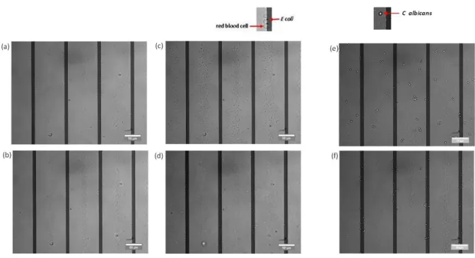
Fig. 3 illusration of negative and positive dielectrophoresis for E coli (a) and C albicans (b)
3
Results and discussion
3.1 micro-organisms separation in a simple buffer 3.1.1 E coli separation in H2O. DEP+ or DEP- depen-ding on the frequency (figure 3(a))
Either in positive or negative DEP regime, we observed that E Coli bacteria ligned up with the field line.
consistent with other papers (ref)
Its own motility seems to prevent it from unspecific adsorp-tion. However it is not the case for other species sush as Sta-phyloccocus epidermidis : the experiments carried out with the strain ATCC 14990 revealed that once adsorbed on the surface of our micro-system, any force from electric field could move the cells away.
3.1.2 C albicans separation in H2O. cross-over frequen-cies different from those with E coli
(figure 3(b))
3.2 pathogens separation from blood cells in a low conductivity medium
As illustrated in figure 4, cells can have differents equili-brium positions. Pictures for different frequencies of the elec-tric signal :
– 50kHz : every cell goes under DEP-– 1MHz : every cell goes under DEP+
– 20 MHz : DEP- for RBC and DEP+ for E. coli
comparison with other articles with experiments with the same conductivity ?
3.2.1 DEP repulsion and reversibility. In these ex-periments, we perform binary sorting between two cell
Fig. 5 (a) E coli speed evolution with the distance from the electrode edges (red lines)
(b) E coli speed comparison with ∇|E|2computed for h=2µm from
the electrodes
populations (micro-organisms and red cell ghosts). In a real contaminated sample, the micro-organisms are far less abondant than blood cells (1 cfu versus 109 cells) : thus
during the sorting, it’s of great importance that blood cells are repelled from the surface of the electrodes in DEP-, therefore it prevents from saturation near the electrodes (pas clair...).
The displacement of the cells, and especially micro-organisms, is reversible : during the experiments bacteria can be attracted or reppeled from the electrodes depending on the frequency as often as required. Thus micro-organisms can be recollected in a buffer after the elimination of the lysed cells to allow further analysis.
3.2.2 average cell velocity. The image interpretation of DEP experiments was performed using the image processing software ImageJ with the particule tracker plugin.
The positions of 40 E coli cells were tracking during the DEP-stage (working frequency = 20MHz) : the computed speed of E coli cells is in good agreement with the evolution of the simulated electric field.
Fig. 4 illusration of negative and positive dielectrophoresis for E coli and red blood cells separation in low conductivity medium
3.2.3 C albicans and blood cells separation. common frequency found for E coli and C albicans (compulsory for your application because we don’t know what king of micro-organisms we are looking for)
3.2.4 blood cell populations. In this study we only take into account red blood cells for it is the most representative po-pulation cell in a blood sample (roughly 109cells/mL). Yet the other cell populations (white blood cells and platelets) have to be considered in more detail because their concentrations will still be far more important than the concentration of the pathogen in the sample. However we can reasonably assume that once lysed, the other blood cells will have the same beha-viour than red cell ghosts.
manip de lyse des globules blancs lundi 19/03 3.2.5 other discussions ?.
Rendement ?
Viability (cell lysis ?)
En statique : only 1.5µL, so it’s necessary to perform this sort with a stream
Indispensable de se placer dans un milieu avec une conducti-vite plus faible que celle des milieux biologiques : ici c’est la dilution qui permet de faire baisser cette conductivite (de 1,5 S/m vers environ 30 mS/m). Si on souhaite supprimer ou tout
du moins diminuer la conductivite, il faudra trouver un autre moyen de faire baisser la conductivite avant l’etape de tri.
4
Conclusions
In this study, we developed a microfluidic device for sepa-ration and concentsepa-ration of pathogens from a blood sample. This method took advantages of the large tolerance of micro-organisms towards osmotic shock to perform dielectrophore-tic separation in a low electric conductivity medium. A well known eletrodes configuration was used to create a highly non-uniform electric field and thus to perform dielectropho-retic sorting. The performance of the device was validated on different pathogen species : indeed the versatility of the me-thod is a key issue, as we don’t know the nature of the pa-thogen prior to its isolation. We believe our device can fill in the critical gap in sample preparation between the collection of potentially contaminated samples and the analysis of patho-gens. In future work, the sorting will be processed with a flui-dic interface, so that we can concentrate the micro-organisms outside the microsystem once separated by DEP. Next step will be to determine the maximum flow rate for which the se-paration is still significant in our microfluidic device to ensure that conventional methods can be challenged.
Références
1 M. P. Hughes, Electrophoresis, 2002, 23, 2569–2582.
2 M. Koklu, S. Park, S. D. Pillai and A. Beskok, Biomicrofluidics, 2010, 4,. 3 S. Park, Y. Zhang, T. H. Wang and S. Yang, Lab on a Chip, 2011, 11,
2893–2900.

![Fig. 2 (a) surface : |E| The red color represents regions with strong electroc field, where particules with Re[ f CM ] > 0 are attracted](https://thumb-eu.123doks.com/thumbv2/123doknet/12880254.369957/4.918.81.409.166.576/surface-color-represents-regions-strong-electroc-particules-attracted.webp)