JON 2900 Katharina Stoeck Klaus Hess Lorenz Amsler Tobias Eckert Dieter Zimmermann Adriano Aguzzi Markus Glatzel
Heightened incidence of sporadic
Creutzfeldt-Jakob disease is associated
with a shift in clinicopathological profiles
Received: 23 February 2007
Received in revised form: 8 February 2008 Accepted: 11 February 2008
Published online: 29 October 2008 Electronic supplementary material The online version of this artiecle (DOI 10.1007/ s00415-008-0900-0) contains supplemen-tary material, which is available to autho-rized users.
K. Stoeck, MD · A. Aguzzi, MD, PhD (쾷) · M. Glatzel, MD
Institute of Neuropathology University Hospital of Zürich Schmelzbergstrasse 12 8091 Zurich, Switzerland Tel.: +41-1/255-2107 Fax: +41-1/255-4402 E-Mail: adriano@pathol.unizh.ch K. Hess, MD Dept. of Neurology University Hospital Zurich Zurich, Switzerland
L. Amsler, MD · T. Eckert, MD Swiss Federal Office of Public Health Bern, Switzerland
D. Zimmermann, PhD Institute of Surgical Pathology University Hospital Zurich Zurich, Switzerland M. Glatzel, MD
Institute of Neuropathology
University Hospital Hamburg-Eppendorf Hamburg-Eppendorf, Germany K. Stoeck, MD
Dept. of Neurology
University Hospital Hamburg-Eppendorf Hamburg-Eppendorf, Germany L. Amsler, MD CSL Behring Bern, Switzerland A. Aguzzi, MD, PhD · M. Glatzel, MD (쾷) Institute of Neuropathology
University Hospital Hamburg-Eppendorf Martinistrasse 52
20246 Hamburg, Germany Tel.: +49-40/4280-37583 Fax: +49-40/4280-34929
E-Mail: m.glatzel@uke.uni-hamburg.de M. Glatzel and A. Aguzzi coordinated the design and operation of the study. Katha-rina Stoeck and Klaus Hess were involved in clinical assessment of patients. M. Glatzel and Dieter Zimmermann were involved in assessment of specimen. All authors con-tributed to the manuscript and approved the final version. M. Glatzel and A. Aguzzi had full access to all data in the study and had final responsibility for the decision to submit for publication.
The study was performed according to established ethical guidelines
This study was supported by grants of the Swiss Federal Office of Public Health and the Swiss National Science Foundation.
■ Abstract Incidences of human transmissible spongiform encepha-lopathies are monitored by na-tional registries in the majority of countries in Western Europe. Dur-ing the past 13 years incidences for Creutzfeldt-Jakob disease (CJD) in Switzerland fluctuated between 0.4 and 2.63 cases/106 inhabitants. We
have compared clinicpathological patient profiles including geo-graphic and gender distribution, age at disease onset, duration of disease, clinical symptoms, and recognized or hypothetical risk factors for CJD, genetic risk factors, biochemical and histopathological data for two cohorts of Swiss sporadic CJD patients from years of regular sporadic CJD incidence (1996–2000, mean incidence 1.3 cases/106 inhabitants, n = 47) to
Swiss sporadic CJD patients from years of elevated sporadic CJD inci-dence (2001–2004, mean inciinci-dence 2.3 cases/106 inhabitants, n = 73).
Sporadic CJD patients from the cohort with elevated sporadic CJD incidence presented with a higher frequency of rare sporadic CJD subtypes. Patients of these sub-types were significantly older and showed a skewed male/female ratio when compared to published patients of identical sporadic CJD-types or to patients from the 1996–2000 cohort and indicates that improved detection of rare sporadic CJD subtypes may have contributed to increased incidence.
■ Key words Creutzfeldt-Jakob disease · prions · dementia · epidemiology
Introduction
Prion diseases or transmissible spongiform encepha-lopathies are fatal neurodegenerative diseases affecting both humans and animals [23]. Neuropathologically, they are characterized by spongiosis, gliosis, neuronal loss and the accumulation of an aberrantly folded iso-form of the normal cellular prion protein, termed PrPSc,
which is an essential component of the infectious agent [22]. The most common human prion disease, sporadic Creutzfeldt-Jakob disease (sCJD) comprises about 85 % of all human prion diseases and is of unknown origin. sCJD may present with a marked clinical heterogeneity. By combining clinical features with histopathological analysis, the status of a polymorphism on the gene en-coding the prion protein as well as biochemical analysis of PrPSc, several sCJD types may be differentiated [14,
19].
About 15 % of human prion diseases are caused by known mutations in the prion protein gene (PRNP) and
may be inherited as autosomal dominant traits [15]. A further subset of human prion diseases are acquired by exposure to infectious prions, in the framework of neu-rosurgical interventions or through hormone substitu-tion, and are referred to as iatrogenic CJD [2]. A novel human prion disease, variant CJD (vCJD), is thought to be caused by exposure to BSE prions via uptake of BSE-contaminated material or by transfusion of vCJD con-taminated blood products [3, 13, 20, 29].
The appearance of vCJD and its development into an epidemic in the UK has led to the establishment of CJD surveillance centres among European countries which carry out active surveillance according to standardized protocols in order to identify and monitor the epidemi-ology of human prion diseases (http://www.eurocjd. ed.ac.uk/).
In 1995, the Swiss National Reference Centre of Prion Diseases (NRPE) was established and active CJD sur-veillance has bee conducted since 1996. This includes clinical and epidemiological assessment of patients, ge-netic analysis, as well as pathological and biochemical analysis of tissue specimens [25].
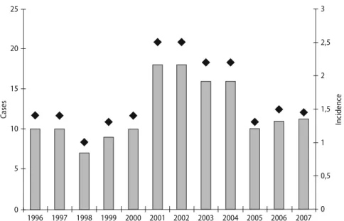
From 1996 to 2000, sCJD affected between seven to 11 patients per year, corresponding to an annual incidence of 1.0 to 1.4 patients per million per year which is well in line with the presumed global incidence of sCJD, about one patient per million per year [11]. However, in 2001 the number of sCJD patients increased to 18, translating to an incidence of 2.5 patients per million per year [10]. In the subsequent three years, the incidence remained elevated, ranging from 1.4 to 2.0 per million per year (Fig. 1).
The aim of the present study was to compare clinical, genetic, biochemical and pathological features of sCJD in two cohorts of patients. The first cohort comprises patients from the years 1996 to 2000 with an average in-cidence of sCJD of 1.3 per million per year, whereas the second cohort comprises patients from the years 2001 to 2004 with an average incidence of sCJD of 2.3 per mil-lion per year.
Patients and methods
■ Patients
In this study, 120 sporadic CJD patients, who were reported to the Swiss National Reference Centre of Prion Diseases (NRPE) and to the Swiss Federal Office of Health between 1996 and 2004, were included. Clinical information including assessment was carried out according to standardized protocols. We collected tissues at necropsy from pa-tients according to established safety and ethical guidelines [4]. The group comprises 109 ‘definite’, neuropathologically-proven, and 11 ‘probable’ CJD patients, corresponding to a proportion of ‘definite
25 20 15 10 5 0 1996 1997 1998 1999 2000 2001 2002 2003 2004 2005 2006 2007 3 2,5 2 1,5 1 0,5 0 Ca se s Incidenc e
Fig. 1 Incidence of sCJD from 1996–2007 in Swit-zerland. From 1996 to 2000, the number of sporadic CJD patients (grey bars) ranged from 8 to 11 per year, corresponding to an annual incidence of sCJD deaths (black diamonds) of 1.3–1.4 cases per million. From 2001 to 2004, the incidence has increased to 2.2–2.5 cases per million per year
CJD’ of 91 %. Clinical diagnosis of ‘probable CJD’ was carried out ac-cording to established criteria [8].
■ Technical investigation
Cerebrospinal fluid (CSF) samples from suspected CJD patients were sent from notifying hospitals. The 14-3-3 test was performed accord-ing to an established protocol [31]. Original electroencephalogram (EEG) recordings were provided from notifying hospitals and evalu-ated for the presence of periodic sharp wave complexes (PSWC) using standardized criteria [26]. Magnet resonance imaging (MRI) scans were analysed for detection of hyperintense basal ganglia on T2-/ FLAIR and diffusion-weighted images (DWI).
■ Genetic analysis
Genetic analysis of PRNP was performed on genomic DNA isolated
from blood or brain tissue according to standard procedures [30]. In order to exclude disease-associatedmutations and to determine the methionine/valine polymorphism oncodon 129 the entire open read-ing frame of the protein was sequenced.
■ Neuropathological examination
Specimen from the following brain regions were examined: frontal, parietal, occipital, and temporal cortex, putamen, thalamus, midbrain, medulla oblongata and cerebellum. Neuropathological examination included assessment of spongiosis, neuronal loss, gliosis and detec-tion of PrP by immunohistochemistry.
■ Immunohistochemistry
Conventional immunohistochemical staining was performed on for-malin-fixed, paraffin embedded tissue. Sections were cut (3 μm), fol-lowing deparaffinization and pre-treatment including hydrolytical autoclaving for 30 min at 121 °C, formic acid for 2.5 min and guani-dinium thiocyanate for 30 min at 4 °C. For detection of PrPSc, sections
were then probed with monoclonal anti-PrP antibody 3F4 according to published methods [25].
■ Western blot analysis
Western blot analysis for detection of PrPSc in brain samples of sCJD
patients was performed according to published protocols [21]. PrPSc
was typed according to the size of protease-resistant unglycosylated PrPSc fragment and designated as either PrPSc type 1 (unglycosylated
fragment running at 21 kDa) or PrPSc type 2 (unglycosylated
frag-ment running at 19 kDa) [5, 7, 19].
■ Triplot of glycoform profiles
Glycoform ratios were compared in a triangular plot correlating the intensities of diglycosylated, monoglycosylated and unglycosylated bands of PrPSc according to published protocols [25]. Densitometric analysis of PrP glycoforms was performed using a one-dimensional software analysis program (Quantity One, Biorad).
■ Statistical analysis
The two cohorts were compared by Student’s t-test for independent samples with unequal variances and chi square test. P-values of < 0.05 were considered significant. As statistical software for calculations, Epi Info(tm) (Version 3.4) and SPSS® (Version 13) were used.
Results
■ Incidence of sCJD in Switzerland from 1996 to 2007 Supplementary Table 1 shows the yearly incidence of sporadic, genetic and iatrogenic CJD from the years 1996 to December 2007. The mean incidence of sCJD in the period 1996 to 2000 was 1.290 cases/106 inhabitants
(95 % C.I. 1.062–1.517) which was significantly higher (p-value 0.000; Wilcoxon rank-sum test) than the mean incidence in the period 2001 to 2004 of 2.318 cases/106
inhabitants (95 % C.I. 2.037–2.599). By including the most recent sCJD incidence data into this analysis, the differences between mean incidences of sCJD from 1996–2000 to 2001–2007 were still statistical significante (from 2001 to 2007, the mean incidence of sCJD was 2.009 cases/106 inhabitants (95 % C.I. 1.592–2.426),
p-value of 0.0185 (Wilcoxon rank-sum test)).
■ Patient characteristics of the cohorts 1996–2000 and 2001–2004
From 1996–2000, 47 sCJD patients were identified, 46 patients were classified as definite and one as probable sCJD. 26 of them were male and 21 were female resulting in a male to female ratio of 1.1:1. From 2001–2004, 73 sCJD patients were diagnosed, 63 definite and 10 prob-able sCJD (Fig. 1). 45 of them were male and 28 were fe-male resulting in a fe-male to fefe-male ratio of 1.6:1 (Fig. 2 A, B). The overrepresentation of male sCJD patients for the years 2001–2004 is statistically significant (p < 0.036, chi square test) when compared to the age-matched normal population of Switzerland (Odds ratio 2.05; 95 % CI 1.22–3.46; p = 0.037, chi square test).
■ Mean age at onset and disease duration
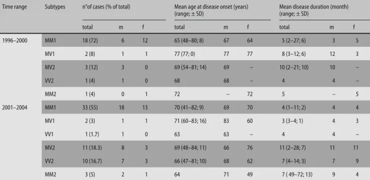
Table 1 presents collective data on age at onset and dis-ease duration of both patient cohorts substratified by sCJD subtypes. From 1996–2000, in depth data on mo-lecular CJD subtypes was available in 25 of 47 patients and from 2001–2004 in 60 of 73 patients.
From 1996–2000, the mean age at onset of disease in sCJD patients was 66 years (n = 47, standard deviation (SD) ± 9 years). Age at onset varied between CJD sub-types: MM1 patients presented the youngest group with a mean age at onset of 65 years (n = 18, SD ± 8 years). Patients of the other groups were older: VV2 and MV2 subtypes showed a mean age at disease onset of 68 (n = 1) and 69 (n = 3) years, whereas for patients of the MM2 and MV1 subtype, age at onset was 72 (n = 1) and 77 (n = 2) years.
Mean disease duration in this time period was 6 months (SD ± 5 months). Disease duration varied among
CJD subtypes. The shortest disease course was observed in the MM1 (5 months, SD ± 6 months) and VV2 sub-group. Patients with MV1 and MV2 phenotype had lon-ger disease durations of 3 and 12 months for MV1 and 10 months for MV2.
In the 2001–2004 cohort, the mean age at disease on-set for sCJD patients was 68 years (n = 73, SD ± 10 years). Patients of the MM1 subgroup had a mean age at disease
onset of 70 years (n = 33, SD ± 9 years), whereas patients belonging to the MV2 and VV2 subgroup of sCJD pa-tients had a mean age at disease onset of 69 years (n = 11, SD ± 11 years) and 66 years (n = 10, SD ± 10 years), re-spectively.
Mean disease duration in the years 2001–2004 was 6 months (SD ± 6 months). MM1 and VV2 patients had short disease durations of 4 months (SD ± 2 months) 2001–2004 1996–2000 65 60 55 50 45 40 35 30 25 20 15 10 5 0 70 60 50 40 30 20 10 0 per cent of C JD pa tients MM MV VV a probable CJD definite CJD c d 2001–2004 1996–2000 2001–2004 1996–2000 70 60 50 40 30 20 10 0 per cent of C JD pa tients MM1 MV1 VV1 MM2 MV2 VV2 b women men 1996–2000 2001–2004 50 45 40 35 30 25 20 15 10 5 0 n ° of C JD cases n ° of C JD cases
Table 1 Clinical characteristics of Swiss CJD patients from 1996–2000 and 2001–2004
Time range Subtypes n°of cases (% of total) Mean age at disease onset (years) (range; ± SD)
Mean disease duration (month) (range; ± SD)
total m f total m f total m f
1996–2000 MM1 18 (72) 6 12 65 (48–80; 8) 67 64 5 (2–27; 6) 3 5 MV1 2 (8) 1 1 77 (77; 0) 77 77 8 (3–12; 6) 12 3 MV2 3 (12) 3 0 69 (54–81; 14) 69 – 10 (2–21; 10) 10 – VV2 1 (4) 1 0 68 68 – 4 4 – MM2 1 (4) 0 1 72 – 72 5 – 5 2001–2004 MM1 33 (55) 18 13 70 (41–82; 9) 69 70 4 (1–11; 2) 4 4 MV1 2 (3) 1 1 71 (60–83; 16) 83 60 3 (3–4; 1) 4 3 VV1 1 (1.7) 1 0 63 63 – 4 4 – MV2 11 (18.3) 8 3 69 (48–84; 11) 66 76 11 (2–28; 7) 11 11 VV2 10 (16.7) 7 3 66 (47–81; 10) 68 62 7 (4–14; 3) 7 9 MM2 3 (5) 2 1 64 71 49 7 ( 49–72; 13) 9 4
Fig. 2 Clinical characterization of sCJD patients from 1996–2000 and 2001–2004 (n = 120). Shown are demographic characteristics of two sCJD cohorts (1996 to 2000 and 2001 to 2004). A Ratios of identified probable to definite sCJD patients from 1996–2000 (n = 47) compared to 2001–2004 (n = 73). B Gender ratios of CJD patients from 1996–2000 and 2001–2004. Note: Increase in male to female ratio (1.6:1) 2001–2004 when compared to the 1996–2000 cohort or other published cohorts. C Distribution of the polymor-phism at PRNP codon 129 (n = 91). sCJD patients are predominantly homozygous for methionine (70 %) or valine (18 %) [1]. Note: Higher frequency of MV and VV patients in the years 2001–2004 when compared to the 1996–2000 cohort or other published cohorts. D Analysis of the molecular CJD subtype based on PrPSc type and codon 129 genotpye (n = 90): Higher frequency of the MV2 and VV2 subtype in 2001–2004 when compared to 1996–2000 or other published cohorts [12, 19]
and 7 months (SD ± 3 months), whereas MV2 patients presented with the longest disease duration of 11 months (SD ± 7 months).
Among all atypical cases combined (i.e. VV1, MM2, MV2 and VV2 subtypes), mean age at onset was 67.3 years (n = 30). In the 1996–2000 cohort (n = 5) it was 69.4 years, and in the 2001–2004 cohort (n = 25) it was 67.0 years.
■ Detailed patients characteristics of the sCJD cohort 2001–2004
A detailed analysis, including clinical signs and results of technical investigations was carried out for the 2001– 2004 cohort. Collective data were available in 51 of 73 patients on clinical signs (neurological and psychiatric) and in 53 of 73 patients on results of technical investiga-tions. We did not find evidence for regional clustering or iatrogenic and zoonotic exposure in this group of pa-tients when assessing recognized or hypothetical risk factors for CJD (data not shown).
■ Neurological and psychiatric signs
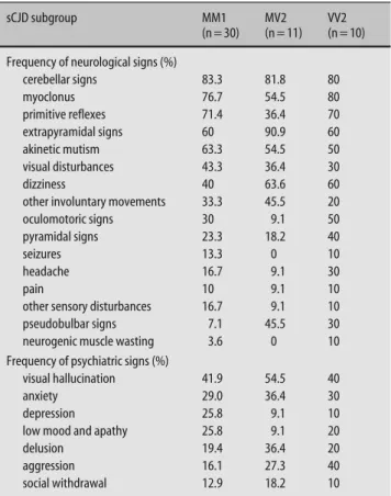
Dementia was the most common clinical sign in all sub-groups (not listed) followed by focal neurological fea-tures such as cerebellar signs, myoclonus, pyramidal and eyxtapyramidal signs, presence of primitive reflexes and akinetic mutism (Table 2). MM1 patients presented mainly with rapid progressive dementia, cerebellar signs, myoclonus and akinetic mutism. MV2 patients showed dominantly extrapyramidal and early cerebellar signs, whereas VV2 patients were mainly characterized by late-onset dementia and early cerebellar signs.
Psychiatric symptoms were frequent in all subtypes. Patients of all subgroups were similarly affected by vis-ual hallucination and anxiety. Depression, low mood and apathy were more frequent in MM1 patients than in MV2 or VV2 patients, whereas these patients showed a higher frequency of delusion and aggressive behaviour than MM1 patients.
■ Results from technical investigations
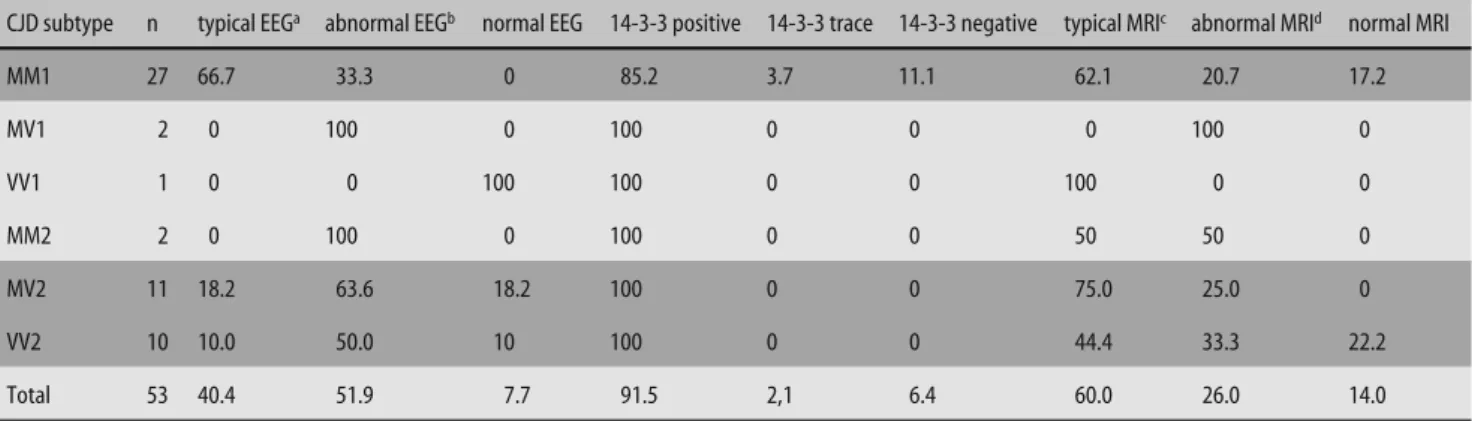
A variety of technical investigations support the clinical diagnosis of sCJD. However, the sensitivity of CJD-typi-cal findings in different investigations, such as MRI (hy-perintense basal ganglia), cerebrospinal fluid analysis (elevation of the 14-3-3 protein) or EEG (PSWC) is lim-ited. In our cohort, CSF analysis was the most sensitive test in all investigated groups with an overall sensitivity of 91.5 % (Table 3). The overall sensitivity of EEG was limited. The highest sensitivity for this examination was
66.7 % for the MM1 group of patients. Sensitivities for patients of the MV2 and VV2 subtype of sCJD were 18 % and 10 %, respectively. Typical MRI changes were mainly observed in MV2 (75 %) and in MM1 (60 %) patients, respectiely, whereas in VV2 patients, CJD-typical MRI changes were less frequently (40 %).
By analysing CJD-typical MRI signs in CJD patients from 2001–2004 including novel imaging methods, such as diffusion-weighted imaging (DWI), we observed an increase of the ratio of CJD-typical MRI in the years 2001–2004 (Fig. 3).
■ Genetic analysis of sCJD patients
Genetic analysis, which included sequencing of PRNP
and determination of a polymorphism on codon 129, was carried out in 91 patients (1996–2000 cohort, 28 of 47 sCJD patients; 2001–2004 cohort, 63 of 73 patients).
In the group of patients from 1996–2000, 20 patients (71.4 %) were homozygous for methionine (MM), 5 pa-tients (17.9 %) were heterozygous for methionine and valine (MV) and 3 patients (10.7 %) were homozygous for valine (VV).
In the group of patients from 2001–2004, 39 patients (62 %) were MM, 14 patients (22.2 %) were MV and 10 patients (15.9 %) were VV (Fig. 2 C).
Table 2 Neurological and psychiatric signs in Swiss sCJD patients 2001–2004
sCJD subgroup MM1 (n = 30) MV2 (n = 11) VV2 (n = 10) Frequency of neurological signs (%)
cerebellar signs 83.3 81.8 80 myoclonus 76.7 54.5 80 primitive reflexes 71.4 36.4 70 extrapyramidal signs 60 90.9 60 akinetic mutism 63.3 54.5 50 visual disturbances 43.3 36.4 30 dizziness 40 63.6 60
other involuntary movements 33.3 45.5 20 oculomotoric signs 30 9.1 50 pyramidal signs 23.3 18.2 40
seizures 13.3 0 10
headache 16.7 9.1 30
pain 10 9.1 10
other sensory disturbances 16.7 9.1 10 pseudobulbar signs 7.1 45.5 30 neurogenic muscle wasting 3.6 0 10 Frequency of psychiatric signs (%)
visual hallucination 41.9 54.5 40
anxiety 29.0 36.4 30
depression 25.8 9.1 10
low mood and apathy 25.8 9.1 20
delusion 19.4 36.4 20
aggression 16.1 27.3 40
■ Molecular sCJD subtypes
The molecular sCJD subtype, based on the combination of codon 129 genotype and PrPSc type in Western blot
and neuropathological analysis, could be assessed in 85 sCJD patients (25 of 47 for the 1996–2000 cohort, 60 of 73 for the 2001–2004 cohort, Table 1).
The most prominent sCJD subtype in the patient group from 1996–2000 was MM1 (72 %, n = 18), followed by MV2 (12 %, n = 3), MV1 (8 %, n = 2) and solitary cases of MM2 (4 %) and VV2 (4 %). From 2001–2004, the ratio in the subtype distribution changed. MM1 was again the most frequent subtype with 55 % (n = 33) of the patients, followed by MV2 (8.3 %, n = 11), VV2 (16,7 %, n = 10), MM2 (5 %, n = 3) as well as cases of MV1 (3 %, n = 2) and VV1 (1,7 %, n = 1). Interestingly, there was a significant increase of atypical cases (VV1, MM2, MV2 and VV2) in the 2001–2004 cohort (43 %) when compared to the 1996–2000 cohort (20 %; n = 83 odds ratio 3.03; 95 % CI
2.47–3.72; p < 0.001, chi square test). Results were similar when taking into account all patients (including the non-specified sCJD subtypes): the ratio increased from 11 % atypical vs. 43 % typical cases in the 1996–2000 co-hort to 34 % atypical vs. 45 % typical cases in the 2001– 2004 cohort (n = 120; odds ratio 3.04; 95 % CI 2.34–3.96; p < 0.001, chi square test).
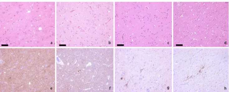
Neuropathological analysis of above mentioned pa-tients confirmed published data [14]. Papa-tients belonging to the MM1 group showed synaptic and perivacuolar PrP deposition, whereas in MV1 and MV2 patients syn-aptic and plaque like deposits could be identified. In pa-tients belonging to the VV2 group, PrP was deposited in band-like arrangements, perineurally and plaque-like (Fig. 4).
■ Triplot analysis of glycoform ratios
Glycoform ratios of sCJD patients from 1996–2000 and from 2001–2004 were compared in a triangular plot cor-relating the intensities of the diglycosylated, monogly-cosylated and unglymonogly-cosylated bands of PrPSc (Fig. 5). Patients with sCJD from 1996–2000 and 2001–2004 clus-ter in the same area of the plot. Non-Swiss control pa-tients of various sCJD subtypes show similar glycoform ratios, whereas patients with vCJD segregate in a distinct region of the plot.
Discussion
From 2001–2004, Switzerland has been reporting an in-creased incidence of sCJD patients, when compared to earlier years of active CJD surveillance, raising ques-tions about the origin of this phenomenon [9, 10]. In this study, we analysed two sCJD cohorts. The first cohort comprised sCJD patients from a period with “regular” sCJD incidence (1996 to 2000). The second cohort com-Table 3 Sensitivity of diagnostic tests in CJD subtypes from 2001–2004 (in %)
CJD subtype n typical EEGa abnormal EEGb normal EEG 14-3-3 positive 14-3-3 trace 14-3-3 negative typical MRIc abnormal MRId normal MRI
MM1 27 66.7 33.3 0 85.2 3.7 11.1 62.1 20.7 17.2 MV1 2 0 100 0 100 0 0 0 100 0 VV1 1 0 0 100 100 0 0 100 0 0 MM2 2 0 100 0 100 0 0 50 50 0 MV2 11 18.2 63.6 18.2 100 0 0 75.0 25.0 0 VV2 10 10.0 50.0 10 100 0 0 44.4 33.3 22.2 Total 53 40.4 51.9 7.7 91.5 2,1 6.4 60.0 26.0 14.0
a generalized PSWC; b mild to moderate EEG changes, diffuse slowing; c hyperintense signal in basal ganglia and /or cortical; d cerebral atrophy
2004 2003 2002 2001 20 18 16 14 12 10 8 6 4 2 0 DWI-MRI typical MRI typical
MRI not typical no MRI
n
°
C
JD cases
Fig. 3 Ratios of MRI results in the years 2001–2004. Shown is the temporal development of imaging results in sCJD patients. Note: increase in CJD-typical MRI over time may be attributed the introduction of (DWI)-MRI
prised sCJD patients from a period with elevated sCJD incidence (2001–2004). We compared differences in clin-icopathological profiles including CJD subtypes between both cohorts and to published data [14, 19]. In an initial
analysis, we formulated several hypotheses concerning the increase in sCJD in Switzerland [9]. One hypothesis comprised that it might be due to statistical fluctuation. We consider this an unlikely explanation for the ob-served increase in incidence since the rise in incidence between the 1996–2000 and 2001–2004 cohorts was sta-tistically significant. Yet, this possibility cannot be dis-missed unequivocally given that the statistical signifi-cance is marginal if most recent incidence data (2001–2007) are included in the analysis.
Another idea implied that the heightened incidence might be related to ascertainment bias due to a tempo-rarily heightened awareness of the disease. In the most recent years, the observed incidence in sCJD deaths (2005: 10 cases; 2006: 11 cases, 2007: 15 cases) dropped to levels just slightly above the 1996–2000 period (http:// www.eurocjd.ed.ac.uk/allcjd.htm). The recent develop-ment may reflect decreased awareness of prion diseases in the general public due to decreased media coverage in recent years. The hypothesis of ascertainment bias is further supported by data from European countries re-porting increased CJD incidences related to heightened awareness due to the introduction of novel diagnostic tools, such as measurement of protein 14-3-3 in the CSF [18, 24]. Typically, increased incidence of CJD attributed to heightened awareness goes along with an increase in the median age of CJD patients [28]. This is believed to be caused by more frequent recognition of CJD in eld-erly demented patients. Although the mean age at onset for the Swiss sCJD cohort 2001–2004 is similar when compared to that of 1996–2000, it is obvious that Swiss sCJD patients of the rare, and clinically-atypical, sCJD subtypes were on average 5 years (VV2) to 10 years (MV2) older than those from large published sCJD co-Fig. 4 Histopathological findings in frontal cortex of sCJD patients. HE stain (A–D) and PrP immunostain (E–H) shows spongiform changes and CJD-type specific PrP deposits. sCJD patients of the MM1 group show synaptic PrP deposition (E), whereas MV1 patients and MV2 patients show additional plaque like or perineural PrP deposits (F, G). In patients belonging to the VV2 group, PrP deposits in band-like arrangements and perineurally (H)
0.8 0.6 0.4 0.2 0.8 0.6 0.6 0.4 0.2 0.4 di-glycosylated 0.2 0.8 un-gly cosyla ted mono -gly cosyla ted 2001–2004 1996–2000 MM1 MV2 VV2 vCJD
Fig. 5 Glycoform profiles of patients with sCJD. The triangular plot correlates the intensities of the diglycosylated (upper), monoglycosylated (middle), and unglycosylated (lower) bands of PrPSc. Patients with sCJD from 1996–2000 (black diamond) and 2001–2004 (gray circles) as well as controls (MM1, MV2 and VV2 patients: white, dark grey and light grey triangle) cluster in the same area of the plot. Instead, control patients with vCJD (black triangle) are segregated in a distinct region of the plot
horts [19, 32]. Although these data do not clearly speak in favour of an ascertainment bias, the fact that the 2001–2004 cohort of sCJD patients showed a higher fre-quency of the less common MV2 subtype when com-pared to the 1996–2000 cohort and to previous publica-tions, in concert with the fact that there is a significant increase in the median age of this patient group is re-markable [12, 19, 32]. This observation might be of par-ticular clinical interest as it highlights the possibility of less common variants such as the MV2 subtype, in older patients with atypical clinical presentations.
A genetic origin for the rise in CJD incidence seems unlikely as there is no increased incidence of genetic CJD from 2001 onwards. Finally, an acquired origin of the rise in incidence, in the framework of iatrogenic or zoonotic transmission which had initially been consid-ered, remains an unlikely option, since neither a iatro-genic nor a zoonotic exposure is evident and we did not observe regional clustering.
Other differences between the two cohorts which do not support or dismantle any of the hypotheses men-tioned above, relate to gender distribution. In 2001–2004 the male to female ratio was (1.6:1), whereas the male to female ratio in the years 1996–2000 was (1.1:1) which is similar to that of published studies [32]. Interestingly, when analysing the gender distribution in different CJD subtypes from 2001–2004, we observed an over-repre-sentation of the MV2 and VV2 subtype in male (70 %) versus female patients (30 %), whereas this phenomenon could not be observed in MM1 patients.
Results in routine investigations in patients from 2001–2004 were similar to those previously described [6, 17]. CSF analysis was the most sensitive test in all inves-tigated groups with an overall sensitivity of 91.5 %. In-terestingly, all MV2 and VV2 patients showed a positive
14-3-3 test. This is surprising, as lower 14-3-3 test sensi-tivities have been described in these subtypes in recent publications [6, 16]. Typical MRI changes were mainly observed in MV2 (75 %) and in MM1 patients (60 %), while in VV2 patients, CJD-typical MRI changes were less frequent (40 %). These results are again comparable to published data and support the importance of MRI as a diagnostic tool for the detection of less common CJD subtypes such as MV2 [16, 27]. Additionally, we show a tendency towards improved diagnostic specificity in sensitive imaging methods, such as DWI-MRI, for the years 2001–2004. This is in agreement with recent stud-ies stressing the importance of sensitive imaging meth-ods in the early diagnosis of sCJD [27]. In summary, this analysis confirms our initial finding of an increased in-cidence of sCJD in Switzerland. Although, the reason for this phenomenon remains unexplained to date, our analysis demonstrates that patients from the years 2001–2004 with increased sCJD incidence differ in sev-eral aspects from published sCJD cohorts. The fact that the MV2 subgroup of patients showed an increase in mean age at disease onset when compared to published cohorts, together with the fact that these patients dem-onstrate distinct features in sensitive imaging methods, may indicate that improved detection of these patients has contributed to the rise in sCJD incidence. Further studies investigating biochemical and genetic aspects will contribute to our understanding of the mechanisms underlying sCJD.
■ Acknowledgements We thank all physicians throughout Switzer-land for referring suspected CJD patients to our reference center. We thank Mauri Peltola for technical support. This study was supported by grants of the Swiss Federal Office of Public Health and the Swiss National Science Foundation.
References
1. Alperovitch A, Zerr I, Pocchiari M, Mitrova E, de Pedro Cuesta J, Hegyi I, Collins S, Kretzschmar H, van Duijn C, Will RG (1999) Codon 129 prion protein genotype and sporadic Creutzfeldt-Jakob disease. Lancet 353:1673–1674
2. Brown P, Preece M, Brandel JP, Sato T, McShane L, Zerr I, Fletcher A, Will RG, Pocchiari M, Cashman NR, d’Aignaux JH, Cervenakova L, Fradkin J, Schon-berger LB, Collins SJ (2000) Iatrogenic Creutzfeldt-Jakob disease at the millennium. Neurology 55:1075–1081
3. Bruce ME, Will RG, Ironside JW, McConnell I, Drummond D, Suttie A, McCardle L, Chree A, Hope J, Birkett C, Cousens S, Fraser H, Bostock CJ (1997) Transmissions to mice indicate that ‘new variant’ CJD is caused by the BSE agent Nature 389:498–501
4. Budka H, Aguzzi A, Brown P, Brucher JM, Bugiani O, Collinge J, Diringer H, Gullotta F, Haltia M, Hauw JJ, et al. (1995) Tissue handling in suspected Creutzfeldt-Jakob disease (CJD) and other human spongiform encephalop-athies (prion diseases). Brain Pathol 5:319–322
5. Cali I, Castellani R, Yuan J, Al-Shekhlee A, Cohen ML, Xiao X, Moleres FJ, Parchi P, Zou WQ, Gambetti P (2006) Classification of sporadic Creutzfeldt-Jakob disease revisited. Brain 129: 2266–2277
6. Castellani RJ, Colucci M, Xie Z, Zou W, Li C, Parchi P, Capellari S, Pastore M, Rahbar MH, Chen SG, Gambetti P (2004) Sensitivity of 14-3-3 protein test varies in subtypes of sporadic Creutzfeldt-Jakob disease. Neurology 63:436–442
7. Collinge J, Sidle KC, Meads J, Ironside J, Hill AF (1996) Molecular analysis of prion strain variation and the aetiol-ogy of ‘new variant’ CJD. Nature 383: 685–690
8. Collins SJ, Sanchez-Juan P, Masters CL, Klug GM, van Duijn C, Poleggi A, Pocchiari M, Almonti S, Cuadrado-Corrales N, de Pedro-Cuesta J, Budka H, Gelpi E, Glatzel M, Tolnay M, Hewer E, Zerr I, Heinemann U, Kretszschmar HA, Jansen GH, Olsen E, Mitrova E, Alperovitch A, Brandel JP, Mackenzie J, Murray K, Will RG (2006) Determi-nants of diagnostic investigation sensi-tivities across the clinical spectrum of sporadic Creutzfeldt-Jakob disease. Brain 129:2278–2287
9. Glatzel M, Ott PM, Lindner T, Gebbers JO, Gmur A, Wuest W, Huber G, Moch H, Podvinec M, Stamm B, Aguzzi A (2003) Human prion diseases: epide-miology and integrated risk assess-ment. The Lancet Neurology 2:757–763 10. Glatzel M, Rogivue C, Ghani A, Streffer
JR, Amsler L, Aguzzi A (2002) Inci-dence of Creutzfeldt-Jakob disease in Switzerland. Lancet 360:139–141 11. Glatzel M, Stoeck K, Seeger H, Luhrs T,
Aguzzi A (2005) Human prion dis-eases: molecular and clinical aspects. Arch Neurol 62:545–552
12. Head MW, Ritchie D, Smith N, McLoughlin V, Nailon W, Samad S, Masson S, Bishop M, McCardle L, Iron-side JW (2004) Peripheral tissue in-volvement in sporadic, iatrogenic, and variant Creutzfeldt-Jakob disease: an immunohistochemical, quantitative, and biochemical study. Am J Pathol 164:143–153
13. Hill AF, Desbruslais M, Joiner S, Sidle KC, Gowland I, Collinge J, Doey LJ, Lantos P (1997) The same prion strain causes vCJD and BSE (letter). Nature 389:448–450
14. Hill AF, Joiner S, Wadsworth JD, Sidle KC, Bell JE, Budka H, Ironside JW, Collinge J (2003) Molecular classifica-tion of sporadic Creutzfeldt-Jakob disease. Brain 126:1333–1346 15. Kovacs GG, Puopolo M, Ladogana A,
Pocchiari M, Budka H, van Duijn C, Collins SJ, Boyd A, Giulivi A, Coulthart M, Delasnerie-Laupretre N, Brandel JP, Zerr I, Kretzschmar HA, de Pedro-Cuesta J, Calero-Lara M, Glatzel M, Aguzzi A, Bishop M, Knight R, Belay G, Will R, Mitrova E (2005) Genetic prion disease: the EUROCJD experience. Hum Genet 118:166–174
16. Krasnianski A, Schulz-Schaeffer WJ, Kallenberg K, Meissner B, Collie DA, Roeber S, Bartl M, Heinemann U, Varges D, Kretzschmar HA, Zerr I (2006) Clinical findings and diagnostic tests in the MV2 subtype of sporadic CJD. Brain 129:2288–2296
17. Meissner B, Kortner K, Bartl M, Jastrow U, Mollenhauer B, Schroter A, Finkenstaedt M, Windl O, Poser S, Kretzschmar HA, Zerr I (2004) Spo-radic Creutzfeldt-Jakob disease: mag-netic resonance imaging and clinical findings. Neurology 63:450–456 18. Mollenhauer B, Zerr I, Ruge D, Krause
G, Mehnert WH, Kretzschmar HA, Poser S (2002) Epidemiology and clini-cal symptomatology of Jakob disease. Dtsch Med Wochenschr 127:312–317
19. Parchi P, Giese A, Capellari S, Brown P, Schulz-Schaeffer W, Windl O, Zerr I, Budka H, Kopp N, Piccardo P, Poser S, Rojiani A, Streichemberger N, Julien J, Vital C, Ghetti B, Gambetti P,
Kretzschmar H (1999) Classification of sporadic Creutzfeldt-Jakob disease based on molecular and phenotypic analysis of 300 subjects. Ann Neurol 46:224–233
20. Peden AH, Head MW, Ritchie DL, Bell JE, Ironside JW (2004) Preclinical vCJD after blood transfusion in a PRNP codon 129 heterozygous patient. Lancet 364:527–529
21. Polymenidou M, Stoeck K, Glatzel M, Vey M, Bellon A, Aguzzi A (2005) Coexistence of multiple PrPSc types in individuals with Creutzfeldt-Jakob disease. Lancet Neurol 4:805–814 22. Prusiner SB (1982) Novel
protein-aceous infectious particles cause scrapie. Science 216:136–144
23. Prusiner SB (2001) Shattuck lecture – neurodegenerative diseases and prions. N Engl J Med 344:1516–1526 24. Saiz A, Nos C, Yague J, Dominguez A,
Graus F, Munoz P (2001) The impact of the introduction of the 14-3-3 protein assay in the surveillance of sporadic Creutzfeldt-Jakob disease in Catalonia. J Neurol 248:592–594
25. Schoch G, Seeger H, Bogousslavsky J, Tolnay M, Janzer RC, Aguzzi A, Glatzel M (2005) Analysis of Prion Strains by PrP(Sc) Profiling in Sporadic Creutzfeldt-Jakob Disease. PLoS Med 3:e14
26. Steinhoff BJ, Zerr I, Glatting M, Schulz-Schaeffer W, Poser S, Kretzschmar HA (2004) Diagnostic value of periodic complexes in Creutzfeldt-Jakob dis-ease. Ann Neurol 56:702–708 27. Ukisu R, Kushihashi T, Kitanosono T,
Fujisawa H, Takenaka H, Ohgiya Y, Gokan T, Munechika H (2005) Serial diffusion-weighted MRI of Creutz-feldt-Jakob disease. AJR Am J Roent-genol 184:560–566
28. Van Everbroeck B, Michotte A, Sciot R, Godfraind C, Deprez M, Quoilin S, Martin JJ, Cras P (2006) Increased incidence of sporadic Jakob disease in the age groups between 70 and 90 years in Belgium. Eur J Epidemiol 21:443–447 29. Will RG, Ironside JW, Zeidler M,
Cousens SN, Estibeiro K, Alperovitch A, Poser S, Pocchiari M, Hofman A, Smith PG (1996) A new variant of Creutzfeldt-Jakob disease in the UK. Lancet 347:921–925
30. Windl O, Giese A, Schulz-Schaeffer W, Zerr I, Skworc K, Arendt S, Oberdieck C, Bodemer M, Poser S, Kretzschmar HA (1999) Molecular genetics of human prion diseases in Germany. Hum Genet 105:244–252
31. Zerr I, Bodemer M, Gefeller O, Otto M, Poser S, Wiltfang J, Windl O,
Kretzschmar HA, Weber T (1998) Detection of 14-3-3 protein in the cerebrospinal fluid supports the diag-nosis of Creutzfeldt-Jakob disease (In Process Citation). Ann Neurol 43:32–40 32. Zerr I, Schulz-Schaeffer WJ, Giese A,
Bodemer M, Schroter A, Henkel K, Tschampa HJ, Windl O, Pfahlberg A, Steinhoff BJ, Gefeller O, Kretzschmar HA, Poser S (2000) Current clinical diagnosis in Creutzfeldt-Jakob disease: identification of uncommon variants. Ann Neurol 48:323–329