T. Schneider H. Rickli V. Gliech M. Maeder
Bland-White-Garland syndrome
and atrial septal defect –
Rare Association and diagnostic challenge
Received: 17 October 2005 Accepted: 30 December 2005 Published online: 8 March 2006
Dr. med. Micha Maeder (
)
) Division of Cardiology University Hospital Basel Petersgraben 44031 Basel, Switzerland Dr. med. Tino Schneider Dr. med. Hans Rickli Kantonsspital St. Gallen Department Innere Medizin Fachbereich Kardiologie Rorschacherstr. 95
9007 St. Gallen, Switzerland Dr. med. Volker Gliech Humaine Klinikum Bad Saarow/Fürstenwalde Pieskowerstr. 33
15526 Bad Saarow, Gemany
n Summary We report on a
40-year-old woman referred for eval-uation of a cardiac murmur and dyspnea on exertion. The electro-cardiogram (ECG) showed incom-plete right bundle branch block, and echocardiography revealed a large atrial septal defect (ASD, os-tium secundum type) with dilated right-sided heart chambers. At cardiac catheterization, a large left-to-right shunt (78% of the pulmonary blood flow) was found, and surprisingly, the additional diagnosis of anomalous origin of the left coronary artery from pul-monary artery (ALCAPA) was es-tablished. After ASD closure and
left coronary artery ligation with implantation of a vein graft to the left anterior descending artery, she had an uneventful 18-years follow-up.
We discuss the interaction of the two associated conditions, and based on the herein reported un-usual combination, we highlight typical features of non-invasive examinations including ausculta-tion, ECG, and echocardiography in adult patients with ALCAPA.
n Key words Shunt –
atrial septal defect – coronary anomaly – non-invasive diagnostics
Inroduction
Anomalous origin of the Left Coronary Artery from
Pulmonary Artery (ALCAPA or Bland-White-Garland
syndrome) is a rare congenital coronary artery anomaly occurring in approximately one in 300 000 live births [3, 22]. Ischemia of the myocardial terri-tory supplied by the left coronary artery occurs as flood flow in the left coronary artery is reversed after birth. Perfusion of the left ventricular myocar-dium therefore depends on collateral flow from the right coronary artery. Up to 90% of children with ALCAPA die during the first year of life because of myocardial ischemia and left ventricular failure.
Sur-vival until adulthood is rare and depends on pre-ex-isting or rapidly developing collateral vessels be-tween the right and the left coronary artery [19, 22, 28].
Whereas ALCAPA usually occurs as an isolated le-sion, there are few reports on its association with other intracardiac defects [20, 24]. We herein de-scribe the case of an adult patient undergoing cardi-ac catheterization for evaluation of large atrial septal defect (ASD), in whom the diagnosis of co-existing ALCAPA was established. Our report focuses on the clinical impact, the diagnostic challenges, and the therapeutic implications encountered in the setting of this unusual combination.
Case report
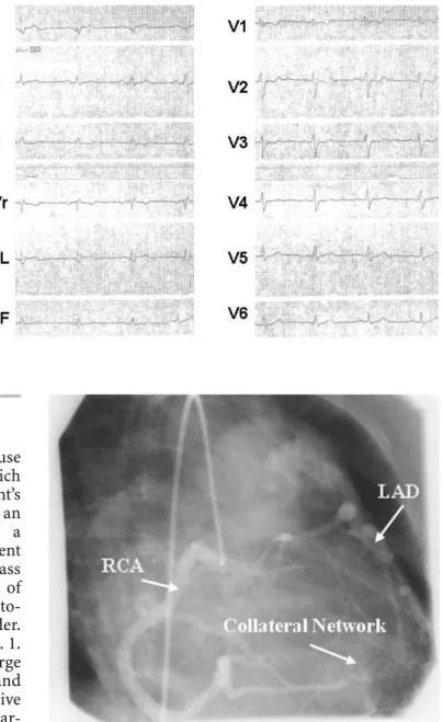
In 1986, a 40-year-old woman was referred because of progressive dyspnea and a cardiac murmur, which had been known for years. Otherwise, the patient’s medical history had been unremarkable. After an uneventful pregnancy she had given birth to a healthy boy 15 years ago. At presentation the patient was in New York Heart Association (NYHA) class III. Physical examination revealed fixed splitting of the second heart sound. Both a systolic and a diasto-lic murmur were audible at the left sternal border. The electrocardiogram (ECG) is shown in Fig. 1. Transthoracic echocardiography revealed a large ASD (ostium secundum type, diameter 14 mm) and dilated right-sided heart chambers. As operative ASD closure was planned, the patient underwent car-diac catheterization. Mean pulmonary artery pres-sure was 27 mmHg, and a large left-to-right-shunt was calculated by dye dilution (78% of the pulmo-nary blood flow). Mild anterolateral hypokinesis of the left ventricle was noted, the global left ventricu-lar ejection fraction being normal. At coronary an-giography, a left coronary ostium could not be iden-tified, but injection into the right coronary artery re-vealed a very large vessel with retrograde collateral filling of the left coronary and entry of the contrast agent into the pulmonary artery (Fig. 2). Thus, the surprising diagnosis of ASD combined with ALCAPA had been established. The patient underwent ASD patch closure, left coronary artery ligation at its ori-gin from the pulmonary artery, and implantation of a vein graft to the left anterior descending artery. She made an uneventful recovery after surgery.
Eight-teen years later, at the age of 58 years the patient was in NYHA class II. Exercise capacity was markedly limited by a concomitant neuromuscular disorder. However, repeated echocardiographic fol-low-up examinations had shown complete normali-zation of systolic left ventricular function, and the left ventricular end-diastolic diameter had decreased from 43 to 38 mm. Dilatation of the right-sided chambers had resolved, and pulmonary hypertension was absent.
Fig. 1 Electrocardiogram (paper speed 50 mm/s) showing
sinus rhythm with incomplete right bundle branch block and negative T-waves in leads V1–3. There are small Q waves in leads I and aVL, and in lead II T waves are negative
Fig. 2 Coronary angiography showing a large right coronary artery (RCA)
with collateral filling of the left anterior descending coronary artery (LAD) and contrast entry into the pulmonary trunk by a collateral network
Discussion
The present example of ALCAPA is unusual with re-spect to both the patient’s less symptomatic course until adulthood and the co-existing ASD with asso-ciated diagnostic and therapeutic pitfalls.
Most patients with ALCAPA become symptomatic a few weeks to months after birth, when the physiolog-ical neonatal pulmonary hypertension has resolved, and closure of the ductus arteriosus has occurred [28, 29]. Flow in the left coronary artery is then direc-ted towards the low-pressure pulmonary artery lead-ing to a steal phenomenon in the myocardial territory supplied by the left coronary artery [2]. Pre-existing or rapidly developing collateral vessels between the right and the left coronary artery may prevent a fatal clinical course early in life, but the left ventricle re-mains chronically hypoperfused resulting in a vari-able degree of left ventricular dysfunction [25]. None-theless, long-term survival in patients with ALCAPA without surgical correction has been well documented [1, 4, 7, 9, 10, 14–18, 21, 23, 27].
In our patient, the large left-to-right shunt across the ASD with subsequently elevated pressure and oxygen saturation in the pulmonary trunk may have improved blood and oxygen supply to the left coro-nary artery by attenuating retrograde left corocoro-nary flow, and hence the beneficial interaction of two po-tentially hazardous conditions may have contributed to the long asymptomatic period including an un-eventful pregnancy. Regarding the significant left-to-right shunt with left-to-right ventricular overload, the indi-cation for ASD closure was evident [5]. However, op-eration without knowledge of the coronary anatomy might have been fatal, as the sudden occurrence of severe left ventricular ischemia due to an abrupt re-duction of pulmonary artery flow and oxygen satu-ration following ASD closure with subsequent coro-nary steal from the left corocoro-nary artery would have been a very probable scenario. An interesting obser-vation underscoring our speculation comes from the
report on a 4-month-old infant with undiagnosed ALCAPA undergoing repair of ventricular septal de-fect (VSD), in whom severe left ventricular failure occurred after VSD closure. After echocardiographic detection of ALCAPA and reimplantation of the left coronary artery into the aorta, the patient could be weaned from cardiopulmonary bypass and recovered [24]. In contrast to ASD, patients with VSD often have marked pulmonary hypertension, which may even become fixed, and therefore a VSD has cer-tainly a more significant impact on co-existing AL-CAPA as compared to an ASD, where volume rather than pressure overload of the pulmonary circulation is present [5]. Nevertheless, in our patient mean pul-monary pressure was somewhat elevated, and hemo-dynamics may have been perfectly adjusted to and depended on the co-existence of ALCAPA and ASD over years, and thus preoperative coronary angiogra-phy probably prevented an adverse outcome. This is a very critical issue, as ASD closure can now be per-formed percutaneously [26], and coronary angiogra-phy is not carried out in all cases.
There are many reports of young adults with yet undiagnosed ALCAPA, who were successfully resus-citated after a cardiac arrest due to ventricular fibril-lation, and in whom only a thorough evaluation led to the correct diagnosis [4, 15, 17]. Therefore, we want to stress the importance of typical findings from non-invasive tests in patients with ALCAPA. First, the patient had a systolic murmur as very often heard in patients with ASD [5]. In addition, there was a diastolic murmur, which was attributed to pulmonary regurgitation as the diagnosis of large ASD was evident. However, the murmur has been more likely due to collateral flow from the right to the left coronary artery, which peaks during diastole. Other auscultatory findings in patients with ALCAPA include systolic murmurs of various degrees due to mitral valve regurgitation following ischemia or in-farction of the anterolateral papillary muscle or sec-ondary to ventricular dilatation [6].
Fig. 3 Electrocardiogram from a 39-year-old
woman with anomalous origin of the left coro-nary artery from pulmocoro-nary artery (case de-scribed in reference [16]) showing Q waves and negatives T waves in leads I and aVL. Note that the Q waves are very small, and that signs of left ventricular hypertrophy are absent
Second, patients with ALCAPA often have a char-acteristic ECG pattern including deep and broad Q waves and/or negative T waves in leads I and aVL and signs of left ventricular hypertrophy [6, 22]. However, classical Q waves are typically less pro-nounced or even absent in adults as compared to neonates or children with ALCAPA [6]. In Fig. 3, a typical ECG of an adult patient with ALCAPA is shown. In the present case, there were ASD-asso-ciated ECG changes, i.e. incomplete right bundle block with T wave abnormalities in the precordial leads, whereas the presence of ALCAPA was not evi-dent. A similar ECG pattern has been reported pre-viously in a 70-day-old boy suffering from both AL-CAPA and ASD [20]. In that patient minimal Q waves and negative T waves in aVL were the only ECG signs of ALCAPA [20].
Third, transthoracic echocardiography is a crucial non-invasive tool as it can definitely establish the di-agnosis of ALCAPA by demonstration of a large right coronary artery and a mainly diastolic flow from an abnormal vessel originating from the pul-monary trunk [6, 17]. In Fig. 4, the typical echocar-diographic aspect of the huge right coronary artery in an adult with ALCAPA is shown, and in Fig. 5, flow from the left coronary artery into the pulmo-nary artery is visualized. In some cases, collateral flow through the interventricular septum can be seen in the transthoracic study [11]. However, these typi-cal features can be easily missed, especially if an-other diagnosis is obvious. Other echocardiographic findings seen in patients with ALCAPA, which are
non-specific however, include regional wall motion abnormalities, most often localized in the anterolat-eral wall. Interestingly, ischemia not always occurs in the myocardial territory supplied by the left coro-nary artery, but also in the infero-posterior wall, as shown by myocardial scintigraphy [13, 18]. The lat-ter finding has been inlat-terpreted as a steal phenome-non in patients with large collateral flow [13]. New imagine techniques such as multislice computed to-mography and magnetic resonance imaging can ac-curately visualize the pathoanatomy of ALCAPA [14], but are not always available.
As long as the diagnosis is established, even oli-gosymptomatic patients with ALCAPA should be op-erated due to an increased risk of sudden cardiac death [1, 8, 10]. Whenever possible, a two-coronary artery system will be established, either by direct implantation of the left coronary artery into the as-cending aorta, or by ligation of the left coronary ar-tery combined with aortocoronary bypass grafting [1, 8]. Isolated ligation of the left coronary artery at its origin from the pulmonary artery has been suc-cessfully performed as an emergency life-saving pro-cedure, but is not recommended for routine applica-tion as it has been shown to be associated with re-sidual silent myocardial ischemia and a less favor-able outcome than the creation of a two-coronary system [8]. Even in patients with significant mitral regurgitation, mitral valve repair or valve replace-ment is rarely indicated as mitral regurgitation often resolves after revascularization [8]. However, most data on surgical results come from operations in
in-Fig. 4 Transthoracic echocardiogram (parasternal
short axis view) obtained from a 15-year-old male who after a collapse was found in ventricu-lar fibrillation and was successfully resuscitated, showing a huge right coronary artery (RCA) aris-ing from the aorta (A). PA = pulmonary artery
fants, where complete recovery of left ventricular function, normalization of left ventricular dilatation, and regression of left ventricular hypertrophy within months after reimplantation of the left coronary ar-tery into the aorta has been demonstrated [12]. These changes however, occurred much faster in in-fants than in older children [12] and may be even slower or incomplete in adults [16]. Therefore the impact of correction of the coronary anatomy on mi-tral valve regurgitation is hard to predict in adults.
In our patient, combined and simultaneous surgi-cal correction of both ASD and ALCAPA was indis-pensable as pointed out above. There are few follow-up data in adult ALCAPA patients after correction.
The present case is an example for an excellent long-term result after surgery for ALCAPA.
Our case study is limited by the fact that our pa-tient was evaluated almost twenty years ago, and that since then significant progress in echocardiography equipment has occurred. Nevertheless, we feel that the present case is a suitable example to highlight the need for a thorough evaluation of patients with congenital heart disease even if the diagnosis seems to be obvious.
n Acknowledgement We are grateful to Franziska Rohner, MD,
and Hans Roelli, MD, for providing the echo tapes. In addition, the excellent technical assistance given by Hubert von Rohr is very much appreciated.
Fig. 5 Transthoracic echocardiogram (parasternal
short axis view) from the same patient showing an abnormal vessel entering the pulmonary trunk (arrow). In this patient, the diagnosis of anoma-lous origin of the left coronary artery from pul-monary artery was established by echocardiogra-phy before transfer to a catheterization center.
A = aorta; PA = pulmonary artery
References
1. Alexi-Meskishvili V, Berger F, Weng Y, Lange PE, Hetzer R (1995) Anoma-lous origin of the left coronary artery from the pulmonary artery in adults. J Card Surg 10:309–315
2. Baue AE, Baum S, Blakemore WS, Zinsser HF (1967) A later stage of anomalous coronary circulation with origin of the left coronary from the pulmonary artery: coronary artery steal. Circulation 36:878–886
3. Bland EF, White PD, Garland J (1933) Congenital anomalies of the coronary arteries. Report of an unusual case associated with cardiac hypertrophy. Am Heart J 8:787–801
4. Braun E, Bieber H, Bienko B, Meyer D, Zwirner K (1991) [Anomalous ori-gin of the left coronary artery from the pulmonary trunk (Bland-White-Garland syndrome) in a 29-year-old man with absolute tachyarrhythmia] Z Kardiol 80:543–548
5. Brickner EM, Hillis LD, Lange RA (2000) Congenital heart disease in adults. First of two parts. N Engl J Med 342:256–263
6. Chu E, Cheitlin MD (1993) Diagnos-tic considerations in patients with suspected coronary artery anomalies. Am Heart J 126:1427–1438
7. Cowie MR, Mahmood S, Ell PJ (1994) The diagnosis and assessment of an adult with anomalous origin of the left coronary artery from the pulmo-nary artery. Eur J Nucl Med 21:1017– 1019
8. Dodge-Khatami A, Mavroudis C, Backer CL (2002) Anomalous origin of the left coronary artery from the pulmonary artery: collective review of surgical therapy. Ann Thorac Surg 74:946–955
9. Fierens C, Budts W, Denef B, Van de Werf F (2000) A 72 year old women with ALCAPA. Heart 83:e2
10. Frapier JM, Leclercq F, Bodino M, Chaptal PA (1999) Malignant ventri-cular arrhythmias revealing anoma-lous origin of the left coronary artery from the pulmonary artery in two adults. Eur J Cardiothorac Surg 15: 539–541
11. Hirota H, Hiraishi H, Nakahata Y (2002) Bland-White-Garland syn-drome with well developed collateral arterial vessels. Cardiol Young 12: 177–178
12. Jin Z, Berger F, Uhlemann F, Schro-der C, Hetzer R, Alexi-Meskhishvili V, Weng Y, Lange PE (1994) Improve-ment in left ventricular dysfunction after aortic reimplantation in 11 con-secutive pediatric patients with anomalous origin of the left coronary artery from the pulmonary artery. Eur Heart J 15:1044–1049
13. Katsuragi M, Yamamoto K, Tashiro T, Nishihara H, Toudou K (1993) Thal-lium-201 myocardial SPECT in Bland-White-Garland syndrome: two adult patients with inferoposterior perfusion defect. J Nucl Med 34: 2182–2184
14. Khanna A, Torigian DA, Ferrari VA, Bross RJ, Rosen MA (2005) Anoma-lous origin of the left coronary artery from the pulmonary artery in adult-hood on CT and MRI. AJR J Roent-genol 185:326–329
15. Kreutzer U, Krülls-Münch J, Angres M, Schiessler A (1998) [Successful re-suscitation from ventricular fibrilla-tion in Bland-White-Garland syn-drome in adulthood – a case report]. Z Kardiol 87:560–565
16. Maeder M, Vogt PR, Ammann P, Rickli H (2004) Bland-White-Garland syndrome in a 39-year-old mother. Ann Thorac Surg 78:1451–1453 17. Maire R, Gallino A, Jenni R (1993)
Initial detection in a teenager of anomalous left coronary artery from the pulmonary artery by color Dop-pler echocardiography. Am Heart J 125:1802–1805
18. Moodie DS, Cook SA, Gill CC, Napoli CA (1980) Thallium-201 myocardial imaging in young adults with anoma-lous origin of the left coronary artery arising from the pulmonary artery. J Nucl Med 21:1076–1079
19. Moodie DS, Fyfe D, Gill CG, Cook SA, Lytle B, Taylor PC, Fitzgerald R, Sheldon WC (1983) Anomalous ori-gin of the left coronary artery from pulmonary artery (Bland-White-Gar-land syndrome) in adult patients: long-term follow-up after surgery. Am Heart J 106:381–388
20. Nakagawa M, Kimura K, Watanabe Y (1996) Atypical electrocardiogram and echocardiogram in a patient with Bland-White-Garland syndrome in association with atrial septal defect. Cardiology 87:358–360
21. Nightingale AK, Burrell CJ, Marshall (1998) Anomalous origin of the left coronary artery from the pulmonary artery: natural history and normal pregnancies. Heart 80:629–631
22. Perlof JK (1994) Anomalous origin of the left coronary artery from the pul-monary trunk. In: Perlof JK (ed) The clinical recognition of congenital heart disease. 4th edition.
Philadel-phia, W.B. Saunders, pp 546–561 23. Purut CM, Sabiston DC (1991) Origin
of the left coronary artery from the pulmonary artery in older adults. J Thorac Cardiovasc Surg 102:566-570. 24. Shanmugan G, McLennan AJ, Pollock
JC, MacArthur KJ (2005) Anomalous left coronary artery, ventricular septal defect, and double aortic arch. Ann Thorac Surg 80:334–336
25. Shivalkar B, Borgers M, Daenen W, Gewillig M, Flameng W (1994) AL-CAPA syndrome: an example of chronic myocardial hypoperfusion. J Am Coll Cardiol 23:772–778
26. Sievert H, Krumsdorf U (2002) Transcatheter closure of intracardiac shunts. Z Kardiol 91(Suppl 3):77–83 27. Tiroke A, Herrmann G, Lins M, el
Mokhtari N, Reinecke A, Wieckhorst A, Cremer J, Simon R (2004) [Bland-White-Garland syndrome in an adult] Z Kardiol 93:58–62
28. von Kodolitisch Y, Franzen O, Lund GK, Koschyk DH, Ito WD, Meinertz T (2005) Coronary artery anomalies. Part II: Recent insights from clinical investigations. Z Kardiol 94:1–13 29. Wilson CL, Dlabal PW, Holeyfiled
RW, Akins CW, Knauf DG (1977) Anomalous origin of left coronary ar-tery from pulmonary arar-tery. Case re-port and review of the literature con-cerning teen-agers and adults. J Thorac Cardiovasc Surg 73:887–893
![Fig. 3 Electrocardiogram from a 39-year-old woman with anomalous origin of the left coro-nary artery from pulmocoro-nary artery (case de-scribed in reference [16]) showing Q waves and negatives T waves in leads I and aVL](https://thumb-eu.123doks.com/thumbv2/123doknet/14875881.642067/3.892.340.816.820.1039/electrocardiogram-anomalous-origin-pulmocoro-scribed-reference-showing-negatives.webp)


