*Corresponding author: Patrick Hunziker, University Hospital Basel, Intensive Care Unit, Petersgraben 4, 4031 Basel, Switzerland E-mail: [email protected]
Previously published online December 20, 2012 Unit, Petersgraben 4, 4031 Basel , Switzerland
Abstract
Over the last decade, the emerging fi eld of nanomedicine has undergone rapid progresses. Different internal and external stimuli like pH, temperature, radiation, ultrasound or light have been introduced to expand the diagnostic and thera-peutic options of various applications within the fi eld. This review focuses on the novel application of light in the fi eld of nanomedicine as a mechanism to control drug delivery, release and biochemical and genetic functionality at the tar-get. The fi eld of functional nanomaterials for medicine, and in particular of light responsive nanocarriers, polymers and biomolecules offer new therapeutic options but also requires substantial further research to render this approach broadly applicable in clinical practice.
Keywords: light; nanoparticles; photodynamic therapy; photothermal therapy.
Introduction
A variety of new nanomaterials such as polymers, liposomes, micelles, dendrimers or metallic nanoparticles have shaped the constant and rapid progressing fi eld of nanomedicine within the last decade. Today, nanoparticles have found their way into the clinical domain as drug delivery systems, for imaging, sensing and therapy. To provide specifi c character-istics, nanoparticles can be tailored into “ intelligent ” nano-particles through stimulus responsiveness. Stimuli responsive nanoparticle for medicine can be classifi ed based on the type of stimuli into locally/internal triggered systems responding to their close environment, and externally triggered stimuli-responsive nanoparticles that can be remote-controlled even from outside the body. Various internal stimuli such as pH, redox potential, enzymatic activity, temperature and external stimuli like ultrasound, magnetic fi eld, temperature and light
principle to design selective and multiplexed activities to be programmed into a material. Therefore, it is not surprising that a signifi cant effort is currently invested into the devel-opment of light responsive nanoparticles, oligonucleotides or peptides.
This review presents a brief overview on light and its applications within the fi eld of nanomedicine. It will describe mechanisms of light-controlled drug delivery, con-trolled drug release, light-concon-trolled activity switching for biochemical mechanisms, gen expression and gene silenc-ing at the target. The aim of this paper is to identify opportu-nities, describe gaps, and thus to stimulate further research, such that light- controlled nanomedical therapies develop into well tolerated, highly effective interventions to the ben-efi t of the patient.
Application of light in nanomedicine
Light for triggered release and activation of drugs and biomolecules
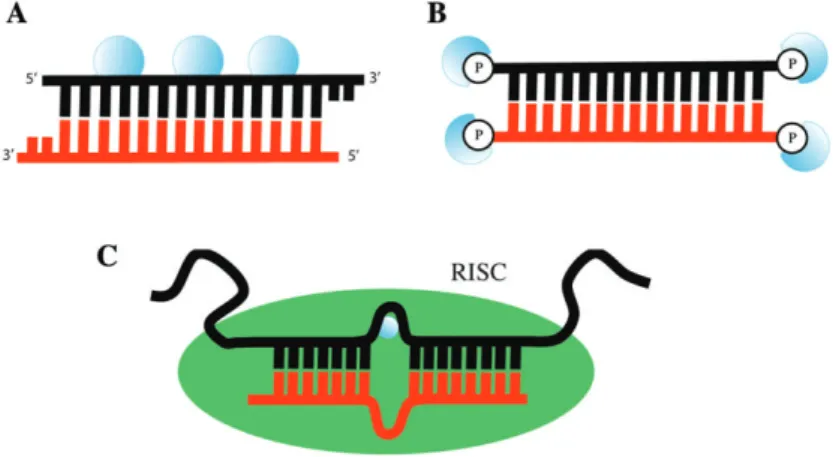
Despite the efforts in drug delivery design and developments, major obstacles such as endosomal escape and effi cient pay-load release within the diseased tissue and cell have to be overcome for effi cient clinical application. Light can be used to enhance drug delivery and payload release by applying light sensitive moieties to drug delivery platforms and of pho-tolabile protecting groups to biologically active molecules by a strategy called caging (Figure 1).
Caging is an attractive way of turning biological molecules e.g., nucleic acids (DNA, RNA), proteins or peptides light sensitive for the investigation of biological processes. Caged biomolecules incorporate a light-removable protecting group, so-called “ caging group ” , which aborts its native biological or biochemical activity. Since caging of ATP was fi rst reported in 1978 by Kaplan et al., several different photolabile groups have been introduced to turn biomolecules temporarily inac-tive (1 – 3) . Examples of caged biomolecules are neurotrans-mitters (4) , nucleotides (4) , peptides (5, 6) , siRNA (7) or DNA (8) . The most widely used caged neurotransmitter so far is glutamate for which different protecting groups have been applied (9) . RNA interference is a mechanism able to inhibit protein translation by gene silencing. Nguyen et al. caged a 1-(2-nirophenyl)ethyl (NPE) group to the 5 ′ terminal phosphate of the siRNA antisense strand, which inactivates
the siRNA activity (10) . They could demonstrate an approxi-mately 70 % effi cient light induced RNA interference using wavelengths between 345 and 385 nm. An alternative form to siRNA mediated control of gene silencing has been reported by Young et al. (11) . They introduced a caging group to DNAzymes to inhibit hybridization with mRNA. DNAzymes are enzymatically active desoxyoligonucleotides, which can cleave RNA in a site-specifi c manner. Translation of mRNA can be aborted upon illumination with UV-light to photo-release the caging group. Caging of DNA has widely been studied as seen from several publications (8, 12, 13) . To ren-der those approaches suitable for future clinical application, extension of the work towards longer wavelengths and there-fore reduced toxicity should be accompanied by identifi ca-tion of suitable in vitro and in vivo disease models of human disease.
Light-responsive materials for drug delivery can be con-structed by the covalent incorporation of specifi c light-sensi-tive chemical groups with the aim to locally release cargo by illumination. The synthesis of a photocleavable amphiphilic block copolymer has been demonstrated by Cabane et al. (14) . As photosensitive molecule they introduced an o-ni-trobenzyl linker between the hydrophobic and hydrophilic blocks, which form vesicles or micelles upon self-assembly in aqueous solution. Successful disruption of the vesicles could be demonstrated after irradiation with UV-light by electron microscopy and dynamic lights scattering data. The design of photocleavable liposomes for drug delivery using different photolabile groups has been reported in several publications (15, 16) . Dvir et al. presented a simple proof of concept by carboxylated polystyrene nanoparticles labeled with the unspecifi c amino acid sequence YIGSR, which
adheres to β 1 integrins present on most cell surfaces (4, 17) . The peptide was caged with a nitrobenzyl group, which could be removed via illumination, leading to nanoparticle binding to the cells. Another approach of light sensitive nanoparticles currently being investigated uses nano-impel-lers. Nano-impellers are nanomechanical systems allowing the spatiotemporal drug release upon illumination, turning them into an attractive application for clinical trials (5, 6, 18, 19) . A clear disadvantage of many published systems is the requirement for light energy in the UV range, limiting their application due to phototoxicity and the very limited penetration range of short wavelength light in biological tissues.
Light induced gene expression and control of gene silencing
Light-mediated control of gene expression and silencing is a powerful and fast growing fi eld in the areas of systems bio-logy, functional genomics and biotechnology. Spatiotemporal and precise gene expression represents the most fundamen-tal level of further complex biological processes such as the control of thousand of proteins and the associated control of metabolic processes. Therefore, light represents a suitable stimulus for in vitro as well as in vivo studies as it is non-in-vasive, sensitive and allows the spatiotemporal and precise application without interfering with metabolic conditions. Light-induced gene expression can either be achieved using caged biomolecules such as plasmid DNA (12, 13) , tran-scription factors (8, 20, 21) or via photoreceptors harboring a chromophore (9, 22, 23) . Several reports focused on caged plasmid DNA ’ s have been published, whereas effective gene Figure 1 (A) Light responsive drug delivery system built through covalent incorporation of specifi c light-sensitive chemical groups (red rectangles) with the aim to locally release cargo (red circles) by illumination. (B) Site-specifi c caging of DNA can be used for light-activated gene expression.
20) . The system is based on two light-inducible fusion pro-teins from Arabidopsis thaliana, GIGANTEA (GI) fused to a Zinc fi nger protein leading the complex to the target DNA sequence and the LOV domain of FKF1 fused to the tran-scriptional activation domain VP16. Illumination with light leads to fusion of the GI and LOV domain, which guides the LOV-VP16 domain to the target gene and enables gene expression.
Beside light induced gene expression, the focus of photo-chemical control of gene function has been directed to RNA interference. RNA interference represents one of the major approaches leading to gene silencing/such as that occurring in embryogenesis) and is being extensively explored as a thera-peutic strategy for different kind of diseases, including can-cer. Two primary approaches for photochemical regulation have been developed. The caging groups are either covalently attached to the phosphate backbone or terminal phosphates or on the nucleotide bases to inhibit the further process of RNA induced silencing (Figure 2). The fi rst report of caged siRNA has been described by Shah et al. using 1-(4, 5-dimethoxy-2-nitrophenyl)ethyl (DMNPE) attached to the phosphate back-bone which only showed a 3 % caging effi ciency (15, 16, 24) . Caging of guanosine and thymidin bases by attaching 2-(2-nitrophenyl)propy (NPP) groups has been reported by Mikat and Heckel (25) . The modifi cations have shown knockdown effi ciency of about 75 % after light irradiation. Jain et al. designed a siRNA caged at the terminal phosphates with a
namic therapy (PDT) was reported at the beginning of the last century by Oscar Raab, a German medical student and his professor Hermann von Tappeiner (28) . The principle of photodynamic therapy involves the administration of a photo sensitizer, which will form highly reactive singlet oxy-gen radical (ROS) from molecular oxyoxy-gen after illumination with light (Figure 3). Singlet oxygen radicals are known to cause severe damage to biological macromolecules such as membrane lipids and proteins (29) . After absorption of light, photosensitizers will change from a ground state into a rela-tively long-lived excited triple state and a short-lived excited single state. The excited single state can return to the ground state by emitting fl uorescence that can be used for clinical detection. In the excited triple state, the photosensitizer mol-ecule can transfer its energy via a type-I or -II reaction. In the type-I reaction, the photosensitizer can react directly with a surrounding substrate to form radicals, which then can further interact with oxygen to produce oxygenated products. In the type-II reaction the energy of the excited photosensitizer can be directly transferred to oxygen to form highly reactive sin-glet oxygen (30) .
Photodynamic therapy has found its way into clinical appli-cations using nanocarrier platforms as delivery system such as photodynamic eye therapy for the treatment of neovascu-larization, abnormal endothelial proliferation or for different cancer treatments (bladder, skin, head and neck, esophageal, or endobronchial cancer) (31, 32) . A number of
Figure 2 Schematic illustration of caged siRNA strategies. (A) Caged phosphate backbone and (B) caged terminal phosphates of siRNA. (C) Introduction of a caged base into a siRNA antisense strand to inhibit RNA interference.
based photodynamic therapies have been approved by the U.S. Food and Drug Administration (FDA) such as e.g., Visudyne ® , Photofrin ® , Levulan ® Kerastick ® opening doors
for future applications and new possible approaches for future therapies (31) . There are several advantages of PDT as a clini-cal application including a single dose requirement for treat-ment followed by illumination compared to radiotherapy and chemotherapy, which both depend on a treatment over several weeks or months. Further, it is a local treatment without inter-fering with the whole organism and retreatment can be sim-ply done in the case of recurrence of a tumor without severe healthy tissue damage. However, further development in the direction of controlled drug release, as well as improved payload capacity of nanoparticle-based delivery systems is warranted.
Photochemical internalization (PCI)
One of the key challenges that still needs to be overcome in order to enable the clinical application of therapeutic delivery of different payloads is endosomal escape. Various strategies have been developed to achieve endosomal escape and these are either based on the characteristic endosomal property of a lower intracellular pH compared to the cytoplasm, incorpora-tion of fusogenic peptides into the endosomal membrane or a strategy called photochemical internalization (PCI). PCI is a site-specifi c method for intracellular drug delivery by induced endolysosomal escape based on photostimulation. The prin-ciple behind PCI relies on photodynamic therapy targeted to endosomes or lysosomes, whereas the vesicular membrane bursts after coming into contact with highly reactive singlet oxygen after illumination of the photosensitizer (Figure 4). In comparison to conventional photodynamic therapy, where the intracellular localization of the photosensitizer does not play an important role because of its complete cellular destruction, PCI is based on the specifi c accumulation of the photosen-sitizer in the endolysosomal compartment to achieve endo-somal escape without harming the rest of the cell (33) .
A fate that may be a consequence to nanocarriers after endocytotic uptake, is the accumulation in the endolysosome, whereas PCI offers a good solution. Lai et al. have Figure 3 Light activation of a photosensitizer leads to the
forma-tion of highly reactive singlet oxygen for selective cell killing. Figure 4 Intracellular drug delivery induced by endolysosomal escape based on photostimulation. Light-irradiation after endocytotic uptake leads to endolysosomal membrane burst upon highly reactive singlet oxygen.
strated the effective delivery of doxorubicin and saporin by photochemical internalization using a poylamidoamine (PAMAM) dendrimer (34, 35) . Recently, Lu et al. reported the overcoming of doxorubicin drug resistance in vivo by applying dendrimer phtalocyanine-encapsulated polymeric micelles combined with doxorubicin into doxorubicin-resis-tant bearing mice (36) . It has also been shown by Nishiyama et al. that PCI can mediate gene transfection, using a com-binational system including polymeric micelles incorporat-ing pDNA and a dendrimer-based photosensitizer (37) . Both polymeric micelles are assumed to be taken up by the cells at the same time. After illumination, a remarkable enhancement of transgene expression could be detected while retaining cell viability. Beside enhancement of gene expression, PCI can also be used for siRNA mediated gene knockdown studies. The fi rst application of PCI to facilitate endosomal escape of siRNA was reported in 2007 by Oliveira et al. (38) . They used TPPS 2a as photosensitizer together with a siRNA able to knock-down epidermal growth factor receptor (EGFR) expression. Complexes of EGFR siRNA and Lipofectamine were applied to the cells. A 10-fold increased effi ciency in EGFR knock-down could be detected after illumination compared to siRNA treatment alone. A recently published study by Varkouhi et al. presents PCI mediated enhancement of gene silencing using a polymer-based nanocarrier platform consisting out of cationic polymethacrylates and N,N,N-trimethylated chitosan (39) . Furthermore, PCI can enhance the effect of targeted protein toxins that have reached the tumors cells (40) . Targeted protein toxins consist of a protein toxin moiety, initiating cytotoxicity and a cell binding moiety, which targets the protein actively to the cell. Denileukin diftitox is the fi rst FDA approved protein toxin for treatment of cutaneous T-cell lymphoma.
Photothermal therapy
Hyperthermia is a non-invasive approach for cancer treatment based on the principle of spatiotemporally increasing the tem-perature to promote selective destruction of cancer cells, which
ible or Near-infrared (NIR) wavelengths (44) . The use of NIR is desirable due to its deep penetrating capacity and minimal interference with water and biomolecules in tissues. The prin-ciple of photothermal therapy is the combination of light and gold nanoparticles (e.g., gold nanospheres, nanorods, nano-shells, nanocages) for clinical treatment. Illumination of gold nanoparticles leads to conversion of absorbed light into ther-mal energy, the resulting heat causes cell and tissue destruc-tion (Figure 5). El-Sayed et al. have shown the use of gold nanorods labeled with an anti-EGFR antibody for selective photothermal treatment of cancer cells (45) . A dual-modality approach for photodynamic and photothermal therapy has been recently published by Kuo et al. (46) . They used gold nanoma-terials conjugated with the hydrophilic photosensitizer, indo-cyanine green, to achieve photothermal therapy (PTT) and photodynamic therapy (PDT). The combination of PTT and PDT showed enhanced destruction of cancer cells in contrast to their single application effectiveness. Photothermal tumor ablation in mice could be proven by O ’ Neal et al. using gold nanoshells (47) . They subcutaneously injected murine colon carcinoma cells into immune-competent mice, followed by injection of gold nanoshells. After 6 h of circulation, tumors were illuminated with NIR. All treated mice looked healthy and tumor free after more then 90 days post-treatment.
Photoswitchable fl uorescent nanoparticles
Over the past decades a huge number of nanoparticles made of different materials have been developed and these have biological and medical applications. Whereas many of those
fl uorescent light at a longer wavelength than that absorbed. Fluorescent nanoparticles such as polymer NPs, silica NPs, gold NPs or quantum dots (QD) gained intensive interest during the last years. They can be produced by doping the material with suitable fl uorescent dyes or luminescent met-als while quantum dots can directly be applied due to their intrinsic fl uorescence properties (48) . The advantages of fl uorescent nanoparticles compared to normal organic dyes are higher brightness due to the fact that a nanoparticle can carry several dye molecules, increase in photostabil-ity because the dyes are entrapped within the nanoparticles, higher specifi city upon their functionalization properties and their long-term-tracking ability.
Understanding cellular networks is the essential key factor to understand the complex structure of certain diseases. To achieve this goal, signifi cant progress has been made in the development of quantum dots for cellular sensing which have been recently reviewed (49) . Sensing quantum dots are based on the principle of the recognition of an analyte, which acts as a fl uorescence quencher, by a receptor or chemosensor caus-ing changes upon emission of the fl uorophore. Various quan-tum dots based on overcoating of the core with ZnS or CdSe to improve their fl uorescence quantum yield and additional modifi cation of the surface properties to increase their emis-sion have been reported (50 – 52) . Furthermore, this concept can be used to prepare glucose or maltose sensing systems, whereas a photoinduced electron transfer (PET) from the coating molecules to the valence band of an excited quantum dot results in emission quenching as shown by Cordes and Sandros et al. (53, 54) .
Figure 5 Photothermal therapy is based on intracellular uptake of gold nanoparticles, which after irradiation with near-infrared light convert absorbed light into thermal energy for specifi c destruction of cancer cells.
and biocompatibility, in particular for wavelengths longer than UV. Furthermore, it has shown advantages regarding its high spatial and temporal precision. However, the major drawback of light is tissue penetration depth, which severely restricts the applications of caged compounds, light sensitive drug delivery systems and light-based therapies into clinical application. Thus, approaches like the usage of NIR linked to two-photon uncaging and up-converting systems seem to be promising but further optimization of these methods is needed to increase the chance of further application in clinical trials.
References
1. Kaplan JH, Forbush B, Hoffman JF. Rapid photolytic release of adenosine 5 ’ -triphosphate from a protected analogue: utilization by the Na:K pump of human red blood cell ghosts. Biochemistry 1978;17:1929 – 35.
2. Pelliccioli AP, Wirz J. Photoremovable protecting groups: reac-tion mechanisms and applicareac-tions. Photochem Photobiol Sci 2002;1:441 – 58.
3. Han G, Mokari T, Ajo-Franklin C, Cohen BE. Caged quantum dots. J Am Chem Soc 2008;130:15811 – 3.
4. Walker JW, Reid GP, McCray JA, Trentham DR. Photolabile 1-(2-nitrophenyl)ethyl phosphate esters of adenine nucleotide analogs. Synthesis and mechanism of photolysis. J Am Chem Soc 1988;110:7170 – 7.
5. Rothman DM, Petersson EJ, V á zquez ME, Brandt GS, Dougherty DA, Imperiali B. Caged Phosphoproteins. J Am Chem Soc 2005;127:846 – 7.
6. Lawrence DS. The preparation and in vivo applications of caged peptides and proteins. Curr Opin Chem Biol 2005;9:570 – 5. 7. Matsushita-Ishiodori Y, Ohtsuki T. Photoinduced RNA
interfer-ence. Acc Chem Res 2012;45:1039 – 47.
8. Ceo LM, Koh JT. Photocaged DNA provides new levels of tran-scription control. ChemBioChem 2012;13:511 – 3.
9. Ellis-Davies GCR. Caged compounds: photorelease techno-logy for control of cellular chemistry and physiotechno-logy. Nat Meth 2007;4:619 – 28.
10. Nguyen QN, Chavli RV, Marques JT, Conrad PG, Wang D, He W, et al. Light controllable siRNAs regulate gene suppression and phenotypes in cells. BBA – Biomembranes 2006;1758:394 – 403. 11. Young DD, Lively MO, Deiters A. Activation and deactivation of DNAzyme and antisense function with light for the photo-chemical regulation of gene expression in mammalian cells. J Am Chem Soc 2010;132:6183 – 93.
12. Yamaguchi S, Chen Y, Nakajima S, Furuta T, Nagamune T. Light-activated gene expression from site-specifi c caged DNA with a biotinylated photolabile protection group. Chem Commun 2010;46:2244 – 6.
13. Monroe WT, McQuain MM, Chang MS, Alexander JS, Haselton FR. Targeting expression with light using caged DNA. J Biol Chem 1999;274:20895 – 900.
14. Cabane E, Malinova V, Meier W. Synthesis of photocleavable amphiphilic block copolymers: toward the design of photosensi-tive nanocarriers. Macromol Chem Phys 2010;211:1847 – 56. 15. Chandra B, Subramaniam R, Mallik S, Srivastava DK.
Formulation of photocleavable liposomes and the mechanism of their content release. Org Biomol Chem 2006;4:1730 – 40. 16. Yavlovich A, Smith B, Gupta K, Blumenthal R, Puri A.
Light-sensitive lipid-based nanoparticles for drug delivery: design prin-ciples and future considerations for biological applications. Mol Membr Biol 2010;27:364 – 81.
Beside the mentioned applications, the most largely exploited photoswitchable fl uorescent sensing mechanism is by fl uorescence resonance energy transfer (FRET) by which an energy transfer from a QD to a fl uorophore will be deter-mined. Until now, several strategies allowing generic on/off photoswitching based on FRET have been reported (55 – 57) . Another more complex approach for photoswitching has been explored by dye doped nanoparticles allowing coact-ive triggering of multiple processes. These dye doped sens-ing nanoparticles were fi rst introduced by Kopelman in the late 1990s and are called photonic explorers for bioanalysis with biologically localized embedding (PEBBLEs) (58) . The insertion of chemosensors into nanoparticles shows several advantages as minimization of interaction with other biomo-lecules within the cells or the introduction of multifunctional sensing schemes as for example by pH sensitivity. A wide variety of PEBBLEs have been reported since their develop-ment, whereas further literature can be found here (59 – 61) .
However, the ability of QDs to combine molecular imaging and therapy can open new doors for clinical application, but the toxicity of especially heavy metals used in QD synthesis such as cadmium is an important concern (62) .
Limiting factors of light
Light as external stimuli for enhanced drug delivery, cargo release, imaging and therapy offers some attractive features such as high sensitivity and spatiotemporal control. However, the major drawback of light is tissue penetration depth, restricting its applications. Solutions to overcome this problem have been made by development of near infrared (NIR) light sensitive photochemical compounds. Near-infrared light at wavelengths of 700 – 1000 nm can penetrate up to several cen-timeters deep into tissues without causing any damage (63) . This renders NIR much more attractive than the often used UV-light regarding its potential for severe tissue damage. The use of two-photon excitation systems as well as application of upconverting nanoparticles, both provide possible solutions of how to overcome the problem of tissue penetration depth. Two-photon excitation depends on the principle of exciting a caged group by absorption of two photons induced via a pulsed laser. This method allows the usage of caged groups, which absorb light in the UV range but can be excited via pulsed NIR. Upconverted nanomaterials are able to convert NIR into UV light, which generates the same benefi t as seen for the two-photon excitation. Practical application of such systems into clinical trials might still need some time due to the fact that most published data using NIR, show irradiation times of hours, which might interfere with clinical practice (64) .
Conclusion
The use of light as an external stimulus is a promising approach for a wide range of applications within the fi eld of nano-medicine based on its attractive properties such as sensitivity
Jaeger K-E. Lights on and action! Controlling microbial gene expression by light. Appl Microbiol Biotechnol 2011;90:23 – 40. 23. van der Horst MA, Hellingwerf KJ. Photoreceptor proteins, “ star
actors of modern times ” : a review of the functional dynamics in the structure of representative members of six different photore-ceptor families. Acc Chem Res 2004;37:13 – 20.
24. Shah S, Rangarajan S, Friedman SH. Light-activated RNA inter-ference. Angew Chem Int Ed Engl 2005;44:1328 – 32.
25. Mikat V, Heckel A. Light-dependent RNA interference with nucleobase-caged siRNAs. Rna 2007;13:2341 – 7.
26. Jain PK, Shah S, Friedman SH. Patterning of gene expression using new photolabile groups applied to light activated RNAi. J Am Chem Soc 2011;133:440 – 6.
27. Ackroyd R, Kelty C, Brown N, Reed M. The history of pho-todetection and photodynamic therapy. Photochem Photobiol 2001;74:656 – 69.
28. Lee Y, Baron ED. Photodynamic therapy: current evidence and applications in dermatology. Semin Cutan Med Surg 2011;30:199 – 209.
29. Pervaiz S, Olivo M. Art and science of photodynamic therapy. Clin Exp Pharmacol Physiol 2006;33:551 – 6.
30. Dolmans DEJGJ, Fukumura D, Jain RK. TIMELINE: photody-namic therapy for cancer. Nat Rev Cancer 2003;3:380 – 7. 31. Christie JG, Kompella UB. Ophthalmic light sensitive
nanocar-rier systems. Drug Discov. Today 2008;13:124 – 34.
32. Triesscheijn M, Baas P, Schellens JHM, Stewart FA. Photody-namic therapy in oncology. The Oncologist 2006;11:1034 – 44. 33. Dougherty TJ, Henderson BW, Gomer CJ, Jori G, Kessel D,
Korbelik M, et al. Photodynamic Therapy. J Natl Cancer Inst 1998;90:889 – 905.
34. Lai P-S, Lou P-J, Peng C-L, Pai C-L, Yen W-N, Huang M-Y, et al. Doxorubicin delivery by polyamidoamine dendrimer con-jugation and photochemical internalization for cancer therapy. J Control Release 2007;122:39 – 46.
35. Lai P-S, Pai C-L, Peng C-L, Shieh M-J, Berg K, Lou P-J. Enhanced cytotoxicity of saporin by polyamidoamine dendrimer conjugation and photochemical internalization. J Biomed Mater Res 2008;87:147 – 55.
36. Lu H-L, Syu W-J, Nishiyama N, Kataoka K, Lai P-S. Dendrimer phthalocyanine-encapsulated polymeric micelle-mediated pho-tochemical internalization extends the effi cacy of photodynamic therapy and overcomes drug-resistance in vivo. J Control Release 2011;155:7 – 7.
37. Nishiyama N, Arnida, Jang W-D, Date K, Miyata K, Kataoka K. Photochemical enhancement of transgene expression by poly-meric micelles incorporating plasmid DNA and dendrimer-based photosensitizer. J Drug Target 2006;14:413 – 24.
38. Oliveira S, Fretz M, H ø gset A, Storm G, Schiffelers RM. Photochemical internalization enhances silencing of epidermal
42. Ohguri T, Imada H, Kato F, Yahara K, Morioka T, Nakano K, et al. Radiotherapy with 8 MHz radiofrequency-capacitive regional hyperthermia for pain relief of unresectable and recur-rent colorectal cancer. Int J Hyperthermia 2006;22:1 – 14. 43. Arthur RM, Straube WL, Trobaugh JW, Moros EG. Non-invasive
estimation of hyperthermia temperatures with ultrasound. Int J Hyperthermia 2005;21:589 – 600.
44. Li J-L, Gu M. Gold-nanoparticle-enhanced cancer photothermal therapy. IEEE J Sel Top Quantum Electron 2010;16:989 – 96. 45. El-Sayed IH, Huang X, El-Sayed MA. Selective laser
photo-thermal therapy of epithelial carcinoma using anti-EGFR antibody conjugated gold nanoparticles. Cancer Lett 2006; 239:7 – 7.
46. Kuo W-S, Chang Y-T, Cho K-C, Chiu K-C, Lien C-H, Yeh C-S, et al. Gold nanomaterials conjugated with indocyanine green for dual-modality photodynamic and photothermal therapy. Biomaterials 2012;33:3270 – 8.
47. O ’ Neal DP, Hirsch LR, Halas NJ, Payne JD, West JL. Photo-thermal tumor ablation in mice using near infrared-absorbing nanoparticles. Cancer Lett 2004;209:6 – 6.
48. Ruedas-Rama MJ, Walters JD, Orte A, Hall EAH. Fluorescent nanoparticles for intracellular sensing: a review. Anal Chim Acta 2012;751:1 – 23.
49. Delehanty JB, Susumu K, Manthe RL, Algar WR, Medintz IL. Active cellular sensing with quantum dots: transitioning from research tool to reality; a review. Anal Chim Acta 2012;750:63 – 81.
50. Dabbousi BO, Rodriguez-Viejo J, Mikulec FV, Heine JR, Mattoussi H, Ober R, et al. (CdSe) ZnS core-shell quantum dots: synthesis and characterization of a size series of highly lumines-cent nanocrystallites. J Phys Chem B 1997;101:9463 – 75. 51. Ji X, Zheng J, Xu J, Rastogi VK, Cheng TC, DeFrank JJ, et al.
(CdSe) ZnS quantum dots and organophosphorus hydrolase bio-conjugate as biosensors for detection of paraoxon. J Phys Chem B 2005;109:3793 – 9.
52. Chen Y, Rosenzweig Z. Luminescent CdS quantum dots as selec-tive ion probes. Anal Chem 2002;74:5132 – 8.
53. Cordes DB, Gamsey S, Singaram B. Fluorescent quantum dots with boronic acid substituted viologens to sense glu-cose in aqueous solution. Angew Chem Int Ed Engl 2006;45: 3829 – 32.
54. Sandros MG, Gao D, Benson DE. A modular nanoparticle-based system for reagentless small molecule biosensing. J Am Chem Soc 2005;127:12198 – 9.
55. Shi L, De Paoli V, Rosenzweig N, Rosenzweig Z. Synthesis and application of quantum dots FRET-based protease sensors. J Am Chem Soc 2006;128:10378 – 9.
56. Zhu L, Zhu M-Q, Hurst JK, Li ADQ. Light-controlled molecu-lar switches modulate nanocrystal fl uorescence. J Am Chem Soc 2005;127:8968 – 70.
Roman Lehner is a 2nd year PhD student in the group of Prof. P. Hunziker, work-ing within the SNF pro-ject “Intelligent Materials”. Roman studied molecular biology at the University of Basel from where he received his MSc in structural bio-logy in 2009. After his mas-ter diploma, he worked as a scientifi c associate at the Institute of Anatomy in Basel and for the spin-off company BioVersys. His work is focused on the design of a polymer- based drug delivery system, which can be functionalized to target particular tissue and combined with a stimulus respon-siveness too light to explore activation of caged compounds. 57. Freeman R, Finder T, Bahshi L, Willner I. β
-cyclodextrin-modifi ed CdSe/ZnS quantum dots for sensing and chiroselective analysis. Nano Lett 2009;9:2073 – 6.
58. Buck SM, Xu H, Brasuel M, Philbert MA, Kopelman R. Nanoscale probes encapsulated by biologically localized embed-ding (PEBBLEs) for ion sensing and imaging in live cells. Talanta 2004;63:41 – 59.
59. Lee Y-EK, Smith R, Kopelman R. Nanoparticle PEBBLE sen-sors in live cells and in vivo. Annu Rev Anal Chem (Palo Alto Calif) 2009;2:57 – 76.
60. Si D, Epstein T, Lee Y-EK, Kopelman R. Nanoparticle PEBBLE sensors for quantitative nanomolar imaging of intracellular free calcium ions. Anal Chem 2012;84:978 – 86.
61. Buck SM, Koo Y-EL, Park E, Xu H, Philbert MA, Brasuel MA, et al. Optochemical nanosensor PEBBLEs: photonic explorers for bioanalysis with biologically localized embedding. Curr Opin Chem Biol 2004;8:540 – 6.
62. Chen N, He Y, Su Y, Li X, Huang Q, Wang H, et al. The cytotoxicity of cadmium-based quantum dots. Biomaterials 2012;33:1238 – 44.
63. Skirtach AG, Antipov AA, Shchukin DG, Sukhorukov GB. Remote activation of capsules containing Ag nanopar ticles and IR dye by laser light. Langmuir 2004;20:6988 – 92.
64. Lehner R, Wang X, Wolf M, Hunziker P. Designing switch-able nanosystems for medical application. J Control Release 2012;161:307 – 16.
Patrick Hunziker has stud ied Medicine at the University of Zurich, Switzerland. He received a doctoral degree based on thesis work in experimental immunology from the University of Zur-ich and did further research in experimental hematol-ogy at University Hospital in Zurich, Switzerland. He earned specialist degrees in Internal Medicine, Car-diology and Intensive Care Medicine. As a fellow of the Massachusetts General Hospital, Harvard Medical School, he worked on cardiac imaging in a joint project with the Massachusetts Institute of Technology, Cambridge. His professional activities in Europe, the U.S., Africa and China gave him a broad insight into the needs for the medicine of the future in a variety of settings. Hunziker became involved in medical applications of Nanoscience in the late 1990s and has been the pioneer physician in Nano-medicine in Switzerland since then. With improved preven-tion, diagnosis and cure of cardiovascular disease as his main research topic, he worked in the nanoscience fi elds of atomic force microscopy, nano-optics, micro/nanofl uidics, nanome-chanical sensors and polymer nanocarriers for targeting. He is the founding president of the European Society of Nano-medicine, cofounder of the European Foundation for Clinical Nanomedicine and coinitiator of the European Conference for Clinical Nanomedicine and is clinically active as deputy head of the Clinic for Intensive Care Medicine at the University Hospital Basel, Switzerland. In November 2008 Patrick Hun-ziker became professor for Cardiology and Intensive Care Medicine at the University of Basel.