HAL Id: hal-03175710
https://hal.archives-ouvertes.fr/hal-03175710
Submitted on 20 Mar 2021HAL is a multi-disciplinary open access archive for the deposit and dissemination of sci-entific research documents, whether they are pub-lished or not. The documents may come from teaching and research institutions in France or abroad, or from public or private research centers.
L’archive ouverte pluridisciplinaire HAL, est destinée au dépôt et à la diffusion de documents scientifiques de niveau recherche, publiés ou non, émanant des établissements d’enseignement et de recherche français ou étrangers, des laboratoires publics ou privés.
Simon task: an electromyographic study
Aurélie Grandjean, Isabel Suarez, Elisa Diaz, Laure Spieser, Boris Burle,
Agnès Blaye, Laurence Casini
To cite this version:
Aurélie Grandjean, Isabel Suarez, Elisa Diaz, Laure Spieser, Boris Burle, et al.. Stronger impulse capture and impaired inhibition of prepotent action in children with ADHD performing a Simon task: an electromyographic study. Neuropsychology, American Psychological Association, inPress. �hal-03175710�
Stronger impulse capture and impaired inhibition of prepotent action in children with ADHD performing a Simon task: an electromyographic study
Aurélie Grandjean1, Isabel Suarez2, Elisa Diaz2, Laure Spieser1, Boris Burle1, Agnès Blaye3, Laurence Casini1
1. Laboratoire de Neurosciences Cognitives, FR 3C, Aix-Marseille Université, CNRS, Marseille, France
2. Universidad del Norte, Baranquilla, Colombia
3. Laboratoire de Psychologie Cognitive, FR 3C, Aix-Marseille Université, CNRS, Marseille, France e-mail addresses : aurelie.grandjean@univ-amu.fr suarez.isabel1982@gmail.com laure.spieser@univ-amu.fr boris.burle@univ-amu.fr agnes.blaye@univ-amu.fr laurence.casini@univ-amu.fr Corresponding author: Laurence Casini Address : LNC, FR 3C, Case C
3 place Victor Hugo
13331 Marseille cedex 3, France
e-mail : laurence.casini@univ-amu.fr Phone : 33413550941
Fax : 33413550958
Note
This work was funded by the French Agence Nationale de la Recherche (ANR-15-CE28-0008, DOPCONTROL). The authors wish to thank Solène Ambrosi for the development of the program and they also warmly thank all the children that accepted to participate in this study as well as the Universidad Del Norte, and the Colegio Sagrado Corazón of Barranquilla for allowing us to conduct this study. The authors also thank Jennifer Coull for her help with English language.
Abstract (200 words)
Objective: A deficit in interference control is commonly reported in children with attention
deficit hyperactivity disorder (ADHD). This has mainly been interpreted as a difficulty in
inhibiting inappropriate responses. However, it could be due to at least two distinct and
independent processes, which are often confounded: the activation or suppression of
impulsive responses. The aim of the present study was to separate the contribution of these
two processes. Method: We compared performance of 26 children with ADHD to that of 26
non-ADHD children using a novel approach based on electromyographic activity (EMG)
analysis. EMG allows two distinct indices to be computed: incorrect activation rate, which is
an index of the intensity of impulse capture and correction rate, which provides a direct
measure of the ability to suppress automatic responses. Results: Children with ADHD were
slower, committed more errors, and had a larger interference effect than non-ADHD children.
Moreover, we observed a greater incorrect activation rate and a lower correction rate in the
ADHD group. Conclusions: Our data suggest that the difficulties in interference control
found in children with ADHD are explained by both impaired inhibitory processes and a
greater propensity to activate automatic responses.
Key Points:
- We investigated how ADHD affects impulsivity control.
- Interference control was impaired in children with ADHD
- Impulsivity and hyperactivity could be due to both a greater propensity to activate impulsive responses and a greater difficulty in inhibiting them.
- Future research should investigate the effect of treatments on these two components of impulsivity control.
Imagine that you are approaching a traffic light that just turned amber. You prepare to brake
but suddenly, in your mirror, you see a huge truck coming up very quickly behind you. The
safest response is actually to accelerate. This situation therefore requires overlearned
automatic actions to be overridden by controlled ones. Every day, we are forced to adapt our
behavior to unexpected environmental changes. This flexibility requires efficient cognitive
control, a term that refers to the set of processes that are engaged when we must stop
inappropriate spontaneous actions in favor of goal-directed actions that are better adapted to
the context.
Experimentally, cognitive control has commonly been studied in conflict tasks
specifically designed to induce a conflict between an automatic tendency to respond to an
irrelevant but salient stimulus and a controlled goal-directed response to a relevant stimulus
(Eriksen & Eriksen, 1974; Simon, 1969; Stroop, 1935). The well-documented Simon task is
particularly suitable for studying cognitive control (Hommel, 2011). In the standard version of
the Simon task, the participants are required to respond as quickly and as accurately as
possible by pressing on a left- or a right-hand key in response to a non-spatial attribute (form
or color, for example) of a stimulus that is presented either on the left or right side of a central
fixation point. Typically, mean reaction times (RT) are longer and errors are more frequent in
trials for which the required response is contralateral to stimulus location (incongruent trials,
IG) than in trials where the required response is ipsilateral to stimulus location (congruent
trials, CG). This behavioral cost is known as the interference effect. The usual interpretation
of these findings is that the position of the stimulus activates a fast and automatic response in
the effector ipsilateral to the stimulus. Since this response is erroneous in IG trials, it must be
suppressed in favor of the response that is correct according to task instructions. The
suppression of the automatic response requiring interference control (De Jong, Liang &
Lauber, 1994; Kornblum, Hasbroucq & Osman, 1990; Kornblum, 1994; Lu & Proctor, 1995).
Several studies (for an overview, see Mullane, Corkum, Klein, & McLaughlin, 2009)
but not all (Borella, de Ribaupierre, Cornoldi, & Chicherio, 2013; Schwartz & Verhaegen,
2008; Van Mourik et al., 2009) have found that children with ADHD manifest poor
performance in conflict tasks. They exhibit longer reaction times, more errors, and larger
interference effects than their typically developing peers (Cao et al., 2013; Homack & Riccio,
2004; Jonkman, et al, 1999; Tsal, Shalev, & Mevorach, 2005; for a review, see Mullane,
Corkum, Klein, & McLaughlin, 2009). ADHD is among the most prevalent and most
extensively studied of the childhood pathologies. Classically, the difficulties that children
with ADHD show in this type of task are considered to be due to difficulties in inhibiting
inappropriate automatic and prepotent responses (Barkley, 1997; Nigg, 2001), even if some
studies more recently suggest that inhibition may also be intact in children with ADHD
(Coghill, Seth, & Matthews, 2014). However, these difficulties could be due to, at least, two
distinct and dynamic processes involved in interference control, which are confounded in
most studies. According to the “activation-suppression model” (Ridderinkhof, 2002), the first
process, which we will call impulse capture, is assumed to reflect the degree to which the
response system is susceptible to activating location-driven automatic responses. The second
process is assumed to reflect inhibitory control, which builds up to suppress the interference
induced by the incorrect action impulse. Therefore, impairments in children with ADHD
could be due either to stronger impulse capture, to less efficient inhibitory processes, or both.
Obviously, a better understanding of difficulties in interference control would help to better
understand ADHD.
The aim of the present study was to disentangle the contribution of these two
distinct indices that provide information on both the intensity of impulse capture and the
ability to inhibit automatic response activation.
EMG activity and interference control
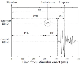
In choice RT tasks, recording EMG activity of the muscles primarily involved in
making a response allows response activation and execution to be studied. It reveals that in
about 20% of correct responses, EMG activity associated with the correct response is
preceded by an earlier sub-threshold EMG activity recorded from the hand associated with the
incorrect response (see Figure 1) (Eriksen et al., 1985; Hasbroucq, Burle, Akamatsu, Vidal &
Possamaï, 2001; Hasbroucq, Possamaï, Bonnet, & Vidal, 1999). This sub-threshold activity is
called a partial error (Burle, Possamaï, Vidal, Bonnet & Hasbroucq, 2002). Such incorrect
response activations are more numerous in incongruent trials (in which the task-irrelevant
affordance primes the incorrect response) than in congruent trials (in which there is response
capture of the correct response). It has been proposed that partial errors are the direct
manifestation of online activation, and subsequent suppression, of an incorrect automatic
response (Burle, Possamaï, Vidal, Bonnet & Hasbroucq, 2002; Van den Wildenberg et al.,
2010). In other words, partial errors correspond to incorrect response activations that have
been detected, stopped and corrected. Therefore, partial errors, which reveal subliminal
activation of the incorrect response effector and its subsequent correction, constitute a key
element in investigating interference control and highlight the utility of EMG recording when
studying processes involved in interference control.
INSERT FIGURE 1 ABOUT HERE
Partial errors provide access to information that is invisible in purely behavioral
experiments. More precisely, they reveal information that is hidden in correct trials and allows
trials in which there is a unique response-related EMG activity in the correct hand, partial
errors correspond to trials with sub-threshold muscle activity in the incorrect hand prior to the
EMG activity associated with the correct response, and overt errors correspond to trials with a
response-related EMG activity in the incorrect hand only. Using these different types of trials,
two different indices highlighting the two processes of interest can be computed. First, the
incorrect activation rate (IAR), corresponding to the sum of partial errors and overt errors
(that is the total number of incorrect activations, corrected or not) divided by the total number
of trials, is considered to provide a direct measure of the intensity of impulse capture,
independent from potential subsequent suppression, when it is calculated for incongruent
trials in which the stimulus position primes the incorrect response. A higher incorrect
activation rate in incongruent trials indicates stronger impulse capture, that is an increased
tendency to activate automatic responses (Hasbroucq, Burle, Vidal & Possamaï, 2009; Van
den Wildenberg et al., 2010). Second, the correction rate, corresponding to the number of
successfully corrected incorrect activations (that is, partial errors) divided by the overall
number of incorrect activations (corrected or not), is considered to provide a direct measure of
the ability to suppress automatic responses in order to prevent response errors (Burle,
Possamaï, Vidal, Bonnet & Hasbroucq, 2002; Van den Wildenberg et al., 2010). A lower
correction rate indicates less efficient suppression of erroneous automatic responses.
We compared performance of children with ADHD to that of children without ADHD
matched in age and education level, and we used classic behavioral measures as well as the
two indices derived from EMG activity to investigate the effect of ADHD on two components
of interference control: activation and suppression of impulsive responses. In addition, since
EMG activity also allows RT to be decomposed into distinct premotor and motor components
(Figure 1), we used it to investigate the effect of ADHD separately on motor and decisional
1. Material and methods 1.1. Participants
We tested two groups of participants: 26 participants with ADHD (mean age = 11.5; 20
males) and 26 children without ADHD (non-ADHD group) (mean age = 11.4; 20 males)
matched in age, school level and socioeconomic status1 to children with ADHD. The primary
language of all participants was Spanish. All participants and their parents gave informed
consent prior to the experiment. The study was approved by the Ethical Committee of the
Universidad del Norte (Barranquilla, Colombia) and was carried out in accordance with the
provisions of the Declaration of Helsinki.
1.1.1. Selection procedure for the ADHD group
Children with ADHD were recruited from a sample of patients who had been referred to the
Instituto Colombiano de Neuropedadogia (Barranquilla, Colombia) by qualified neurologists
and who continue to be regularly followed by psychiatrists or neurologists of the institute.
They all met DSM IV diagnostic criteria for ADHD (American Psychiatric Association,
2000). The assessment was conducted separately with teachers, children and their parents, and
made on the basis of a semi-structured clinical diagnostic interview (DSM IV checklist) by
trained neurologists specialized in ADHD. Final diagnoses were reached by a consensus on
the basis of the results of structured interviews, collateral historical information, different
behavioral scales rated by parents and teachers, and clinical interviews of parents and children
by a committee of clinicians, all of whom have extensive experience with ADHD. The
Instituto Colombiano de Neuropedadogia is known to the Caribbean community for
providing comprehensive diagnostic and psychoeducational services. Moreover, it is approved
1Colombian neighbourhoods are classified by socio-economic status. All children who
by the Colombian government as a neuroscience institution conducting developmental and
clinical child and adolescent research.
In addition, in order to compare control and ADHD groups, the parents of each child filled out
a behavioral rating scale, the EDAH scale (Evaluacion Deficit de Atencion e Hiperactividad,
Farré & Narbona, 2013; Sánchez, Ramos, Díaz & Simón, 2010).
For this study, only children who had never received medication were contacted and solicited
for their participation: even with a 24-48h wash out period, it is difficult to exclude the
possibility that medication could affect cognitive performance. Three children had comorbid
oppositional defiant disorder (ODD).
1.1.2. Inclusion criteria for the control group
Children from the non-ADHD group were recruited via local schools in Barranquilla. They all
attended normal classes corresponding to their age level. Inclusion criteria were the following:
1/ absence of current or previous diagnosis of ADHD determined by completion of the
EDAH, 2/ absence of emotional disturbance or learning disabilities based on teachers’ and
academic psychologists’ reports, 3/ no concurrent treatment with psychotropic medication.
1.1.3. Exclusion criteria for both groups
Exclusion criteria included (1) a diagnosis of any additional psychiatric disorder (major
depression, panic disorder, suicide risk, anxiety, substance abuse, psychoactive substance use,
psychotic disorders) on the basis of a Spanish version of the structured psychiatric interview
(Children’s Interview for Psychiatric Syndromes, CHIPS) (Weller, Weller, Fristad, Rooney &
Schecter, 2000) performed by a neurologist before the experiment, (2) absence of consent or
parental consent, (3) an intelligence quotient (IQ) < 70, assessed by the vocabulary-block
WISC III (Wechsler, 1991) was used and IQ was calculated with reference to Mexican norms
since Colombian norms do not exist. In addition, all participants also performed the working
memory index from the WISC III (Spanish version with Mexican norms) (Wechsler, 1991),
which comprised arithmetic, digit span, and letter-number sequencing subtests to assess
working memory.
1.2. Simon choice RT task
Stimuli and apparatus. Participants were comfortably seated facing a black computer screen,
located 80 cm away, upon which stimuli appeared. A plastic board was placed in front of the
participant. Two plastic cylinders were fixed on the board and served as handgrips. One
press-button was fixed to the top of each cylinder. The participant had to maintain the distal
phalanxes of each thumb on the response buttons. Responses were given by pressing the top
of one of the two cylinders either with the right or left thumb. All stimuli and responses were
controlled using components of Psychopy (Peirce, 2007). RTs were recorded to the nearest
millisecond.
Task and procedure. Throughout the test, two experimenters were present. One sat near the
participant and the other one managed the computer programs. Each trial started with the
appearance of a central fixation point that participants had to fixate during the entire trial.
After a delay of 500 ms, a picture appeared on either the right or left of the fixation point.
Children had to press the left or right button as a function of the presented picture, as quickly
and accurately as possible. The stimuli remained on the screen until children responded. Two
types of trials were defined: Congruent trials (CG) in which the required response was
ipsilateral to the stimulus location, and incongruent trials (IG) in which the required response
Three different sets of stimuli were used in order to keep the subject engaged in the task (set
1: banana versus carrot; set 2: pig versus frog; set 3: strawberry versus hazelnut)
(Smigasiewicz, Ambrosi, Blaye, & Burle, 2020). Each participant performed the task with the
three sets. The set-order and the picture-response mapping (for example, press right for a
banana and press left for a carrot) were counterbalanced across participants. For each set,
participants performed one training session of 34 trials corresponding to 17 IG trials and 17
CG trials, presented in random order, to familiarize themselves with the task. They then
performed four experimental blocks of 25 trials. The first trial of each block was chosen
randomly and not further analyzed. The remaining 24 trials corresponded to 12 IG trials and
12 CG trials, presented in random order. To summarize, each child performed three sets of
100 trials each. The sets were separated by a pause of about 5 minutes and the entire
experiment lasted about 60 minutes.
1.3. EMG signal recording and processing
The EMG activity of the flexor pollicis brevis was recorded bipolarly by means of surface Ag/
AgCl electrodes, 6 mm in diameter, fixed about 10 mm apart on the skin of the thenar
eminence. EMG activity was amplified, digitized on-line at 2048 Hz, and offline high-pass
filtered at 10Hz. In order to facilitate off-line EMG onset detection, the EMG signal was
continuously monitored by the experimenter to minimize the influence of background activity
that could interfere with small bursts of muscular activation during the reaction period. If the
signal became noisy, the experimenter immediately asked the child to relax his/her muscles.
He also regularly reminded the child to keep hands on the handgrips.
To detect very small incorrect muscular activation, onsets of EMG activity were
detected by a home-made custom program written in Python to facilitate visual inspection
license, is accessible upon request). EMG traces of each participant were then inspected
visually and corrected manually in the case of inaccurate detection by the program. The
processing of EMG data was performed by an experimenter who was blind to diagnostic
group.
1.4. EMG data analysis
The analysis of EMG data had two main objectives: (1) classification of responses into three
trial types in order to compute incorrect activation rate (IAR) and correction rate (CR), and
(2) chronometric analysis of data to decompose RT into decisional and motor subcomponents.
Computation of IAR and CR. First, correct trials were sorted into two categories, labeled “pure
correct” and “partial error” trials. “Partial error” trials contained activation of the agonist
muscle involved in the incorrect response prior to activation of the agonist involved in the correct response. Importantly, to be classified as a “partial error”, EMG signal detection had
to be phasic and to return to baseline (rest) before the onset of the response-related EMG
activity. Response errors and partial errors were detected and counted and the two variables
were then calculated: IAR = (partial errors + overt errors)/total number of non rejected trials,
and CR = PE / (PE+E) where PE is the number of partial errors and E the number of overt
errors.
Chronometric analysis. The EMG analysis also provided access to several different
chronometric indices as illustrated in Figure 1. Reaction time (RT) was defined as the latency
between stimulus onset and the button press response. It was broken down into two
components: premotor time (PMT, from stimulus onset to EMG onset), and motor time (MT,
defined: Partial error latency (PEL) corresponding to the interval between stimulus onset and
EMG onset of the incorrect response, and correction time (CT) corresponding to the latency
between incorrect EMG onset and correct EMG onset (Figure 1).
Two-way ANOVAs, with the between-subjects factor of Group (ADHD versus non-ADHD)
and the within-subject factor of Congruency (CG versus IG), were performed on the different
variables. Proportional scores (error rate, incorrect activation rate, correction rate), in
particular when they are rather high (or low), have non-Gaussian distributions because of
ceiling (or floor) effects. Therefore, to normalize distributions, data were arcsine transformed
before being entered into the ANOVA. This nonlinear but monotonic transformation allows a
Gaussian distribution to be obtained so that the required conditions for the ANOVA are met
(Winer, 1970).
Cohen’s d was used to compute effect size (standardized difference between the two means)
for the two group comparisons; effect sizes are commonly classified as small (d = 0.2),
medium (d = 0.5), or large (d = 0.8) (Cohen, 1988).
2. Results
Three children with ADHD and one non-ADHD child were excluded from analyses because
their EMG signal was too noisy (participants did not succeed in relaxing their muscles).
Analyses were thus performed with data from 23 children with ADHD and 25 non-ADHD
children.
2.1. Demographic and neuropsychological variables
Differences between demographic characteristics of non-ADHD and ADHD groups were
no difference in age, children with ADHD had smaller IQ scores and working memory index.
Scores on the EDAH were significantly different.
INSERT TABLE 1 ABOUT HERE
2.2. Simon task
Extreme RT values, either excessively fast (< 150 ms, so-called anticipatory errors) or slow (>
3 standard deviations [SDs]), were removed from the analysis. On average, this led to the
exclusion of fewer than 1% of trials per participant. In addition, specifically for EMG data,
some trials (2.02 %) were rejected due to tonic activity or artifacts preceding the contraction
involved in the response.
2.2.1. Behavioral data
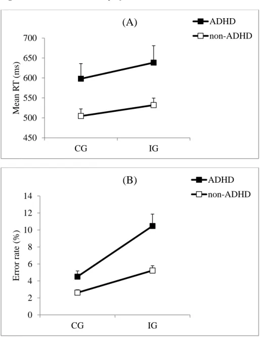
Mean Reaction times. As shown in Figure 2A, mean RTs were longer for children with
ADHD (617 ms) compared with non-ADHD children (518 ms) (F1,46 = 5.47; p =.02; Cohen’s
d = .66), and longer in IG trials (583 ms) than in CG trials (549 ms) (F1,46 = 97.99; p < .0001).
The difference between groups for the interference effect was medium in magnitude
(Cohen’s2 d = .54; ADHD group = 41 ms, non-ADHD group = 28 ms), but the difference was not statistically significant (Group x Congruency interaction: F1,46 = 3.57; p = .06).
Intra-subject RT variability. The intra-subject RT variability (SD) was larger in children with
ADHD (199 ms) compared to non-ADHD children (113 ms) (F1,46 = 12.1; p = .001; Cohen’s d
= .99) and larger in CG trials (156 ms) than in IG trials (149 ms) (F1,46 = 4.8; p = .03). There
was no statistically significant Group x Congruency interaction (F1,46 = 2.3; p = .10).
2 Cohen’s d refers to the comparison of interference effects (mean CG RT – mean IG
Error rate. As shown in Figure 2B, children with ADHD committed more errors (7.5%) than
non-ADHD children (3.9%) (F1,46 = 12.8; p < .0001; Cohen’s d = 1.02). As usual, the error
rate was higher in IG trials (7.7%) compared to CG trials (3.5%) (F1,46 = 52.7; p < .0001). The
interference effect was larger in children with ADHD (6%) compared with non-ADHD
children (2.6%) (Group x Congruency interaction: F1,46 = 8.08; p = .006; Cohen’s3 d = .81).
INSERT FIGURE 2 ABOUT HERE
2.2.2. Analysis from partial errors
From EMG activity, three categories of trials were determined: pure correct, partial error and
real error. The percentage of each trial type for each group is presented in Table 2. The
percentage of pure correct trials was lower for ADHD group than for non-ADHD group but
children with ADHD did not commit more partial errors than non-ADHD children.
INSERT TABLE 2 ABOUT HERE
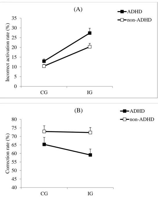
Incorrect activation rate. The IAR data, combining real errors and partial errors, revealed that
children with ADHD made more incorrect EMG activations (19.9%) than non-ADHD
children (15.2%) (F1,46 = 5.53; p = .023; Cohen’s d = .69) (Figure 3A). IAR was larger for IG
trials (23.7%) than for CG trials (11.6%) (F1,46 = 99.73; p < .0001). The difference between
groups for the interference effect was medium in magnitude (Cohen’s d = .53; ADHD group = 14.4%, non-ADHD group = 9.9%) but the difference was not statistically significant
(Congruency x Group: F1,46 = 3.44; p = .07).
Even more relevant for our purpose, IAR in IG trials, considered an index of the strength of
impulse capture, was larger in children with ADHD (27.4%) than in non-ADHD children
(20.6%) (t46 = 2.62; p = .011; Cohen’s d = .67).
3Cohen’s d refers to the comparison of interference effects ( CG accuracy rate – IG accuracy
Correction rate. The CR was lower for the ADHD group (60.7%) than for the non-ADHD
group (73.1%) (F1,46 = 6.84; p = .012; Cohen’s d = .94) (Figure 3B). There was no significant
difference between CG (69.2%) and IG trials (65.9%) (F1,46 = 1.35; p = .25), as classically
observed in previous studies (Burle et al, 2002; Burle, Spieser, Servant & Hasbroucq, 2014).
The Group X Congruency interaction was not significant (F1,46 = .9; p = .35).
INSERT FIGURE 3 ABOUT HERE
2.2.3. Chronometric analysis
2.2.3.1. Reaction time components in pure correct trials
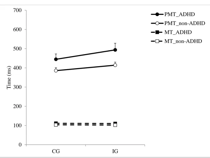
Premotor times (PMT). Mean PMT was longer for the ADHD group (467 ms) than for the
non-ADHD group (399 ms) (F1,46 = 4.14; p = .048; Cohen’s d = .57) and mean PMT was
longer in IG trials (452 ms) than in CG trials (414 ms) (F1,46 = 71.61; p < .0001) (Figure 4).
The Group x Congruency interaction was statistically significant (F1,46 = 5.03; p = .03;
Cohen’s d = .63) revealing that the interference effect was larger in the ADHD group (49 ms)
than in the non-ADHD group (28 ms).
In addition, intra-subject PMT variability was larger in children with ADHD (175 ms)
than non-ADHD children (103 ms) (F1,46 = 10.65; p = .002; Cohen’s d = .92), but intra-subject
PMT variability was no different between CG and IG trials (F1,46 = .35; p = .55). The
difference in variability between IG and CG trials was significantly larger in children with
ADHD (14 ms) than in non-ADHD children (9 ms), as indicated by the significant Group x
Congruency interaction (F1,46 = 7.81; p = .007; Cohen’s d = .78).
Motor times (MT). For mean MT, there was no difference between groups (F1,46 = .77; p =
(Burle et al, 2002; Burle, Spieser, Servant, & Hasbroucq, 2014), and no Group X Congruency
interaction (F1,46 = .01; p = .92) (Figure 4). Concerning intra-subject MT variability, there was
no difference between groups (F1,46 = .23; p = .63), nor between CG and IG trials (F1,46 = .1; p
= .75). There was no significant Group x Congruency interaction (F1,46 = .007; p = .93).
INSERT FIGURE 4 ABOUT HERE
2.2.3.2. PMT components in partial error trials
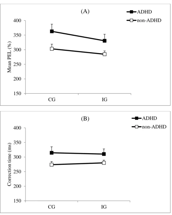
Partial error latency (PEL). Mean PEL was longer for the ADHD group (340 ms) than for the
non-ADHD group (291 ms) (F1,46 = 4.67; p = .036; Cohen’s d = .59) (Figure 5A). It was
longer for CG (332 ms) than for IG trials (307 ms) (F1,46 = 5.47; p = .023), as is typically
observed (Burle et al, 2002; Burle, Spieser, Servant, & Hasbroucq, 2014). The Group x
Congruency interaction was not significant (F1,46 = .41; p = .53). In addition, intra-subject
PEL variability was larger in children with ADHD (141 ms) than in non-ADHD children (93
ms) (F1,46 = 6.73; p = .01; Cohen’s d = .65) and larger in CG trials (127 ms) than in IG trials
(100 ms) (F1,46 = 4.31; p = .04). There was no significant Group x Congruency interaction
(F1,46 = .22; p = .64) for intra-subject PEL variability.
Correction time (CT). The difference between groups was medium in magnitude (Cohen’s d =
.45; ADHD group: 312 ms; non-ADHD group: 280 ms) but the difference was not statistically significant (F1,46 = 3.04; p = .088) (Figure 5B). CT was not affected by congruency (F1,46 =
.02; p = .89) and there was no significant Group x Congruency interaction (F1,46 = .74; p =
.39). Moreover, intra-subject CT variability was no different between groups (F1,46 = 2.46; p =
.13), nor between CG and IG trials (F1,46 = 2.56; p = .12) but there was a significant Group x
2.3. Correlation analyses
Correlation coefficients were computed between our two indices of interest (IAR and CR) and
working memory indices and IQ, for each group. No statistically significant correlations were
found (see Table 3).
INSERT TABLE 3 ABOUT HERE
3. Discussion
The aim of the present study was to investigate interference control in children with
ADHD when performing a Simon task. Using EMG activity recordings, we were able to
distinguish two components of interference control and so separately evaluate the intensity of
impulse capture and the ability to inhibit automatic responses.
3.1. Children with ADHD have impaired behavioral performance in conflict tasks
A traditional analysis of RTs and error rates revealed two main results. Firstly, we
observed greater intra-individual RT variability in children with ADHD compared to
non-ADHD children. This is consistent with data found in prior studies, which report increased
intra-individual RT variability in a variety of different RT tasks (Klein, Wendling, Huettner,
Ruder & Peper, 2006; Lipszyc & Schachtar, 2010; Tamm, Nakonezny & Hughes, 2012).
Nonetheless, it remains open to debate whether this result is ubiquitous and so could represent
a stable trait of ADHD, or whether it might also be characteristic of other populations, (Klein,
Wendling, Huettner, Ruder & Peper, 2006; Tamm, Nakonezny & Hughes, 2012; for review,
Kofler et al., 2013). At a cognitive level, the high intra-individual RT variability of patients
with ADHD likely reflects impairments in information processing and difficulties in
maintaining attentional control. However, the nature of mechanisms and processes
Kofler et al., 2013; Kofler et al., 2014; Sonuga-Barke & Castellanos, 2007; Tamm,
Nakonezny & Hughes, 2012).
Secondly, children with ADHD were slower, committed more errors than non-ADHD
children, and exhibited a larger interference effect, which suggests that children with ADHD
were globally impaired on the Simon RT task compared to non-ADHD children. These data
confirm the results of other studies in the literature (Cao et al., 2013; Homack & Riccio, 2004;
Jonkman, et al, 1999; Tsal, Shalev, & Mevorach, 2005; for a review, see Mullane, Corkum,
Klein, & McLaughlin, 2009) indicating that these children have difficulties in performing
conflict tasks. The larger interference effect could arise from difficulties in ignoring the
irrelevant feature of the stimulus, its position, and/or in maintaining attention on the relevant
feature, its color. In most studies, a larger interference effect is interpreted as being due to
poor inhibition (Barkley, 1997; Nigg, 2001). However, the difficulties of children with
ADHD could have at least two causes: stronger impulse capture, that is a stronger propensity
to activate automatic responses or, alternatively, poorer inhibition of these automatic
responses. Analysis of EMG recordings, which allows these two processes to be dissociated,
is a convenient way to tackle this issue.
3.2. What can we learn from partial errors?
As mentioned in the Introduction, analysis of EMG activity allows us to dissociate two
types of trials embedded within correct response trials, pure correct responses and partial
errors, and then to compute two indices: incorrect activation rate (IAR) and correction rate
(CR). The first index (IAR) expresses all incorrect activations, whether they resulted in an
incorrect response or not (if they were corrected) and can be considered an index of the
strength of impulse capture, particularly in incongruent (IG) trials in which stimulus position
proportion of incorrect activations that were corrected before making an overt error and can
be considered an index of the efficiency of automatic response inhibition.
Our results revealed a larger IAR in children with ADHD than in non-ADHD children
and, more specifically, we observed a larger IAR in IG trials. This indicates that children with
ADHD are more likely to automatically activate the response ipsilateral to stimulus position.
In other words, they have more difficulty not being influenced by salient yet irrelevant
information. Consequently, these children would be more prone to selecting the response that
corresponds to the most frequent association between the location of the stimulus and the
response, that is, the ipsilateral response. In addition, we observed a lower correction rate in
children with ADHD. This second result suggests that children with ADHD had more
difficulty stopping and correcting automatic, but erroneous, activations than non-ADHD
children. Since incorrect activations were detected and stopped less often, these children
committed more overt errors, which contributed to an increased IAR. Therefore, these data
provide new and more direct evidence in favor of inhibition deficits in children with ADHD.
Response inhibition includes a variety of processes, such as inhibiting prepotent
responses, inhibiting all ongoing responses (global inhibition) and selective inhibition (in
interference control tasks). The ability to inhibit prepotent and ongoing responses, as assessed
by the stop task, have been extensively studied in ADHD. Most studies report decreased
performance in children with ADHD than in their typically developing peers and some
authors have concluded that children with ADHD have deficient inhibitory control (for
reviews, see Barkley, 1997; Crosbie et al., 2013; Matzke, Verbruggen & Logan, 2018; Nigg,
2001; Osterlaan, Logan, & Sergeant, 1998; Sergeant, Geurts, & Oosterlaan, 2002).
Nonetheless, others who dissected the stop-signal reaction time (SSRT) have proposed that
the data reveal an underlying attentional deficit rather than deficient inhibitory control
Kenemans, Verbaten, & van Engeland, 2005). However, the authors also concluded that the
data do not rule out the possibility that other aspects of inhibitory control, such as interference
control could be deficient in ADHD. Indeed, even though global inhibition is at play in the
stop-task, selective inhibition (stopping the incorrect response and continuing to execute the
correct one) is required in conflict tasks that require interference control, such as the Simon
task used here. These two types of inhibition have been shown to partially differ in terms of
neural mechanisms and substrates (Aron & Verbruggen, 2008; Aron, 2011). Our findings
therefore suggest that selective inhibition is impaired in ADHD. Inhibition can be used to
refer to a variety of different processes and it seems important to more precisely identify
which one is impaired in ADHD. Accordingly, Weigard and collaborators (2019) used a
Bayesian parametric approach for estimating stop-signal RT distribution in order to better
identify the cognitive processes responsible for impaired performance in children with ADHD
in stop-tasks. They established that difficulty in stopping a prepotent response was mainly due
to a failure to trigger the inhibition process rather than an impairment in the inhibition process
itself (Weigard, Heathcote, Matzke, & Huang-Pollock, 2019). In our study, the correction rate
directly reflects the proportion of incorrect activations that have been stopped and corrected
or, in other words, successful inhibition via the action cancellation subcomponent of
interference control. A decrease in correction rate, as observed in the ADHD group, may
indicate failure to trigger the inhibition process as proposed in Weigard et al’s study (2019).
Impairments in children with ADHD could then be more precisely explained by a difficulty in
initiating inhibition rather than by a difficulty in inhibiting the response.
Moreover, impulsive responses entail at least two processes: an impulse or urge to act,
whose strength depends on the context, and a lack of inhibition (DeYoung, 2013). Our data
suggest that the difficulties observed in conflict tasks in children with ADHD are also
incorrect activation rate), which is rarely considered in most studies. Our results are partially
consistent with those presented in one another behavioral study that investigates both
activation and suppression of impulse capture in children with ADHD engaged in a conflict
task (Ridderinkhof, Scheres, Oosterlan & Sergeant, 2005). The authors reported that children
with ADHD had difficulty in suppressing automatic response but did not make more
impulsive responses in the first place. However, at least two differences with our own
experiment may be noted. First, Ridderinkhof et al (2005) used dynamic analysis of
performance (delta-plot analysis of reaction times and accuracy) to dissociate the two
complementary components of interference, whereas we based our conclusions on indices
derived from EMG data, which give more direct access to the activation and suppression of
response impulses (Van den Wildenberg et al., 2010). Second, children in the previous study
performed a flanker task in which the nature of interference (perceptive interference) is
different to that in the Simon task (motor interference). Data from a recent study comparing
the two tasks have suggested that the control of inappropriate responses is more difficult to
demonstrate, and less stable, in the flanker task (Burle, Spieser, Servant, & Hasbroucq, 2014).
From a neurobiological point of view, it is interesting to note that some brain
structures found to be dysfunctional in ADHD are also involved in interference control.
Indeed, anatomical and functional observations converge to indicate that ADHD is associated
with a dysfunction of the prefrontal cortex and its connections, in particular with basal ganglia
(Dickstein, Bannon, Castellanos & Milham, 2006; Durston, Van Belle & De Zeeuw, 2011;
Konrad & Eickhoff, 2010), cerebral structures that are also known to be involved in
interference control (Chambers, Garavan & Bellgroce, 2009; Fassbender & Schweitzer, 2006;
Forstmann, Van den Wildenberg & Ridderinkhof, 2008; Spieser, van den Wildenberg,
Hasbroucq, Ridderinkhof, & Burle, 2015). More specifically, it has been shown that during
hypoactivation primarily of inferior frontal cortex (IFC), dorsolateral prefrontal cortex
(DLPFC), anterior cingulate cortex (ACC) and caudate nucleus (Bush et al., 1999; Cubillo,
Halari, Giampietro, Taylor & Rubia, 2011; Cubillo, Halari, Smith, Taylor & Rubia, 2012 ;
Rubia et al., 2009; Rubia et al., 2010; Vaidya et al., 2005). Our results might therefore
represent the behavioral outcome of functional alterations in structures known to be involved
in interference control and which are impaired in individuals with ADHD.
To summarize, our data suggest that the difficulty experienced by children with
ADHD in tasks requiring interference control could be explained by both stronger impulse
capture and impaired inhibition, two independent processes that can be differentially impaired
in certain cases (Fluchère et al., 2015; 2018; Ramdani et al., 2015). For example, subthalamic
nucleus stimulation delivered to patients with Parkinson’s disease selectively impairs
suppression of response impulsivity but does not increase the strength of impulse capture
(Fluchère et al., 2018). These two functions are probably anatomically dissociated and this
dissociation could have important implications not only for refining explanations of ADHD
but also for classifying patients according to different symptoms and determining suitable
treatments.
3.3. What do chronometric indices reveal?
EMG activity analysis also provided several different chronometric indices, such as premotor
time (PMT) and motor time (MT) (PMT + MT = RT). We observed slower and more variable
PMT for children with ADHD than for non-ADHD children, whereas no difference was found
for the mean or variability of MTs between groups. These results confirm that the slowed RTs
observed in children with ADHD was not due to motor slowing, therefore arguing against an
explanation in terms of motor execution deficits. In contrast, they suggest that RT slowing
preparation. This is in line with data from the modeling literature. According to computational
modeling (such as drift-diffusion model, DDM, Ratcliff & McKoon, 2008; linear ballistic
accumulation model, LBA, Brown & Heathcote, 2008) accuracy and response time
distributions depend on decision and non-decision parameters. Non-decision parameters
represent extra-decisional components, such as encoding and motor processes. Among them,
one important variable is the drift rate, which reflects task difficulty or information processing
ability. It corresponds to the speed with which the accumulation of evidence approaches the
response boundary. High drift rates correspond to faster and more accurate responses. A
second variable is the boundary separation, which describes the amount of accumulated
evidence needed until a decision is made (also called the decision threshold). The majority of
studies have found reduced drift in ADHD (Huang-Pollock et al., 2017; Metin et al., 2013;
Weigard & Huang-Pollock, 2014; for a review, see Ziegler, Pedersen, Movinckel, & Biele,
2016) whereas effects on decision threshold are unclear (Ziegler, Pedersen, Movinckel, &
Biele, 2016). In addition, residual non-decision time is very often reported to be unaffected
(Ziegler, Pedersen, Movinckel, & Biele, 2016) though not always (Metin et al., 2013).
Non-decision time includes both perceptual and motor times. In an EMG decomposition of RT,
MT can indisputably be linked to motor execution meaning our results are consistent with
data from modeling studies. However, PMT comprises both encoding and decision times.
Assuming that encoding time is similar in ADHD and non-ADHD groups (as suggested by
modeling data), the lengthening of PMT would reflect a lower drift rate. It has to be noted
however that standard models (DDM or LBA) cannot correctly fit data from tasks such as
Simon, Stroop or Eriksen flanker tasks (Hübner, Steinhauser, & Lehle, 2010; White, Ratcliff,
& Starns, 2011) because non-constant drift rate models are required to fit conflict data (White,
Servant, & Logan, 2018) and, to the best of our knowledge, such models have not yet been
EMG activity analysis also provided chronometric indices specific to partial error
trials, such as partial error latency and correction time. We observed larger and more variable
partial error latencies in the ADHD group compared to the non-ADHD group, suggesting that
children with ADHD were slower than non-ADHD children even when committing a partial
error, that is, when an incorrect activation had been triggered. They also tended to be slower
to correct these erroneous activations, suggesting that all processes, correct or incorrect, are
delayed in children with ADHD. This delay, observed at each stage of processing, again
speaks in favor of fluctuations in sustained attention.
Interestingly, these results appear to be in line with the recent neuro-energetic theory
(NeT) of ADHD (Killeen, Russell & Sergeant, 2013; Killeen, 2013), which proposes that an
insufficient neuronal energy supply, due to a deficit in lactate supplementation, could impair
all forms of effortful responses. Impairment at any point in the neurobiological cascade of
lactate synthesis would produce neuronal fatigue, which in turn would cause fluctuations in
sustained attention. Lapses of attention could cause children with ADHD to have difficulty in
focusing their attention on the relevant stimulus feature, since non-salient task-dependent
features require controlled attention. These children would then be more prone to initiating
automatic location-driven responses. Moreover, some studies suggest that suppressing
automatic responses may be under top-down control and depend upon attentional resources
(Suarez, Vidal, Burle & Casini, 2015). Inattention and difficulties in cognitive control may
therefore be linked (Alderson, Rapport, & Kofler, 2007; Weigard, Heathcote, Matzke, &
Huang-Pollock, 2019).
3.4. Limitations and conclusion
There are at least two limitations to the current study. The first limitation refers to the
present study. More intriguing is that both groups and particularly the non-ADHD group had
relatively high IQ scores and WMI compared to other studies. But, as mentioned in the
Methods section, IQ scores and WMI were evaluated with the Spanish version of the WISC
III using Mexican norms because Colombian norms are not available. It is therefore possible
that IQ scores and WMI were overestimated for both groups compared with studies using
more recent versions of the WISC and norms developed in the country where studies were
conducted. Nonetheless, the difference in IQ raises a question as to whether our results would
persist after controlling for IQ. However, it has to be noted that IQ was used only as an
inclusion criterion and was evaluated by only two sub-tests of the WISC. Moreover, we
believe that IQ should not be included as a covariate in cognitive studies for at least two
reasons: 1/IQ measures aptitude and potential, rather than performance; 2/IQ does not fulfill
the methodological and statistical requirements of a covariate (for detailed arguments, see
Dennis et al., 2009). In addition, even though some studies indicate a mediating effect of IQ
on inhibition (Mahone et al., 2002), others have shown that not all executive functions,
particularly inhibition, are related to IQ (Friedman et al., 2006, Bitsakou et al., 2008).
Similarly, it has also been proposed that WMI and inhibitory processes could be related
(Miyake et al., 2000; Karr et al., 2018). It might then be hypothesized that the superior
working memory of the non-ADHD group could have artificially inflated estimates of an
inhibition deficit in ADHD. We observed no statistically significant correlation between WMI
and correction rate in either group (Table 3). However, due to our sample size, correlations
suggest a modest relationship between WMI and correction rate for the non-ADHD group. A
last point that can be mentioned is that studies investigating potential relationships between
inhibition and IQ or WMI present at least three noticeable differences with the present study:
1/ a full-scale IQ test is usually used, not simply two subtests of the WISC; 2/ cognitive
or neuropsychological tests are used to measure inhibition but no study has yet used a variable
as precise and directly linked to inhibitory processes as the correction rate. Nonetheless, our
data merit confirmation in future studies in which both groups of children would be matched
in age, gender, WMI and IQ scores. In this regard,we would like to mention that we carried
out a sensitivity analysis that compared two groups matched for IQ and WMI scores (non-ADHD children with very high scores were excluded) and that the findings were very comparable to those reported in the present manuscript. Data are available in Supplementary Materials (Table S1).
The second limitation refers to the fact that because ADHD is so heterogeneous,
further studies are needed to fully understand this developmental disorder. For example, the
question of whether ADHD is due to a central deficit or to multiple deficits, as proposed more
and more frequently (Castellanos & Tannock, 2002; Castellanos, Sonuga-Barke, Milham, &
Tannock, 2006; Nigg, Willcutt, Doyle & Sonuga-Barke, 2005), should be a crucial question in
developing further studies. Comparing the performance of a group of predominantly
inattentive children with a group of predominantly hyperactive children using EMG activity
and the indices described here would be of great interest.
To conclude, our data suggest that the deficit in interference control in children with
ADHD can be explained by both stronger impulse capture and difficulty in inhibiting such
impulses, which are two independent processes. Moreover, the slowing of all processes along
the sensorimotor chain suggests difficulties in sustained attention, which could also
exacerbate difficulties in interference control. One interesting follow-up to the current study
could be to evaluate how different treatments, such as stimulant medication or behavioral
therapy, separately affect impulse capture and inhibitory control. Although children with
affect one of the two processes, which could shed light on the heterogeneity of treatment
References
Alderson, R.M., Rapport, M.D., & Kofler, M.J. (2007). Attention-deficit/hyperactivity
disorder and behavioral inhibition: a meta-analytic review of the stop-signal paradigm.
Journal of Abnormal Child Psychology, 35, 745-758.
Alderson, R.M., Rapport, M.D., Sarver, D.E., & Kofler, M.J. (2008). ADHD and behavioral
inhibition: A re-examination of the Stop-signal task. Journal of Abnormal Child Psychology,
36, 989–998.
American Psychiatric Association. (2000). Diagnostic and statistical manual of mental
disorders. Forth Edition Revised (DSM IV-TR). Washington DC.
Aron, A.R. (2011). From reactive to proactive and selective control: developing a richer
model for stopping inappropriate responses. Biological Psychiatry. 69(12): e55–e68.
Aron, A.R., & Verbruggen, F. (2008). Stop the presses: dissociating a selective from a global
mechanism for stopping. Psychological Science, 19, 1146-1153.
Barkley, R.R. (1997). Behavioral inhibition, sustained attention, and executive functions:
constructing a unifying theory of ADHD. Psychological Bulletin, 121, 65-94.
Bitsakou, P., Psychogiou, L., Thompson, M., & Sonuga-Barke, E.J.S. (2008). Inhibitory
deficits in attention-deficit/hyperactivity disorder are independent of basic processing
Borella, E., de Ribaupierre, A., Cornoldi, C., & Chicherio, C. (2013). Beyond interference
control impairment in ADHD: Evidence from increased intraindividual variability in the
color-stroop test. Child Neuropsychology, 19(5), 495-515.
Brown, S.D., & Heathcote, A. (2008). The simplest complete model of choice response time:
Linear ballistic accumulation. Cognitive Psychology, 57,153-178.
Burle, B., Possamaï, C.A., Vidal, F., Bonnet, M., & Hasbroucq, T. (2002). Executive control
in Simon effect: an electromyographic ans distributional analysis. Psychological Research,
66, 324-336.
Burle, B., Spieser, L., Servant, M., & Hasbroucq, T. (2014). Distributional reaction time
properties in the Eriksen task: marked differences or hidden similarities with the Simon task?
Psychonomic Bulletin and Review, 21, 1003-1010.
Bush, G., Frazier, J. A., Rauch, S. L., Seidman, L. J., Whalen, P. J., Jenike, M. A., Rosen,
B.R., & Biederman, J. (1999). Anterior cingulate cortex dysfunction in
attention-deficit/hyperactivity disorder revealed by fMRI and the counting stroop. Biological
Psychiatry, 45, 1542–1552.
Cao, J., Wang, S., Ren, Y., Zhang, Y., Cai, J., Tu, W., Shen, H., Dong, X., & Xia, Y. (2013).
Interference control in 6-11 year-old children with and without ADHD: behavioral and ERP
Castellanos, F.X., & Tannock, R. (2002). Neuroscience of attention-deficit/hyperactivity
disorder: the search for endophenotypes. Nature Reviews Neuroscience, 3, 617-628.
Castellanos, F.X., Sonuga-Barke, E.J.S., Milham, M.P & Tannock, R. (2006). Characterizing
cognition in ADHD: beyond executive dysfunction. Trends in Cognitive Sciences, 10,
117-123.
Chambers, C.D., Garavan, H., & Bellgroce, M.A. (2009). Insights into the neural basis of
response inhibition from cognitive and clinical neuroscience. Neuroscience and
Biobehavioral Reviews, 33, 631-646.
Coghill, D.R., Seth, S., & Matthews, K. (2014). A comprehensive assessment of memory,
delay aversion, timing, inhibition, decision making and variability in attention deficit
hyperactivity disorder: advancing beyond the three-pathway models. Psychological Medicine,
44(9),1-13.
Cohen, J. (1988). Statistical power analysis for the behavioral sciences (2nd ed.). Hillsdale,
NJ: Lawrence Earlbaum Associates.
Crosbie, J., Arnold, P., Paterson, A., Swanson, J., Dupuis, A., Li, X., Shan, J., Goodal, T.,
Strug, L, & Schachar, R.J. (2013). Response inhibition and ADHD traits: correlates and
heritability in a community sample. Journal of Abnormal Child Psychology, 41, 497–507.
Cubillo, A., Halari, R., Giampietro, V., Taylor, E., & Rubia, K. (2011). Fronto-striatal
with attention deficit/hyperactivity disorder and persistent symptoms. Psychiatry Research:
Neuroimaging, 193, 17-27.
Cubillo, A., Halari, R., Smith, A., Taylor, E., & Rubia, K. (2012). A review of fronto-striatal
and fronto-cortical brain abnormalities in children and adults with Attention Deficit
Hyperactivity Disorder (ADHD) and new evidence for dysfunction in adults with ADHD
during motivation and attention. Cortex, 48, 194-215.
De Jong, R., Liang, C-C., & Lauber, E. (1994). Conditional and Unconditional Automaticity:
A Dual-Process Model of Effects of Spatial Stimulus-Response Correspondence. Journal of
Experimental Psychology: Human Perception and Performance, 20, 731-750.
Dennis, M., Francis, D.J., Cirino, P.T., Schachar, R., Barnes, M.A., & Fletcher, J.M. (2009).
Why IQ is not a covariate in cognitive studies of neurodevelopmental disorders. Journal of
the International Neuropsychological Society, 15, 331-343.
DeYoung, C. (2013). The neuromodulator of exploration: A unifying theory of the role of
dopamine in personality. Frontiers in Human Neuroscience. doi: 10.3389/fnhum.2013.00762
Dickstein, S.G., Bannon, K., Castellanos, X., & Milham, M.P. (2006). The neural correlates
of attention deficit hyperacticity disorder: an ALE meta-analysis. Journal of Child Psychology
Durston, S., Van Belle, J., & De Zeeuw, P. (2011). Differenciating frontostriatal and
fronto-cerebellar circuits in attention-deficit/hyperactivity disorder. Biological Psychiatry, 69,
1178-1184.
Eriksen, C. W., Coles, M. G., Morris, L. R., & O'Hara, W. P. (1985). An electromyographic
examination of response competition. Bulletin of the Psychonomic Society, 23, 165-168.
Eriksen, B. A., & Eriksen, C. W. (1974). Effects of noise letters upon the identification of a
target letter in a nonsearch task. Attention, Perception, & Psychophysics, 16, 143-149.
Farré A., & Narbona, J. (1998). EDAH. Escalas para la evaluación del trastorno por déficit de
atención con hiperactividad. (TEA). Madrid.
Fassbender, C., & Schweitzer, J.B. (2006). Is there evidence for neural compensation in
attention deficit hyperactivity disorder? A review of the functional neuroimaging literature.
Clinical Psychology Review, 26, 445-465.
Fluchere, F., Deveaux, M., Burle, B., Vidal, F., van den Wildenberg, W.P.M., Witjas, T.,
Eusebio, A. Azulay, J.P., Hasbroucq, T. (2015). Dopa therapy and action impulsivity: subthreshold error activation and suppression in Parkinson’s disease. Psychopharmacology,
232, 1735-1746.
Fluchere, F., Burle, B., Vidal, F., van den Wilbenberg, W.P.M., Witjas, T., Eusebio, A.,
Azulay, J.P., Hasbroucq, T. (2018). Subthalamic nucleus stimulation, dopaminergic treatment
Forstmann, B.U., Van den Wildenberg, W.P.M., & Ridderinkhof, K.R. (2008). Neural
mechanisms, temporal dynamics, and individual differences in interference control. Journal of
Cognitive Neuroscience, 20, 1854-1865.
Friedman, N. P., Miyake, A., Corley, R. P., Young, S. E., De Fries, J. C., & Hewitt, J. K.
(2006). Not all executive functions are related to intelligence. Psychological Science, 17(2),
172–179.
Hasbroucq, T., Possamaï, C.A., Bonnet, M., & Vidal, F. (1999). Effect of irrelevant location
of the response signal on choice reaction time: an electromyographic study in humans.
Psychophysiology, 36, 522-526.
Hasbroucq, T., Burle, B., Akamatsu, M., Vidal, F., & Possamaï, C.A. (2001). An
electromyographic investigation of the effect of stimulus-response mapping on choice
reaction time. Psychophysiology, 38, 157-162
Hasbroucq, T., Burle, B., Vidal, F., & Possamaï, CA. (2009). Stimulus-hand correspondence
and direct response activation: An electromyographic analysis. Psychophysiology, 46,
1160-1169.
Homack, S., & Riccio, C.A. (2004). A meta-analysis of the sensitivity and specificity of the
Stroop color and word test with children. Archives of Clinical Neuropsychology, 19,
Hommel, B. (2011). The Simon effect as tool and heuristic. Acta Psychologica, 136, 189-202.
Huang-Pollock, C., Ratcliff, R., McKoon, G., Shapiro, Z., Weigard, A., & Galloway-Long, H.
(2017).Using the diffusion model to explain cognitive deficits in attention deficit
hyperactivity disorder. Journal of Abnormal Psychology, 45, 57-68.
Hübner, R., Steinhauser, M., & Lehle, C. (2010). A dual-stage two-phase model of selective
attention. Psychological Review, 117(3), 759-784.
Jonkman, L.M., Kemner, C., Verbaten, M.N., Van Engeland, H., Kenemans, J.L.,
Gamfferman, G., Buitelaar, J.K., & Koelega, H.S. (1999). Perceptual and response
interference in children with attention-deficit hyperactivity disorder, and the effects of
methylphenidate. Psychophysiology, 36, 419-429.
Karr, J., Areshenkoff, C., Rast, P., Hofer, S., Iverson, G., & Garcia-Barrera, M. (2018). The
unity and diversity of executive functions: A systematic review and re-analysis of latent
variable studies. Psychological bulletin, 144(11), 1147-1185.
Killeen, P.R. (2013). Absent without leave; a neuroenergetic theory of mind wandering.
Frontiers in Psychology, 4.
Killeen, P.R., Russell, V.A., & Sergeant, J.A. (2013). A behavioral neuroenergetics theory of
Klein, C., Wendling, K., Huettner, P., Ruder, H., & Peper, M. (2006). Intrasubject variability
in attention-deficit hyperactivity disorder. Biological Psychiatry, 60, 1088–1097.
Kofler, M.J., Rapport, M.D., Sarver, D.E., Raiker, J.S., Orban, S.A., Friedman, L.M., &
Kolomeyer, E.G. (2013). Reaction time variability in ADHD: A meta-analytic review of 319
studies. Clinical Psychology Review, 33, 795-811.
Kofler, M.J., Alderson, R.M., Raiker, J.S., Bolden, J., Sarver, D.E., Rapport, M.D. (2014).
Working memory and intra-individual variability as neurocognitive indicators in ADHD:
examining competing model predictions. Neuropsychology, 28, 459-471.
Konrad, K., & Eickhoff, S.B. (2010). Is the ADHD Brain Wired Differently? A Review on
Structural and Functional Connectivity in Attention Deficit Hyperactivity Disorder. Human
Brain Mapping, 31, 904–916.
Kornblum, S., Hasbroucq, T., & Osman, A. (1990). Dimensional Overlap: cognitive basis for
stimulus-response compatibility-a model and taxonomy. Psychological Review, 97, 253-270.
Kornblum, S., (1994). The way irrelevant dimensions are processed depends on what they
overlap with: The case of Stroop- and Simon-like stimuli. Psychological Research, 56,
130-135.
Lijffijt, M., Kenemans, L., Verbaten, M. N., & van Engeland, H. (2005). A meta-analytic
review of stopping performance in attention-deficit/hyperactivity disorder: deficient inhibitory
Lipszyc, J., & Schachtar, R. (2010). Inhibitory control and psychopathology: A meta-analysis
of studies using the stop signal task. Journal of the International Neuropsychological Society,
16, 1064–1076.
Lu, C., & Proctor, R. W. (1995). The influence of irrelevant location information on
performance: A review of the Simon and spatial Stroop effects. Psychonomic Bulletin &
Review, 2, 174–207.
Mahone, E.M., Hagelthorn, K.M., Cutting, L.E., Schuerholz, L.J., Pelletier, S.F., Rawlins, C.,
Singer, H.S., & Denckla, M.B. (2002). Effects of IQ on executive functions measures in
children with ADHD. Child Neuropsychology, 8:1, 52-65.
Matzke, D., Verbruggen, F., & Logan, G. (2018). The stop signal paradigm. In J.T. Wixted &
E.J. Wagenmakers (Eds.), Stevens’ handbook of experimental psychology and cognitive
neuroscience (pp. 383–427). Hoboken, NJ: John Wiley & Sons, Inc.
Metin, B., Roeyers, H., Wiersema, J.R., van der Meere, J.J., Thompson, M., Sonuga-Barke, E.
(2013). ADHD performance reflects inefficient but not impulsive information processing: a
diffusion model analysis. Neuropsychology, 27, 193-200.
Miyake, A., Friedman, N.P., Emerson, M.J., Witzki, A.H., Howerter, A., & Wager, T.D.,
(2000). The unity and diversity of executive functions and their contributions to complex ‘‘Frontal Lobe’’ tasks: A latent variable analysis. Cognitive Psychology 41, 49–100.
Mullane, J.C., Corkum, P.V., Klein, R.M., & McLaughlin, E. (2009). Interference control in
children with ADHD: A systematic review of flanker and Simon task performance. Child
Neuropsychology, 15, 321-342.
Nigg, J.T. (2001). Is ADHD a disinhibitory disorder? Psychological Bulletin, 127, 571-598.
Nigg, J.T., Willcutt, E.G., Doyle, A.E. & Sonuga-Barke, E.J.S. (2005). Causal heterogeneity
in Attention-Deficit/Hyperactivity disorder: do we need neuropsychologically impaired
subtypes? Biological Psychiatry, 57, 1224-1230.
Oosterlaan, J., Logan, G. D., & Sergeant, J. A. (1998). Response inhibition in AD/HD, CD,
comorbid AD/HD+CD, anxious, and control children: A meta-analysis of studies with the
stop task. Journal of Child Psychology and Psychiatry, 39, 411–425.
Peirce, J.W. (2007). PsychoPy–Psychophysics software in Python. Journal of Neuroscience
Methods, 162, 8–13.
Ramdani, C., Carbonnell, L., Vidal, F., Béranger, C., Dagher, A., Hasbroucq, T. (2015).
Dopamine precursors depletion impairs impulse control in healthy volunteers.
Psychopharmacology, 232, 477–487.
Ratcliff, R., & McKoon, G. (2008). The diffusion decision model: theory and data for
two-choice decision tasks. Neural Computation, 20, 873-922.
clarification through distributional analyses. In W. Prinz, & B. Hommel (Eds.), Attention and
Performance, Vol. XIX, Common Mechanisms in Perception and Action (pp. 494–519).
Oxford: Oxford University Press.
Ridderinkhof, K.R., Scheres, A., Oosterlan, J., & Sergeant, J.A. (2005 ). Delta plots in the
study of individual differences: new tools reveal response inhibition deficits in AD/HD that
are eliminated by methylphenidate treatment. Journal of abnormal psychology, 114, 197-215.
Rubia, K., Halari, R., Smith, A.B., Mohammad, M., Scott, S., & Brammer, M.J. (2009).
Shared and disorder-specific prefrontal abnormalities in boys with pure attention
deficit/hyperactivity disorder compared to boys with pure CD during interference inhibition
and attention allocation. The Journal of Child Psychology and Psychiatry, 50, 669-678.
Rubia, K., Cubillo, A., Smith, A.B., Woolley, J., Heyman, I., & Brammer, M.J. (2010).
Disorder-specific dysfunction in right inferior prefrontal cortex during two inhibition tasks in
boys with attention-deficit hyperactivity disorder compared to boys with
obsessive-compulsive disorder. Human Brain Mapping, 31, 287-299.
Sánchez, C.R., Ramos, C., Díaz, F., & Simón, M. (2010). Validación de la escala de
evaluación del trastorno por déficit de atención/hiperactividad (EDAH) en población
adolescente. Revista de Neurología, 50, 283-290.
Schwartz, K., & Verhaeghen, P. (2008). ADHD and Stroop interfenrence from age 9 to 41
Sergeant, J. A., Geurts, H., & Oosterlaan, J. (2002). How specific is a deficit of executive
functioning for attention-deficit/hyperactivity disorder? Behavioural Brain Research, 130, 3–
28.
Simon, J.R. (1969). Reactions toward the source of stimulation. Journal of Experimental
Psychology, 81, 174-176.
Smigasiewicz, K., Ambrosi, S., Blaye, A., & Burle, B. (2020). Inhibiting errors while they are
produced: direct evidence for error monitoring and inhibitory control in children.
Developmental Cognitive Neuroscience, 41, doi: 100742.
Sonuga-Barke, E.J.S., & Castellanos, F.X. (2007). Spontaneous attentional fluctuations in
impaired states and pathological conditions: A neurobiological hypothesis. Neuroscience and
Biobehavioral Reviews, 31, 977-986.
Spieser, L., van den Wildenberg, W., Hasbroucq, T., Ridderinkhof, K.R., & Burle, B. (2015).
Controlling your impulses: electrical stimulation of the human supplementary motor complex
prevents impulsive errors. The journal of Neuroscience, 35, 3010-3015.
Stroop, J. R. (1935). Studies of interference in serial verbal reactions. Journal of experimental
psychology, 18, 643-662.
Suarez, I., Vidal, F., Burle, B., & Casini, L. (2015). A dual-task paradigm to study the