“Damage Control” in Severely Injured Patients
Why, When, and How?
Marius Keel, Ludwig Labler, Otmar Trentz
1Ab stract
The concept of “damage control” is established in the management of severely injured patients. This strategy saves life by deferring repair of anatomic lesions and focusing on restoring the physiology. The “lethal triad” hypothermia, coagulopathy, and acidosis are physiolog-ical criteria in the selection of injured patients for “damage control”. Other criteria, such as scoring of in-jury severity or the time required to accomplish defini-tive repair, are also useful in determining the need for “damage control”. The staged sequential procedures of “damage control” include, after the selection of pa-tients (stage 1), “damage control surgery” or “damage control orthopedics” (stage 2), resuscitation in the in-tensive care unit (stage 3), “second-look” operations or scheduled definitive surgery (stage 4), and the second-ary reconstructive surgery (stage 5). The concept of “damage control” was carried out in a third of 622 se-verely injured patients in our division. Although level I evidence is lacking, the incidence of posttraumatic complications and the mortality rate were reduced. However, better understanding of the significance and kinetics of physiological parameters including inflam-matory mediators could help to optimize the “damage control” concept concerning the selection of patients and the time points of staged sequential surgery.
Key Words
Trauma · Injury · Host defense response · Primary survey · Life-saving surgery · Emergency room tho-racotomy · Damage control · Damage control sur-gery · Damage control orthopedics · Bail-out proce-dure · Vacuum-assisted closure · Abdominal compartment syndrome · Systemic inflammatory response syndrome · Mortality
Eur J Trau ma 2005;31:212–21
DOI 10.1007/s00068-005-2034-8
Introduction
The term “damage control” (DC) was coined by the US Navy and refers to keeping afloat a badly damaged ship by procedures to limit flooding, stabilize the vessel, iso-late fires and explosions and avoid their spreading [1]. These measures permit damage assessment and gain time to establish plans for definitive salvage. The anal-ogy to the care of a severely injured patient with im-pending physiological exhaustion is evident and the ex-pression DC was adopted by civilian trauma centers [1, 2].
Battlefield victims with exsanguinating extremity injuries have undergone rapid amputation or deep bleeding wounds have been treated with tamponade packing for hundreds of years. At the beginning of the 20th century, Pringle and Halsted described the digital compression of the portal triad and the use of packing for severe liver injury [3, 4]. These techniques fell out of favor and reappeared with success in the 1970s and 1980s [5–8]. The earlier the liver packing was used the better survival was observed [7]. The concept of abbre-viated laparotomy was first described by Stone et al. in 1983 [8]. In this report, abdominal hemorrhage was con-trolled by tamponade, bowel injuries were resected with oversewing of the bowel ends, noncritical vessels and injured ureters were ligated, and biliopancreatic injuries were drained. The definitive repair of these injuries was carried out after correction of the coagulopathy. The term “damage control” for a successful treatment of penetrating abdominal injuries was popularized by Ro-tondo et al. in 1993 [2]. This strategy has become the
1 Division of Trauma Surgery, University Hospital Zurich, Zurich,
Switzerland.
standard of care for abdominal trauma of severely in-jured patients and was defined as rapid abbreviated laparotomy to stop hemorrhage and peritoneal soiling and staged sequential repair after ongoing resuscitation and recovery from the lethal triad hypothermia, acido-sis, and coagulopathy [9].
Based on the DC concept for abdominal injuries, the application of the same principles to the manage-ment of multiply injured patients with associated frac-tures of the long bones and pelvic fracfrac-tures was named “damage control orthopedics” (DCO) [10]. This term was introduced in the 1990s after description of in-creased incidence of adult respiratory distress syndrome (ARDS) related to the early definitive stabilization (“early total care” [ETC]) of femoral fractures with reamed intramedullary nailing [11, 12]. These pulmo-nary complications mostly developed in patients with severe chest injuries and after severe hemorrhagic shock [12].
During the last decade several reviews about the DC concept for abdominal as well as for thoracic and orthopedic injuries were published [1, 10, 11, 13, 14]. The aim of this review article is to summarize some physiological considerations and the work-up for se-verely injured patients with the integration of the DC concept. Answers to the questions why, when and how DC should be done are given. In addition, some data about the own experiences in DC procedures in severely injured patients are presented.
Why? – Pathophysiological Considerations for “Damage Control”
The trauma impact itself determines primary organ or soft-tissue injuries and fractures (first hit, trauma load) with local tissue damages as well as a systemic inflam-mation with release of pro-inflammatory (“systemic inflammatory response syndrome” [SIRS]) and anti-inflammatory (“compensatory anti-anti-inflammatory re-sponse syndrome” [CARS]) cytokines, complement factors, proteins of the contact phase and coagulation systems, acute-phase proteins, neuroendocrine media-tors, and an accumulation of immunocompetent cells at the local side of tissue damage (host defense response) [15]. In addition, respiratory distress with hypoxia, un-controlled hemorrhage with cardiovascular instability, ischemia/reperfusion injuries, avital tissues and contam-inations act early as endogenous (antigenic load) sec-ond hits. First and secsec-ond hits can result in the devel-opment of the triad hypothermia (core temperature
< 35 °C), coagulopathy, and acidosis [1, 16–20]. Each of these life-threatening abnormalities exacerbates the others, contributing to spiraling cycle with cellular hyp-oxia and failure of the coagulation system. The core temperature of trauma patients decreases rapidly through a prolonged “on-scene time”. This is aggravat-ed by the administration of cold fluids, the presence of extended abdominal or chest wounds, and the removal of clothing in the emergency room [17]. Hypothermia will shift the oxygen dissociation curve to the left, re-duces oxygen delivery and the liver’s ability to metabo-lize citrate and lactate and may decrease the heart rate, cardiac output, or glomerular filtration rate, and in-crease systemic vascular resistance and arrhythmias [1, 20]. The failure to normalize either an abnormal lactate serum level or base deficit by 48 h after trauma has been correlated with mortalities ranging from 86% to 100% [18].
Furthermore, prolonged surgical interventions with severe tissue damages, evaporative heat loss or blood loss, inadequate or delayed surgical or intensive care af-ter neglected or missed injuries as well as massive trans-fusions represent exogenous second hits (interventional or surgical load) with exacerbation of the systemic in-flammation and lethal triad [15]. Therefore, the philoso-phy of DC is to abbreviate surgical interventions by deferring repair of anatomic lesions before the develment of irreversible physiological endpoints. This op-erative concept reduces the mortality rate and the inci-dence of posttraumatic complications (host defense failure) such as sepsis, ARDS, multiple organ dysfunc-tion syndrome (MODS) or failure (MOF) of severely injured patients [1, 10, 11].
Initial Management – Life-Saving Surgery
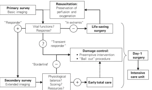
Physicians initially treating injured patients must con-duct a systemic work-up. According to the Advanced Trauma Life Support (ATLS®) course patients undergo
the primary survey of airway, breathing, circulation, neurologic status and core temperature [21]. Patients with extensive trauma who are unconscious (Glasgow Coma Scale [GCS] < 9 points) or in shock benefit from immediate endotracheal intubation and oxygenation. On rare occasions such as severe maxillofacial injuries or laryngeal fractures, patients require a surgical airway management (cricothyroidotomy or tracheostomy) as life-saving procedure (Figure 1). Simultaneous with air-way management, a quick assessment of the patient will determine the degree of shock present. A patient with a
systolic blood pressure < 90 mmHg, a thready pulse and flat neck veins is assumed to have hypovolemic shock until proven otherwise. If the neck veins are distended, tension pneumothorax or pericardial tamponade are the most common diagnoses. For tension pneumotho-rax a needle decompression into the second intercostal space in the midclavicular line followed by a tube thora-costomy represents the life-saving procedure (Figure 1). Pericardial tamponade is mostly observed in patients with penetrating injuries to the torso. A left anterolat-eral emergency room thoracotomy (ERT) with opening of the pericardium can be life-saving [13].
If the patient’s primary problem in shock is blood loss, the intention is to stop the bleeding and replace the blood loss. Obvious and occult blood loss should be de-tected immediately. An external bleeding of an open fracture or central amputation as well as closed fractures of long bones should be clinically obvious, whereas hid-den blood loss in pleural cavities and the abdomen in-clusive of the retroperitoneum and the pelvis are tested by the basic imaging during the primary survey includ-ing chest X-ray, ultrasound of abdomen and retroperi-toneum and a plain film of the pelvis. Blood loss through vascular injuries in fractures or central amputations should be stopped by manual compression followed by clamping or ligation.
As soon as possible blood work is obtained that in-cludes arterial blood gas analysis, hematocrit, hemoglo-bin, lactate level, base deficit, pH, toxicology, blood type and cross-match, and a screening battery of other laboratory tests including coagulation parameters. The
fluid used to resuscitate a hypoten-sive patient and the further work-up will depend on the patient’s response to initial fluid load (2 l crystalloids), the laboratory and further clinical analyses [1, 21]. All fluids need to be at body temperature or above. In addition, to prevent a hypothermia patients can be placed on warming mattresses and their environment kept warm using warm air blankets. The “rapid responder” may require no more that crystalloid to replace the volume deficit and progress to the secondary survey, which focuses on a complete physical examination that directs further diagnostic stud-ies (extended imaging) such as CT scan trauma protocol (Figure 1). The “transient re-sponder” may need the addition of blood. In exsangui-nating patients, type 0 blood should be given, whereas in more stable patients it is prudent to wait for typed and cross-matched blood. With extensive hemorrhage and massive transfusions, component therapy must be di-rected by monitoring specific coagulative defects. The application of platelets, stored (previously thawed) fresh frozen plasma (FFP) or fibrinogen are well estab-lished, whereas the adjunctive treatment of coagulopa-thy with recombinant activated factor VII (rFVIIa) in trauma patients is undergoing trials [22].
Bickell et al. found that the survival in patients with penetrating torso trauma was improved, if fluid replace-ment was delayed after immediate surgical control of bleeding [23]. They suggested that immediate volume replacement in these patients might disrupt blood clot that had obliterated a bleeding vessel. The left antero-lateral ERT with thoracic aortic cross-clamping and open cardiopulmonary resuscitation as life-saving inter-vention represents an accepted indication for patients sustaining penetrating cardiac injuries that arrive in trauma centers after a short scene/transport time with witnessed and/or objectively measured vital parameters (patients “in extremis”; Figure 1) [24]. Cardiac injuries may be temporized by digital pressure or the use of a Foley catheter to tamponade bleeding. The pericardium is then opened longitudinally above the phrenic nerve and the cardiac injury repaired. In addition, this access allows to cannulate the right atrium with a catheter for massive resuscitation. For patients sustaining penetrat-Primary survey Basic imaging Resuscitation: Preservation of perfusion and oxygenation Vital functions? Response? Secondary survey Extended imaging Damage control: • Preemptive intervention • “Bail- out” procedure
Life-saving surgery
Early total care
Intensive care unit Physiological balance? Scoring? Resources? + + __ ? ? + + Day-1 surgery _ _ “Transient responder” ”In extremis” “Responder“ “Borderline”
ing noncardiac thoracic injuries or as an adjunct to an emergency room laparotomy (“crash”-laparotomy) for repair of exsanguinating abdominal vascular injuries the ERT or sternotomy should be performed selectively due to its low survival rate (10%) [24]. In addition, in patients sustaining cardiopulmonary arrest secondary to blunt trauma ERT should be carried out only rarely due to its very low survival rate (1.5%) [24]. In patients with exsanguinating abdominal hemorrhage after pen-etrating or blunt trauma and without response to fluids “crash”-laparotomy can be needed to control the ab-dominal aorta either digitally at the aortic hiatus or by placement of an aortic infradiaphragmatic cross clamp [25].
When? – Indications for “Damage Control”: Stage 1 If patients “in extremis” survive life-saving procedures, DC interventions are used for associated injuries. On the basis of clinical and laboratory findings during pri-mary or secondary survey a decision for DC as “pre-emptive intervention” should be made: stage 1 of DC (Figure 1) [26]. Patients with a “transient response” to resuscitation with a hypotension (< 90 mmHg) in excess of 70 min or a transfusion rate of 10–15 units of packed red blood cells should be transferred to the operating room (OR) without delay and undergo DC procedures [1]. In addition, attempts have been made to define physiological criteria for the initiation of DC based on hypothermia (< 34 °C), coagulopathy (prothrombin time > 19 s or partial thromboplastin time > 60 s; platelet count < 90,000) and acidosis (pH < 7.2 or lactate serum level > 5 mmol/l), but this has still not been standardized and validated by prospective studies [1, 26, 27]. Further cited indications especially for DCO concern type and severity of injury (Injury Severity Score [ISS] > 35 points; severe head injury AIS [Abbreviated Injury Scale] > 2 points; multiple injuries with an ISS > 20 points and additional thoracic trauma AIS > 2 points; multiple injuries with abdominal/pelvic trauma and hemorrhagic shock; radiographic evidence of bilateral pulmonary contusion) as well as type of surgery (pre-sumed operation time > 60 min and expected major blood loss) [10, 11, 27, 28]. These first-hit and second-hit phenomena predispose these patients “at risk” or “bor-derline” to deterioration after surgery and justify the decision for DC.
During ETC interventions intraoperative problems can arise or unexpected associated injuries are found. Inability to achieve hemostasis due to coagulopathy,
in-accessible major venous injury, time-consuming proce-dures in a patient with suboptimal response to resuscita-tion, reassessment of intraabdominal contents, and inability to reapproximate abdominal fascia due to vis-ceral edema are reasons for turning to the DC concept as “bail-out” procedure (Figure 1) [1].
Furthermore, ancillary issues indicating benefits of DC are limited resources in a mass casuality, a limited experience of the surgical team in complex injuries, or a fatigued and overwhelmed surgical team. However, se-lecting DC too careless may mean an unnecessarily pre-mature termination of surgery in patients who would otherwise have recovered from a single definitive pro-cedure. It would subject the patients to risks and ex-pense of multiple surgical interventions.
How? – Staged Sequential Procedures
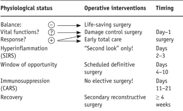
“Damage control surgery” (DCS) for nonorthopedic injuries and DCO for musculoskeletal injuries, respec-tively, can be described as staged sequential procedures [1, 10, 11, 26]. In stage 2 of DC an abbreviated surgery for rapid control of hemorrhage and contamination, sta-bilization of long bones or large joints and pelvic ring, provisional closure of wounds or abdominal cavity is carried out [26]. Thereafter, the patient is moved to the intensive care unit (ICU), where stage 3 consists of on-going core rewarming, correction of coagulopathy, fluid resuscitation and optimization of hemodynamic status with correction of the acidosis, reexamination of the pa-tient (“tertiary survey”) to diagnose missed injuries as well as specific management of patients with traumatic brain injury (TBI). When normal physiology has been restored, “second looks” or staged definitive surgery (stage 4) can be undertaken usually within 24–72 h after trauma [1, 26] (Table 1). Concerning definitive fracture repair there is a “window of opportunity” between days 4 and 10 after trauma [10, 11, 27]. Extensive secondary reconstructive surgery (stage 5) is recommended after recovering from the status of immunosuppression (CARS) and from a katabolic to an anabolic metabo-lism (≥ 4 weeks), respectively (Table 1) [15].
“Damage Control Surgery”: Stage 2
Thoracic injuries. The anterolateral thoracotomy per-mits rapid access to the thoracic cavity [1, 13, 24]. If a pulmonary hilar hematoma or active hemorrhage are present, cross-clamping of the pulmonary hilum may be necessary. Stapled, nonanatomic wedge resections of the lung can achieve hemostasis and control of air leaks.
Pulmonary tractotomy using long clamps or stapler may be an effective way to control hemorrhage in penetrat-ing lung injury [29]. Resection of the affected lobe or lung for injuries involving hilar bronchi or vessels is preferable to extensive repairs. Packing the thoracic cavity is useful for chest wall or diffuse bleeding from the pulmonary parenchyma (Figure 2) [1, 13].
Abdominal injuries. The DC laparotomy includes five components: control of hemorrhage, exploration, con-trol of contamination, definitive packing, and rapid ab-dominal closure [1, 26]. The incision of choice is midline from xiphoid process to pubic symphysis.
To control hemorrhage, blood and clot are quickly removed digitally and by suction. Thereafter, lateral
re-traction of abdominal wall is performed to enable initial resuscitative four-quadrant packing. If bleeding seems controlled with packing, this is an excellent time to al-low the anesthesia team to stabilize the patient with vol-ume therapy. Temporary infradiaphragmatic aortic oc-clusion or balloon catheter tamponade may be necessary for completion of hemorrhage control. The exact explo-ration follows pack removal, beginning from the sus-pected sites of injury. Abdominal vascular injuries are managed as described below. Techniques to control liver bleeding include, besides the workhorse of perihe-patic packing after Pringle maneuver, direct ligation of bleeding vessels, hepatorrhaphy, cauterization, topical hemostatic agents, partial resection, hepatic artery liga-tion, catheter balloon tamponade or angiographic em-bolization [3–7]. Splenic injuries require mostly an im-mediate splenectomy. Attempts at splenorrhaphy or partial resection should be reserved for stable patients. Occasionally, the splenic fossa needs packing to allow tamponade of small vessels until coagulopathy is re-versed. If no compelling bleeding source has been found, then retroperitoneal vascular, renal or pelvic injuries are likely sources [1]. Severe renal injury in the exsan-guinating patient is best dealt by nephrectomy, if a con-tralateral kidney is palpable [1]. Alternatives are retro-peritoneal packing or postoperative embolization.
Hollow viscus injury must be controlled with clamps, staples, suturing, or resection without anastomosis. In-juries to the pancreas should be primarily managed by
Table 1. Operative phases. See text for details and explanations. Physiological status Operative interventions Timing
Balance: – Life-saving surgery
Vital functions? ? Damage control surgery Day–1 Response? + Early total care surgery Hyperinflammation “Second look” only! Days
(SIRS) 2–3
Window of opportunity Scheduled definitive Days
surgery 4–10
Immunosuppression No elective surgery! Days
(CARS) 11–21
Recovery Secondary reconstructive ≥ 4
surgery weeks
Figures 2a and 2b. Chest X-ray of a multiply injured patient with flail chest at admission (a) and 24 h after right anterolateral thoracotomy with
packing of the thoracic cavity (b).
drains and packing. Urethral and bladder injuries of un-stable patients are managed temporarily with splinting and/or suprapubic urinary diversion [1].
After temporary control of hemorrhage and contami-nation a decision for definitive repair of a part or all in-traabdominal injuries should be made according to the physiological parameters. In the face of the “lethal triad” a definitive packing is followed by a rapid skin closure [1, 26]. Leaving the fascia open limits the risk of abdominal compartment syndrome (ACS) and preserves the fascial edges [1, 26, 30]. Massive visceral edema makes the closure sometimes impossible and the open abdomen can be tem-porarily closed with a prosthetic material, zipper (Ethizip®),
plastic sheet or a vacuum pack technique such as the ab-dominal vacuum-assisted closure (V.A.C.®) [31].
Vascular injuries. Percutaneous vascular control using bal-loon tamponade through the wound site has been de-scribed. Simple lateral repair of vascular injury is a rapid technique, whereas end-to-end anastomosis or graft inter-position are time-consuming [1]. As “bail-out” procedure most arteries and veins can be ligated to save the patient’s life. However, ligation of the aorta, vena cava, superior mesenteric artery, or common or external iliac artery often precipitates significant ischemia with a high mortality and should be reserved only for desperate situations [1]. An alternative to ligation may be the rapid placement of tem-porary arterial or venous shunts [32, 33].
Pelvic fractures. The management of multiply injured patients with pelvic ring disruption and severe hem-orrhage is still under debate [34, 35]. It is well accept-ed that the displacaccept-ed pelvic ring injury must rapidly be reduced and stabilized by external fixator for the anterior pelvic ring and C-clamp for the posterior ring
(Figure 3) [34, 36]. The methods by which control of hemorrhagic shock is achieved vary from laparotomy with pelvic packing to angiographic embolization. The rationale for pelvic packing is the following: bleeding from the venous plexus can only be effec-tively controlled by local packing with the pelvic ring as stable abutment; arterial bleeding can also be suc-cessfully treated by pelvic packs; bleeding from large-bore vessels can be controlled surgically; com-plex pelvic injuries are often combined with intraper-itoneal lesions [34]. However, in rare situations with persistent hemorrhagic shock combined procedures with intra- or postoperative angiographic emboliza-tion are necessary.
Extremity fractures and soft-tissue injuries. Closed or open fractures of long bones or highly unstable large joints should be temporarily stabilized by external fix-ators [10, 11]. For some closed or open fractures the fast application of locking compression plates (LCPs) as in-ternal fixators in a minimally invasive technique repre-sents an alternative (Figure 4). Open wounds and frac-tures should undergo a debridement with resection of avital tissues to limit the antigenic load. Fasciotomy should be performed liberally in the settings of ischemia, imminent or manifest compartment syndromes. Wounds are temporarily closed by Epigard® or V.A.C.® [37].
However, in patients with a massive coagulopathy tam-ponades as temporary closure are preferable to avoid persistent bleeding. In mangled extremity, very little time and blood should be spent debating the limb sal-vageability [1].
Head injuries. Although the aim of neurotrauma proce-dures is to avoid secondary brain damages, placement
Figures 3a to 3c. Plain films of the pelvis of a multiply injured patient with vertical shear injury of the right side preoperatively (a) and
postopera-tively (b) after application of a pelvic clamp and external fixator as well as pelvic packing and abdominal vacuum-assisted closure after laparoto-my. Physiological restoration of the patient in the ICU (c).
of a ventricular catheter through a burr hole, craniotomy or craniecto-my (removal of the bone flap) with arrest of intracranial bleeding and evacuation of intracranial hemato-ma, or a decompressive craniectomy are not abbreviated surgical inter-ventions and belong to the ETC con-cept. However, an immediate de-compressive craniectomy for an epidural hematoma is life-saving [38].
Resuscitation in ICU: Stage 3 Priorities in the ICU focus on resto-ration of the lethal triad hypother-mia, coagulopathy, and acidosis as well as an optimization of the oxygen delivery [1, 16–19, 25]. Endpoints in-clude a core temperature > 35 °C, normalization of the prothrombin time, and a systemic lactate level < 2.5 mmol/l within 12 h [1, 26].
Additionally, an array of sup-portive therapies are established to avoid secondary hits and organ
dam-ages [15]. Secondary brain injuries with elevated intracra-nial pressure (ICP) due to cerebral edema or ischemia/ reper fusion injuries can be limited by applying different neuroprotective strategies, such as optimization of the cerebral perfusion pressure (CPP) through increase of mean arterial pressure and release of cerebrospinal fluid (CSF), controlled hyperventilation, moderate hypother-mia, and whenever these therapeutic regimens fail to re-duce ICP, intravenous administration of barbiturate may become necessary [15, 38]. An early enteral nutrition through gastric or duodenal tubes reduces the accumula-tion of pathogenic bacteria in the intestinal tract and avoids an atrophy of intestinal mucosa, an essential bac-terial barrier. Immune-enhanced enteral nutrition (IEEN; e.g., arginine, glutamine) reduces the posttrau-matic hypermetabolism and improves the immunocom-petence [15].
A common, serious, and often insidious complica-tion of abbreviated laparotomy is the ACS with pro-gressive oliguria and advancing hypoxemia [1, 26, 30]. The most accurate and simple method to detect an evolving ACS is measuring bladder pressure via the Foley catheter [1, 26, 30].
“Second Look” and Scheduled Definitive Surgery: Stage 4
Timing for return to the OR is governed by the injury pattern, the planned operative procedure, the physio-logical response in the ICU, or the development of com-plications [1, 26]. Patients, who have been packed for hemorrhage control, are returned to the OR within 24 h post-injury as “second look” for removal of packs, clot-ted blood, and fluid collections, debridement of avital tissue, reconstructions of digestive tract, urethral or bladder injuries, colostomy formation, duodenal feed-ing access, and rarely extensive procedures such as pan-creaticoduodenectomy. Recurrent or persistent bleed-ing (more than 10 units of packed red blood cells in the early postoperative period) will necessitate immediate repacking or angiographic embolization. Patients who develop ACS or intestinal necrosis undergo relaparoto-my without delay. After temporary closure of the abdo-men by V.A.C.® because of extensive
reperfusion-in-duced gut distension, “second looks” are delayed 48–72 h, awaiting sufficient edema resorption to allow abdom-inal fascial closure if possible, otherwise a new V.A.C.®
is applied to reduce the edema [31].
Figures 4a to 4c. X-rays of the left multiply fractured femur of a severely injured patient after
stabilization with a locking compression plate (LCP) as internal fixator through small incisions (a), after definitive stabilization with an antegrade femoral nail (b), and 1 year after trauma with complete consolidation of the fracture (c).
Definitive operations (nailing, plating) of extremity or pelvic fractures as well as plastic reconstructive sur-gery for the closure of open wounds (e.g., muscle trans-fer) of severely injured patients should be delayed until after the 4th day from initial surgery [10–12]. An immi-nent or manifest compartment syndrome of extremities in the posttraumatic course or after fracture stabiliza-tion requires an immediate fasciotomy.
Secondary Reconstructive Surgery: Stage 5
Reconstructive operations after abdominal injuries in-clude abdominal wall reconstruction or anastomosis af-ter colostomy [1, 26]. Staged laparotomy can be compli-cated by open abdomen incisional hernia. Fascial approximation is precluded by fascial retraction after multiple delayed procedures or abdominal wall loss through the trauma itself or infection. The usual ap-proach to abdominal wall reconstruction is to bridge the fascial defect with a synthetic mesh template to facili-tate secondary wound healing or a bilateral fascial re-lease, rarely a local muscle flap rotation is necessary [1, 26]. However, the use of abdominal V.A.C.® has
result-ed in higher fascial closure rates (90%), obviating the need for subsequent hernia repair in most patients [31]. The utility of the vacuum-assisted fascial closure tech-nique is not limited to the early postoperative period, but can be successful as much as ≥ 4 weeks after the ini-tial operation [31].
Complex reconstructive procedures of ligamentary joint injuries, secondary joint prosthesis or nerve recon-structions can be planned in this stage. Furthermore, the bone defect after craniectomy is repaired or the bone flap replaced in secondary cranioplasty procedure once all the brain swelling has subsided ≥ 6 weeks after trauma.
Own Experiences in “Damage Control”
In a 6-year period 622 severely injured patients with an ISS > 16 points were included in a retrospective analy-sis, if they arrived at the ICU after surviving day-1 sur-gery. 205 patients (33%) were classified to the DC group, 417 (67%) to the ETC group (Table 2). The ISS and the injury pattern were different in the two groups, while the age was comparable. The ISS was significantly higher, and the injury pattern was more complex in the DC group (Table 2). Concerning physiological param-eters at admission, core temperature, prothrombin time %, serum lactate level, and hemoglobin were more pathologic in the DC group (Table 2). In addition, a higher proportion of severe hemorrhagic shock (grades
III or IV according to the ATLS® classification [21, 24])
could be observed in the DC group (Table 2).
Patients surviving the first 3 days on ICU developed more infectious or septic complications in the DC group, whereas the incidence of severe SIRS was comparable
Table 2. Demographic data and physiological parameters at admission
of severely injured patients managed by “damage control” or “early total care”. Data are presented in % or in mean ± SEM (standard errror of mean). ATLS®: Advanced Trauma Life Support; ISS: Injury Severity Score.
“Damage “Early total
control” care” Demographic data • Patients (n) 205 (33%) 417 (67%) • Age (years) 40.2 ± 1.3 39.8 ± 0.8 • ISS (points) 36.1 ± 0.9 31.8 ± 0.6 Injury pattern • Head 56% 84% • Thorax 64% 55% • Abdomen 46% 22% • Pelvis 37% 15% • Extremity 86% 46% Penetrating injuries 15% 7%
Physiological parameters at admission
• Core temperature (°C) 34.6 ± 0.1 35.6 ± 0.1 • Prothrombin time % (PT%) 69.2 ± 1.5 82.3 ± 0.9 • Serum lactate level (mmol/l) 4.1 ± 0.9 2.9 ± 0.1 • Hemoglobin (g/dl) 9.4 ± 0.7 11.8 ± 0.1 • Severe hemorrhagic shock (grades III
or IV according to ATLS® classifi- 19% 7% cation [21])
Table 3. Posttraumatic morbidity and mortality of severely injured
pa-tients after “damage control” or “early total care” management and arriving on intensive care unit (ICU). Data are presented in % or in mean ± SEM (standard error of mean). MODS: multiple organ dysfunc-tion syndrome; SIRS: systemic inflammatory response syndrome.
“Damage “Early total
control” care”
Morbiditya
• Severe SIRS (3 or 4 SIRS criteria are 46% 41% fulfilled [15])
• Sepsis (4 SIRS criteria and detected 30% 19% infectious focus [15])
• Infections 57% 42%
• Pneumonia 31% 27%
• MODS score (Marshall et al. [39]) 7.9 ± 0.3 6.3 ± 0.2 (points)
Mortality 27% 27%
• Head injury (tentorial herniation) 49% 77% • Hemorrhagic shock 16% 2% • Multiple organ failure 35% 21%
(Table 3). In accordance with the septic complications the mean multiple organ dysfunction score was higher in the DC group compared with the ETC group [39]. However, the mortality rate was equal in both groups (Table 3). Head injury was the killer in the ETC group, whereas in the DC group the causes of death were more heterogeneous.
Although the ISS and the incidence of infectious and septic complications are higher in the DC group, the mortality rate is identical. These data emphasize the benefit of the decision for the DC concept for severely injured patients with a complex injury pattern. Howev-er, head injury represents the leading killer after DC and ETC management of severely injured patients.
Conclusion
The concept of DC is well established for the manage-ment of thoracic, abdominal, vascular, pelvic, extremity and soft-tissue injuries in severely injured patients. Al-though level I evidence is lacking, reasonably clear indi-cations and defined endpoints for each stage were evolved and the mortality rate was reduced [1, 26]. How-ever, better understanding of the significance and kinet-ics of physiological parameters including inflammatory mediators (“beside immunomonitoring”) could help us in the decision-making for selection of patients and the optimal time points of staged sequential surgery to limit second hits and therefore to optimize the DC concept [10, 11, 13, 15].
References
1. Shapiro MB, Jenkins DH, Schwab CW, et al. Damage control: col-lective review. J Trauma 2000;49:969–78.
2. Rotondo MF, Schwab W, McGonigal MD, et al. Damage control: an approach for improved survival in exsanguinating penetrating abdominal injury. J Trauma 1993;35:375–83.
3. Pringle J. Notes on the arrest of hepatic hemorrhage due to trau-ma. Ann Surg 1908;48:541–9.
4. Halsted WS. Ligature and suture material. JAMA 1913;LX:1119–26. 5. Walt AJ. The mythology of hepatic trauma – or Babel revisited.
Am J Surg 1978;135:12–8.
6. Feliciano DV, Mattox KL, Jordan GL Jr. Intra-abdominal packing for control of hepatic hemorrhage: a reappraisal. J Trauma
1981;21:285–90.
7. Garrison J, Richardson D, Hilakos A, et al. Predicting the need to pack early for severe intra-abdominal hemorrhage. J Trauma 1996;40:923–9.
8. Stone HH, Strom PR, Mullins RJ. Management of the major coagulopathy with onset during laparotomy. Ann Surg 983;197: 532–5.
9. Burch J, Oritz V, Richardson R, et al. Abbreviated laparotomy and planned reoperation for critically injured patients. Ann Surg 1992;215:476–84.
10. Giannoudis PV. Aspects of current management: surgical priori-ties in damage control in polytrauma. J Bone Joint Surg Br 2003; 85:478–83.
11. Pape HC, Giannoudis PV, Krettek C. The timing of fracture treat-ment in polytrauma patients: relevance of damage control ortho-paedic surgery. Am J Surg 2002;183:622–9.
12. Pape HC, Hildebrand F, Perschy S, et al. Changes in the manage-ment of femoral shaft fractures in polytrauma patients: from ear-ly total care to damage control orthopedic surgery. J Orthop Trau-ma 2004;18:Suppl:S13–23.
13. Wall MJ, Soltero E. Damage control for thoracic injuries. Surg Clin North Am 1997;77:863–78.
14. Porter JM, Ivatury RR, Nassoura ZE. Extending the horizons of “damage control” in unstable trauma patients beyond the abdo-men and gastrointestinal tract. J Trauma 1997;42:559–61. 15. Keel M, Trentz O. Pathophysiology of polytrauma: a review. Injury
2005:in press.
16. Ferrara A, MacArthur J, Wright H, et al. Hypothermia and acidosis worsen coagulopathy in the patient requiring massive transfu-sion. Am J Surg 1990;160:515–8.
17. Gregory J, Flancbaum L, Townsend M, et al. Incidence and timing of hypothermia in seriously injured patients. J Trauma 1991;31: 795–800.
18. Abramson D, Scalea T, Hitchcock R, et al. Lactate clearance and survival following injury. J Trauma 1993;35:584–9.
19. Gubler K, Gentilello L, Hassantash S, et al. The impact of hypo-thermia on dilutional coagulopathy. J Trauma 1994;36:847–51. 20. Cosgrif N, Moore EE, Sauaia A, et al. Predicting life-threatening
coagulopathy in the massively transfused patient: hypothermia and acidoses revisited. J Trauma 1997;42:857–62.
21. Collicott PE, Hughes I. Training in Advanced Trauma Life Support. JAMA 1980;243:1156–9.
22. Parr MJA, Alabdi T. Damage control surgery and intensive care. In-jury 2004;35:713–22.
23. Bickell WH, Wall MJ Jr, Pepe PE, et al. Immediate versus delayed fluid resuscitation for hypotensive patients with penetrating tor-so injuries. N Engl J Med 1994;331:1105–9.
24. Demetriades D, Asensio JA. Trauma management. Georgetown: Landes Bioscience, 2000.
25. Carrillo EH, Bergamini TM, Miller FB, et al. Abdominal vascular in-juries. J Trauma 1997;43:164–71.
26. Moore EE. Staged laparotomy for the hypothermia, acidosis, and coagulopathy syndrome. Am J Surg 1996;172:405–10.
27. Pape HC, Giannoudis PV, Krettek C, et al. Timing of fixation of major fractures in blunt polytrauma – the role of conventional indicators in clinical decision making. J Orthop Trauma 2005:in press. 28. Giannoudis PV, Veysi VT, Pape HC, et al. When should we operate
on major fractures in patients with severe head trauma? Am J Surg 2002;183:261–7.
29. Wall MJ, Villavicencio RT, Miller CC, et al. Pulmonary tractotomy as an abbreviated thoracotomy technique. J Trauma 1998;45: 1015–23.
30. Ertel W, Oberholzer A, Platz A, et al. Incidence and clinical pattern of the abdominal compartment syndrome after “damage control”-laparotomy in 311 patients with severe abdominal and/ or pelvic trauma. Crit Care Med 2000;28:1747–53.
31. Miller PR, Meredith JW, Johnson JC, et al. Prospecitve evaluation of vacuum-assisted fascial closure after open abdomen. Planned ventral hernia rate is substantially reduced. Ann Surg 2004;239: 608–16.
32. Reilly P, Rotondo M, Carpenter J, et al. Temporary vascular conti-nuity during damage control: intraluminal shunting for proximal superior mesenteric artery injury. J Trauma 1995;39:757–60.
33. Reber PU, Patel AG, Sapio NL, et al. Selective use of temporary in-travascular shunts in coincident vascular and orthopedic upper and lower limb trauma. J Trauma 1999;47:72–6.
34. Ertel W, Eid K, Keel M, et al. Therapeutical strategies and outcome of polytraumatized patients with pelvic injuries – a six-year expe-rience. Eur J Trauma 2000;6:14–7.
35. Ertel W, Keel M, Eid K, et al. Control of severe hemorrhage using C-clamp and pelvic packing in multiply injured patients with pel-vic ring disruption. J Orthop Trauma 2001;15:468–74.
36. Ganz R, Krushell R, Jakob R, et al. The antishock pelvic clamp. Clin Orthop 1991;267:71–8.
37. Labler L, Keel M, Trentz O. Vacuum-assisted closure (V.A.C.®) for temporary soft tissue coverage in type III open fracture of lower extremities. Eur J Trauma 2004;5:305–12.
38. Rosenfeld JV. Damage control neurosurgery. Injury 2004;35: 655–60.
39. Marshall JC, Cook DJ, Christou NV, et al. Multiple organ dysfunc-tion score: a reliable descriptor of a complex clinical outcome. Crit Care Med 1995;23:1638–52.
Address for Correspondence
Marius Keel, MD
Division of Trauma Surgery University Hospital Zurich Rämistraße 100
8091 Zürich Switzerland
Phone (+:41/44) 255-3657, Fax -4406 e-mail: [email protected]