E
ffects of miltefosine treatment in fibroblast cell cultures
and in mice experimentally infected with Neospora caninum
tachyzoites
KARIM DEBACHE and ANDREW HEMPHILL*
Institute of Parasitology, Vetsuisse Faculty, University of Berne, Länggass-Strasse 122, CH-3012 Berne, Switzerland
(Received 5 October 2011; revised 24 November and 12 December 2011; accepted 22 December 2011; first published online 6 February 2012)
S U M M A R Y
Miltefosine was investigated for its activity against Neospora caninum tachyzoites in vitro, and was shown to inhibit the proliferation of N. caninum tachyzoites cultured in human foreskinfibroblasts (HFF) with an IC50of 5·2μM. Treatment of infected cells with 25μM miltefosine for a period of 10 h had only a parasitostatic effect, while after 20 h of treatment parasiticidal effects were observed. This was confirmed by transmission electron microscopy of N. caninum-infected and miltefosine-treated HFF. Administration of miltefosine to N. caninum-infected Balb/c female mice at 40 mg/kg/day for 14 days resulted in 6 out of 10 mice exhibiting weight loss, ruffled coat and apathy between days 7 and 13 post-infection. In the group that received placebo, only 2 out of 8 mice succumbed to infection, but the cerebral burden was significantly higher compared to the miltefosine treatment group. In a second experiment, the time-span of treatment was reduced to 5 days, and mice were maintained without further treatment for 4 weeks. Only 2 out of 9 mice in the miltefosine treatment group exhibited signs of disease, while 8 out of 10 mice succumbed to infection in the placebo group. These results showed that miltefosine hampered the dissemination of parasites into the CNS during experimental N. caninum infection in mice. Key words: miltefosine, Neospora caninum, tachyzoites, culturedfibroblasts, proliferation.
I N T R O D U C T I O N
Neospora caninum, an apicomplexan parasite that was initially described associated with severe neuromus-cular disease in dogs (Dubey et al.1988), represents one of the most important causative agents of infectious bovine abortion. Neosporosis is respon-sible for important economic loss in both dairy and beef cattle worldwide, also involving reduced milk yield and diminished post-weaning weight gain in beef calves (Dubey et al.2007; Dubey and Schares, 2011). The worldwide serious economic impact of neosporosis upon the cattle industry has inflamed the interest for an effective control strategy as well as for prophylactic or metaphylactic measures to prevent and treat bovine neosporosis infection. At the present time, there is no vaccine available to induce complete protective immunity (Monney et al. 2011). Therefore, chemotherapeutic measures using pharmacologically active compounds might rep-resent economically viable options, provided that suitable compounds are identified (Häsler et al. 2006a,b).
To date, no specific treatment against neosporosis is available. However, several studies on chemother-apeutically interesting compounds have been carried out. These include lasalocid, monensin, pirithrexim,
pyrimethamine, clindamycin, robenidine and
trimethoprim (Lindsay et al. 1994), artemisinin (Kim et al. 2002), depudecin (Kwon et al. 2003), toltrazuril, ponazuril (Darius et al. 2004), nitro-and bromo-thiazolides (Esposito et al. 2005; 2007a,b), pentamidine analogues (Leepin et al.2008; Debache et al.2011) and alcoholic herbal extracts (Youn et al. 2004). Only few compounds have been investigated for anti-Neospora activity using experimental in vivo models. A low efficacy in Neospora-infected mice was reported for sulfadiazine and amprolium (Lindsay and Dubey,1990). Toltrazuril, a triazinone derivate that inhibits the electron transfer in the mitochon-drial respiratory chain, was shown to exhibit effective anti-parasitic activity against N. caninum in mice (Gottstein et al. 2001), while results in bovines did not allow definitive conclusions (Kritzner et al. 2002; Haerdi et al.2006). Arylimidamides also seem to be interesting compounds for chemotherapeutical intervention. For instance, DB750, a phenylated pentamidine analogue, significantly improved the survival rate and reduced the cerebral parasite burden in experimentally infected mice when applied intraperitoneally. In contrast, nitazoxanide, which had shown promising activity against N. caninum in vitro, was not active in vivo, (Debache et al.2011).
Miltefosine (hexadecylphosphocholine, HePC) is an alkylphospholipid originally developed as an anti-cancer agent for local treatment of skin metastases. It was later found to show potent leishmanicidal activity as a consequence of its interference in the parasite’s metabolic pathways (Unger et al.1989; Barratt et al.
* Corresponding author: andrew.hemphill@vetsuisse. unibe.ch
2009). Miltfosine is commercialized in Germany as Impavido(R) for leishmaniasis treatment, is also registered for the treatment of visceral and cutaneous leishmaniasis in India and Colombia, and is under-going clinical trials for registration in several other countries, including Brazil and Guatemala. It cur-rently represents the only effective oral treatment for leishmaniasis. The mode of action of miltefosine against Leishmania parasites involves the interaction with membrane phospholipids and sterols, and the drug interferes with the functionality of a number of enzymes involved in phospholipid metabolism such as protein kinase C and the phospholipases A, C and D, finally leading to apoptosis (Barratt et al. 2009). Hexadecyltrimethylammonium bromide, a compound structurally similar to miltefosine, was demonstrated to exhibit potent in vitro activity against Plasmodium falciparum (Walochnik et al. 2002; Schuster et al.2006; Choubey et al.2007).
In this study, we investigated the effects of miltefosine against N. caninum tachyzoites in cul-tured fibroblasts. In experimentally infected mice, miltefosine treatment at 40 mg/kg/day for 17 days affected the disseminiation of the parasite into the CNS, but 50% of the mice succumbed to infec-tion, indicating that miltefosine treatment rendered mice more susceptible to neosporosis. However, reducing the treatment time to 5 days severely inhibited N. caninum dissemination and cerebral infection.
M A T E R I A L S A N D M E T H O D S
Unless otherwise stated, cell culture reagents were supplied by Gibco-BRL (Zürich, Switzerland) and chemicals were purchased from Sigma (St Louis, MO, USA).
Cell culture and Neospora caninum purification Vero cells (green monkey kidney epithelial) and human foreskin fibroblasts (HFF) were routinely cultured in RPMI 1640 medium supplemented with 10% heat-inactivated FCS, 2 mM glutamine, 50 U of penicillin/ml and 50μg of streptomycin/ml at 37 °C/ 5% CO2 in tissue-culture flasks. Cultures were passaged at least once per week. N. caninum tachyzoites of the Nc1 strain (Dubey et al. 1988) were maintained by serial passages in Vero cells or HFF during which time FCS was replaced with 5% immunoglobulin G (IgG)-free horse serum (HS). Parasites were harvested as described previously (Hemphill, 1996). Infected cells were trypsinized, washed twice in cold RPMI 1640 medium, and the resulting pellet was re-suspended in 2 ml of cold RPMI 1640 medium. Cells were repeatedly passaged through a 25 G-needle and liberated
tachyzoites were purified by passage through
Sephadex-G25 columns (Amersham Biosciences,
Otelfingen, Switzerland), previously equilibrated with cold RPMI 1640 medium. Purified tachyzoites were obtained after centrifugation at 600 g/10 min/4 ° C, and re-suspended in 1 ml of RPMI 1640 contain-ing 5% FCS, 50 U of penicillin/ml and 50μg of streptomycin/ml. They were counted in a Neubauer chamber and used to infect HFF monolayers as described below.
In vitro miltefosine treatments
In vitro drug treatment assays were carried out in triplicate essentially as described by Leepin et al. (2008). HFF cell monolayers were grown to con fl-uency in 24-well tissue-culture plates, and infec-tion was carried out for 1 h by addiinfec-tion of 2 × 10⁴cell culture-derived N. caninum tachyzoites per well. Infected monolayers were washed 3 times with cold RPMI 1640, and fresh medium was added, containing miltefosine, previously prepared as a stock solution in dimethyl sulfoxide (DMSO), at concen-trations ranging from 0·1μg/ml to 50 μg/ml. Controls contained DMSO solvent alone. The cultures were maintained for 72 h at 37 °C/5% CO2 and were inspected daily by light microscopy. After 72 h the medium was removed, and attached cells were suspended in 200μl of lysis buffer (Qiagen, Basel, Switzerland) and stored at −20 °C for subsequent N. caninum real-time PCR analysis (Müller et al. 2002).
IC50 values were calculated after logit –log-transformation of the relative growth data (RG; control = 1) according to the formula ln[(RG/(1− RG)] = a × ln(drug concentration) + b. Subsequent regression analysis was performed using the corre-sponding software tool contained in the Excel soft-ware package (Microsoft, Seattle, WA, USA).
To determine the time-span required for miltefo-sine to exert a true parasiticidal activity, infected HFF monolayers were treated with 25μM milte-fosine (5 times IC50) for 10 h or 20 h. The drug-containing medium was then removed and replaced with fresh culture medium without drug and changed every 2 days. Samples were collected on a daily basis by adding 200μl of Lysis buffer (Qiagen, Basel, Switzerland) until up to 9 days post-treatment and stored at−20 °C for subsequent N. caninum real-time PCR analysis.
Transmission electron microscopy (TEM)
HFF cell layers were grown to confluency in Corning cell-culture flasks, infected with N. caninum tachy-zoites for 2 days and treated with miltefosine at 24·5μM. After 10 h or 20 h, respectively, samples were rinsed with sterile PBS, washed in 100 mM sodium cacodylate buffer (pH 7·2), and fixed in cacodylate buffer containing 2·5% glutaraldehyde for 2 h at room temperature. Cells were scraped
with a rubber policeman and centrifuged at 1000g for 10 min at 4 °C and, and the resulting pellet was further fixed at 4 °C overnight, followed by post-fixation in 2% OsO4in cacodylate buffer for 4 h at 4 °C. Subsequently, specimens were washed in water and pre-stained in 1% uranyl acetate in water for 1 h at 4 °C, followed by extensive washing in water. Following dehydration in a graded series of ethanol (30, 50, 70, 90, and 100%), they were embedded in Epon 820 epoxy resin over a period of 2 days with 3 resin changes. The resin was polymerized at 65 °C for 24 h. Ultrathin sections were cut on a Reichert and Jung ultramicrotome and were loaded onto 300-mesh copper grids (Plano GmbH, Marburg, Germany). Staining with uranyl acetate and lead citrate was performed as described by Leepin et al. (2008).
In vivo Experiment 1: long-term miltefosine treatments in Neospora caninum-infected Balb/c mice
Female Balb/c mice (6 weeks of age) were purchased
from Charles River Laboratories (Sulzheim,
Germany) and housed under conventional day/ night conditions according to the standards set up by the animal welfare legislation of the Swiss Veterinary Office. At the age of 8–9 weeks, mice were randomly divided into 4 groups of 8 or 10 mice each (Table 1) and the serological status (Neospora-negative) was checked by enzyme-linked immuno-sorbent assay (ELISA). Miltefosine suspensions were prepared in 0·5% carboxy-methylcellulose (CMC) at 20 and 40 mg/kg/day, respectively, con-trols received CMC without drug. The drug and control suspensions were sterilefiltered and applied by intragastric inoculation (100μl per mouse/day). At days 1–3, all mice received the corresponding dose of miltefosine or CMC, at day 4 all mice were infected intraperitoneally with freshly purified N. caninum tachyzoites (2 × 10⁶) resuspended in 100μl RPMI, with the exception of the group receiving miltefosine 40 mg/kg/day without infection (see Table 1). Treatments were continued until 14 days post-infection (p.i.). Mice were weighed daily to assess potential toxicity. Animals exhibiting signs of acute disease (ruffled coat/inability to reach up for food and water/apathy) or neuronal symptoms (head tilt, running in circles, hind limb paralysis) were eu-thanized at the onset of these symptoms. Latest on day 18 p.i., all mice were sacrificed by CO2 -euthanasia. Brains were collected using individual sterile instruments and immediately frozen at−20 °C for subsequent quantitative PCR analysis.
In vivo Experiment 2: short-term miltefosine
treatments in Neospora caninum-infected Balb/c mice
Female mice (Charles River Laboratories, Sulzheim, Germany) were housed under conditions as described
above, and were divided into 2 groups of 10 (placebo) and 9 (miltefosine, 40 mg/kg/day), respectively. Treatments and infection were done as described above, but miltefosine treatment was discontinued at day 5 p.i., and mice were observed for another 30 days. Animals exhibiting signs of disease were euthanized at the onset of these symptoms and on day 35 p.i. at the latest, all mice were sacrificed by CO2-euthanasia. Collection of tissue samples and storage was done as for Experiment 1
DNA extraction and real-time-PCR
DNA purification from in vitro cultures and cerebral tissues was performed using the DNeasy®Blood & Tissue Kit (Qiagen, Basel, Switzerland) according
to the standard protocol suitable for animal
tissues. Frozen samples were lysed overnight at 56 ° C in 1 ml of ATL buffer containing proteinase K,
and 100μl of the suspension were used for
DNA purification. DNA was eluted in 50 μl of AE buffer and boiled at 95 °C. The DNA concentrations in all samples were determined by UV spectropho-tometry and adjusted to 100 ng/μl with sterile DNAse-free water. The assessments of N. caninum
tachyzoite loads were performed using a
LightCycler™Instrument (Roche Diagnostic, Basel, Switzerland) as previously described (Cannas et al. 2003a,b; Alaeddine et al.2005; Debache et al.2009). The parasite counts were calculated by interpolation from a standard curve with DNA equivalents from 1000, 100 and 10 parasites included in each run.
Statistical analysis
The significance of the differences between values of the control and experimental assays in parasite load assessment in vitro assay was determined by Student’s t-test using the Microsoft Excel program. P values of <0·01 and <0·05 were considered statistically significant. Cerebral parasite loads in different treatment and control groups were analysed by Tukey-Kramer Multiple-Comparison (P < 0·05). To determine the significance of mean weight loss prior and after treatment in the different experimen-tal groups, one-way ANOVA followed by Duncan’s multiple range test were employed.
R E S U L T S
In vitro activity of miltefosine in HFF and Vero cells
In order to investigate potential susceptibility of the host cells used for culture of N. caninum tachyzoites, namely Vero cells and HFF, respectively, non-infected confluent monolayers were incubated with different concentrations of miltefosine (1·5–122 μM) for 48 h, and cultures were stained with Trypan blue. Light microsocpical inspection revealed that miltefo-sine had no effect on HFF monolayers, while Vero cell
monolayers contained numerous (up to 50%) Trypan blue positive cells even at the lowest miltefosine concentration used (data not shown). Thus, HFF monolayers were used for further assessments on the effects of miltefosine against N. caninum.
In vitro activity of miltefosine against Neospora caninum tachyzoites
Different concentrations of miltefosine (1·5–122 μM) were added to N. caninum-infected HFF monolayers, and N. caninum proliferation was analysed after 3 days. Miltefosine inhibited the multiplication of tachyzoites with an IC50 of 5·2μM, and parasite proliferation was completely abolished at 50μM miltefosine (Fig. 1A).
Subsequently, infected cells were treated with 25μM miltefosine for 10 h and 20 h, respectively and, after washing in medium, cultures were main-tained for an additional 9 days (Fig. 1B). A pro-nounced inhibition of parasite proliferation was observed in both treatment groups during a period
of 3 days following treatment. Subsequently, tachy-zoites in those cultures treated for 10 h resumed proliferation. In contrast, in those specimens treated for 20 h the numbers of tachyzoites decreased continuously until day 9, indicating that in this case miltefosine exhibited parasiticidal efficacy.
Effects of miltefosine treatment on the ultrastructure of Neospora caninum tachyzoites
The morphological and structural alterations associ-ated with miltefosine treatments in N.
caninum-infected HFF were visualized by TEM. In
non-treated control cultures, intact N. caninum tachyzoites were localized within a parasitophorous vacuole, surrounded by a parasitophorous vacuole membrane, and characteristic organelles such as micronemes, rhoptries and dense granules, were clearly identified (Fig. 2A). Upon miltefosine treat-ment of infected cultures for 10 h, prominent alterations were noted within the tachyzoite cyto-plasm, such membrane stacks and an increasing
Fig. 1. Effects of miltefosine on the proliferation of Neospora caninum tachyzoites in HFF monolayers in vitro. (A) Dose-dependent inhibition of tachyzoite proliferation in HFF. The experiment was carried out in triplicate for each concentration and results are presented as means of number of tachyzoites ± S.D. Asterisks indicate those parasite numbers significantly differing from the parasite as assessed in the infection control (0 μg/ml) (P<0·01). (B) Parasiticidal efficacy of miltefosine against N. caninum tachyzoites proliferation. Miltefosine (25 μM) was applied for 10 h and 20 h respectively, cultures were washed and further incubated in medium for up to 9 days. From day 6 onwards, all parasite numbers assessed from 20 h post-treatment samples were significantly lower compared to tachyzoite numbers in the infection control and the 10 h post treatment samples. Results are presented as means of number of tachyzoites ± S.D.
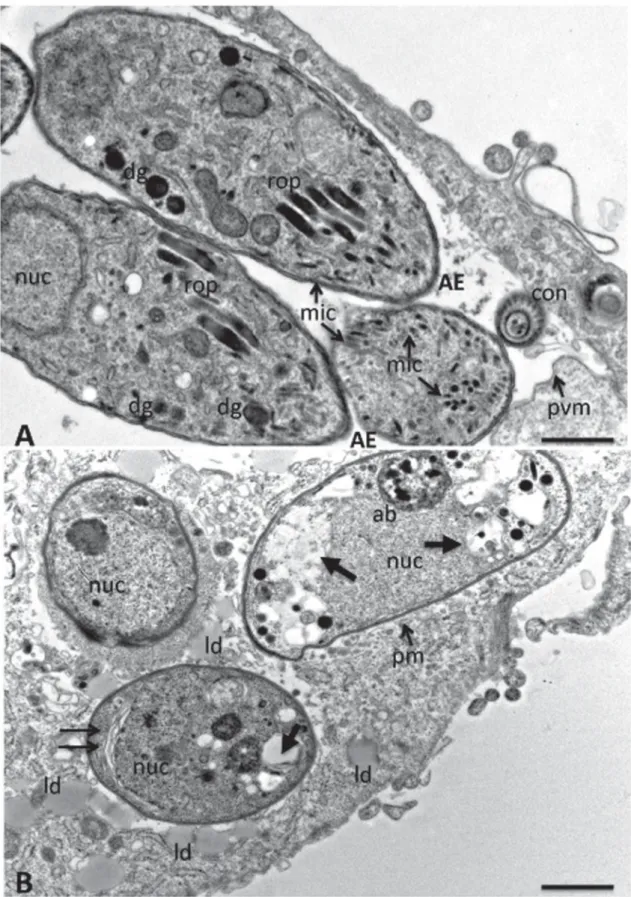
Fig. 2. TEM of Neospora caninum tachyzoites grown in HFF. (A) Non-treated control culture, with tachyzoites within a parasitophorous vacuole that is delineated by a parasitophorous vacuole membrane (pvm). The hallmarks of
apicomplexan parasites such as rhoptries (rop), micronemes (mic) and dense granules (dg) are clearly visible. AE, anterior end; con, cross-section through a conoid; nuc, nucleus. Scale bar = 0·32μm. (B) TEM after treatment of infected monolayers with 25μM miltefosine for 10 h. Note the presence of an apoptotic body (ab) in one tachyzoite, membrane stacks are indicated by 2 thin horizontal arrows, increased vacuolization is indicated by thick arrows, ld, lipid droplets; pm, plasma membrane. Scale bar = 0·38μm.
number of vacuoles within the parasite cytoplasm,
some of which were filled with membranous and
granular material of unknown origin, electron-dense inclusions, and the presence of structures resembling apoptotic bodies in some parasites (Fig. 2B). In addition, lipid droplets formed in the close vicinity of the parasitophorous vacuoles, although clearly localized within the cytoplasm of the host cells (Fig. 2B). Such lipid droplets were not found in uninfectedfibroblasts (not shown). Upon incubation of infected cultures with miltefosine for 20 h, the alterations became much more pronounced. Most host cells harboured remnants of parasitophorous vacuoles that werefilled with a dense granular matrix, containing completely distorted tachyzoites, and the micronemes, rhoptries and dense granule organelles
were not discernible anymore. The parasite cytoplasm was lacking any distinct subcellular organization; however, in all cases the parasite membrane surround-ing the tachyzoites still appeared to be intact (Fig. 3A, B). Few tachyzoites were found that were still localized within a parasitophorous vacuole sur-rounded by a distinct parasitophorous vacuole mem-brane, but they also contained vacuoles filled with membrane stacks and other unidentified material, and large lipid droplet were localized just adjacent to the parasitophorous vacuole membrane (Fig. 3C). Many liberated, but clearly distorted tachyzoites were identified in the extracellular space, often surrounded by cellular debris of unknown origin (Fig. 3D). Thus, miltefosine treatment had a profound effect on the ultrastructure of N. caninum tachyzoites.
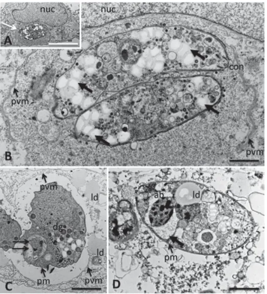
Fig. 3. TEM of Neospora caninum tachyzoites grown in HFF and treated with 25μM miltefosine for 20 h. (A) Overview and (B) corresponding higher magnification view of 2 intracellular tachyzoites. Parasites are largely surrounded by a parasitophorous vacuole (pvm), and embedded in a dense granular matrix. Thick arrows indicate extensive vacuolization, con, conoid. Scale bar in (A) = 8μm; in (B)=0·4 μm. (C) Intracellular tachyzoite with alterations such as cytoplasmic membrane stacks (horizontal arrows), still intact plasma membrane (pm) and parasitophorous vacuole membrane (pvm), and lipid droplets (ld) just adjacent to the pvm; dg, dense granules. Scale bar = 0·38μm. (D) Extracellular, non-viable tachyzoite with distorted cytoplasmic organization, containing apoptotic body (ab), lipid droplet (ld) and cytoplasmic vacuolesfilled with material of unknown origing (thick arrow). The plasma membrane still appears intact (pm). Scale bar = 0·38μm.
Miltefosine treatment in mice experimentally infected with Neospora caninum tachyzoites
In Experiment 1, miltefosine was orally applied to N. caninum-infected Balb/c mice at 20 and 40 mg/kg/ day for a period of 14 days p. i., respectively, a placebo control group received PBS/CMC only, and a fourth group remained uninfected and was treated with 40 mg/kg/day of miltefosine to assess a poten-tial impact of medication on the health of the ani-mals. After the 14 days treatment period, mice were euthanized and the cerebral parasite burden was assessed. In the placebo group, 2 out of 8 mice succumbed to infection at days 11 and 12 p.i., while in the group treated with miltefosine at 20 mg/kg/day 1 mouse died at day 12 p.i. No significant average weight loss was recorded during the 14 days of treatment (Table 1). Comparison of the cerebral parasite load in the 2 groups did not reveal any major differences (Fig. 4A, Supplementary Table 1, online version only). However, when N. caninum-infected mice were treated with 40 mg/kg/day of miltefosine, 6 out of 10 mice exhibited ruffled coat, general weakness and apathy, and they were not able to feed anymore. Thus they had to be euthanized at the onset of these symptoms between days 7 and 13 p.i.. None of the mice exhibited neurological disorders such as tilting and hind limb paralysis. In addition, a significant mean weight loss of over 10% was noted for that group within that 14-day period. Application of miltefosine at 40 mg/kg/day in non-infected mice, however, did not result in any weight loss or signs of toxicity, indicating that the drug per se did not exert any toxic effects (Table 1). Mice treated with
40 mg/kg/day for 14 days exhibited significantly lower cerebral parasite burdens compared to the placebo group (Fig. 4A). This result suggested that miltefosine treatment, although not causing any harm by itself, rendered mice more susceptible to acute N. caninum infection, and the cause of death of these animals was due to infection of other organs.
In Experiment 2, N. caninum-infected mice were treated with 40 mg/kg/day, but only for a period of 5 days p.i., and were then left untreated for 30 days. As indicated in Table 1, miltefosine treatment had a profound impact, with only 2 out of 9 mice succumbing to infection on days 5 (due to weakness and apathy) and 26 (neurological signs such as head tilt and running in circles), respectively. On the other hand, 8 out of 10 mice had to be euthanized in the placebo group between days 16 and 31. These mice exhibited neurological symptoms (head tilt, hind limb paralysis, circular movements, ruffled coat), indicative for CNS involvement. The overall cerebral parasite burden in the miltefosine-treated mice was significantly lower compared to the placebo group (Fig. 4B, Supplementary Table 1, online version only). This showed that the drug inhibited the proliferation of N. caninum and dissemination of tachyzoites into the cerebral tissue.
D I S C U S S I O N
Miltefosine, originally developed as an anti-cancer agent, is used for the treatment of skin metastases
of mammacarcinoma (Leonard et al. 2001).
Subsequently it was found that miltefosine is also Table 1. In vivo studies: summary of experimental groups and outcome of experiments in terms of
symptomatic mice, time of death and median body weights at day 0 and at the end of the experiments, day 14 for Exp. 1 and day 30 for Exp. 2
(Significant weight loss is found only in the experimentally infected mice treated with miltefosine (40 mg/kg/day for 14 days) and indicated by *.)
Groups Number of mice
Number of sympt. mice Time of death (days p.i.)
Weight at day p.i.
0 14 or 30 Experiment 1 Non-infected miltefosine 40 mg/kg/day 14 days 8 – – 18·95 ± 0·80 18·86 ± 0·56 Infected placebo CMC only 14 days 8 2 11/12 19·07 ± 0·99 18·18 ± 0·56 Infected miltefosine 20 mg/kg/day 14 days 8 1 12 17·9 ± 0·92 17·3 ± 1·59 Infected miltefosine 40 mg/kg/day 14 days 10 6 7/10/11/12/12/13 20·09 ± 0·73 17·47 ± 1·52* Experiment 2
Placebo CMC only 5 days 10 8 16/19/22/22/25/26/
27/31
19·5 ± 0·86 18·9 ± 0·42
Miltefosine 40 mg/kg/day 5 days
active against visceral and cutanous leishmaniasis, and can be applied orally or topically, respectively (Croft and Engel, 2006; Bhattacharya et al. 2007). The drug was also active against Trypanosoma brucei, Entamoeba histolytica, Acanthamoeba spp. and T. cruzi (Croft et al. 1996; Konstantinov et al. 1997; Seifert et al.2001; Walochnik et al.2002), and it was shown that miltefosine affects apicomplexan parasites such as Plasmodium falciparum and Cryptosporidium (Pessi et al. 2004; Shahiduzzaman et al. 2009). Similar to many of the above-mentioned protozoan parasites, Neospora occupies an intracellular niche,
indicating that miltefosine might also be useful for the treatment neosporosis.
In vitro cytotoxicity assays demonstrated that Vero cells, which represent an immortalized and highly proliferative cell line that does not undergo contact inhibition, are highly susceptible to the drug. In contrast, human foreskin fibroblasts, which do not proliferate further once a confluent monolayer is formed, were not affected by miltefosine treatment at concentrations up to 122μM. This reflects the anti-proliferative properties of miltefosine (Leonard et al. 2001). The drug inhibited the proliferation of N. caninum tachyzoites with an IC50 of 5·2μM, which is in a similar range to that reported for the thiazolide nitazoxanide (IC50= 4·23μM; Esposito et al. 2007a), but substantially higher than the dicationic pentamidine derivative DB750 (IC50= 0·23μM; Leepin et al. 2008). Miltefosine appeared to act swiftly: at 25μM an incubation period of 20 h was sufficient to exert a parasiticidal activity within N. caninum-infected HFF. In comparison,
nitazoxanide added to infected HFF at 30μM
required a treatment duration of 5 days in order to exert parasiticidal activity in vitro (Esposito et al. 2005), and for toltrazuril, a 10-day treatment at a concentration of 70·5μM had only a parasitostatic effect, and a total of 14 days were required to exert a parasiticidal effect (Strohbusch et al.2009).
Toltrazuril (Darius et al.2004), nitazoxanide and other thiazoides (Esposito et al.2005,2007a,b), and DB750 (Leepin et al. 2008), exerted profound ultrastructural changes in N. caninum tachyzoites in vitro. The same accounted for miltefosine in this study. While we could not detect any major differences between treated and untreated uninfected HFF monolayers (data not shown), clear signs of N. caninum tachyzoite deterioration were already detectable after 10 h of treatment, which included profound alterations in the organization of some of the membrane-based compartments of the cyto-plasm. Rhoptries and micronemes were mostly no longer visible after 10 h of treatment, while the electron-dense granules were apparently affected by a lower degree, and in many cells rounded structures resembling apoptotic bodies were evident. The tachyzoite cytoplasm exhibited increased vacuoliza-tion but, in most cases, no damage could be detected on nuclear membranes and parasite plasma mem-branes. In several tachyzoites layers of membrane stacks localized near the parasite nucleus were observed. An impressive number of lipid droplets accumulated within the host cell cytoplasm in close vicinity of the parasitophorous vacuole membrane. After 20 h of miltefosine treatment, the majority of tachyzoites were found either intracellularly, obviously non-viable and often embedded in a rather electron-dense granular matrix filling the parasito-phorous vacuole, or were present in the extracellular space, without any clearly discernible organized
Fig. 4. Box plots showing the effects of miltefosine treatments on the cerebral parasite burden in mice experimentally infected with Neospora caninum
tachyzoites. (A) Treatment with miltefosine (20 or 40 mg/ kg/day for 14 days) or placebo. The y-axis depicts the number of detected parasites by real time PCR /100 ng of DNA. The placebo group was treated identically but with the solvent carboxy-methylcellulose (CMC) only. * Indicates a significant reduction in parasite load (P < 0·05). (B) Treatment with miltefosine (40 mg/kg/day) for a period of 5 days p.i. only, followed by maintenance of mice for 30 days without treatment. * Indicates a significant reduction in parasite load (P<0·05) in the treatment group. For more details on cerebral parasite burden of each individual mouse refer to supplementary Table 1, online version only.
cytoplasmic features, with the exception of lipid droplets. For the parasites located intracellularly, the electron-dense parasitophorous vacuolar matrix clo-sely resembled the cyst-wall associated structures observed during in vitro development of N. caninum bradyzoites induced by sodium nitroprusside treat-ment (Vonlaufen et al. 2002, 2004). This matrix could be formed due to stress-related increased secretory activity, most notably from the parasite dense granule organelles (Vonlaufen et al.2004). The presence of extracellular tachyzoites could be a result of increased, drug-induced egress and the inability of the parasite to subsequently invade another host cell successfully, as has previously been demonstrated for a series of thiazolides (Esposito et al.2007a).
The presence of increased numbers of lipid droplets within the host cell and tachyzoite cytoplasm could be caused by disturbances in fatty acid and sterol metabolism as previously reported for L. donovani by Rakotomanga et al. (2007). It is not clear whether the increased presence of lipid droplets in the vicinity of parasitophorous vacuoles reflects a similar effect in host cells, or whether they are a consequence of stress that is induced by the presence of tachyzoites. More prolonged TEM investigations covering more time-points would be required to study the effects of deteriorating parasites on host cell survival. Similar studies undertaken with nitazox-anide and related compounds (Esposito et al.2005) had suggested that parasite death also had a negative impact on host cell ultrastructure and survival. However, in specimens incubated with miltefosine for 20 h, the vast majority of observed tachyzoites exhibited a clearly discernible plasma membrane, which would be indicative of apoptotic events taking place during drug treatment. This would be in accordance with previous studies on L. donovani (Paris et al.2004).
Perez-Victoria et al. (2006) had reported that uptake of miltefosine is mediated by a plasma membrane P-type ATPase aminophospholipid trans-locase. Suggested targets of miltefosine in Leishmania include perturbation of ether-lipid metabolism, glycosylphosphatidylinositol (GPI) anchor biosyn-thesis and signal transduction as well as inhibition of the glycosomal located alkyl-specific acyl-Co-A acyltransferase, an enzyme involved in lipid remodel-ling (Lux et al. 2000). However, more detailed investigations are required in order to define the mechanisms that account for the effects of miltefosine against N. caninum tachyzoites.
The relatively low host cell toxicity and clear parasiticidal activity in vitro prompted us to under-take an in vivo study on the effects of miltefosine treatment on Neospora infection in mice. Afirst set of experimental groups were treated with either 20 mg/kg/day or 40 mg/kg/day for a period of 14 days, after which the animals were euthanized and analysed, similar to previous studies employing the
dicationic compound DB750 (Debache et al.2011). The goal was to investigate whether miltefosine affected cerebral infection. Our results show that in N. caninum-infected mice treatment with miltefosine at 20 mg/kg/day did not reduce the cerebral parasite burden compared to the placebo group, while treatment at 40 mg/kg/day did, but those mice could obviously not cope with the infection. In human patients, adverse side effects of miltefosine treatment have been reported such as vomiting, nausea, kinetosis, headache, diarrhoea, and a mild to moderate increase in aminotransferases and crea-tinine (reviewed by Oliveira et al.2011). In contrast, application of miltefosine at 40 mg/kg/day for only 5 days did not exert any adverse effects. Following treatment, mice were monitored for a period of 30 days, and subsequent analysis showed a significantly reduced number of fatalities and a significantly decreased cerebral parasite burden in the miltefosine treatment group. When used for the treatment of Leishmania parasites in Balb/c mice, miltefosine treatment for 5 consecutive days at 30 mg/kg/day was highly effective, as was the case when treatment was performed in Leishmania-infected scid mice (Escobar et al.2001).
The results obtained in N. caninum infected Balb/c mice suggest that miltefosine treatment can limit the dissemination of tachyzoites into the CNS. However, further studies are needed to demonstrate the potential of this compound in cases where the CNS has already been invaded by the parasite. Since miltefosine is an amphipatic molecule, it is likely to cross the blood-brain barrier, and further studies will show whether miltefosine can also exert an effect against parasites within the CNS. Since this drug is already in use for the treatment of canine leishma-niasis, its application in cases of canine neosporosis might be an attractive option.
A C K N O W L E D G E M E N T S
This work was supported by the Swiss National Science Foundation (Grant No. 31003A_127374/1). The authors thank Norbert Müller for helpful comments on the manuscript, and Joachim Müller for help in statistics. J.P. Dubey is gratefully acknowledged for providing us with the Nc-1 isolate of Neospora caninum. Many thanks are addressed to Dr Andreas Obwaller (Orphanidis Pharma Research GmbH) for providing us with miltefosine.
R E F E R E N C E S
Alaeddine, F., Keller, N., Leepin, A. and Hemphill, A. (2005). Reduced infection and protection from clinical signs of cerebral neosporosis in C57BL/6 mice vaccinated with recombinant microneme antigen NcMIC1. Journal of Parasitology91, 657–665.
Barratt, G., Saint-Pierre-Chazalet, M. and Loiseau, P. M. (2009). Cellular transport and lipid interactions of miltefosine. Current Drug Metabolism10, 247–255.
Bhattacharya, S. K., Sinha, P. K., Sundar, S., Thakur, C. P., Jha, T. K., Pandey, K., Das, V. R., Kumar, N., Lal, C., Verma, N., Singh, V. P., Ranjan, A., Verma, R. B., Anders, G., Sinderman, H. and Ganguly, N. K. (2007). Phase 4 trial of miltefosine for the treatment of Indian visceral leishmaniasis. Journal of Infectious Diseases196, 591–598.
Cannas, A., Naguleswaran, A., Müller, N., Gottstein, B., Eperon, S. and Hemphill, A. (2003a). Vaccination of mice against experimental N. caninum infection using NcSAG1- and NcSRS2-based recombinant antigens and DNA-vaccines. Parasitology126, 303–312.
Cannas, A., Naguleswaran, A., Müller, N., Gottstein, B. and Hemphill, A. (2003b). Reduced cerebral infection of Neospora caninum-infected mice after vaccination with recombinant microneme protein NcMIC3 and ribi adjuvant. Journal of Parasitology89, 44–50.
Choubey, V., Maity, P., Guha, M., Kumar, S., Srivastava, K., Puri, S. K. and Bandyopadhyay, U. (2007). Inhibition of Plasmodium falciparum choline kinase by hexadecyltrimethylammonium bromide: a possible antimalarial mechanism. Antimicrobial Agents and Chemotherapy 51, 696–706.
Croft, S. L., Snowdon, D. and Yardley, V. (1996). The activities of four anticancer alkyllysophospholipids against Leishmania donovani, Trypanosoma cruzi and Trypanosoma brucei. Antimicrobial Agents and Chemotherapy6, 1041–7.
Croft, S. L., Engel, J. (2006). Miltefosine–discovery of the antileishmanial activity of phospholipid derivatives. Transactions of the Royal Society of Tropical Medicine and Hygiene100, S4–S8.
Darius, A. K., Mehlhorn, H. and Heydorn, A. O. (2004). Effects of toltrazuril and ponazuril on the fine structure and multipli-cation of tachyzoites of the NC-1 strain of Neospora caninum (a synonym of Hammondia heydorni) in cell cultures. Parasitology Research 92, 453–458.
Debache, K., Alaeddine, F., Guionaud, C., Monney, T., Müller, J., Strohbusch, M., Leib, S. L., Grandgirard, D. and Hemphill, A. (2009). Vaccination with recombinant NcROP2 combined with recombinant NcMIC1 and NcMIC3 reduces cerebral infection and vertical transmission in mice experimentally infected with Neospora caninum tachyzoites. International Journal for Parasitology39, 1373–1384.
Debache, K., Guionaud, C., Kropf, C., Boykin, D. and Hemphill, A. (2011). Experimental treatment of Neospora caninum-infected mice with the arylimidamide DB750 and the thiazolide nitazoxanide. Experimental Parasitology129, 95–100.
Dubey, J. P., Hattel, A. L., Lindsay, D. S. and Topper, M. J. (1988). Neonatal Neospora caninum infection in dogs: isolation of the causative agent and experimental transmission. Journal of the American Veterinary Medical Association193, 1259–1263.
Dubey, J.P. and Schares, G. (2011). Neosporosis in animals the lastfive years. Veterinary Parasitology180, 90–108.
Dubey, J. P., Schares, G. and Ortega-Mora, L. M. (2007). Epidemiology and control of neosporosis and Neospora caninum. Clinical Microbiology. Microbiology Reviews20, 323–367.
Escobar, P., Yardely, V., and Croft, S. L. (2001). Activities of hexadecylphosphocholine (Miltefosine), AmBisome, and sodium stiboglu-conate (Pentostam) against Leishmania donovani in immunodeficient scid mice. Antimicrobial Agents and Chemotherapy45, 1872–1875.
Esposito, M., Moores, S., Naguleswaran, A., Mueller, J. and Hemphill, A. (2007a). Induction of tachyzoite egress from cells infected with the protozoan Neospora caninum by nitro- and bromo-thiazolides, a class of broad-spectrum anti-parasitic drugs. International Journal for Parasitology37, 1143–1152.
Esposito, M., Muller, N. and Hemphill, A. (2007b). Structure-activity relationships from in vitro efficacies of the thiazolide series against the intracellular apicomplexan protozoan Neospora caninum. International Journal for Parasitology37, 183–190.
Esposito, M., Stettler, R., Moores, S. L., Pidathala, C., Muller, N., Stachulski, A., Berry, N. G., Rossignol, J. F. and Hemphill, A. (2005). In vitro efficacies of nitazoxanide and other thiazolides against Neospora caninum tachyzoites reveal antiparasitic activity independent of the nitro group. Antimicrobial Agents and Chemotherapy49, 3715–3723.
Gottstein, B., Eperon, S., Dai, W. J., Cannas, A., Hemphill, A. and Greif, G. (2001). Efficacy of toltrazuril and ponazuril against experimental Neospora caninum infection in mice. Parasitology Research 87, 43–48.
Haerdi, C., Haessig, M., Sager, H., Greif, G., Staubli, D. and Gottstein, B. (2006). Humoral immune reaction of newborn calves congenitally infected with Neospora caninum and experimentally treated with toltrazuril. Parasitology Research99, 534–540.
Häsler, B., Regula, G., Stärk, K. D., Sager, H., Gottstein, B. and Reist, M. (2006b). Financial analysis of various strategies for the control of Neospora caninum in dairy cattle in Switzerland. Preventive Veterinary Medicine77, 230–253.
Häsler, B., Stärk, K. D., Sager, H., Gottstein, B. and Reist, M. (2006a). Simulating the impact of four control strategies on the population dynamics of Neospora caninum infection in Swiss dairy cattle. Preventive Veterinary Medicine77, 254–283.
Hemphill, A. (1996). Subcellular localization and functional characteriz-ation of Nc-p43, a major Neospora caninum tachyzoite surface protein. Infection and Immunity64, 4279–4287.
Kim, J. T., Park, J. Y., Seo, H. S., Oh, H. G., Noh, J. W., Kim, J. H., Kim, D. Y. and Youn, H. J. (2002). In vitro antiprotozoal effects of artemisinin on Neospora caninum. Veterinary Parasitology103, 53–63. Konstantinov, S. M., Kaminsky, R., Brun, R., Berger, M. R. and Zillmann, U. (1997). Efficacy of anticancer alkylphosphocholines in Trypanosoma brucei subspecies. Acta Tropica64, 145–154.
Kritzner, S., Sager, H., Blum, J., Krebber, R., Greif, G. and Gottstein, B. (2002). An explorative study to assess the efficacy of toltrazuril-sulfone (ponazuril) in calves experimentally infected with Neospora caninum. Annals of Clinical Microbiology and Antimicrobials 1, 4–10.
Kwon, H. J., Kim, J. H., Kim, M., Lee, J. K., Hwang, W. S. and Kim, D. Y. (2003). Anti-parasitic activity of depudecin on Neospora caninum via the inhibition of histone deacetylase. Veterinary Parasitology 112, 269–276.
Leepin, A., Studli, A., Brun, R., Stephens, C. E., Boykin, D. W. and Hemphill, A. (2008). Host cells participate in the in vitro effects of novel diamidine analogues against tachyzoites of the intracellular apicomplexan parasites Neospora caninum and Toxoplasma gondii. Antimicrobial Agents and Chemotherapy52, 1999–2008.
Leonard, R., Hardy, J., van Tienhoven, G., Houston, S., Simmonds, P., David, M. and Mansi, J. (2001). Randomized, double-blind, placebo-controlled, multicenter trial of 6% miltefosine solution, a topical chemotherapy in cutaneous metastases from breast cancer. Journal of Clinical Oncology19, 4150–4159.
Lindsay, D. S. and Dubey, J. P. (1989). Evaluation of anti-coccidial drugs’ inhibition of Neospora caninum development in cell cultures. Journal of Parasitology75, 990–992.
Lindsay, D. S. and Dubey, J. P. (1990). Effects of sulfadiazine and amprolium on Neospora caninum (Protozoa: Apicomplexa) infections in mice. Journal of Parasitology76, 177–179.
Lindsay, D. S., Rippey, N. S., Cole, R. A., Parsons, L. C., Dubey, J. P., Tidwell, R. R. and Blagburn, B. L. (1994). Examination of the activities of 43 chemotherapeutic agents against Neospora caninum tachyzoites in cultured cells. American Journal of Veterinary Research55, 976–981. Lux, H., Heise, N., Klenner, T., Hart, D. and Opperdoes, F. R. (2000). Ether–lipid (alkyl-phospholipid) metabolism and the mechanism of action of ether–lipid analogues in Leishmania. Molecular and Biochemical Parasitology111, 1–14.
Monney, T., Debache, K. and Hemphill, A. (2011). Vaccines against a major cause of abortion in cattle, Neospora caninum infection. Animals1, 306–325.
Müller, N., Vonlaufen, N., Gianinazzi, C., Leib, S. L. and Hemphill, A. (2002). Application of real time fluorescent PCR for quantitative assessment of Neospora caninum infections in organotypic slice cultures of rat central nervous tissue. Journal of Clinical Microbiology 40, 252–255.
Oliveira, L. F., Schubach, A. O., Martins, M. M., Passos, S. L., Oliveira, R. V., Marzochi, M. C. and Andrade, C. A. (2011). Systematic review of the adverse effects of cutaneous Leishmaniasis treatment in the New World. Acta Tropica118, 87–96.
Paris, C., Loiseau, P. M., Bories, C. and Bréard, J. (2004). Miltefosine induces apoptosis-like death in Leishmania donovani promastigotes. Antimicrobial Agents and Chemotherapy48, 852–859.
Perez-Victoria, F. J., Sanchez-Canete, M. P., Seifert, K., Croft, S. L., Sundar, S., Castanys, S. and Gamarro, F. (2006). Mechanisms of experimental resistance of Leishmania to miltefosine: Implications for clinical use. Drug Resistance Updates9, 26–39.
Pessi, G., Kociubinski, G. and Mamoun, C. B. (2004). A pathway for phosphatidylcholine biosynthesis in Plasmodium falciparum involving phosphoethanolamine methylation. Proceedings of the National Academy of Sciences, USA101, 6206–6211.
Rakotomanga, M., Blanc, S., Gaudin, K., Chaminade, P. and Loiseau, P. M. (2007). Miltefosine affects lipid metabolism in Leishmania donovani promastigotes. Antimicrobial Agents and Chemotherapy 51, 1425–1430.
Schuster, F. L., Guglielmo, B. J. and Visvesvara, G. S. (2006). In-vitro activity of miltefosine and voriconazole on clinical isolates of free-living amebas: Balamuthia mandrillaris, Acanthamoeba spp., and Naegleria fowleri. Journal of Eukaryotic Microbiology53, 121–126.
Seifert, K., Duchêne, M., Wernsdorfer, W. H., Kollaritsch, H., Scheiner, O., Wiedermann, G., Hottkowitz, T. and Eibl, H. (2001). Effects of miltefosine and other alkylphosphocholines on human intestinal parasite Entamoeba histolytica. Antimicrobial Agents and Chemotherapy45, 1505–1510.
Shahiduzzaman, M., Dyachenko, V., Obwaller, A., Unglaube, S. and Daugschies, A. (2009). Combination of cell culture and quantitative PCR for screening of drugs against Cryptosporidium parvum. Veterinary Parasitology162, 271–277.
Strohbusch, M., Müller, N., Hemphill, A., Krebber, R., Greif, G. and Gottstein, B. (2009). Toltrazuril treatment of congenitally acquired Neospora caninum infection in newborn mice. Parasitology Research104, 1335–1343.
Unger, C., Damenz, W., Fleer, E. A., Kim, D. J., Breiser, A., Hilgard, P., Engel, J., Nagel, G. and Eibl, H. (1989). Hexadecylphosphocholine, a new ether lipid analogue. Studies on the antineoplastic activity in vitro and in vivo. Acta Oncologica28, 213–217. Vonlaufen, N., Guetg, N., Naguleswaran, A., Müller, N., Björkman, C., Schares, G., von Blumroeder, D., Ellis, J. and Hemphill, A. (2004). In vitro induction of Neospora caninum bradyzoites in vero cells reveals differential antigen expression, localization, and host-cell
recognition of tachyzoites and bradyzoites. Infection and Immunity 72, 576–583.
Vonlaufen, N., Müller, N., Keller, N., Naguleswaran, A., Bohne, W., McAllister, M. M., Björkman, C., Müller, E., Caldelari, R. and Hemphill, A. (2002). Exogenous nitric oxide triggers Neospora caninum tachyzoite-to-bradyzoite stage conversion in murine epidermal keratinocyte cell cultures. International Journal for Parasitology32, 1253–1265. Walochnik, J., Duchene, M., Seifert, K., Obwaller, A., Hottkowitz, T., Wiedermann, G., Eibl, H. and Aspöck, H. (2002). Cytotoxic activities of alkylphosphocholines against clinical isolates of Acanthamoeba spp. Antimicrobial Agents and Chemotherapy46, 695–701.
Youn, H. J., Lakritz, J., Rottinghaus, G. E., Seo, H. S., Kim, D. Y., Cho, M. H. and Marsh, A. E. (2004). Anti-protozoal efficacy of high performance liquid chromatography fractions of Torilis japonica and Sophoraflavescens extracts on Neospora caninum and Toxoplasma gondii. Veterinary Parasitology125, 409–414.