of the early enzymes in the Campylobacter
jejuni N-linked glycosylation pathway
The MIT Faculty has made this article openly available. Please share how this access benefits you. Your story matters.Citation Morrison, James P.; Troutman, Jerry M. and Imperiali, Barbara.
“Development of a Multicomponent Kinetic Assay of the Early Enzymes in the Campylobacter Jejuni N-Linked Glycosylation Pathway.” Bioorganic & Medicinal Chemistry 18, no. 23 (December 2010): 8167–8171. © 2010 Elsevier Ltd
As Published http://dx.doi.org/10.1016/j.bmc.2010.10.020
Publisher Elsevier
Version Author's final manuscript
Citable link http://hdl.handle.net/1721.1/108116
Terms of Use Creative Commons Attribution-NonCommercial-NoDerivs License
In Vitro Pathway Screening of the Early Enzymes in the Campylobacter jejuni N-Linked
Glycosylation Pathway
James P. Morrisona, Jerry M. Troutmana, and Barbara Imperialia,b,*
Departments of a Chemistry and b Biology, Massachusetts Institute of Technology, 77 Massachusetts Avenue, Cambridge, Massachusetts 02139.
Abstract
The human pathogen Campylobacter jejuni possesses a general N-linked glycosylation system that is known to play a role in pathogenicity; however, a detailed understanding of this role remains elusive. A considerable hindrance to studying bacterial N-glycosylation in vivo is the absence of small molecule inhibitors to reversibly control the process. This report describes a pathway-screening assay that targets the early enzymes of C. jejuni N-glycan biosynthesis that would enable identification of inhibitors to the first four steps in the pathway. The assay includes PglF, PglE, PglD, PglC and PglA; the enzymes involved in the biosynthesis of an undecaprenyl diphosphate-linked disaccharide and monitors the transfer [3H]GalNAc from the hydrophilic UDP-linked carrier to the lipophilic UndPP-diNAcBac (2,4-diacetamido-2,4,6-trideoxyglucose). The optimized assay has a Z'-factor calculated to be 0.77, indicating a robust assay suitable for screening. The diacylglycerol kinase from Streptococcus mutans, which provides a convenient method for phosphorylating undecaprenol, has been included in a modified version of the assay thereby allowing the screen to be conducted with entirely commercially available substrates.
Abbreviations: Streptococcus mutans diacylglycerol kinase, DGK;
tris(2-carboxyethyl)phosphine, TCEP; UndOH, undecaprenol; UndP, undecaprenyl phosphate; diNAcBac, 2,4-diacetamido-2,4,6-trideoxyglucose; dimethyl sulfoxide, DMSO, disintegrations per minute, DPM; pure solvent upper phase, PSUP
1. Introduction
The general N-linked protein glycosylation (pgl) pathway of Campylobacter jejuni is the first discovered bacterial system and has been demonstrated to play an important role in the pathogenicity of this organism. The human pathogen C. jejuni is a major cause of gastroenteritis and is the most common antecedent to Guillain-Barré syndrome, a major cause of non-trauma-induced paralysis.1-3 N-linked glycosylation occurs in the periplasm of the Gram-negative pathogen, with the en bloc transfer of a heptasaccharide from an undecaprenyl diphosphate (UndPP) carrier to an asparagine in the acceptor protein. Over 40 proteins have been demonstrated to be N-glycosylated in C. jejuni, and it is predicted that more than 150 of the 340 secreted C. jejuni proteins may be glycosylated based on the presence of the recognition sequence D/E-X1-N-X2-S/T (where X1 and X2 may be any residue except proline).4-6 The role of
this post-translational modification and how it affects pathogenicity are areas of active investigation. While a detailed understanding of how N-glycosylation enables pathogenicity is lacking, blocking N-glycosylation using genetic approaches renders C. jejuni dramatically less pathogenic, as has been determined in several studies employing chick colonization pathogenicity models.7-10 The genes mutated in these studies include the oligosaccharyl transferase PglB and those involved in the biosynthesis of the undecaprenyl diphosphate-heptasaccharide; this strongly suggests that an inhibitor of one of the biosynthetic enzymes along this pathway would block N-glycosylation and potentially modulate pathogenicity.
The biosynthesis of the C. jejuni heptasaccharide involves the sequential addition of sugars to generate a heptasaccharide linked to undecaprenyl diphosphate (Und-PP) anchored to the cytoplasmic face of the inner membrane (Figure 1A). The first sugar in the heptasaccharide is the unusual sugar 2,4-diacetamido-2,4,6-trideoxyglucose (diNAcBac), which is transferred from UDP-diNAcBac and is only known to exist in certain strains of bacteria.11 In addition to the involvement in C. jejuni N-glycosylation, UDP-diNAcBac is also a known intermediate in the biosynthesis of legionaminic acid in Legionella pneumophilia (causative agent of Legionnaire’s disease)12 and is putatively a component sugar in O-linked protein glycosylation in Neisseria
gonorrhoeae (causative agent of gonorrhea).13 In C. jejuni, UDP-diNAcBac is derived from UDP-GlcNAc by the sequential action of the 4,6-dehydratase PglF, the 4-aminotransferase PglE and the 4-acetyltransferase PglD.14, 15 The glycosyl-1-phosphate transferase PglC then uses UDP-diNAcBac and undecaprenyl phosphate (UndP) to generate UDP-diNAcBac-PP-Und.16 The
glycosyltransferase PglA then adds GalNAc (via an 1,3 linkage), then PglJ adds the second GalNAc ( 1,4-linkage), and PglH adds three additional GalNAc sugars ( 1,4-linkages).17-19 PglI adds a branching glucose to the third GalNAc ( 1,3-linkage) to afford the complete Und-PP-heptasaccharide.18 The undecaprenyl diphosphate-linked glycan is then flipped to the periplasmic face of the inner membrane by the flippase PglK,20 where the oligosaccharyltransferase PglB transfers the glycan to asparagine residues within the consensus sequence of proteins in the periplasm. A high-resolution magic angle spinning nuclear magnetic resonance study comparing
C. jejuni mutants found that N-linked glycosylation was greatly reduced in pglF, pglE, pglD, pglA, pglJ, pglH, pglK and pglB mutants, while the pglI mutant is unaffected.10 A pglC mutant was unavailable as this mutation appears to be lethal.10, 21 The essential nature of the entire biosynthetic pathway (except PglI) strongly suggests one could target any one of the enzymes with inhibitors to reduce the flux of substrates through the N-glycosylation pathway of C. jejuni.
In this report we describe a multicomponent kinetic assay for the early enzymes in the UndPP-heptasaccharide biosynthetic pathway (Figure 1B). PglF, PglE, PglD, PglC, and PglA are assembled in vitro and the conversion of UDP-GlcNAc into UndPP-diNAcBac-[3H]GalNAc is quantified by monitoring the transfer of [3H]GalNAc from the water soluble UDP carrier to the lipophilic UndPP-diNAcBac acceptor. Under the conditions developed, this assay targets enzymes involved in the biosynthesis of the unusual bacterial sugar diNAcBac and the transfer of diNAcBac-phosphate to UndP. We also describe a modification of this assay, which incorporates the diacylglycerol kinase from Streptococcus mutans (DGK) which allows for use of the readily available undecaprenol in lieu of UndP,22 such that screening of five Pgl enzymes in a single assay can be carried out using entirely commercially available substrates.
2. Results
2.1 Assembled activities of PglF, PglE, PglD, PglC, PglA and DGK
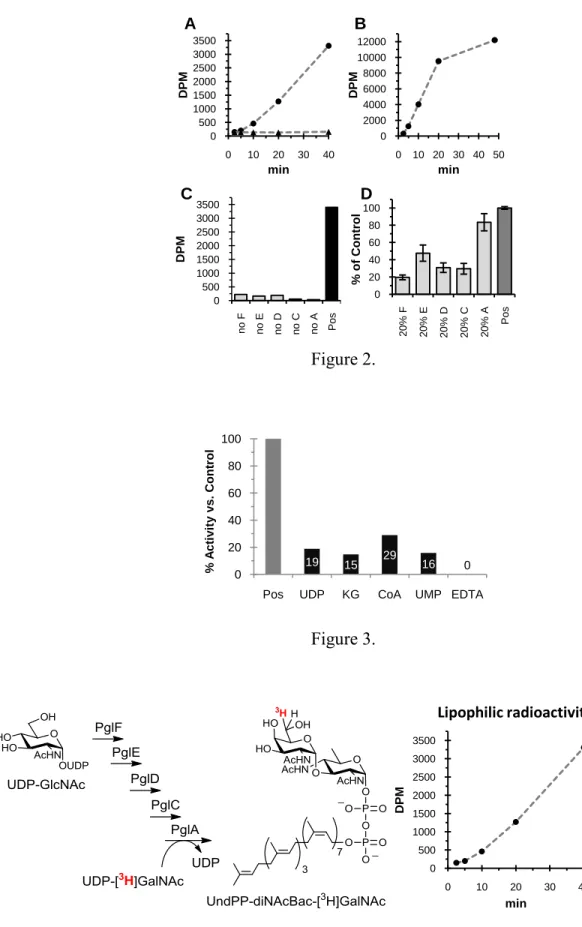
The five pgl pathway enzymes with the appropriate cofactors and substrates, including UndP and UDP-[3H]GalNAc, were assembled for the quenched time-point assay. PglF and PglC are integral membrane proteins purified from cell envelope fractions, and therefore the assays are performed in the presence of the detergent Triton X-100. Quenched time-points were extracted with a chloroform-methanol-water extraction system, allowing measurement of the transfer of [3H]GalNAc by PglA to the organic soluble UndPP-diNAcBac, which is assembled by PglF,
PglE, PglD and PglA. As anticipated, a time-dependent transfer of radioactivity to the organic phase was observed that did not occur in the absence of UndP (Figure 2A). In a second experiment where DGK, ATP, and undecaprenol (UndOH) were used in place of UndP, similar results were observed (Figure 2B). In both of these experiments an initial lag period was observed, consistent with a coupled assay with several partially rate-limiting steps. This was followed by a linear phase representing the reaction progress from ~5 to 30% conversion. At higher conversion, the reaction rate decelerates due to substrate depletion (Figure 2B). These results are consistent with formation of the final PglA product UndPP-diNAcBac-[3H]GalNAc,18,
19, 22 as is the requirement of each enzyme for these observations (Figure 2C). The systematic
removal of individual enzymes led to observation of only background levels of radioactivity in the organic phase from the quenched assay aliquot, confirming these observations measure the formation of the final product of these five enzymes, UndPP-diNAcBac-GalNAc.
2.2 Balancing enzyme concentrations for kinetic sensitivity to PglF, PglE, PglD and PglC We have chosen to include the first five enzymes in the UndPP-heptasaccharide pathway, as this represents several enzymes intimately involved with recognition of the unusual sugar diNAcBac or its precursor intermediates. Although PglA falls into this category, the primary role of this enzyme is to deliver the [3H] labeled GalNAc to UndPP-diNAcBac and drive the phosphoglycosyl transferase reaction forward and therefore, it is used in excess in the assay. In order to make the assay simultaneously sensitive to inhibitors of PglF, PglE, PglD, and PglC, the concentrations of these enzymes have been balanced such that the activity of each of these is partially rate determining. This was accomplished by systematically lowering the concentration of each enzyme until a significant effect on the overall rate was observed. A simulated inhibition of these enzymes demonstrates that this balanced state has been achieved; when the concentration of PglF, PglE, PglD or PglC is lowered five-fold a lower overall rate results (Figure 2C).
2.3 Effects of Triton X-100 and DMSO
Variation of the detergent concentration has a significant effect on observed activity. Maximum activity was observed with 0.04-0.06% Triton X-100, near the critical micelle concentration of this detergent (Figure S1). This activity was 30-fold greater than that that
measured at 1% Triton X-100 and no activity was observed below the critical micelle concentration of the detergent. These observations are consistent with the activity of known membrane-associated enzymes.23
Dimethyl sulfoxide (DMSO) is a common small molecule carrier in library screening and as such its compatibility with the assay was assessed. Up to 5% DMSO did not have a noticeable effect on the overall activity, and only a minor effect on background signal was observed. When PglF, PglE, or PglD was absent, higher background radioactivity was observed than when PglC, PglA or DGK was absent (Figure 2). These results suggest a minor effect of DMSO on the substrate specificity on PglC and PglA, however this effect does not have a significant impact on the reliability of the assay at these concentrations.
2.4 Volume-minimized assay for screening and inhibition with substrate analogues
For screening purposes the reaction volume has been minimized to allow for economical use of reagents and time. Reaction volumes of 20 L, which allow for a single time point evaluation of reaction progress, have been deemed sufficient for screening purposes. Conditions are such that at 30 min ~10% conversion to final product is observed, which is in the linear phase of reaction progress. The assay was evaluated by running positive and negative controls at three different times. The average signal-to-background ratio was 56-fold and the standard deviation for the positive and negative controls was 7.3 and 0.27%, respectively. This data was used to calculate the Z'-factor,24 which is a statistical parameter used to evaluate assay quality. The Z'-factor takes into consideration the signal dynamic range and data reproducibility, and should be between 0.5 and 1 for an assay to be suitable for screening. The Z'-factor for the minimized assay was calculated to be 0.77.
To demonstrate inhibition in the context of the assay, the effects of several products and product analogues and the divalent metal ion chelator EDTA were investigated. UDP, -ketoglutarate, CoASH, and UMP were all found to inhibit the assay (Figure 3). EDTA was also found to inhibit the assay, presumably by sequestering the divalent magnesium required for PglC and PglA, which are known to require a divalent cation for activity.16, 25
3. Discussion
A recurring theme in prokaryotic glycoconjugate assembly is the sequential transfer of sugars to a membrane-bound isoprene-linked carrier during glycan biosynthesis. Notable examples of this include the biosynthesis of GlcNAc-MurNAc-pentapetide subunits of peptidoglycan, the outer O-antigens of lipopolysaccharide (LPS), capsular polysaccharides and lipooligosaccharides. More recently discovered examples include bacterial glycosylation pathways, such as the O-glycosylation pathway of N. gonorrheae and the N-glycosylation pathway of C. jejuni. Having established an assay for C. jejuni, which takes advantage of the lipophilicity of the undecaprenyl diphosphate-linked glycan, it is anticipated that the assay described here could easily be adapted to these other pathways.
The development of this lipophilicity-based pathway screen is a useful tool to address the present need for inhibitors of C. jejuni N-glycosylation. The absence of such inhibitors is currently a hindrance for researchers studying the roles of this pathway in vivo. This is in contrast with eukaryotic N-glycosylation, which is inhibited by tunicamycin, the utility of which is demonstrated by more than 2500 peer-reviewed journal articles citing its use. Tunicamycin targets the GlcNAc phosphototransferase, which transfers GlcNAc-1-P from UDP-GlcNAc to dolichyl-phosphate. Tunicamycin is also known to inhibit the transfer of GlcNAc-1-P to UndP and the formation of the undecaprenyl-PP-MurNAc pentapeptide involved in bacterial peptidoglycan biosynthesis. However, tunicamycin does not inhibit C. jejuni PglC, as described previously in a study in which this lack of inhibition was contrasted with inhibition of
Escherichia coli GlcNAc-1-phosphate transferase WecA.16 The observed difference in tunicamycin sensitivity suggests the feasibility of targeting the C. jejuni pathway selectively.
In the search for inhibitors of C. jejuni N-glycosylation, pathway screening offers several advantages over parallel screening of each individual enzyme. Chiefly, the efficiency of simultaneously screening multiple enzymes reduces the total number of assays required. It is estimated that one could easily screen 25 potential inhibitory interactions in a single tube with a cocktail of five compounds using the present assay. This would require additional experiments to identify the particular inhibitor/enzyme combination of interest, but does not necessarily require individual assays for each enzyme as was demonstrated in a similar pathway screen of the ADP-heptose biosynthetic pathway of LPS.26 In the case of PglF, PglE, PgD, PglC and PglA, however, individual assays are established.14-16, 25 A second benefit of the pathway screen is the use of
commercially available substrates, which is particularly relevant here where every sugar intermediate beyond UDP-GlcNAc is rare and requires special preparation and handling. Finally, assaying the pathway also allows identification of potential modulators of enzyme complex formation.
4. Conclusion
In this report we have described how the lipophilic nature of the isoprene-linked glycan can be harnessed to screen multiple enzymes involved with unusual sugar biosynthesis. This assay has been optimized for maximum sensitivity to inhibition of PglF, PglE, PglD, and PglC by balancing the enzyme concentrations such that each is partially rate determining. The assay has been optimized for maximum activity, which was found at 0.04-0.06% Triton X-100, and the assay volume has been minimized to 20 L to economize on reagent use. The robustness of the optimized assay has been demonstrated by the measured Z'-factor of 0.77, well within the desired range of 0.5 to 1 for reliable screening.
Acknowledgements
We thank Dr. Nelson Oliver, Dr. Mark Chen, Meredith Hartley, and Michael Morrison for helpful discussions and Angelyn Larkin for helpful discussions and critical reading of the manuscript. This work was supported by NIH Grant GM39334 to B.I., a NIH postdoctoral fellowship to J.M.T. 5F32GM080794 and an NSERC postdoctoral fellowship to J.P.M.
5. Experimental 5.1 Materials
All chemicals were purchased from Sigma-Aldrich unless otherwise noted. Undecaprenol (UndOH) and UDP-[3H]GalNAc were purchased from American Radiolabeled Chemicals Inc. Undecaprenyl phosphate (UndP) had been previously27 prepared from undecaprenol using phosphoramidite chemistry. The pure solvent upper phase (PSUP) used in the assay contains 49% MeOH, 48% 100 mM aqueous KCl, and 3% CHCl3.
5.2 Enzyme preparation and storage
Recombinant enzymes were purified by affinity chromatography as N-terminal GST-tagged (PglF) or N-terminal His-tagged/C-terminal T7-tagged (PglE, PglD, PglC, PglA, DGK) constructs as described previously.14, 16, 18, 22 His6-tagged enzymes were dialyzed into 50 mM
triethanolamine pH 7.8 with 1 mM tris(2-carboxyethyl)phosphine (TCEP) added while GST-tagged enzymes were stored in the elution buffer containing 10 mM glutathione (and 0.1% Triton X-100 in the case of PglF). Purified enzyme aliquots were stored at -80 °C with 15% (PglE, PglD, PglC, PglA, DGK) or 30% (PglF) glycerol.
5.3 Optimized PglF, PglE, PglD, PglC, PglA Assay Protocol
Reactions were performed in parallel on a 20 L scale in 1.65 mL Eppendorf tubes at 25 ºC. Reactions were initiated by addition of an enzyme stock to a solution of reagents and substrate. For control reactions containing no enzyme an equivalent volume of buffer was added instead. Assay components were 42 nM PglF, 146 nM PglE, 1.2 nM PglD, 53 nM PglC, 444 nM PglA, 500 M UDP-GlcNAc, 10 M UDP-[3H]GalNAc (specific activity of 225 nCi nmol-1), 3 M undecaprenyl phosphate, 20 mM L-glutamate, 1.68 mM AcCoA, 450 M NAD+, 300 M PLP,
10 mM MgCl2, 0.04% Triton X-100, 2% DMSO, 50 mM pH 7.8 triethanolamine. Immediately
after addition of enzyme the solution was gently vortexed, spun down, then incubated. After 30 min 18 L was quenched by addition to a biphasic mixture containing 800 L 2:1 CHCl3/MeOH
and 200 L of PSUP (pure solvent upper phase) and vortexed vigorously. The lower organic layer was washed with 3 x 200 L PSUP, then dried under a stream of nitrogen. The residue was dissolved with 200 L DMSO and 5 mL EcoLite scintillation fluid (MP Biomedicals) was then added. Each vial was subjected to a 5 min scintillation count (maximum theoretical conversion = 27 000 DPM [3H]).
5.4 Optimized PglF, PglE, PglD, PglC, PglA, DGK Assay Protocol
The assay including DGK was performed as described in section 5.3 with the following changes. UndP was replaced with 3 M UndOH, and 1 mM ATP and 106 nM DGK.
5.5 Time-dependent Assays
Time-dependent assays were performed as described in section 5.3 with the following modifications. Assays were scaled up according to how many time points were to be acquired, and 18 L aliquots were removed at the appropriate times. Other changes to particular experiments are noted in figure captions.
Figure Captions
Figure 1. A) The C. jejuni biosynthesis of UndPP-heptasaccharide and the en bloc transfer of the glycan to asparagine residues of target proteins in the periplasm. B) The lipophilicity-based pathway assay is composed of the first five enzymes of the C. jejuni UndPP-heptasaccharide biosynthesis pathway, which can be coupled with Streptococcus mutans diacylglycerol kinase (DGK).
Figure 2. Transfer of radioactivity to an organic extract in the Pgl pathway screen (27000 DPM = theoretical yield). A) Initial activity of PglF, PglE, PglD, PglC, PglA assay with UndP (circles) and without UndP (triangles) measured with the lipophilicity-based assay (1% Triton X-100, 709 nM PglF, 2.39 M PglE, 2.46 M PglD, 400 nM PglC, 444 nM PglA). B) Activity of PglF, PglE, PglD, PglC, PglA, DGK assay with UndOH (0.04% Triton X-100, 84 nM PglF, 700 nM PglE, 675 nM PglD, 105 nM PglC, 222 nM PglA, 272 nM DGK). C) Transfer of radioactivity to organic phase is dependent on each enzyme. Radioactivity transferred to the organic phase at 30 min by PglF, PglE, PglD, PglC, PglA assay with UndP (control) compared to when individual enzymes are removed (0.04% Triton X-100, 42 nM PglF, 146 nM PglE, 1.2 nM PglD, 53 nM PglC, 444 nM PglA). Similar results were observed when UndOH and DGK used instead of UndP (not show). D. Assay is optimized for maximum sensitivity to inhibition of PglF, PglE, PglD, and PglC. Lipophilic radioactivity when each enzyme is individually lowered five-fold from control ([Triton X-100], [enzyme] as in C). Data normalized from three separate runs where lipophilic radioactivity in control was 2000 to 2500 DPM. Error bars represent the standard error of the mean (n = 3 for 20% enzyme data, n = 9 for control).
Figure 3. Inhibition of PglF, PglE, PglD, PglC, PglA assay with substrate analogs and EDTA. Numbers indicate percent remaining activity relative to the positive control. Concentrations used: 1 mM UDP, 20 mM -ketoglutarate, 3.75 mM CoASH, 1 mM UMP, 18 mM EDTA. Background (no enzymes control) subtracted.
Figure 2. Figure 3. Graphical abstract 0 500 1000 1500 2000 2500 3000 3500 0 10 20 30 40 D P M min A 0 2000 4000 6000 8000 10000 12000 0 10 20 30 40 50 D P M min B 0 500 1000 1500 2000 2500 3000 3500 n o F n o E n o D n o C n o A P o s DP M C 0 20 40 60 80 100 2 0 % F 2 0 % E 2 0 % D 2 0 % C 2 0 % A P o s % of C ontr ol D 19 15 29 16 0 0 20 40 60 80 100
Pos UDP KG CoA UMP EDTA
% A cti vi ty vs. Con tro l 0 500 1000 1500 2000 2500 3000 3500 0 10 20 30 40 D P M min Lipophilic radioactivity
References
1. Zilbauer, M.; Dorrell, N.; Wren, B. W. T. Roy. Soc. Trop. Med. H. 2008, 102, 123. 2. Kaida, K.; Ariga, T.; Yu, R. K. Glycobiology 2009, 19, 676.
3. Douglas, M. R.; Winer, J. B. Expert Rev. Neurotherapeutics 2006, 6, 1569.
4. Nothaft, H.; Amber, S.; Aebi, M.; Szymanski, C. In Campylobacter; Nachamkin, I., Szymanski, C. M., and M. J. Baser, Ed.; ASM Press: Washington, DC, 2008, pp. 447.
5. Chen, M. M.; Glover, K. J.; Imperiali, B. Biochemistry 2007, 46, 5579.
6. Kowarik, M.; Young, N. M.; Numao, S.; Schulz, B. L.; Hug, I.; Callewaert, N.; Mills, D. C.; Watson, D. C.; Hernandez, M.; Kelly, J. F.; Wacker, M.; Aebi, M. EMBO J 2006, 25, 1957. 7. Hendrixson, D. R.; DiRita, V. J. Mol. Microbiol. 2004, 52, 471.
8. Karlyshev, A. V.; Everest, P.; Linton, D.; Cawthraw, S.; Newell, D. G.; Wren, B. W.
Microbiology 2004, 150, 1957.
9. Jones, M. A.; Marston, K. L.; Woodall, C. A.; Maskell, D. J.; Linton, D.; Karlyshev, A. V.; Dorrell, N.; Wren, B. W.; Barrow, P. A. Infect. Immun. 2004, 72, 3769.
10. Kelly, J.; Jarrell, H.; Millar, L.; Tessier, T.; Fiori, L. M.; Lau, P. C.; Allan, B.; Szymanski, C., M. J. Bacteriol. 2006, 188, 2427.
11. Sharon, N. Glycobiology 2007, 17, 1150.
12. Glaze, P. A.; Watson, D. C.; Young, N. M.; Tanner, M. E. Biochemistry 2008, 47, 3272. 13. Vik, Å.; Aas, F. E.; Anonsen, J. H.; Bilsborough, S.; Schneider, A.; Egge-Jacobson, W.; Koomey, M. Proc. Nat. Acad. Sci. U.S.A. 2009, 106, 4447.
14. Olivier, N. B.; Chen, M. M.; Behr, J. R.; Imperiali, B. Biochemistry 2006, 45, 13659. 15. Schoenhofen, I. C.; McNally, D. J.; Vinogradov, E.; Whitfield, D.; Young, N. M.; Dick, S.; Wakarchuk, W. W.; Brisson, J.-R.; Logan, S. M. J. Biol. Chem. 2006, 281, 723.
16. Glover, K. J.; Weerapana, E.; Chen, M. M.; Imperiali, B. Biochemistry 2006, 45, 5343. 17. Linton, D.; Dorrell, N.; Hitchen, P. G.; Amber, S.; Karlyshev, A. V.; Morrison, H. R.; Dell, A.; Valvano, M. A.; Aebi, M.; Wren, B. W. Mol. Microbiol. 2005, 55, 1695.
18. Glover, K. J.; Weerapana, E.; Imperiali, B. Proc. Natl. Acad. Sci. U.S.A 2005, 102, 14255.
19. Troutman, J. M.; Imperiali, B. Biochemistry 2009, 48, 2807.
20. Alaimo, C.; Catrein, I.; Morf, L.; Marolda, C. L.; Callewaert, N.; Valvano, M. A.; Feldman, M. F.; Aebi, M. EMBO J. 2006, 25, 967.
21. Wood, A. C.; Oldfield, N. J.; O'Dwyer, C. A.; Ketley, J. M. Microbiology 1999, 145, 379.
22. Hartley, M. D.; Larkin, A.; Imperiali, B. Bioorg. Med. Chem. 2008, 16, 5149.
23. Womack, M. D.; Kendall, D. A.; MacDonald, R. C. Biochim. Biophys. Acta, Biomembr. 1983, 733, 210.
24. Zhang, J.-H.; Chung, T. D.; Oldenburg, K. R. J. Biomol. Screen. 1999, 4, 67.
25. Weerapana, E.; Glover, K. J.; Chen, M. M.; Imperiali, B. J. Am. Chem. Soc. 2005, 127, 13766.
26. De Leon, G. P.; Elowe, N. H.; Koteva, K. P.; Valvano, M. A.; Wright, G. D. Chem. Biol. 2006, 13, 437.
27. Chen, M. M.; Weerapana, E.; Ciepichal, E.; Stupak, J.; Reid, C. W.; Swiezewska, E.; Imperiali, B. Biochemistry 2007, 46, 14342.
!["Benjamin Carrión. Correspondencia I. Cartas a Benjamín", Préface de Jorge Enrique Adoum, sélection et notes de Gustavo Salazar [compte-rendu]](data:image/gif;base64,R0lGODlhAQABAIAAAP///wAAACH5BAEAAAAALAAAAAABAAEAAAICRAEAOw==)