HAL Id: hal-02403399
https://hal.archives-ouvertes.fr/hal-02403399
Submitted on 10 Dec 2019
HAL is a multi-disciplinary open access
archive for the deposit and dissemination of
sci-entific research documents, whether they are
pub-lished or not. The documents may come from
teaching and research institutions in France or
abroad, or from public or private research centers.
L’archive ouverte pluridisciplinaire HAL, est
destinée au dépôt et à la diffusion de documents
scientifiques de niveau recherche, publiés ou non,
émanant des établissements d’enseignement et de
recherche français ou étrangers, des laboratoires
publics ou privés.
erythrocytosis
Florence Pasquier, Caroline Marty, Thomas Balligrand, Frédérique Verdier,
Sarah Grosjean, Vitalina Gryshkova, Hana Raslova, Stefan N Constantinescu,
Nicole Casadevall, William Vainchenker, et al.
To cite this version:
Florence Pasquier, Caroline Marty, Thomas Balligrand, Frédérique Verdier, Sarah Grosjean, et al..
New pathogenic mechanisms induced by germline erythropoietin receptor mutations in primary
ery-throcytosis. Haematologica, Ferrata Storti Foundation, 2017, �10.3324/haematol.2017.176370�.
�hal-02403399�
Received: July 12, 2017. Accepted: December 21, 2017. Pre-published: December 21, 2017.
©2018 Ferrata Storti Foundation
Material published in Haematologica is covered by copyright. All rights are reserved to the Ferrata Storti Foundation. Use of published material is allowed under the following terms and conditions:
https://creativecommons.org/licenses/by-nc/4.0/legalcode. Copies of published material are allowed for personal or inter-nal use. Sharing published material for non-commercial pur-poses is subject to the following conditions:
https://creativecommons.org/licenses/by-nc/4.0/legalcode, sect. 3. Reproducing and sharing published material for com-mercial purposes is not allowed without permission in writing from the publisher.
Correspondence:
isabelle.plo@gustaveroussy.fr or christine.bellanne-chantelot@aphp.fr Ferrata Storti FoundationHaematologica
2018
Volume 103(4):575-586
doi:10.3324/haematol.2017.176370 Check the online version for the most updated information on this article, online supplements, and information on authorship & disclosures: www.haematologica.org/content/103/4/575Introduction
Primary erythrocytosis (also known as primary familial and congenital poly-cythemia, PFCP) is a pathology of erythroid progenitors, which display
hypersen-sitivity to erythropoietin.1-14This rare inherited entity is usually passed down with
an autosomal dominant pattern with complete penetrance.2-6,8-14PFCP is
character-ized by an isolated primary polycythemia in which an increased red cell mass is associated with subnormal serum erythropoietin levels. It can therefore mimic the clinical presentation of polycythemia vera. However, the hematopoiesis is
poly-P
rimary familial and congenital polycythemia is characterized by
ery-thropoietin hypersensitivity of erythroid progenitors due to
germline nonsense or frameshift mutations in the erythropoietin
receptor gene. All mutations so far described lead to the truncation of the
C-terminal receptor sequence that contains negative regulatory domains.
Their removal is presented as sufficient to cause the erythropoietin
hypersensitivity phenotype. Here we provide evidence for a new
mech-anism whereby the presence of novel sequences generated by frameshift
mutations is required for the phenotype rather than just extensive
trun-cation resulting from nonsense mutations. We show that the
erythropoi-etin hypersensitivity induced by a new erythropoierythropoi-etin receptor mutant,
p.Gln434Profs*11, could not be explained by the loss of negative
signal-ing and of the internalization domains, but rather by the appearance of a
new C-terminal tail. The latter, by increasing erythropoietin receptor
dimerization, stability and cell-surface localization, causes pre-activation
of erythropoietin receptor and JAK2, constitutive signaling and
hypersen-sitivity to erythropoietin. Similar results were obtained with another
mutant, p.Pro438Metfs*6, which shares the same last five amino acid
residues (MDTVP) with erythropoietin receptor p.Gln434Profs*11,
con-firming the involvement of the new peptide sequence in the
erythropoi-etin hypersensitivity phenotype. These results suggest a new mechanism
that might be common to erythropoietin receptor frameshift mutations.
In summary, we show that primary familial and congenital polycythemia
is more complex than expected since distinct mechanisms are involved in
the erythropoietin hypersensitivity phenotype, according to the type of
erythropoietin receptor mutation.
New pathogenic mechanisms induced by
germline erythropoietin receptor mutations
in primary erythrocytosis
Florence Pasquier,1,2,3,4Caroline Marty,1,2,4Thomas Balligand,5
Frédérique Verdier,4,6Sarah Grosjean,1,2,4Vitalina Gryshkova,5Hana Raslova,1,2,4 Stefan N. Constantinescu,5Nicole Casadevall,1,7William Vainchenker,1,2,4 Christine Bellanné-Chantelot1,8* and Isabelle Plo1,2,4*
1INSERM, UMR 1170, Gustave Roussy, Laboratoire d’Excellence GR-Ex, Villejuif, France; 2Université Paris-Sud, UMR 1170, Gustave Roussy, Villejuif, France; 3Service d’Hématologie,
Département d’Oncologie Médicale, Gustave Roussy, Villejuif, France; 4Laboratoire
d’Excellence GR-Ex, Paris, France; 5Ludwig Institute for Cancer Research, and Université
Catholique de Louvain, de Duve Institute, Brussels, Belgium; 6INSERM U1016, Institut
Cochin, CNRS UMR8104, Université Paris Descartes, France; 7Laboratoire d'Hématologie,
Hôpital Saint Antoine, Assistance Publique Hôpitaux de Paris, France and 8Département de
Génétique, Hôpital Universitaire Pitié-Salpêtrière, Assistance Publique Hôpitaux de Paris, France
* CB-C and IP contributed equally to this work.
clonal and PFCP patients do not show any risk of their dis-ease transforming into myelofibrosis or leukemia.
Moreover, somatic JAK2V617F15 and JAK2 exon1216
muta-tions, hallmarks of polycythemia vera, are not found in
PFCP,12,13which is, however, characterized by the presence
of germline mutations in the erythropoietin receptor gene (EPOR).
Erythropoietin receptor (EPOR) is a type I cytokine
receptor,17 essentially expressed by erythroid progenitors
in hematopoietic tissue. The binding of erythropoietin to its receptor at the cell surface leads to the transient
activa-tion of preformed EPOR-JAK2 complexes18,19 and
down-stream signaling pathways, including signal transducer
and activators of transcription (STAT),20
phosphatidylinos-itol 3 kinase/Akt21 and mitogen-activated protein kinase
pathways. Erythropoietin-induced signaling is crucial for the proliferation and the survival of erythroid progenitors
as well as for terminal erythroid differentiation.22
Around 20 germline heterozygous nonsense and frameshift mutations located in exon 8 of EPOR have been described so far in PFCP, all leading to the truncation of the C-terminal part of the receptor.3-11,13,14,23-28Interestingly,
sim-ilar EPOR truncations have been described in BCR-ABL1-like acute lymphoblastic leukemia, due to acquired rearrangements of EPOR with immunoglobulin chain
loci.29The C-terminal part of the receptor includes several
conserved tyrosine residues that are docking sites for pos-itive and negative regulators of EPOR signaling. The ery-thropoietin hypersensitivity of PFCP progenitors is, there-fore, usually explained by the disappearance of negative regulatory domains located in the truncated part of the
receptor.30,31 A few missense EPOR mutations have also
been described, but their involvement in the PFCP pheno-type is not yet clear.4,7,13,26-28,32Previous studies have
suggest-ed that truncatsuggest-ed EPOR mutations might not be equiva-lent in term of underlying mechanisms leading to EPOR
activation,14,27 but relatively few functional studies have
been carried out. We, therefore, investigated the mecha-nism of some EPOR mutations in PFCP.
We identified and extensively studied a new germline frameshift EPOR mutation, c.1300dup (p.Gln434Profs*11), responsible for marked erythropoietin hypersensitivity as
in JAK2V617F-positive polycythemia vera. We modeled
EPOR p.Gln434Profs*11 and several other EPOR mutants already described in the Ba/F3 and UT-7 cell line and demonstrated that different mechanisms are involved in the erythropoietin hypersensitivity phenotype, according to the type of EPOR mutation, highlighting that PFCP is a more complex pathology than usually considered.
Methods
Materials
Human recombinant erythropoietin and interleukin-3 were generous gifts from Amgen (Neuilly, France). Stem cell factor was purchased from Biovitrum AB (Stockholm, Sweden).
Patients, cell purification and erythroblast cultures
Peripheral blood samples from the patient or healthy donors were collected by leukapheresis. Written informed consent was obtained from the patient in accordance with the Declaration of Helsinki and the study was approved by the ethics committee of La Pitié-Salpétrière Hospital. Mononuclear cells were separated over a Ficoll density gradient and CD34+cells were purified by a
double-positive magnetic cell sorting system (AutoMACS; Miltenyi Biotec, Paris, France). CD34+cells were amplified in
ery-throid conditions for 7-10 days in Iscove modified Dulbecco medi-um with penicillin/streptomycin/glutamine, alpha-thioglycerol, bovine serum albumin, a mixture of sonicated lipids and insulin-transferrin in the presence of recombinant human cytokines (25 ng/mL stem cell factor, 100 U/mL interleukin-3, 1 U/mL erythro-poietin).
EPOR sequencing
Genomic DNA was extracted from blood, nails and hair using standard procedures. Exon 8 of EPOR was amplified and sequenced in both directions. Mutations are numbered as recom-mended by the Human Genome Variation Society (http://www.hgvs.org/) using the reference sequence NM_000121.3. Participants gave written informed consent to the genetic study.
Quantification of clonogenic progenitors in semi-solid
cultures
Colony assays were performed with 500 purified CD34+
pro-genitors per culture dish in duplicate in H4100 Methocult media (StemCell Technologies, Grenoble, France) supplemented with 12% bovine serum albumin, 30% or no fetal bovine serum, 2- β-mercaptoethanol (1 mM), 1% L-glutamine, stem cell factor (25 ng/mL), interleukin-3 (100 U/mL) and in the absence or presence of increasing concentrations of erythropoietin (0.001; 0.01; 0.1 and 1 U/mL). Burst-forming units-erythroid (BFU-E)-derived colonies were counted on day 14.
DNA manipulation and retrovirus production
EPOR mutations were introduced into the pMX-HA-human
EPOR WT-IRES-GFP plasmid by the QuikChange site-directed mutagenesis method using the PfuUltra high-fidelity DNA poly-merase (Agilent Technologies, Stratagene, Les Ulis, France): c.1300dup (p.Gln434Profs*11, EPOR FS); c.1330G>T (p.Gln444*, EPOR STOP); c.1303_1304delinsGC (p.Leu435Ala, EPOR WTdiL or EPOR STOPdiL); c.1195G>T (p.Glu399*); c.1273G>T
(p.Glu425*); c.1311_1312del (p.Pro438Metfs*6);
c.1327_1329delinsTAA (p.Pro443*); c.[1300dup; 1311G>C] (p.[Gln434Profs*11; Ala437Arg]) (Table 1). Full-length EPOR mutant cDNA was verified by sequencing.
Cell lines
The murine Ba/F3 and human UT-7 cells were grown in Dulbecco modified Eagle medium supplemented with 10% fetal bovine serum (StemCell Technologies, Grenoble, France) and 5% WEHI-conditioned medium as a source of murine interleukin-3 or granulocyte-macrophage colony-stimulating factor (10 ng/mL). Cells were transduced with pMX-IRES-GFP retrovirus to stably express the human wild-type EPOR or EPOR mutants, carrying a N-terminal HA-tag. GFP+cells were subsequently sorted by flow
cytometry (Influx, Beckton-Dickinson, Le Pont-de-claix, France).
Proliferation assays
The premixed WST-1 cell proliferation assay was carried out according to the manufacturer’s instructions (Takara Bio Europe, Clontech, Saint-Germain-en-Laye, France). Experiments were per-formed in triplicate. Dose-response curves to erythropoietin were expressed as percentages of viability of the maximal response.
Western blot analysis
For signaling studies, cells were starved for 5 h and then incu-bated with increasing concentrations of erythropoietin for 15 min or stimulated with 1 U/mL erythropoietin for 15 min and washed
three times in phosphate-buffered saline 1X to remove the cytokine.
Signaling studies were performed using polyclonal antibodies against the phosphorylated forms of JAK2 (Tyr 1007/1008), STAT5 (Tyr 694), ERK1/2 (Thr 202/Tyr 204) and Akt (Ser 473) and against the pan proteins (Cell Signaling Technology, Ozyme, Montigny -le-Bretonneux, France). β-actin (Sigma, Saint Quentin-Fallaviers) was used as a loading control.
For glycosidase digestion, cell lysates were incubated with either endoglycosidase H (Endo H) or peptide:N-glycosidase F (PNGase F) (1,000 U) at 37°C for 16 h according to the recommen-dations of the supplier (New England Biolabs, Evry, France).
Dimerization of human erythropoietin receptor
monomers assessed by split Gaussia luciferase assay.
A split Gaussia subunit, Gluc1 or Gluc2, was fused to the C-ter-minus of each EPOR construct.33When an EPOR monomer with a
Gluc1 fusion subunit dimerizes with another EPOR monomer with a Gluc2 fusion subunit, the two Gluc subunit proteins recom-bine into a catalytically active luciferase that is able to degrade coelenterazine, thus emitting light. Both EPOR constructs fused to a C-terminal Gluc1 and a Gluc2 subunit were transiently transfect-ed at a 50:50 ratio into HEK cells using Transit-LT1 (Mirus, Euromedex, Souffelweyersheim, France) as a transfecting agent. The pGL3-control vector (Promega, Charbonnières-les-Bains, France) that constitutively expresses the firefly luciferase was co-transfected in each condition as a transfection control reporter. After 48 h, luciferase signals were read by a GloMax discovery system (Promega) after addition of coelenterazine and firefly luciferin to each well. A 530LP filter was used to discriminate the luminescence of the firefly luciferase from that of the Gaussia luciferase.
Erythropoietin receptor stability assay
Ba/F3 cells were incubated for different periods of time with 50 ng/mL cycloheximide (Sigma). After cell lysis, western blotting was performed with anti-HA antibody. HA-tagged EPOR was quantified with Image J software, using β-actin as a loading con-trol. The half-life of the receptor was calculated using GraphPad PRISM software.
Cell-surface localization of HA-tagged erythropoietin
receptor by flow cytometry
Cells were labeled with monoclonal mouse anti-HA antibody conjugated to phycoerythrin (Miltenyi Biotecs) and processed by flow cytometry (FACSCanto, Beckton-Dickinson, Le Pont-de-claix, France).
Erythropoietin labeling and binding and erythropoietin
receptor internalization studies
Erythropoietin labeling using IODO-GEN (Pierce, Rockford, IL, USA), erythropoietin binding and EPOR internalization studies were performed as previously described.34-36Nonspecific
binding was determined using a 250-fold excess of unlabeled erythropoietin and was less than 5% in each case. All reported data represent specific binding. The number of cell-surface receptors was determined by comparing the radioactivity of Ba/F3-EPOR cells to the radioactivity of the reference UT-7 cell line.34For internalization experiments, after incubation with 125
I-erythropoietin, 5 x 10⁶cells per condition were washed twice at
4°C to remove unbound ligand. An acidic wash was then per-formed to separate cell surface–bound from internalized ery-thropoietin. Cells were incubated in 0.5 mL acidic buffer (150 mM NaCl, 50 mM sodium acetate, pH 3.5) for 3 min at 4°C. The pH was then adjusted to 7.4 using 1 M Tris-HCl, pH 9 and the cell suspension was centrifuged. The radioactivity of the supernatant (cell surface–bound erythropoietin) and of the cell pellet (internalized erythropoietin) was determined. When 125
I-erythropoietin was bound to the cells at 4°C to inhibit erythro-poietin internalization, more than 95% of cell-bound 125
I-ery-thropoietin was recovered in the acidic wash supernatant using this method. Each experiment was performed three times with similar results.
Results
Identification of a new germline EPOR mutation
responsible for marked erythropoietin hypersensitivity
Primary polycythemia was diagnosed in a 28-year old woman without a history of thrombosis. She had high hemoglobin concentration (21 g/dL) and hematocrit (60%) and an increased red cell mass (65%) with a low erythro-poietin level (1.2 mU/mL; laboratory standard: 5-25 mU/mL). She had no splenomegaly at physical examina-tion. Leukocyte and platelet counts in the peripheral blood as well as bone marrow aspiration and biopsy were
strict-ly normal. Later no JAK2V617For JAK2 exon12 mutation was
found, rendering the diagnosis of polycythemia vera unlikely. The search for abnormal hemoglobin affinity and for VHL, PHD1/2, SH2B3 (LNK) pathological muta-tions was negative. EPOR sequencing identified a new germline heterozygous frameshift mutation, c.1300dup (p.Gln434Profs*11), which generates a new ten-amino acid C-terminal tail and a stop codon at position 444,
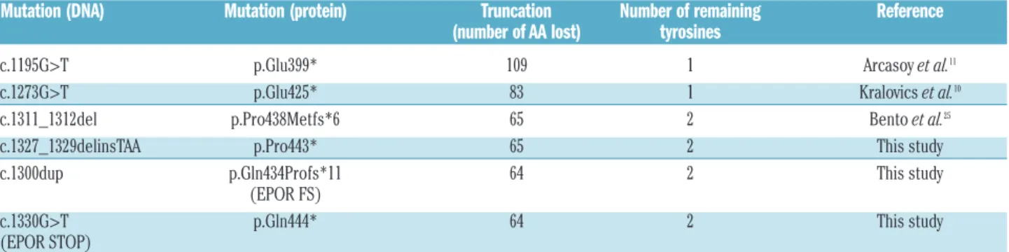
lead-Table 1. EPOR mutations investigated in this study and their functional consequences.
Mutation (DNA) Mutation (protein) Truncation Number of remaining Reference (number of AA lost) tyrosines
c.1195G>T p.Glu399* 109 1 Arcasoy et al.11
c.1273G>T p.Glu425* 83 1 Kralovics et al.10
c.1311_1312del p.Pro438Metfs*6 65 2 Bento et al.25
c.1327_1329delinsTAA p.Pro443* 65 2 This study c.1300dup p.Gln434Profs*11 64 2 This study (EPOR FS)
c.1330G>T p.Gln444* 64 2 This study (EPOR STOP)
ing to the truncation of 64 amino acids of the wild-type receptor and the loss of the six last conserved tyrosine residues (Table 1, Figure 1A,B). The mutation was found
in neutrophils and CD3+T lymphocytes as well as in
non-hematopoietic tissues such as nails and hair allowing the diagnosis of PFCP to be made. There was no familial
his-tory of polycythemia. The blood counts of the mother and two children were strictly normal. The father’s blood count was not available.
Erythroid progenitors displayed autonomous growth when cultured in the presence of serum (Figure 1C), an effect probably explained by residual erythropoietin in the
Figure 1. Study of the EPOR c.1300dup (p.Gln434Profs*11) in primary cells.(A) Electropherograms showing the EPOR c.1300dup mutation in hematopoietic cells (CD3+T-lymphocytes and neutrophils) and in non-hematopoietic tissues (nails, hair and buccal swab). (B) Scheme of EPOR wild-type and of the new frameshift EPOR
mutants (EPOR FS). Negative signaling regulators (on the left) and internalization/degradation sites (on the right) are indicated. The tyrosine (Y) number of the mature EPOR is also indicated in parentheses. (C, D) Effect of erythropoietin (EPO) concentration on erythroid colony formation in the presence (C) or absence (D) of fetal bovine serum. BFU-E colonies were counted at day 14. Each experiment was performed twice in duplicate. The results are expressed in percentages of the number of colonies at 1 U/mL of EPO. (E) Effect of EPO concentration on EPOR signaling in erythroblasts. After 7 to 10 days in culture with stem cell factor (SCF), interleukin-3 (IL3) and EPO, CD34+cells from the patient or healthy donors were cytokine-starved for 5 h then stimulated for 15 min with increasing concentrations of EPO. JAK2 and STAT5
phosphorylations were examined by western blotting. One of three independent experiments is presented and fold activation is indicated below. (F) Persistence of JAK2 and STAT5 phosphorylation in erythroblasts. After 7 to 10 days in culture with SCF, IL3 and EPO, CD34+cells from the patient or healthy donors were cytokine-starved
for 5 h prior to 15 min of stimulation with EPO 1 U/mL. Cells were then washed to remove the EPO and cultured in the absence of cytokine or serum. JAK2 and STAT5 phosphorylations were examined by western blotting in a time-dependent manner: after 5 h of starvation (-), after the EPO stimulation (+EPO) and at different times after EPO removal (10 min, 30 min, 1 h and 4 h). One out of two independent experiments is presented and fold activation is indicated below.
A B
C D
serum, as in the absence of serum, no spontaneous growth was observed, but there was greater erythropoietin hyper-sensitivity (a nearly 5-fold increase) compared to that of control cells from a healthy donor (Figure 1D).
We performed signaling experiments with primary
CD34+cells from the PFCP patient which were cultured in
vitro for 7-10 days in the presence of interleukin-3, stem
cell factor and erythropoietin. Unlike control cells, PFCP erythroid progenitors exhibited constitutive and persistent phosphorylation of JAK2 as well as constitutive activation of STAT5 with a hypersensitive erythropoietin dose-response (Figure 1E,F). This erythropoietin-induced JAK2
Figure 2. Functional study of EPOR c.1300dup (p.Gln434Profs*11) in the Ba/F3 cell line.(A) Ba/F3 cells were transduced with pMX-HA-huEPOR-IRES-GFP retrovirus to stably express the wild-type receptor (EPOR WT), a truncated mutant at position 444 (p.Gln444*, EPOR STOP) or the frameshift mutant EPOR c.1300dup (p.Gln434Profs*11, EPOR FS). (B) Proliferation was assessed 48 h after culturing Ba/F3-EPOR cells in the absence or presence of increasing doses of erythropoietin (EPO) (0.01, 0.02, 0.03, 0.05, 0.1, 0.3, and 1 U/mL) by a WST-1 proliferation assay. Dose-response curves are means expressed in percentages of maximum growth value ± SEM (n = 3 in triplicate). Two-tailed t-test, *P<0.05, **P<0.01, ***P<0.001, ****P<0.0001. (C) Effect of EPO concentration on EPOR signaling. Ba/F3 cells expressing different EPOR constructs were examined by western blotting for the presence and phosphorylation status of various signaling molecules. Cells were serum- and cytokine-starved for 5 h prior to stimulation for 15 min with increasing doses of EPO (0, 0.001, 0.01, 0.1 and 1 U/mL). Expression of β-actin was used as a loading control. One of three independent experiments is presented. (D) Phosho-STAT5/actin spontaneous phosphorylation was quantified compared to WT using Image J. Results represent the mean ± SEM (n = 3). Two-tailed t-test, *P<0.05, ***P<0.001, (E) Persistence of STAT5 phosphorylation. Ba/F3-EPOR cells were serum- and cytokine-starved for 5 h prior to 15 min of stimulation with EPO 1 U/mL. Cells were then washed to remove the EPO and cultured in the absence of cytokine or serum. STAT5 phosphorylation was examined by western blotting in a time-dependent manner: after 5 h of starvation (-), after EPO stimulation (+EPO) and at different times after EPO removal (5 min, 30 min, 1 h, 2 h and 4 h). One out of two independent experiments is presented.
A
B C
and STAT5 activation also persisted for 4 h after removal of erythropoietin from the PFCP cells, whereas it was much more transient in control cells (Figure 1F).
Here we show that EPOR c.1300dup (p.Gln434Profs*11) is a strong gain-of-function mutation, which induces major erythropoietin hypersensitivity in primary ery-throid progenitors, similar to that observed in patients with polycythemia vera, as well as constitutive and per-sistent activation of JAK2 and STAT5.
Functional analysis of EPOR c.1300dup
(p.Gln434Profs*11) in the Ba/F3 cell line
In order to study the functional impact of the new frameshift mutant, Ba/F3 cells were transduced to express different HA-tagged human EPOR: the wild-type receptor (EPOR WT), EPOR p.Gln434Profs*11 (EPOR FS), identical to the patient’s mutation or EPOR p.Gln444* (EPOR STOP) generating a truncated receptor at position 444 (Table 1, Figure 2A). EPOR FS and EPOR STOP differed by the nature of the C-terminal ten amino acid residues, namely the new sequence PALASMDTVP in EPOR FS and the natural sequence QLLRPWTLCP in EPOR STOP. These cells expressed quite similar levels of exogenous EPOR, as detected with anti-HA antibody (Figure 2C). Interestingly, EPOR FS migrated slightly above EPOR STOP, suggesting differences in post-trans-lational modification. However, the glycosylation states of EPOR WT, EPOR STOP and EPOR FS were similar after using Endo H and PGNase F (Online Supplementary
Figure S1).
MTT-like assays were performed to investigate the potential effects of EPOR STOP and EPOR FS on cell prolif-eration. None of these mutants was able to induce cytokine-independent cell growth. However, EPOR FS con-ferred a 4- to 5-fold greater erythropoietin hypersensitivity to Ba/F3 cells compared to EPOR WT. Interestingly the ery-thropoietin-induced growth of EPOR STOP Ba/F3 cells was similar to that of EPOR WT cells (Figure 2B). To confirm these results in a human setting, we transduced EPOR WT, STOP and FS in the human UT-7 cell line, which expresses endogenous EPOR and found similar results as in the Ba/F3 cell line (Online Supplementary Figure S2A).
We checked the signaling pathways and observed that EPOR FS induced constitutive phosphorylation of STAT5, AKT and ERK compared to EPOR WT and to a higher extent than EPOR STOP in Ba/F3 cells (Figure 2C) and UT-7 cells (Online Supplementary Figure S2B). Semi-quantitative analysis of spontaneous STAT5 phosphorylation showed a significant increase in EPOR FS compared to EPOR WT and EPOR STOP (Figure 2D). Moreover, we observed a similar persistent STAT5 activation in both EPOR FS and EPOR STOP Ba/F3 cells compared to EPOR WT cells (4 h, 2 h and 30 min, respectively, after erythropoietin removal) (Figure 2E).
These results show that EPOR FS confers a similar ery-thropoietin hypersensitivity to Ba/F3 and UT-7 cells as that observed in PFCP erythroid progenitors, while the truncated mutant EPOR STOP did not confer such ery-thropoietin hypersensitivity to these cells. The greater erythropoietin hypersensitivity induced by EPOR p.Gln434Profs*11 cannot, therefore, be explained by the receptor truncation itself and the loss of the two SHP-1 and SOCS3 binding sites which are responsible for a per-sistent activation, but rather by the appearance of a new C-terminal tail that confers spontaneous signaling.
Effects of the c.1300dup (p.Gln434Profs*11)
mutation on erythropoietin receptor stability, cell
sur-face expression and dimerization
To further characterize this new EPOR mutant, the cell surface expression of the wild-type receptor and both mutants was studied by flow cytometry. At similar levels of GFP (transduction of cells with IRES-GFP retroviruses), EPOR FS was significantly more abundant at the cell sur-face (more than 2-fold, P=0.0002) than EPOR WT and EPOR STOP in both Ba/F3 and UT-7 cell lines (Figure 3A and Online Supplementary Figure S3). These results were
further confirmed in Ba/F3 cells using radiolabeled 125
I-ery-thropoietin (2,412±494 receptors for EPOR FS, P=0.015, 1,018±284 receptors for EPOR STOP and 1,201±304 receptors for EPOR WT) (Figure 3b). We next investigated the stability of the receptors using treatment with the pro-tein synthesis inhibitor cycloheximide. EPOR FS was more stable than both EPOR WT and EPOR STOP (half-life of 2 h and 1 h, respectively) (Figure 3C). We also stud-ied the dimerization of human EPOR monomers by split Gaussia luciferase assay in steady-state conditions in HEK cells, in the absence of erythropoietin (Figure 3D). Close proximity between the C-terminal cytosolic domains of EPOR was increased by 4-fold with EPOR FS compared to control and EPOR STOP, as assessed by reconstitution of the split Gaussia luciferase activity (Figure 3E).
The increased stability and cell surface localization of EPOR FS may also result in a defect in the receptor inter-nalization pattern due to the loss of specific domains located in the C-terminal part of the wild-type receptor. However, EPOR WT, STOP and FS displayed the same
internalization pattern as assessed by 125I-erythropoietin
labeling experiments (Figure 4A,B). Furthermore, we noted that a dileucine motif, known to be a potential
clathrin-dependent endocytosis signal,37 was lost in the
new C-terminal tail of the EPOR FS. We assumed that this particular modification could be involved in the increased cell surface expression of EPOR FS. Thus, based on EPOR WT and EPOR STOP constructs, we generated two other mutants, EPOR WT/diL and EPOR STOP/diL, where the dileucine motif was removed (Figure 4C). However its abrogation did not modify either the erythropoietin sensi-tivity of EPOR WT or STOP Ba/F3 cells (Figure 4D) or the localization of the receptors at the cell surface (Figure 4E). Likewise, EPOR WT/diL and EPOR STOP/diL Ba/F3 cells displayed a similar signaling pattern to that of EPOR WT and EPOR STOP cells, respectively (Figure 4F).
Collectively these results show that p.Gln434Profs*11 increases EPOR stability, dimerization and localization at the cell surface without modifying internalization of the receptor.
Different mechanisms are involved in the
erythropoietin hypersensitivity phenotype in primary
erythrocytosis according to the type of EPOR mutation
We wondered whether this model could be extended to other EPOR mutations already described in PFCP. We therefore investigated the impact of the frameshift EPOR
c.1311_1312del (p.Pro438Metfs*6) mutation25 and its
designed nonsense mutant counterpart, EPOR
c.1327_1329delinsTAA (p.Pro443*) (Figure 5A) on the pro-liferation rate of Ba/F3 cells. These mutants lack the last 65 amino acids of the receptor, retaining two of the eight conserved tyrosine residues, Tyr-368 and Tyr-426 (Table 1, Figure 6A,C,D). Interestingly, EPOR p.Pro438Metfs*6 and
EPOR p.Gln434Profs*11 (EPOR FS) share the same five-amino acid terminal sequence (MDTVP). MTT-like assays showed that erythropoietin hypersensitivity was induced by EPOR p.Pro438Metfs*6, but not by EPOR p.Pro443* (Figure 5B), similarly to the results obtained with EPOR p.Gln434Profs*11 (EPOR FS) and EPOR p.Gln444* (EPOR STOP), respectively. We also studied two proximal
non-sense mutations, EPOR c.1195G>T (p.Glu399*)11 and
c.1273G>T (p.Glu425*)10(Figure 5A), which are
responsi-ble for more extensive truncations (109 and 83 amino acids, respectively) and the loss of seven of the eight cyto-plasmic tyrosine residues, retaining only Tyr-368 (Table 1, Figure 6A,B). Interestingly, these nonsense mutants, unlike EPOR p.Pro434* and p.Gln444*, were able to confer ery-thropoietin hypersensitivity to Ba/F3 cells (Figure 5C). These results show that extensive truncations, unlike shorter ones, are sufficient per se to induce the erythropoi-etin hypersensitivity phenotype. In comparison, erythro-poietin hypersensitivity induced by frameshift EPOR
mutations is due to a distinct common mechanism based on the appearance of new amino acid sequences.
Discussion
In this study, we identified a new germline heterozygous
EPOR mutation, c.1300dup (p.Gln434Profs*11) in a patient
suffering from PFCP. Like the other mutations already described in this pathology, c.1300dup is located in exon 8 of EPOR. The frameshift generates a new cytoplasmic tail of ten amino acids and a premature stop codon at position 444 leading to the loss of 64 amino acids in the C-terminal part of the receptor. Two other mutations, c.1281dup
(p.Ile428Tyrfs*17)7 and c.1288dup (p.Asp430Glyfs*15),4
leading to a similar truncation at position 444 had been previously reported but, as for most other EPOR mutants, no extensive functional study had been carried out. Here we show that EPOR p.Gln434Profs*11 is a strong
gain-of-Figure 3. Effects of c.1300dup (p.Gln434Profs*11) mutation on EPOR stability, dimerization and cell surface expression.(A) Cell-surface expression of the different EPOR was assessed by flow cytometry using PE fluorescent labeling of the extracellular HA-tag. The histogram shows the ratio of mean fluorescence intensiy (MFI) of PE-labeled cell-surface erythropoietin (EPO) on the respective MFI of GFP. Results are the mean ± SEM of seven independent experiments. (B) Cell-surface expression of the different EPOR was assessed with radiolabeled 125
I-EPO. The results are expressed in cpm normalized to EPOR WT. The number of cell-surface receptors was determined by comparison between the radioactivity of transduced Ba/F3 cells and parental UT-7 cells that express 7000 receptors. (C) EPOR stability. Ba/F3-EPOR cells were incubated with cycloheximide for different times (0 min, 15 min, 30 min, 1 h, 2 h, 4 h, 6 h) and HA expression was studied by western blotting. HA-EPOR and β-actin were quantified using Image J software. The curves represent the HA/β-actin ratios. Three independent experiments were done. (D) Schematic representation of split Gaussia princeps luciferase com-plementation assay used to test EPOR dimerization in HEK293-derived BOSC cells. (E) Dimerization of human EPOR monomers was assessed by split Gaussia luciferase assay in steady-state condi-tions, in the absence of EPO in HEK cells. Close proximity between the C-terminal cytosolic domains of EPOR is significantly promoted by EPOR FS. The results repre-sent the mean ± SEM from three inde-pendent experiments, each performed with eight biological replicates per condi-tion. For each experiment, raw values were normalized to the average of the EPOR WT condition before pooling the data of all experiments together. Unpaired two-tailed t-test with Welch correction, ****P<0.0001.
A B
C D
Figure 4. Role of diLeucine motif (diL) loss in the EPOR c.1300dup (p.Gln434Profs*11, EPOR FS) mechanism.(A, B) EPOR internalization was performed with radio -labeled 125I-erythropoietin (EPO). Cells were incubated with 125I-EPO and washed at 4°C to remove unbound ligand. An acidic wash was then performed to separate
cell surface–bound from internalized EPO. The radioactivity levels of the (A) supernatant (cell surface–bound EPO) and of (B) the cell pellet (internalized EPO) were determined. Each experiment was performed three times with similar results. (C) To study the potential role of the dileucine motif loss in the EPO hypersensitivity phenotype induced by EPOR FS, Ba/F3 cells were transduced with pMX-HA-huEPOR-IRES-GFP retrovirus to stably express EPOR WT or EPOR STOP receptor with abro-gation of the dileucine motif EPOR WT/diL and EPOR STOP/diL respectively. (D) Proliferation was assessed 48 h after culturing Ba/F3-EPOR cells in the absence or presence of increasing doses of EPO (0.01, 0.02, 0.03, 0.05, 0.1, 0.3, and 1 U/mL) by a WST-1 proliferation assay. Dose-response curves are means expressed in percentages of maximum growth value ± SEM (n = 3 in triplicate). Two-tailed t-test, *P<0.05, **P<0.01, ***P<0.001, ****P<0.0001. (E) Cell-surface expression of the different EPOR was assessed by flow cytometry using PE fluorescence labeling of the extracellular HA-tag. The histogram shows the ratio of mean fluorescence intensity (MFI) of PE-labeled cell-surface EPOR on the respective MFI of GFP. Results are the mean ± SEM of three independent experiments. (F) Effect of EPO con-centration on EPOR signaling. Ba/F3 cells expressing different EPOR constructs were examined by western blotting for the presence and phosphorylation status of various signaling molecules. Cells were serum- and cytokine-starved for 5 h prior to stimulation for 15 min with increasing doses of EPO (0, 0.001, 0.01, 0.1 and 1 U/mL). Expression of β-actin was used as a loading control. One of three independent experiments is presented and fold activation is indicated below.
A
C
D E
F
function mutant that induces major erythropoietin hyper-sensitivity in primary cells as well as in transduced Ba/F3 cells, without spontaneous growth, in accordance with the diagnosis of PCFP.
Previous reports assumed that the erythropoietin hyper-sensitivity phenotype observed in PFCP is due to the loss of negative regulatory domains located in the C-terminal part of the receptor, especially some conserved tyrosine
residues.30,31Both the magnitude and duration of EPOR
sig-naling are indeed crucial for erythropoiesis and are there-fore tightly regulated by several mechanisms induced as soon as erythropoietin binds to its cognate receptor. A classic negative feedback loop is achieved by signaling
inhibitors such as the tyrosine phosphatase SHP-138 and
the suppressor of cytokine signaling (SOCS) proteins
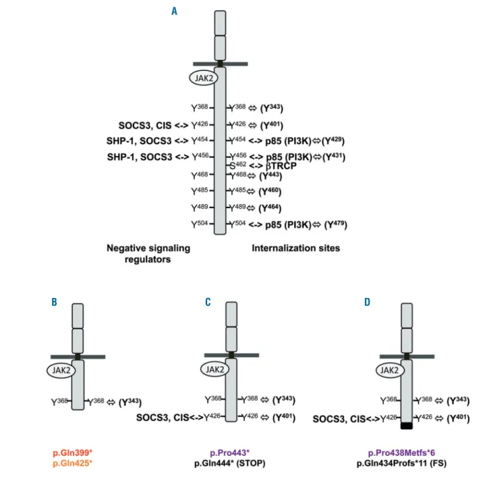
SOCS3 and CIS,39which bind the conserved Tyr-426, -454
and -456 on the cytoplasmic tail of EPOR (Figure 6A). Rapid internalization and degradation of the receptor
induced by erythropoietin binding34also contribute to the
negative regulation of EPOR signaling. Several possible cooperative and partially redundant mechanisms are involved in this process, but their relative contributions remain unclear. The C-terminal part of EPOR is degraded
at the cell surface by the proteasome.34,35This step requires
binding of the E3-ligase βTrcp to a conserved motif
Asp461-Ser462-Gly463 of EPOR (Figure 6A).36 The
ery-Figure 5. Different mechanisms are involved in the erythropoietin hypersensitivity phenotype of primary familial and congenital polycythemia depending on the type of EPOR mutation.(A) Ba/F3 cells were transduced with pMX-HA-huEPOR-IRES-GFP retrovirus to stably express different kind of EPOR mutations already described: the frameshift EPOR c.1311_1312del mutant (p.Pro438Metfs*6) or its nonsense designed counterpart EPOR c.1327_1329delinsTAA (p.Pro443*), more proximal truncations due to the nonsense mutants EPOR c.1195G>T (p.Glu399*) and c.1273G>T (p.Glu425*). (B, C) Proliferation was assessed 48 h after culturing EPOR p.Pro438Metfs*6 and p.Pro443* Ba/F3 cells (B) or EPOR p.Glu399* and p.Glu425* Ba/F3 cells (C) in the absence or presence of increasing doses of EPO (0.01, 0.02, 0.03, 0.05, 0.1, 0.3, and 1 U/mL) and compared to EPOR WT and EPOR FS growth by a WST-1 proliferation assay. Dose-response curves are means expressed in percentages of maximum growth value ± SEM (n = 3 in triplicate). Two-tailed t-test, *P<0.05, **P<0.01, ***P<0.001, ****P<0.0001.
A
thropoietin-EPOR complex is then internalized in a process dependent on Tyr-454, -456 and -504 and the p85 subunit of phosphatidylinositol 3 kinase, and targeted to
the lysosomes for degradation (Figure 6A).30,31,35The loss
of these negative signaling regulatory and degradation domains due to EPOR truncation is usually thought to
explain erythropoietin hypersensitivity in PFCP.
6,7,14,27,29-31,36,40In the present study, we studied two extensive
trun-cations, p.Glu399* and p.Glu425*, which lack seven of the eight conserved tyrosine residues (Table 1, Figure 6B) and all sequences that constitute the EPOR negative reg-ulatory domains. These mutants conferred greater ery-thropoietin hypersensitivity to Ba/F3 cells compared to EPOR WT. Thus, confirming previous reports, we showed that extensive truncations lacking all EPOR
neg-ative regulatory sites are sufficient in themselves to induce the PFCP phenotype.
Our results highlight that a different mechanism under-lies erythropoietin hypersensitivity due to frameshift
EPOR mutations. The study of EPOR p.Gln434Profs*11
(EPOR FS) and EPOR p.Pro438Metfs*6 and their designed nonsense counterparts, EPOR p.Gln444* (EPOR STOP) and EPOR p.Pro434*, respectively, allowed us to discrimi-nate between the effects due to the truncations them-selves leading to the loss of SHP-1 and SOCS3 binding sites and those due to the appearance of a new cytoplas-mic tail. In accordance with the loss of negative regulatory
sites and previous reports,9,14,27,29EPOR FS and EPOR STOP
Ba/F3 cells displayed persistent phosphorylation of STAT5 after erythropoietin removal, but only EPOR FS was able
Figure 6. Regulation of EPOR signaling and positions of the EPOR truncations.(A) Negative regulators of EPOR signaling and their binding sites in the C-terminal part of the receptor: negative signaling regulators (on the left) and internalization/degradation sites (on the right). The tyrosine (Y) number of the mature EPOR is also indicated in parentheses. (B) Proximal truncations due to EPOR p.Glu399* and p.Glu425* lack all negative regulation sites of the receptor. (C and D) More dis-tal truncations due to frameshift EPOR mutants and their designed counterparts retain the Tyr-426.
D C
B
to induce constitutive STAT5 phosphorylation and ery-thropoietin hypersensitivity. In a similar way to EPOR p.Gln434Profs*11 (EPOR FS), Ba/F3 cells expressing EPOR p.Pro438Metfs*6 displayed erythropoietin hypersensitivi-ty, unlike EPOR p.Pro443*, suggesting a common mecha-nism for the frameshift EPOR mutations in PFCP due to the presence of a new, partially common cytoplasmic tail (MDTVP). Of note EPOR p.Pro443* and p.Gln444* have never been identified in PFCP patients. Furthermore, a pre-viously described murine mutant that lacks the last 40 amino acids and has a new cytoplasmic tail, which does not include the MDTVP motif, is also responsible for ery-thropoietin hypersensitivity. This suggests that other sequences of amino acids could be involved in
erythropoi-etin hypersensitivity.41 Furthermore, another mutant,
EPOR p.Trp439*, was found in a large family with ery-throcytosis, with a mild phenotype probably associated
with incomplete penetrance of the mutation.3This finding
suggests that our in vitro cellular model may be insuffi-ciently sensitive to unmask weak functional effects or that the wild-type WTLCP motif may act as a negative regula-tor of erythropoietin sensitivity.
We also demonstrated that the increased stability and the greater localization at the cell surface of EPOR FS compared to EPOR WT and EPOR STOP were not due to the loss of internalization sites located in the cytoplasmic tail of the protein as these receptors displayed similar internalization patterns. These results suggest better addressing to the cell surface, increased stability or greater recycling of EPOR FS. Moreover, we found that EPOR FS increased the basal dimerization of the receptor, correlating with the spontaneous activation of JAK2 and downstream signaling. Two recent studies have empha-sized the role of EPOR dimerization and conformation in signal transduction. Indeed, while the EPO R150Q mutant was unable to mediate full signaling due to
decreased EPOR dimerization and JAK2 activation,42
alter-ation of EPOR conformalter-ation by diabodies was able to
finely tune the receptor signaling.43 The conformation of
EPOR is indeed crucial for its optimal activation. In the absence of ligand, inactive EPOR dimers are pre-formed
at the cell surface44 through their transmembrane
domains.45Erythropoietin binding to its receptor induces
conformational changes, such as the formation of a 120° angle between the D1 domains of the two EPOR
mole-cules46and the reorientation of the continuous rigid alpha
helix formed by the transmembrane domain and the cytosolic juxtamembrane region that contains a rigid
hydrophobic motif, composed of residues Leu278, Ile282
and Trp283.47-49It has been shown that the orientation of
this conserved motif is crucial for JAK2 activation and
JAK2-induced EPOR phosphorylation.47 Other active
EPOR conformations have been described but this partic-ular dimer orientation is the optimal one for signaling.50
As EPOR is not permissive for self-activation in terms of conformation, the appearance of a new cytoplasmic tail due to frameshift mutations might induce reorientation of EPOR transmembrane and/or juxtamembrane domains, leading to pre-activation of the EPOR-JAK2 complex at the cell surface.
To our knowledge this is the first extensive functional study of EPOR mutations in PFCP. We demonstrated here that different mechanisms, depending on the type and location of EPOR mutations, contribute to the erythropoi-etin hypersensitivity phenotype, as suggested previously. Extensive truncations lacking all negative regulatory and degradation domains are sufficient by themselves to con-fer erythropoietin hypersensitivity, whereas more distal truncations induced by frameshift mutants confer erythro-poietin hypersensitivity that depends on the appearance of a new C-terminal tail. The latter, by increasing EPOR dimerization and stability at the cell surface, cause pre-activation of EPOR and JAK2, constitutive signaling and hypersensitivity to erythropoietin similar to that occurring
in JAK2V617F-positive polycythemia vera.
Acknowledgments
We are deeply grateful to the patient involved in the study. We thank the Imaging and Cytometry Platform (PFIC) of Gustave Roussy, especially Philippe Rameau and Yann Lecluse. We also thank Gwendoline Leroy for the sequencing.
This work was supported by grants from l’Agence Nationale de la Recherche (ANR-13-JVSV1-GERMPN-01), the Laurette Fugain foundation, the GIS-Institute for rare diseases for high throughput sequencing (AO9102LS), the Association de Recherche sur la Moelle Osseuse (ARMO), the Association pour la Recherche contre le Cancer (ARC) (Fondation ARC libre 2012) and the regional PHRC AOR07014. The “Investissements d’avenir” program is funding the Labex GR-Ex (IP, WV and FV). FP was supported by Ph.D. grants from the ARC. CM was sup-ported by ANR-Blanc 2013 GERMPN. WV is a recipient of a research fellowship from IGR INSERM (contrats d’interface). TB is a Télévie PhD fellow and SNC is supported by the Ludwig Institute for Cancer, Fondation Contre le Cancer, Fondation Salus Sanguinis, FNRS-FRSM-PDR, Project ARC10/15-027 and PAI Belgian Medical Genetics Initiative Project.
References
1. Yoshimura A, Longmore G, Lodish HF. Point mutation in the exoplasmic domain of the erythropoietin receptor resulting in hormone-independent activation and tumorigenicity. Nature. 1990;348(6302): 647-649.
2. Juvonen E, Ikkala E, Fyhrquist F, Ruutu T. Autosomal dominant erythrocytosis caused by increased sensitivity to erythro-poietin. Blood. 1991;78(11):3066-3069. 3. de la Chapelle A, Traskelin AL, Juvonen E.
Truncated erythropoietin receptor causes dominantly inherited benign human
ery-throcytosis. Proc Natl Acad Sci USA. 1993;90(10):4495-4499.
4. Sokol L, Luhovy M, Guan Y, Prchal JF, Semenza GL, Prchal JT. Primary familial polycythemia: a frameshift mutation in the erythropoietin receptor gene and increased sensitivity of erythroid progenitors to ery-thropoietin. Blood. 1995;86(1):15-22. 5. Arcasoy MO, Degar BA, Harris KW, Forget
BG. Familial erythrocytosis associated with a short deletion in the erythropoietin recep-tor gene. Blood. 1997;89(12):4628-4635. 6. Furukawa T, Narita M, Sakaue M, et al.
Primary familial polycythaemia associated with a novel point mutation in the
erythro-poietin receptor. Br J Haematol. 1997;99(1): 222-227.
7. Kralovics R, Indrak K, Stopka T, Berman BW, Prchal JF, Prchal JT. Two new EPO receptor mutations: truncated EPO recep-tors are most frequently associated with primary familial and congenital poly-cythemias. Blood. 1997;90(5):2057-2061. 8. Kralovics R, Sokol L, Prchal JT. Absence of
polycythemia in a child with a unique ery-thropoietin receptor mutation in a family with autosomal dominant primary poly-cythemia. J Clin Invest. 1998;102(1):124-129. 9. Watowich SS, Xie X, Klingmuller U, et al. Erythropoietin receptor mutations
associat-hypersensitivity to erythropoietin in the heterozygous state. Blood. 1999;94(7): 2530-2532.
10. Kralovics R, Prchal JT. Genetic heterogene-ity of primary familial and congenital poly-cythemia. Am J Hematol. 2001;68(2):115-121.
11. Arcasoy MO, Karayal AF, Segal HM, Sinning JG, Forget BG. A novel mutation in the erythropoietin receptor gene is associ-ated with familial erythrocytosis. Blood. 2002;99(8):3066-3069.
12. Rives S, Pahl HL, Florensa L, et al. Molecular genetic analyses in familial and sporadic congenital primary erythrocytosis. Haematologica. 2007;92(5):674-677. 13. Al-Sheikh M, Mazurier E, Gardie B, et al. A
study of 36 unrelated cases with pure ery-throcytosis revealed three new mutations in the erythropoietin receptor gene. Haematologica. 2008;93(7):1072-1075. 14. Perrotta S, Cucciolla V, Ferraro M, et al.
EPO receptor gain-of-function causes hereditary polycythemia, alters CD34 cell differentiation and increases circulating endothelial precursors. PLoS One. 2010;5(8):e12015.
15. James C, Ugo V, Le Couedic JP, et al. A unique clonal JAK2 mutation leading to constitutive signalling causes poly-cythaemia vera. Nature. 2005;434(7037): 1144-1148.
16. Scott LM, Tong W, Levine RL, et al. JAK2 exon 12 mutations in polycythemia vera and idiopathic erythrocytosis. N Engl J Med. 2007;356(5):459-468.
17. D'Andrea AD, Fasman GD, Lodish HF. Erythropoietin receptor and interleukin-2 receptor beta chain: a new receptor family. Cell. 1989;58(6):1023-1024.
18. Huang LJ, Constantinescu SN, Lodish HF. The N-terminal domain of Janus kinase 2 is required for Golgi processing and cell sur-face expression of erythropoietin receptor. Mol Cell. 2001;8(6):1327-1338.
19. Witthuhn BA, Quelle FW, Silvennoinen O, et al. JAK2 associates with the erythropoi-etin receptor and is tyrosine phosphorylat-ed and activatphosphorylat-ed following stimulation with erythropoietin. Cell. 1993;74(2):227-236.
20. Socolovsky M, Nam H, Fleming MD, Haase VH, Brugnara C, Lodish HF. Ineffective ery-thropoiesis in Stat5a(-/-)5b(-/-) mice due to decreased survival of early erythroblasts. Blood. 2001;98(12):3261-3273.
21. Bouscary D, Pene F, Claessens YE, et al. Critical role for PI 3-kinase in the control of erythropoietin-induced erythroid progeni-tor proliferation. Blood. 2003;101(9):3436-3443.
22. Wu H, Liu X, Jaenisch R, Lodish HF. Generation of committed erythroid BFU-E and CFU-E progenitors does not require erythropoietin or the erythropoietin recep-tor. Cell. 1995;83(1):59-67.
23. Petersen KB, Hokland P, Petersen GB, Nyvold CG. Erythropoietin receptor defect: a cause of primary polycythaemia. Br J
24. O'Rourke K, Fairbairn DJ, Jackson KA, Morris KL, Tey SK, Kennedy GA. A novel mutation of the erythropoietin receptor gene associated with primary familial and congenital polycythaemia. Int J Hematol. 2011;93(4):542-544.
25. Bento C, Almeida H, Maia TM, et al. Molecular study of congenital erythrocyto-sis in 70 unrelated patients revealed a potential causal mutation in less than half of the cases (where is/are the missing gene(s)?). Eur J Haematol. 2013;91(4):361-368.
26. Bento C, Percy MJ, Gardie B, et al. Genetic basis of congenital erythrocytosis: muta-tion update and online databases. Hum Mutat. 2014;35(1):15-26.
27. Gross M, Ben-Califa N, McMullin MF, et al. Polycythaemia-inducing mutations in the erythropoietin receptor (EPOR): mecha-nism and function as elucidated by epider-mal growth factor receptor-EPOR chimeras. Br J Haematol. 2014;165(4):519-528.
28. Chauveau A, Luque Paz D, Lecucq L, et al. A new point mutation in EPOR inducing a short deletion in congenital erythrocytosis. Br J Haematol. 2016;172(3):475-477. 29. Iacobucci I, Li Y, Roberts KG, et al.
Truncating erythropoietin receptor rearrangements in acute lymphoblastic leukemia. Cancer Cell. 2016;29(2):186-200. 30. Sulahian R, Cleaver O, Huang LJ. Ligand-induced EpoR internalization is mediated by JAK2 and p85 and is impaired by muta-tions responsible for primary familial and congenital polycythemia. Blood. 2009;113(21):5287-5297.
31. Bulut GB, Sulahian R, Yao H, Huang LJ. Cbl ubiquitination of p85 is essential for Epo-induced EpoR endocytosis. Blood. 2013;122(24):3964-3972.
32. Le Couedic JP, Mitjavila MT, Villeval JL, et al. Missense mutation of the erythropoietin receptor is a rare event in human erythroid malignancies. Blood. 1996;87(4):1502-1511. 33. Remy I, Michnick SW. A highly sensitive protein-protein interaction assay based on Gaussia luciferase. Nat Methods. 2006;3(12):977-979.
34. Verdier F, Walrafen P, Hubert N, et al. Proteasomes regulate the duration of ery-thropoietin receptor activation by control-ling down-regulation of cell surface recep-tors. J Biol Chem. 2000;275(24):18375-18381.
35. Walrafen P, Verdier F, Kadri Z, Chretien S, Lacombe C, Mayeux P. Both proteasomes and lysosomes degrade the activated ery-thropoietin receptor. Blood. 2005;105(2): 600-608.
36. Meyer L, Deau B, Forejtnikova H, et al. beta-Trcp mediates ubiquitination and degradation of the erythropoietin receptor and controls cell proliferation. Blood. 2007;109(12):5215-5222.
37. Letourneur F, Klausner RD. A novel di-leucine motif and a tyrosine-based motif independently mediate lysosomal targeting
1992;69(7):1143-1157.
38. Jiao H, Berrada K, Yang W, Tabrizi M, Platanias LC, Yi T. Direct association with and dephosphorylation of Jak2 kinase by the SH2-domain-containing protein tyro-sine phosphatase SHP-1. Mol Cell Biol. 1996;16(12):6985-6992.
39. Sasaki A, Yasukawa H, Shouda T, Kitamura T, Dikic I, Yoshimura A. CIS3/SOCS-3 sup-presses erythropoietin (EPO) signaling by binding the EPO receptor and JAK2. J Biol Chem. 2000;275(38):29338-29347. 40. Arcasoy MO, Harris KW, Forget BG. A
human erythropoietin receptor gene mutant causing familial erythrocytosis is associated with deregulation of the rates of Jak2 and Stat5 inactivation. Exp Hematol. 1999;27(1):63-74.
41. D'Andrea AD, Yoshimura A, Youssoufian H, Zon LI, Koo JW, Lodish HF. The cyto-plasmic region of the erythropoietin recep-tor contains nonoverlapping positive and negative growth-regulatory domains. Mol Cell Biol. 1991;11(4):1980-1987.
42. Kim AR, Ulirsch JC, Wilmes S, et al. Functional selectivity in cytokine signaling revealed through a pathogenic EPO muta-tion. Cell. 2017;168(6):1053-1064 e1015. 43. Moraga I, Wernig G, Wilmes S, et al.
Tuning cytokine receptor signaling by re-orienting dimer geometry with surrogate ligands. Cell. 2015;160(6):1196-1208. 44. Livnah O, Stura EA, Middleton SA,
Johnson DL, Jolliffe LK, Wilson IA. Crystallographic evidence for preformed dimers of erythropoietin receptor before ligand activation. Science. 1999;283 (5404):987-990.
45. Constantinescu SN, Keren T, Socolovsky M, Nam H, Henis YI, Lodish HF. Ligand-independent oligomerization of cell-surface erythropoietin receptor is mediated by the transmembrane domain. Proc Natl Acad Sci USA. 2001;98(8):4379-4384.
46. Syed RS, Reid SW, Li C, et al. Efficiency of signalling through cytokine receptors depends critically on receptor orientation. Nature. 1998;395(6701):511-516.
47. Constantinescu SN, Huang LJ, Nam H, Lodish HF. The erythropoietin receptor cytosolic juxtamembrane domain contains an essential, precisely oriented, hydropho-bic motif. Mol Cell. 2001;7(2):377-385. 48. Lu X, Gross AW, Lodish HF. Active
confor-mation of the erythropoietin receptor: ran-dom and cysteine-scanning mutagenesis of the extracellular juxtamembrane and trans-membrane domains. J Biol Chem. 2006;281 (11):7002-7011.
49. Li Q, Wong YL, Huang Q, Kang C. Structural insight into the transmembrane domain and the juxtamembrane region of the erythropoietin receptor in micelles. Biophys J. 2014;107(10):2325-2336. 50. Seubert N, Royer Y, Staerk J, et al. Active
and inactive orientations of the transmem-brane and cytosolic domains of the ery-thropoietin receptor dimer. Mol Cell. 2003;12(5):1239-1250.