HAL Id: hal-03016985
https://hal.uca.fr/hal-03016985
Submitted on 20 Nov 2020
HAL is a multi-disciplinary open access
archive for the deposit and dissemination of sci-entific research documents, whether they are pub-lished or not. The documents may come from teaching and research institutions in France or abroad, or from public or private research centers.
L’archive ouverte pluridisciplinaire HAL, est destinée au dépôt et à la diffusion de documents scientifiques de niveau recherche, publiés ou non, émanant des établissements d’enseignement et de recherche français ou étrangers, des laboratoires publics ou privés.
Distributed under a Creative Commons Attribution| 4.0 International License
Reliability study under thermal and photonic stresses of
sulforhodamine B (SRB) confined in layered double
hydroxide (LDH)
Paul Legentil, Fabrice Leroux, Sandrine Therias, Damien Boyer, François
Réveret, Geneviève Chadeyron
To cite this version:
Paul Legentil, Fabrice Leroux, Sandrine Therias, Damien Boyer, François Réveret, et al.. Reliability study under thermal and photonic stresses of sulforhodamine B (SRB) confined in layered double hydroxide (LDH). Applied Clay Science, Elsevier, 2020, pp.105922. �10.1016/j.clay.2020.105922�. �hal-03016985�
1
Reliability study under thermal and photonic stresses of sulforhodamine B
1(SRB) confined in layered double hydroxide (LDH).
2Paul Legentil1, Fabrice. Leroux1*, Sandrine Therias1, Damien Boyer1, François Reveret2,Geneviève 3
Chadeyron1* 4
1
Université Clermont Auvergne, CNRS, SIGMA Clermont, ICCF, F-63000 Clermont–Ferrand,
5
France.
6
2
Université Clermont Auvergne, CNRS, SIGMA Clermont, Institut Pascal, F-63000 Clermont–
7
Ferrand, France.
8
*e-mail genevieve.chadeyron@sigma-clermont.fr, fabrice.leroux@uca.fr
9
ABSTRACT
10When sulforhodamine B (SRB) is entrapped in a tightly packed hybrid material composed of 11
dodecylsulfate anions interleaved in layered double hydroxide, the organic dye is considered as a 12
possible red-emitting phosphor for white light-emitting diodes (WLEDs). To confirm such promising 13
potential, a reliability study is here undertaken and the materials and their associated silicone films 14
are subjected to different thermal and photonic stresses. Optical properties, photoluminescence 15
quantum yields and emission spectra are recorded after photo-aging studies. Interestingly, the 16
composite silicone film is found to be completely stable under blue LED excitation, while in the 17
absence of the hybrid LDH the emission of SRB decays rapidly, thus underlining the protective role of 18
the LDH hybrid cargo. At this stage, these results confirm the potential offered by a system consisting 19
of such an emitter film and a blue LED. This system also opens up new possibilities for interesting 20
organic dyes that are sensitive to photonic and/or thermal stresses. 21 22 23 24 25 26 27 28 29 30 31 32 33
2
1. Introduction
34
In lighting applications, white light-emitting diodes (WLED) are now emerging very strongly and 35
constitute a reference in solid-state lighting sources because of their excellent properties, such as 36
high light efficiency, energy-saving properties, long lifetime and absence of toxic mercury or other 37
heavy metals. A commercial WLED consists of a blue chip, emitting between 450 and 480 nm, 38
combined with a yellow-emitting phosphor (Nair et al., 2020), YAG:Ce3+ (yttrium aluminium garnet 39
doped with Ce3+ cations), and a red-emitting phosphor is commonly added to improve the 40
colorimetric characteristics of light (Xia et al., 2016). A recent study concerning a well-known organic 41
dye, sulforhodamine B (SRB), underlined its interest as a red phosphor and its potential under 42
commercial blue LED excitation when highly dispersed in a host structure, layered double hydroxide 43
(LDH) (Legentil et al., 2020). Indeed, the inorganic LDH vessel intercalated with surfactant molecules 44
(dodecylsulfate (DS)) is found to display a suitable arrangement for SRB to be well ensconced, 45
avoiding intermolecular interactions deleterious for the red emission as well as providing an affinity 46
with silicone in the preparation of homogeneous films. The properties of an optimized system 47
composed of alternating a blue chip, a YAG: Ce3+/silicone composite film and a LDH-DS-SRB/silicone 48
composite film are satisfactorily met, with a correlated color temperature (T(K)) associated with a 49
color rendering index (CRI) suitable for a commercial WLED application. 50
LEDs are known to have an operating temperature of up to 100 or 120°C, and hence phosphors 51
must have the ability to sustain such harsh conditions without any degradation 52
Several studies have been undertaken on inorganic phosphors. Da Lago et al.(Dal Lago et al., 53
2012) published one of the first reliability studies concerning a commercial YAG: Ce phosphor under 54
thermal stresses (between 85 and 145 °C) for remote-phosphor technology. Thus, it was 55
demonstrated that the heat generated can significantly limit the lifetime as well as the performance 56
of the phosphor. We can also mention the works of Shao et al (Shao et al., 2014), which have shown 57
the modification of the color coordinates and the spectral shift of BaSiO4: Eu2+ emission (a green-58
emitting phosphor under blue LED excitation) with respect to temperature. In order to provide better 59
thermal stability, they suggested substituting the Ba sites by Sr atoms in this phosphor, and in this 60
way they succeeded in lowering thermal quenching. Since then, other works have reported the 61
limited stability of phosphors formulated with rare earth elements under thermal stresses (Yazdan 62
Mehr et al., 2014; Zhou et al., 2014). 63
However, the reliability of organic phosphors has scarcely been reported so far. Indeed, the use 64
of organic dyes for WLED applications is appealing in terms of their luminescence properties but the 65
stability studies are only superficial (Boonsin et al., 2015; Kajjam et al., 2018). As mentioned before, 66
to further address a practical application in commercial lighting devices, a major specification 67
required of the fluorophore and its associated system is to present long-term stability with respect to 68
various types of stress in real operating conditions. Several published works mentioned the use of 69
fluorophore with a potential application for replacing traditional phosphors with rare earth elements 70
in commercial WLED, such as fluorescein, pyranine, rhodamine or triphenylamine derivatives for 71
instance (Das and Manam, 2018; Legentil et al., 2019; Nyalosaso et al., 2019; Zhang et al., 2008). 72
However, the authors do not usually pay attention to the reliability of the dyes in stressful 73
conditions. 74
Traditionally, LDH materials find applications as precursors in catalysis and as scavengers in 75
environmental science, but are also finding renewed interest in the domain of energy (Wu et al., 76
2018; Yin et al., 2019) as well as in biology as vessels for drug delivery (Conterosito et al., 2016; 77
Lonkar et al., 2013; Yan et al., 2014). Their general chemical formula is as follows 78
3 [M(II)1−xM(III)x(OH)2]x+[An−]x/n·mH2O where M(II) is a divalent cation such as Zn2+, Mg2+, Fe2+, etc… and 79
M(III) a trivalent cation such as Al3+, Fe3+, etc. and An− is an interleaved anion. The anisotropic two-80
dimensional structure is built from edge-sharing octahedral. Compared to brucite Mg(OH)2, the 81
partial substitution of divalent cations by trivalent cations implies for LDH sheets a net positive 82
charge. To ensure electroneutrality, anions of interest, here the dye molecules together with the 83
surfactant molecules, are located in the interlayer space of the LDH with the presence of water 84
molecules. 85
Importantly, organic dyes are known to be sensitive to different stresses (thermal, photonic…) 86
(Lamouche et al., 1999; Sultana, 2018). To overcome such issues, an elegant solution consists in 87
immobilizing the SRB molecules in an inorganic host lattice like layered double hydroxide (LDH) as 88
previously reported (Legentil et al., 2020). Pioneer research has underlined the benefit of confining 89
photoactive species to a restricted space such as a two-dimensional host in terms of 1) their strong 90
adsorption through host-guest interaction to avoid possible migration and aggregation within a 91
polymer, and 2) their dispersion with the presence of co-adsorbates to avoid as far as possible guest-92
guest interactions (Ogawa and Kuroda, 1995). However, a reliability study of such a hybrid organic 93
dye/LDH host matrix for LED applications must be conducted before considering any practical 94
application; this aspect has never been published to the best of our knowledge. 95
In the following, a reliability study of the SRB dye in the presence of thermal and photonic 96
stresses in different conditions when placed in a LED set-up is presented in order to confirm the 97
promising features of the synthetized hybrid LDH-SRB. To address this issue, comparative studies 98
were performed, first on the hybrid LDH/SRB powder and then on a composite film by dispersing the 99
powder in silicone polymer. In the first case only temperature effects were studied. Then, both 100
thermal and photonic stresses were concomitantly applied and recorded for silicone films. Behaviour 101
in accelerated photo-aging was also observed. Photoluminescence properties such as emission 102
spectra and photoluminescence quantum yields were scrutinized during all the different tests to 103
better understand the mechanisms involved in the loss of optical properties. 104
2. Experimental section
1052.1. Materials
106
Sulforhodamine B sodium salt C27H30N2NaO7S2, sodium dodecylsulfate CH3(CH2)11OSO3Na and 107
terephthalic acid C8H6O4 were purchased from Sigma-Aldrich, and YAG:Ce3+ from Phosphortech. 108
Zn(NO3)2 .6H2O (purity 99.9+%), Al(NO3 )3 .9H2O (purity 99.9+%) and NaOH (97%) were obtained from 109
Sigma Aldrich. The two-component silicone elastomer, Bluesil RTV 141 part A and part B, was 110
supplied by Elkem. 111
2.2. Synthesis procedure for LDH hybrid materials using the coprecipitation method
112
The sulforhodamine B (SRB)-dodecylsulfate (DS) LDH phase, Zn2Al–DS-SRB, called LDH-DS-SRB, was 113
prepared using the coprecipitation method. The synthesis of [Zn2Al1(OH)6]+[DS-]1-x[SRB−]x·mH2O was 114
performed using 50 mL of sulforhodamine B and dodecylsulfate aqueous solution (with de-ionized 115
water). 50 mL of an aqueous solution of Zn2+ (3.2 mmol) and Al+3 (1.6 mmol) was added dropwise 116
over a period of 3 hours under magnetic stirring. To synthesize the [Zn2Al1(OH)6]+[DS -117
]0.9995[SRB−]0.0005·mH2O sample, 0.8x10⁻³ mmol and 1.5992 mmol were used for SRB salt and sodium 118
dodecylsulfate, respectively. This formulation was determined in a previous article as that leading to 119
the best optical performance under commercial blue LED and UV excitation (Legentil et al., 2020). 120
4 The pH was maintained at 8.5 by adding 0.25 M NaOH during the synthesis process. 121
Coprecipitation was performed under nitrogen at 20 °C. The mixture was centrifuged at 5,000 rpm 122
for 5 minutes and the slurry on the bottom of the flask was then washed several times with de-123
ionized water until a clear and transparent supernatant was obtained. A paste was recovered and 124
dried overnight at room temperature to obtain the LDH-DS-SRB powder. 125
The post-synthesis hydrothermal treatment was performed by dispersing the paste, obtained 126
before the drying step, in 20 ml of de-ionized water in a sealed container at 110 °C under autogenous 127
pressure during 48 hours. The slurry was then centrifuged (5 minutes at 5,000 rpm) and the paste 128
was dried overnight at room temperature to obtain the LDH-DS-SRB-TH powder. 129
Similarly, the terephthalate LDH phase, Zn2Al–TP-SRB, was prepared by following the method 130
previously described for LDH-DS-SRB by replacing DS by terephthalic acid (TP). However, TP being 131
dianionic, the input of TP was half the molar concentration compared to DS. 132
2.3. Elaboration of Silicone/HDL-DS-SRB composite films
133
The LDH-DS-SRB powder was used to produce the silicone/hybrid composite material. A loading 134
rate of 40 wt. % was chosen (beyond this loading rate, it was difficult to obtain a homogeneous 135
distribution of the powder in the polymer matrix, as shown in figure S1). The two-component silicone 136
elastomer (silicone Bluesil-RTV 141 A&B) was composed of a viscous liquid, called part A, cured by a 137
polyaddition reaction with a catalyser, part B. 138
The silicone/hybrid composite material was manufactured by mixing the LDH powder with part 139
A of the silicone elastomer using a mechanical mixer (“Thinky Mixer”) for 10 min at 1200 rpm. Then 140
the obtained mixture was processed through a three-roll Exakt80E (spacing of 30 m between the 141
first two rolls and 50 m between the last two rolls) to achieve a better dispersion of the LDH 142
platelets by shearing and to obtain a homogeneous hybrid component. Part B was added at 10phr 143
and further homogenised using the mechanical mixer for 10 min at 1200 rpm. 144
The silicone/hybrid composite film (Si-LDH) was prepared by casting onto a Teflon surface using 145
an Elcometer 4340 automatic film applicator. The knife blade height was set at 200 µm and the 146
casting speed was 30 mm/s. This two-component silicone film was cured at 80 °C for 2 hours. 147
A reference film composed of SRB powder dispersed in the silicone, called Si-SRB, was also 148
performed by dispersing 100 μL of an SRB solution in ethylene glycol (10 g.L-1) in 4.45 g of part A of 149
the silicone elastomer using the “Thinky Mixer” mechanical mixer for 10 min at 1200 rpm. Then 0.45 150
g of part B was added and homogenised with the mechanical mixer for 10 min at 1200 rpm. The film 151
was prepared by casting onto a Teflon surface in the same way as for Si-LDH mentioned above. 152
Film thickness was measured using an Elcometer 456 coating thickness gauge. 153
2.4. Characterisation
154
2.4.1. X-ray diffraction
155
LDH powders were characterised by X-Ray Diffraction; the XRD patterns were recorded with a 156
Philips X-Pert Pro diffractometer operating with Cu-Kα radiation (λ = 1.5418 Å). The data were 157
collected in a 2θ range between 5° and 70° with a step size of 1°/min. 158
5
2.4.2. Thermogravimetric analysis
159
Thermogravimetric (TG) analyses were performed on a Setaram TGA 92 instrument with a linear 160
heating rate of 5 °C.min-1 under air. 161
2.4.3. UV-Visible absorption
162
The UV–visible absorption spectra of the samples were recorded in a wavelength range of 200 163
to 800 nm with a UV–vis spectrophotometer (SP-3000 Plus) equipped with an integrating sphere and 164
UV-Probe software. 165
2.4.4. Luminescence
166
Quantum yield efficiencies and emission spectra were measured using a C9920−02G PL-QY 167
integrating sphere measurement system from Hamamatsu Photonics. The setup consisted of a 150 W 168
monochromatized Xe lamp, an integrating sphere (Spectralon coating, ⌀= 3.3 in.) and a high-169
sensitivity CCD camera. 170
2.4.5. Emission stability of films under LED irradiation
171
A photo-aging study was performed in a SEPAP 12/24 unit. This system was designed to achieve 172
accelerated artificial weathering conditions related to natural ageing. The composite films were 173
placed on a rotating carousel positioned in the centre, and four polychromatic mercury lamps (400 174
W) with wavelengths higher than 300 nm (90 W.m-2) were installed at the corners of the chamber. 175
The temperature was set at 60 °C. 176
Reliability studies were carried out using a home-made setup consisting of a power-controlled blue 177
LED emitting at 460 nm as the excitation source and a HR4000 high resolution spectrometer (Ocean 178
Optics) as the PL analyser. The samples were positioned on a heating element whose temperature 179
was adjusted to 80°C. The emission spectra of the composite films were acquired every 20 min for 24 180
hours. Their area was integrated to obtain the total emission intensity. LED power was measured 181
using a Scientech Model Mentor MA 10 with a MC2501 calorimetric head unit (25.4 mm aperture). 182
The measurement was performed by centring the head unit over the LED source and measuring the 183
power of the LED light emitted through the aperture. The blue LED power was 183 mW. The LED 184
power density can be expressed in W/m2 and was calculated by LED power (in watt) per unit surface 185
of the sample (0.25 cm2). The power density of the blue LED was 9300 W/m2; for our experiments, a 186
filter was used in order to reduce this value to 2480 W/m2. 187
3. Results and discussion
1883.1. Hydrothermal post-treatment
189
3.1.1. Structural effect
190
X-ray diffractogram patterns of both LDH-DS-SRB and LDH-DS-SRB-HT powders before and after 191
hydrothermal treatment respectively are plotted in Fig. 1. The two patterns closely overlap, and the 192
associated diffraction peaks are located at similar angles in 2θ. The observed interlamellar space is 193
2.58 nm for both powders. However, thinner diffraction peaks are observed for LDH-DS-SRB-HT than 194
for LDH-DS-SRB powder, in particular the peaks with l Miller peak indices, underlining the fact that 195
the hydrothermal treatment induced more ordered platelet stacking. 196
6 0 5 10 15 20 25 30 35 40 45 50 55 60 65 70 (009 ) (006 ) 2 (°) LDH-DS-SRB-HT LDH-DS-SRB I (a.u.) (003 ) 197
Figure 1. X-ray patterns of LDH-DS-SRB and LDH-DS-SRB-HT powders and on the right an idealized 198
scheme of the LDH-DS-SRB. 199
200
The average size of crystallites can be calculated using the Scherrer equation L = kλ/(B.cosθ) 201
where B is the FWHM (Full Width at Half Maximum) of diffraction peak (003), λ is the Cu Kα radiation 202
wavelength in nm, θ is the diffraction angle of (003) in radian et k is the shape factor (k = 0,9) (Cullity, 203
1978). The sizes calculated for LDH-DS-SRB and LDH-DS-SRB-HT are 27.2 nm and 42.3 nm 204
respectively, these values corresponding roughly to 11 and 16 structurally coherent stacked layers, 205
thus confirming a better crystallisation in the stacking direction for LDH-DS-SRB-HT. 206
As shown in a previous paper (Legentil et al., 2020) and illustrated on the scheme of the 207
Fig.1(right), the sulfonate groups are in the vicinity of the hydroxyl-covered LDH layers, thus leaving 208
the benzene and xanthene rings close to the surfactant alkyl chains. The relative amount of SRB per 209
DS is very small, SRB is highly diluted in the LDH-DS environment. 210
3.1.2. Optical properties
211
Internal photoluminescence quantum yields (PL QYint) were recorded from the samples 212
before and after hydrothermal treatment to know whether a change in the ordered stacking may 213
affect the optical response of the interleaved dye molecules (Table 1). Rather counter-intuitively, 214
LDH-DS-SRB-HT powder exhibits lower PL QYint than LDH-DS-SRB. Even if the hydrothermal treatment 215
leaves intact the confinement of the SRB molecules inside the host structure (similar basal spacing is 216
observed), a significant decrease in the luminescence properties is observed for the three excitation 217
wavelengths of interest (570 nm is the excitation wavelength leading to the best PL QYab and int, 218
(Legentil et al., 2020) (365 nm and 480 nm wavelengths correspond to UV and commercial blue 219
LED, respectively). 220
Table 1. PL QYint of LDH-DS-SRB and LDH-DS-SRB-HT powder for three excitation wavelengths (570 221
nm, 480 nm and 365 nm). Three wavelengths are of interest: the 365 nm and 480 nm wavelengths 222
correspond to UV and commercial blue LED respectively, while 570 nm is the excitation wavelength 223
leading to the best PL QYab. 224 Excitation wavelength (nm)(((nm)nm) LDH-DS-SRB LDH-DS-SRB-TH PL QYint (%) 570 nm 73.1 ± 3.5 52.6 ± 2.5 480 nm 68.9 ± 3.5 42.8 ± 2.1 365 nm 73.1 ± 3.7 39.0 ± 2.0
7 We can note that the PL QYab recorded for these samples before and after hydrothermal 225
treatment (which corresponds to the ratio between the number of photons emitted by the phosphor 226
and the number of photons emitted by the excitation source) leads to the same conclusion (Table 227
S2). 228
To better understand the modification undergone by the dye molecule, the emission spectra 229
under blue excitation (480 nm) were recorded for both LDH-DS-SRB and LDH-DS-SRB-HT powders 230
(Fig. 2). The emission spectra being similar for the other two excitation wavelengths, they are not 231
presented here. The maximum intensity of the emission band is shifted towards higher wavelengths 232
(from 608 to 614 nm) after the hydrothermal treatment, thus indicating a modification of the 233
environment for the SRB molecules. In detail, the intensity ratio between the main emission peak 234
and the associated shoulder is also modified, with the values of Ishoulder1/ Ipeak1 = 0.58 and Ishoulder2/ Ipeak2 235
= 0.67 before and after the hydrothermal treatment, respectively. Since the main peak is attributed 236
to SRB monomer molecules and the shoulder peak to SRB aggregates (dimer and trimer) (Ray and 237
Nakahara, 2002; Yan et al., 2009), the hydrothermal treatment leads to a greater aggregation of the 238
SRB molecules into the LDH-DS hybrid material. 239 550 600 650 700 750 800 0 100 200 300 400 500 600 700 800 Ishoulder2 Ishoulder1 Ipeak2 614 nm I ( a.u .) Wavelength(nm) LDH-DS-SRB LDH-DS-SRB-HT 608 nm Ipeak1 240
Figure 2. Emission spectra of LDH-DS-SRB and LDH-DS-SRB-HT for λexc = 480 nm 241
From these observations, the LDH-DS-SRB sample without hydrothermal treatment is selected in the 242
following. 243
3.2. Effect of thermal stress
244
3.2.1. Powder LDS-DS-SRB
245 246
Thermogravimetric analyses (TGA) 247
Thermogravimetric analysis shows that the SRB molecule is stable up to about 220 °C (Fig. 3a). 248
Below 100 °C, the mass loss can be attributed to the departure of the weakly-adsorbed water 249
8 molecules (weight loss n°1 of 6,8% around 80-100 °C). After 220 °C, decomposition of the SRB 250
molecules (weight loss n°2, 3 and 4) occurs until the final mass loss observed at 500 °C. 251 252 0 100 200 300 400 500 600 700 800 35 40 45 50 55 60 65 70 75 80 85 90 95 100 105 Temperature (°C) Weight loss ( %) -2 -1 0 1 2 3 4 5 6 7 8 9 3 4 2 Heat flow ( W/g) 1 a) 253 0 100 200 300 400 500 600 700 800 900 35 40 45 50 55 60 65 70 75 80 85 90 95 100 105 5 4 3 1 Temperature (°C) Weight loss ( %) 2 b) -1.0 -0.8 -0.6 -0.4 -0.2 0.0 0.2 0.4 0.6 0.8 1.0 Heat F low( W/g) 254
Figure 3. Thermogravimetric analysis and heat flow of a) SRB (2 °C/min, 25 °C- 800 °C, under air) and
255
b) LDH-DS-SRB powder (2 °C/min, 25 °C-1000 °C, under air). 256
For the hybrid LDH-DS-SRB material, several mass losses are observed. After the water 257
molecules that are weakly bound to the surface of the HDL platelets have been removed (weight loss 258
1), deshydroxylation of the LDH layers occurs from 180 up to 300 °C, concomitantly with the 259
decomposition of the interleaved organic molecules (weight losses 2 and 3). This is consistent with 260
DS decomposition, well documented in the literature (Degoutin et al., 2008; El-kharrag et al., 2011). 261
The associated mass loss starts at 200 °C and continues up to 400 °C. The mass loss observed at 500 262
°C is attributed to the final decomposition of the organic residue, while the mass loss at around 740 263
°C is assigned to the decomposition of ZnSO4 formed during the thermal treatment with the collapse 264
of the LDH layers reacting with the functionalized head of the surfactant (as well as possibly SRB). 265
Thermal stress in oven (120 °C) 266
The thermal stability study was completed in static conditions by placing the LDH-DS-SRB in an 267
oven at 120 °C for 24 hours. X-ray diffraction patterns were recorded after 5 and 24 hours (Fig. 4). 268
9
0 5 10 15 20 25 30 35 40 45 50 55 60 65 70
Stacked layers loss
I ( a.u .) 2 (°) LDH-DS-SRB LDH-DS-SRB 5h@120°C LDH-DS-SRB 24h@120°C
Lamellar structure conservation
269
Figure 4. X-ray diffraction patterns of LDH-DS-SRB powder before after thermal treatment (5 and 24
270
hours) 271
After both exposure times at 120 °C, the LDH-DS-SRB powder exhibits a similar XRD pattern. As 272
the peak at 61 ° in 2θ assigned to the diffraction peak (110) merged with (113) is still observed, this is 273
indicative that the LDH layer structure is maintained. However, the harmonic peaks (00l), visible at 274
angles lower than 25 ° (2θ) attributed to the strongly pronounced stacking for LDH-DS-SRB disappear 275
after the thermal treatment at 120 °C. Only a few peaks of relatively low intensity are observed; the 276
first peak located at 2 = 7.5 ° corresponds to a basal spacing of 11.8 nm. Unexpectedly at this low 277
temperature, this strong modification can be explained by the decomposition of the interleaved 278
organic DS molecules. Even if the decomposition of DS occurs at slightly higher temperatures under 279
dynamic mode (TGA), the surfactant is found to be thermally unstable in static conditions. A basal 280
spacing of 11 to 12 nm for the degraded hybrid phase seems to indicate the presence of interleaved 281
sulfate anions (Sotiles and Wypych, 2019) in association with a significant loss of coherent stacking, 282
which can be further interpreted by a process of exfoliation. 283
Variations in internal photoluminescence quantum yields (PL QYint) as a function of the 284
excitation wavelengths are shown for the samples treated 5 and 24 h at 120°C and for 285
pristine LDH-DS-SRB powder in Fig. 5. 286 300 325 350 375 400 425 450 475 500 525 550 575 600 0 5 10 15 20 25 30 35 40 45 50 55 60 65 70 75 80 85 90 95 100 PL QY int (%) Excitation wavelength (nm) LDH-DS-SRB LDH-DS-SRB 5 h@120 °C LDH-DS-SRB 24 h@120 °C 287
Figure 5. Variation in PL QYint as a function of the excitation wavelength for LDH-DS-SRB powders, 288
before and after thermal treatment at 120 °C. 289
10 The PL QYint slightly decreases after the thermal treatment (for both exposure times) while the 290
SRB molecule is not expected to decompose in this temperature range (Fig. 3 a). The partial loss of 291
the stacked structure reduces the optical properties, most probably by changing the local 292
environment of the SRB molecules. Indeed, initially stabilised by the LDH host structure as well as 293
well dispersed and ensconced within the space supplied by the interleaved DS, the modification may 294
cause the SRB molecules to move closer to each other. This could explain the decrease in the 295
photoluminescence properties due to the non-radiative de-excitations. 296
Emission spectra of the LDH-DS-SRB powder before and after thermal treatment are compared 297
(Fig. 6). In both cases, a wide and asymmetric emission band is observed. The maximum intensity is 298
strongly shifted (by about 15 nm) after treatment at 120 °C. Moreover, emission intensity is found to 299
decrease and the contribution associated with the SRB aggregates increases. At this temperature the 300
loss of the lamellar structure, as explained before, leads to significant interactions between the SRB 301
molecules, which are close to each other and are prone to aggregation by π-stacking (parallel 302
stacking) due to conjugated xanthene rings or by hydrogen bonding (head-to-tail). Xanthene dyes 303
such as SRB or fluorescein are well known to interact mutually, resulting in luminescence quenching 304
(De and Kundu, 2011). 305 306 550 600 650 700 750 800 I ( a.u .) Wavelength (nm) LDH-DS-SRB LDH-DS-SRB 5 h@120 °C LDH-DS-SRB 24 h@120 °C = 480 nm 307
Figure 6. Emission spectra of the LDH-DS-SRB powder before and after thermal treatment at 120 °C
308
for 5 h and 24 h (λexc = 480 nm). 309
The LDH-DS-SRB powders treated at 120 °C for 5 and 24 hours are shown in Fig. 7 under daylight 310
and UV excitation (365 nm). The heat-treated powder exposed for 24 hours appears slightly darker 311
under daylight (Fig. 7 c) than the initial powder or the sample treated for 5 hours (Fig. 7 a) and b), 312
respectively). Under UV radiation (Fig. 7 d), e) and f), the three powders are photoluminescent with 313
no real distinction in terms of emission intensity. 314
11 315
Figure 7. Initial LDH-DS-SRB powder and powders treated at 120 °C for 5 and 24 hours under daylight
316
(a), b) and c), respectively) and under UV radiation (365 nm) (d), e) and f), respectively). 317
Replacing dodecylsulfate (DS) by terephthalate anions (TP) 318
With the aim of providing more stability, a study was performed by replacing the mono-319
functional surfactant DS by terephthalate anions (TP). Pioneering work showed the pillaring effect of 320
such a di-anion form (Drezdzon, 1988), which should be of interest here in stabilizing the host 321
structure. However, the amount of interleaved TP was half the quantity of DS, while the amount of 322
SRB was not modified. The LDH TP SRB sample was treated at 120 °C in an oven for 24 hours. The 323
XRD patterns before and after this thermal treatment are presented in Fig. 8 a). Before thermal 324
treatment, the powder is well crystallized, leading to a basal spacing of 1.41 nm (diffraction peak 325
(003) at 6.3 ° in 2θ), in agreement with the literature (Drezdzon, 1988). As before in the case of DS, 326
the presence of SRB molecules cannot be detected, due to the very small amount used. After 327
treatment, the pattern is strongly modified but the diffraction peak (003) at 6.3 ° in 2θ is still 328
observed, meaning that the lamellar structure, even if largely degraded, is still somehow present. As 329
expected, the 2D host structure is more robust under thermal stress thanks to a pillaring effect 330
supplied by the TP molecules. Indeed, bi-tethered TP molecules allow an increase in the cohesion 331
strength between layers. 332 0 5 10 15 20 25 30 35 40 45 50 55 60 65 70 LDH-TA-SRB LDH-TA-SRB 24h@120 °C I ( a.u .) 2 (°) 6.26° : 1.41 nm a) 300 350 400 450 500 550 600 0 5 10 15 20 25 30 35 40 45 50 55 60 65 70 75 80 85 90 95 100 PL QY int (%) Wavelength (nm) LDH-TA-SRB LDH-TA-SRB 24h@120 °C b) 333
Figure 8. a) XRD patterns of LDH-TP-SRB powder and b) PL QYint before and after thermal treatment 334
at 120 °C for 24h. 335
12 PL QYint was recorded before and after the thermal treatment for excitation wavelengths 336
between 300 and 600 nm (Fig. 8 b). Before thermal treatment, the optical performances of the 337
sample LDH-TP-SRB are generally weaker than for LDH-DS-SRB (68.1 % and 73.8 % at 570 nm 338
respectively, for example). Moreover, photoluminescence is also thermally more affected, and the PL 339
QYint of LDH-TP-SRB decreases significantly after the treatment at 120°C; this is even more 340
pronounced below 450 nm. At first glance, the structural cohesion supplied by TP molecules does not 341
help to stabilize the photoluminescence properties more than in the case of the DS spacer. This is 342
tentatively interpreted by the fact that the interlayer molecular packing is halved when replacing DS 343
by TP, leaving the SRB molecules more space and more potential to diffuse freely, possibly causing 344
their intermolecular interaction and thus optical quenching. For this reason, the focus is on LDH DS 345
SRB in the following. 346
HTK-XRD analysis: thermal stability up to 220 °C 347
To unravel more precisely the structural changes occurring in the temperature domain between 348
25 and 220 °C, the LDH-DS-SRB powder pattern was recorded with a progressive temperature rise. 349
The series of stacked XRD patterns recorded for each 20 °C step is shown in Fig. 9. 350 5 10 15 20 25 30 35 40 45 50 55 60 65 70 I ( a.u .) 2 (°) 25 °C 40 °C 60 °C 80 °C 100 °C 120°C 140 °C 160 °C 180 °C 200 °C 220 °C 25 °C : sample holder 351
Figure 9. XRD patterns of LDH-DS-SRB powder as a function of temperature (HTK-XRD): temperature
352
elevation of 5 °C/min followed by 15 minutes of temperature stabilisation then 1 hour of acquisition. 353
Cooling, at 15 °C/min until reaching 25 °C, is followed by a 1-hour stabilisation period; then a final 354
acquisition for another hour is recorded. The three intense peaks at 40°, 46° and 65 ° in 2θ are due to 355
the platinum holder. 356
Between 25 and 60 °C, the XRD patterns are superimposed, then the intensity of the 357
diffraction peaks (00l) decreases with the temperature above 80°C, underlining that the LDH matrix 358
is gradually losing its crystallinity in the stacking direction. Above 160°C, the diffraction peaks (00l) 359
observed below 30 ° in 2θ disappear, thus showing the absence of any stacking. However, the LDH 360
sheets remain intact, with the presence of diffraction peaks at 35 and 60° in 2θ. This study confirms 361
that the layer structure of the LDH hybrid material is lost with the decomposition of the surfactant 362
molecules, while the LDH sheets remain after treatment at 220°C. 363
Photographs of LDH-DS-SRB powder under daylight and UV excitation at 365 nm before and 364
after HTK-XRD are presented in Fig. 10. In daylight, the powder treated at 220 °C (b) is still pink, but 365
13 slightly less colourful than the initial powder (a). Under UV radiation (365 nm), photoluminescence is 366
also still observed (d) but the emission is much weaker than initially (c). Moreover, the colour 367
emitted by the powder excited under UV radiation is slightly different, as shown in Fig. 11. Indeed, 368
the thermal treatment has an effect on the trichromatic coordinates, with a pronounced shift in the 369
XY CIE diagram. 370
371
Figure 10. LDH-DS-SRB powder in daylight before (a) and after (b) HTK-XRD analysis (220 °C then
372
cooled to 25 °C) and under UV radiation at 365 nm before (c) and after (d) this analysis. 373
374
Figure 11. XY CIE 1931 diagram representing the trichromatic coordinates of the LDH-DS-SRB powder
375
before and after HTK analysis. 376
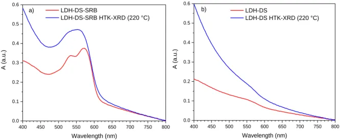
The effect of the thermal treatment on the UV-vis absorption spectra of LDH-DS-SRB is shown in 377
Fig. 12 a. The main absorption band of the SRB molecule, centred at 570 nm, is still observed after 378
the thermal treatment at 220°C. However, the overall absorption is found after such heat treatment 379
to increase significantly in the 400-600 nm range. This could be explained by the formation of 380
absorbent residual product from the decomposition of the DS molecules from 120 to 150°C (Fig. 3 & 381
4). This is confirmed by the UV-vis absorption spectra of the LDH-DS powder (Fig. 12 b). The LDH-DS 382
powder initially presents a white colour, whereas after heat treatment at 220°C it is black, which is 383
consistent with the formation of carbonaceous products. 384
14 400 450 500 550 600 650 700 750 800 0.0 0.1 0.2 0.3 0.4 0.5 0.6 A (a. u.) Wavelength (nm) LDH-DS-SRB LDH-DS-SRB HTK-XRD (220 °C) a) 400 450 500 550 600 650 700 750 800 0.0 0.1 0.2 0.3 0.4 0.5 0.6 A (a. u.) Wavelength (nm) LDH-DS LDH-DS HTK-XRD (220 °C) b) 385
Figure 12. UV-visible spectra of a) LDH-DS-SRB and b) LDH-DS powders before and after HTK-XRD
386
analysis. 387
The PL QYint of the LDH-DS-SRB powders before and after thermal treatment at 220°C are 388
reported in Table 2 for λexc = 480 and 365 nm. The drop in optical performance is clearly shown by 389
these PL QYint values. 390
391
Table 2. PL QYint for λexc = 480 nm and 365 nm of LDH-DS-SRB powder before and after the HTK-XRD 392
analysis (after cooling to 25 °C) 393 Excitation wavelength LDH-DS-SRB 25 °C LDH-DS-SRB 220°C then cooled to 25°C PL QYint (%) λexc = 480 nm 68.9 ± 3.5 15.0 ± 0.8 λexc = 365 nm 73.1 ± 3.7 4.3 ± 0.2 394
3.2.2. Silicone/LDH-DS-SRB composite film (Si-LDS) behaviour under thermal stress
395
The thermal stability of the Si-LDS composite film with the optimal formulation was further 396
studied at 120 °C for 24 h. 397
X-ray diffraction 398
XRD patterns of the Si-LDS composite film before and after the thermal treatment are presented 399
in Fig. 13 and were compared to the pattern of the LDH-DS-SRB reference powder. First, the LDH 400
matrix relative diffraction peaks are observed on the composite film pattern. After the thermal 401
treatment the harmonic peaks (00l) disappear, indicating that the dispersion of the LDH-DS-SRB 402
powder in silicone does not improve its thermal stability in terms of structural 2D integrity. 403
15 0 5 10 15 20 25 30 35 40 45 50 55 60 65 70 I ( a.u .) 2 (°) LDH-DS-SRB Si-LDS Si-LDS 24h@120°C 404
Figure 13. XRD patterns of the Si-LDS composite film before and after thermal treatment as well as
405
the pattern of the LDH-DS-SRB powder 406
Optical properties 407
The PL QYint of the composite film were recorded as a function of the excitation wavelengths 408
before and after the thermal treatment in an oven at 120 °C for 24 hours, and are shown for each 409
sample in Fig. 14 a. After the thermal treatment, the PL QYint decreases significantly in the studied 410
spectral range of 300-600 nm. 411
The emission spectrum of the composite film is shifted after the thermal treatment towards 412
higher wavelengths (Fig. 14 b). This is the result of the aggregation of SRB molecules, as already 413
observed with the LDH-DS-SRB reference powder (Fig. 6). The UV-Vis emission spectra also support 414
these findings, with a significant broadening and an increase in absorption over the 550-350 nm 415
range for the sample heat-treated at 120°C. Finally, for this thermal treatment, the PL QYint of the Si-416
LDS composite film and of the LDH-DS-SRB powder (Fig. 5), exhibit the same change in their spectral 417
features. However, the PL QYint values recorded for the film are lower than for powder. This is in part 418
explained by the dilution effect provided by the silicone matrix. 419
A film composed exclusively of silicone was also produced; it underwent the same 24-hour heat 420
treatment at 120°C as the Si-LDS film. Recording the UV-Vis and infrared spectra led to the 421
conclusion that the silicone does not undergo any modification after this treatment. The decrease in 422
optical performance after the heat treatment at 120°C is thus explained, as for the powder, by an 423
aggregation phenomenon generated by temperature. 424
16 300 350 400 450 500 550 600 0 10 20 30 40 50 60 70 80 90 100 PL QY int (%) Wavelength (nm) Si-LDS Si-LDS 24h@120°C a) 425 500 550 600 650 700 750 800 0 50 100 150 200 250 300 350 I ( a.u .) Wavelength (nm) Si-LDS Si-LDS 24h@120°C b) 350 400 450 500 550 600 650 700 750 800 0.00 0.05 0.10 0.15 0.20 0.25 0.30 535 nm A (a. u.) Wavelength (nm) t 0 24h@120°C 570 nm = 480 nm 426
Figure 14. a) Variation in PL QYint as a function of the excitation wavelength for the Si-LDS composite 427
film before and after thermal treatment. b) Emission spectra (exc = 480 nm), and UV-visible spectra 428
(inset) of the composite film before and after thermal treatment. 429
3.3. Composite film: durability under photonic and thermal stresses
430
3.3.1. Accelerated photo-aging in a SEPAP 12/24 unit
431
The photo-aging of the Si-LDS composite film was studied using a SEPAP 12/24 unit. In order to 432
have a reference, a second Si-SRB composite film was produced by dispersing 0.05 %wt. pure SRB in 433
the silicone matrix. Thus, the amounts of SRB in both films are comparable. 434
Fig. 15 plots PL QYint as a function of irradiation time for three excitation wavelengths of interest 435
(365, 480 and 570 nm) for Si-LDS composite film. Only the variation in the PL QYint for excitation at 436
570 nm is provided for the S-SRB film because its luminescence is initially very low or even non-437
existent for excitations at 365 and 470 nm. At 570 nm, after only 3 hours, the Si-SRB film has entirely 438
lost its luminescence, whereas the luminescence of the Si-LDS film remains identical to that observed 439
initially, showing the benefit of the inorganic vessel for stabilizing the SRB optical properties. 440
Quantitatively, after 35 hours the PL QYint of the Si-LDS film is still above 30 %. For excitation at 365 441
and 480 nm, the kinetic of luminescence extinction is close to that at 570 nm. 442
17 0 5 10 15 20 25 30 35 0 5 10 15 20 25 30 35 40 45 50 55 60 65 70 75 80 85 90 95 100 PL QY int (%) Irradiation time (h) Si-LDS (exc = 365 nm) Si-LDS (exc = 480 nm) Si-LDS (exc = 570 nm) Si-SRB (exc = 570 nm) 443
Figure 15. Variation in PL QYint as a function of irradiation time in the SEPAP 12/24 unit for both Si-444
SRB and Si-LDS composite films for several excitation wavelengths (365, 480 and 570 nm). 445
In order to have a better understanding of these observations, UV-vis spectra were recorded 446
after photo-aging. The asymmetric band relative to SRB molecules is observed for both composites 447
before the irradiation process starts. Maximum absorbance is the same for both composites, which 448
confirms the similar quantity of SRB molecules in both composites. However, the maximum is shifted 449
by 20 nm between the two, and can safely be explained by a higher amount of SRB aggregates in Si-450 SRB than in Si-LDS. 451 400 500 600 700 800 0.0 0.1 0.2 0.3 Si-SRB 4h Si-LDS 34h Si-SRB 0h A (a. u.) Wavelength (nm) Si-LDS 0h a) 0 5 10 15 20 25 30 35 0.00 0.05 0.10 0.15 0.20 0.25 0.30 A (a. u.) Irradiation time (h) Si-SRB Si-LDS b) 452
Figure 16. Variation in UV-visible spectra (a) and maximum absorbance (b) for Si-SRB and Si-LDS
453
composites as a function of irradiation time. 454
During the irradiation process, the main absorbance band decreases for both composites (Fig. 455
16 a). The SRB molecules are irreversibly damaged by irradiation in the SEPAP 12/24 unit. However, 456
the degradation kinetic is considerably faster for the Si-SRB film (Fig. 16 b) than for Si-LDS. Indeed, 457
absorbance is almost extinguished after only 4 hours for the Si-SRB film, whereas a value still subsists 458
after 34 hours for Si-LDS. This observation matches the decrease in the PL QYint described above (Fig. 459
15). 460
In conclusion, the LDH host matrix makes it possible to elaborate a homogeneous composite 461
luminescent film, but also to significantly increase SRB stability under stresses (thermal and 462
18 photonic). The conditions in the SEPAP 12/24 unit were very drastic ( > 300 nm and T = 60 °C) and 463
are not well representative of the photo-aging of composites associated with commercial blue LEDs. 464
Nevertheless, these experiments provide information on the stability of the Si-LDS film under UV LED 465
excitation. In the following section, the behaviour of these composites is studied in direct association 466
with a commercial blue LED. 467
3.3.2. Aging under blue LED
468
In order to assess the stability of the Si-LDS composite film in operating conditions close to 469
those encountered in LED devices, the Si-LDS film and the Si-SRB composite film were irradiated by a 470
powerful blue LED (460 nm, power: 2480 W/m2) for 1400 minutes at 80°C in a black chamber. A 471
spectrophotometer recorded the emission spectra every 20 minutes for 24 hours. 472
The variation in the integrated areas of the emission spectra as a function of irradiation time is 473
illustrated in Fig.17. 474
The Si-LDS composite film, using the LDH hosting the SRB molecules, is not affected at all by the 475
stresses, and photoluminescence intensity is stable. On the contrary, the intensity of Si-SRB 476
decreases significantly with irradiation time. A loss of 32 % was observed at the end of the first 24 477
hours. The beneficial effect of the LDH matrix is thus clearly demonstrated in such real conditions of 478
use, and confirms that this Si-LDS film is fully compatible with a blue LED device. 479 0 200 400 600 800 1000 1200 1400 0 20 40 60 80 100 120 Si-SRB Norm alized inte nsity ( %)
Irradiation time (min)
Si-LDS
480
Figure 17. Variation in normalized photoluminescence intensity with respect to irradiation time for
481
the Si-LDS and Si-SRB composite films heated to 80 °C and irradiated by a commercial blue LED (460 482
nm, 2480 W.m-2). 483
Photographs of both composite films, Si-SRB and Si-SDS, are presented in Fig.18 before and after 484
the 24 h irradiation process. The colour fading of Si-SRB reveals the degradation of the SRB 485
molecules. The aspect of Si-LDS is not modified by photo-aging; the role of LDH is well highlighted 486
here again. 487
19
Figure 18. Photographs of Si-SRB and Si-LDS composite films before (a) and c), respectively)
489
and after (b) and d), respectively) aging under photonic (blue LED) and thermal (80°C) stresses. 490
4. Conclusions
491This study emphasises the ability of a LDH host structure to protect the SRB luminescent dye 492
against various stresses. The LDH interior, filled with spacer, impedes SRB intermolecular interaction. 493
This effect is found to be more pronounced for tightly-packed interlayer space obtained from a 494
mono-functional co-intercalate such as DS than from TP. In the latter case, SRB molecules are freer to 495
diffuse and thus to quench the optical properties. It is pointed out that the hybrid LDH-DS-SRB and 496
the associated composite film exhibit a suitable orange - red emission under blue LED excitation. 497
The progressive loss in the optical properties of the LDH-DS-SRB hybrid powder after a 498
treatment at 120 °C is explained by a breakdown of the LDH stacking structure, as confirmed by XRD 499
analysis in temperature. This structural disruptive effect modifies the environment of the SRB 500
molecules, most probably provoking their partial aggregations and thus leading to non-radiative 501
energy transfers. Satisfactorily, a significant photoluminescence quantum yield is still observed after 502
the thermal treatment. 503
Similarly, the LDH-DS-SRB/silicone composite film exhibits luminescence after thermal exposure, 504
and this is also the case after being exposed to severe photo-aging conditions. Interestingly, the 505
emission of the composite film when irradiated by a commercial blue LED at 80 °C to reproduce “real 506
conditions of use” is completely stable after 24 hours. These results highlight the stability provided 507
by the LDH host structure for dyes, and the potential of LDH-DS-SRB for commercial lighting 508
applications based on a blue LED. 509
CONFLICTS OF INTEREST
510There is no conflict to declare. 511
FORMATTING OF FUNDING SOURCES
512This work was supported by CPER DEFI MMASYF through its 2016 «MetaProfile» project. Thus, 513
the authors would like to thank the European Union in the framework of the European Regional 514
Development Fund (ERDF), and the Région Auvergne Rhône-Alpes, which co-funded this project. 515
REFERENCES
516Boonsin, R., Chadeyron, G., Roblin, J.-P., Boyer, D., Mahiou, R., 2015. Development of rare-earth-free 517
phosphors for eco-energy lighting based LEDs. Journal of Materials Chemistry C 3, 9580-9587. 518
Conterosito, E., Benesperi, I., Toson, V., Saccone, D., Barbero, N., Palin, L., Barolo, C., Gianotti, V., 519
Milanesio, M., 2016. High-Throughput Preparation of New Photoactive Nanocomposites. 520
ChemSUSchem 9, 1279-1289. 521
Cullity, B.D., 1978. Elements of X-ray Diffraction. Addison-Wesley Publishing Company. 522
Dal Lago, M., Meneghini, M., Trivellin, N., Mura, G., Vanzi, M., Meneghesso, G., Zanoni, E., 2012. 523
Phosphors for LED-based light sources: Thermal properties and reliability issues. Microelectronics 524
Reliability 52, 2164-2167. 525
Das, S., Manam, J., 2018. Fluorescein isothiocyanate and rhodamine B dye encapsulated mesoporous 526
SiO2 for applications of blue LED excited white LED. Optical Materials 79, 259-263. 527
20 De, S., Kundu, R., 2011. Spectroscopic studies with fluorescein dye—Protonation, aggregation and 528
interaction with nanoparticles. Journal of Photochemistry and Photobiology A: Chemistry 223, 71-81. 529
Degoutin, S., Bacquet, M., Morcellet, M., 2008. Organosilica Mesoporous Materials with Double 530
Functionality: Amino Groups and β-Cyclodextrin Synthesis and Properties, pp. 213-221. 531
Drezdzon, M.A., 1988. Synthesis of isopolymetalate-pillared hydrotalcite via organic-anion-pillared 532
precursors. Inorganic Chemistry 27, 4628-4632. 533
El-kharrag, R., Amin, A., Greish, Y., 2011. Synthesis and Characterization of Mesoporous Sodium 534
Dodecyl Sulfate-Coated Magnetite Nanoparticles. Journal of Ceramic Science and Technology 2, 203-535
210. 536
Kajjam, A.B., Kumar, P.S.V., Subramanian, V., Vaidyanathan, S., 2018. Triphenylamine based 537
yellowish-orange light emitting organic dyes (donor–π–acceptor) for hybrid WLEDs and OLEDs: 538
synthesis, characterization and theoretical study. Physical Chemistry Chemical Physics 20, 4490-4501. 539
Lamouche, G., Lavallard, P., Gacoin, T., 1999. Optical properties of dye molecules as a function of the 540
surrounding dielectric medium. Physical Review A 59, 4668-4674. 541
Legentil, P., Leroux, F., Therias, S., Boyer, D., Chadeyron, G., 2020. Sulforhodamine B-LDH composite 542
as a rare-earth-free red-emitting phosphor for LED lighting. Journal of Materials Chemistry C, DOI: 543
10.1039/D0TC02802A 544
Legentil, P., Leroux, F., Therias, S., Mahiou, R., Chadeyron, G., 2019. Revisiting fluorescein and 545
layered double hydroxide using a synergistic approach: A complete optical study. Journal of 546
Luminescence 215, 116634. 547
Lonkar, S.P., Kutlu, B., Leuteritz, A., Heinrich, G., 2013. Nanohybrids of phenolic antioxidant 548
intercalated into MgAl-layered double hydroxide clay. Applied Clay Science 71, 8-14. 549
Nair, G.B., Swart, H.C., Dhoble, S.J., 2020. A review on the advancements in phosphor-converted light 550
emitting diodes (pc-LEDs): Phosphor synthesis, device fabrication and characterization. Progress in 551
Materials Science 109, 100622. 552
Nyalosaso, J.L., Boonsin, R., Vialat, P., Boyer, D., Chadeyron, G., Mahiou, R., Leroux, F., 2019. Towards 553
rare-earth-free white light-emitting diode devices based on the combination of dicyanomethylene 554
and pyranine as organic dyes supported on zinc single-layered hydroxide. Beilstein Journal of 555
Nanotechnology 10, 760-770. 556
Ogawa, M., Kuroda, K., 1995. Photofunctions of Intercalation Compounds. Chemical Reviews 95, 399-557
438. 558
Ray, K., Nakahara, H., 2002. Adsorption of Sulforhodamine Dyes in Cationic Langmuir−Blodgett Films: 559
Spectroscopic and Structural Studies. The Journal of Physical Chemistry B 106, 92-100. 560
Shao, Q., Lin, H., Dong, Y., Jiang, J., 2014. Temperature-dependent photoluminescence properties of 561
(Ba,Sr)2SiO4:Eu2+ phosphors for white LEDs applications. Journal of Luminescence, 165-169. 562
Sotiles, A.R., Wypych, F., 2019. Converting Mn/Al layered double hydroxide anion exchangers into 563
cation exchangers by topotactic reactions using alkali metal sulfate solutions. Chemical 564
communications 55, 7824-7827. 565
Sultana, N., 2018. Role of ammonium ion on the aggregation and adsorption properties of sodium 566
dodecylsulfate. Journal of Dispersion Science and Technology 39, 92-99. 567
Wu, M.J., Wu, J.Z., Zhang, J., Chen, H., Zhou, J.Z., Qian, G.R., Xu, Z.P., Du, Z., Rao, Q.L., 2018. A review 568
on fabricating heterostructures from layered double hydroxides for enhanced photocatalytic 569
activities. Catalysis Science & Technology 8, 1207-1228. 570
Xia, Z., Xu, Z., Chen, M., Liu, Q., 2016. Recent developments in the new inorganic solid-state LED 571
phosphors. Dalton transactions 45, 11214-11232. 572
Yan, D., Lu, J., Wei, M., G Evans, D., Duan, X., 2009. Sulforhodamine B Intercalated Layered Double 573
Hydroxide Thin Film with Polarized Photoluminescence. 574
Yan, L., Wang, Y., Li, J., Kalytchuk, S., Susha, A.S., Kershaw, S.V., Yan, F., Rogach, A.L., Chen, X., 2014. 575
Highly luminescent covalently bonded layered double hydroxide–fluorescent dye nanohybrids. 576
Journal of Materials Chemistry C 2, 4490. 577
Yazdan Mehr, M., van Driel, W.D., Zhang, G.Q., 2014. Accelerated life time testing and optical 578
degradation of remote phosphor plates. Microelectronics Reliability 54, 1544-1548. 579
21 Yin, Q., Li, D., Zhang, J., Zhao, Y., Wang, C., Han, J., 2019. CoNi-layered double hydroxide array on 580
graphene-based fiber as a new electrode material for microsupercapacitor. Applied Surface Science 581
487, 1-8. 582
Zhang, L., Li, B., Lei, B., Hong, Z., Li, W., 2008. A triphenylamine derivative as an efficient organic light 583
color-conversion material for white LEDs. Journal of Luminescence 128, 67-73. 584
Zhou, Z., Zhao, L., Lu, P., Zheng, H., Wang, J., Zeng, Y., 2014. Thermal influence of phosphor to GaN-585
based white LEDs. SPIE. 586