HAL Id: hal-03220929
https://hal.univ-lorraine.fr/hal-03220929
Submitted on 20 May 2021
HAL is a multi-disciplinary open access
archive for the deposit and dissemination of
sci-entific research documents, whether they are
pub-lished or not. The documents may come from
teaching and research institutions in France or
abroad, or from public or private research centers.
L’archive ouverte pluridisciplinaire HAL, est
destinée au dépôt et à la diffusion de documents
scientifiques de niveau recherche, publiés ou non,
émanant des établissements d’enseignement et de
recherche français ou étrangers, des laboratoires
publics ou privés.
Magnetic Resonance Imaging or Computed Tomography
Before Treatment in Acute Ischemic Stroke: Effect on
Workflow and Functional Outcome
Corentin Provost, Marc Soudant, Laurence Legrand, Ben Hassen, Yu Xie,
Sébastien Soize, Romain Bourcier, Joseph Benzakoun, Myriam Edjlali,
Grégoire Boulouis, et al.
To cite this version:
Corentin Provost, Marc Soudant, Laurence Legrand, Ben Hassen, Yu Xie, et al.. Magnetic
Res-onance Imaging or Computed Tomography Before Treatment in Acute Ischemic Stroke: Effect on
Workflow and Functional Outcome. Stroke, American Heart Association, 2019, 50 (3), pp.659-664.
�10.1161/STROKEAHA.118.023882�. �hal-03220929�
659
S
ince the benefits of recanalization are highly time-depen-dent in acute ischemic stroke,1–4 minimizing the workflow duration is crucial. Computed tomography (CT) is the most widely used diagnostic tool because of its availability and rapid acquisition time. However, magnetic resonance imaging (MRI) has advantages over CT. Diffusion-weighted imaging (DWI) best detects acute ischemia,5 stroke-like events6–8 and is the reference for infarct core extent measurement, which is crucial in selecting patients for endovascular treatment in thelater time window.9,10 In patients with unknown onset time, MRI identifies strokes that occurred approximately within the previous 4.5 hours and could benefit from thrombolysis.11 MRI drawbacks theoretically include screening time for con-traindications, limited access, additional time needed to clear the MRI scan and place the patient on the table, and longer scan duration.
The recent trials showing the efficacy of endovascular therapy in acute ischemic stroke used primarily CT-based
Background and Purpose—The acute management of stroke patients requires a fast and efficient screening imaging modality. We compared workflow and functional outcome in acute ischemic stroke patients screened by magnetic resonance imaging (MRI) or computed tomography (CT) before treatment in the THRACE trial (Thrombectomie des Artères Cérébrales), with the emphasis on the duration of the imaging step.
Methods—The THRACE randomized trial (June 2010 to February 2015) evaluated the efficacy of mechanical thrombectomy
after intravenous tPA (tissue-type plasminogen activator) in ischemic stroke patients with proximal occlusion. The choice of screening imaging modality was left to each enrolling center. Differences between MRI and CT groups were assessed using univariable analysis and the impact of imaging modality on favorable 3-month functional outcome (modified Rankin Scale score of ≤2) was tested using multivariable logistic regression.
Results—Four hundred one patients were included (25 centers), comprising 299 MRI-selected and 102 CT-selected patients.
Median baseline National Institutes of Health Stroke Scale score was 18 in both groups. MRI scan duration (median [interquartile range]) was longer than CT (MRI: 13 minutes [10–16]; CT: 9 minutes [7–12]; P<0.001). Stroke-imaging time (MRI: median 114 minutes [interquartile range, 89–138]; CT: 107 minutes [88–139]; P=0.19), onset-to-intravenous tPA time (MRI: 150 minutes [124–179]; CT: 150 minutes [123–180]; P=0.38) and onset-to-angiography-suite time (MRI: 200 minutes [170–250]; CT: 213 minutes [180–246]; P=0.57) did not differ between groups. Imaging modality was not significantly associated with functional outcome in the multivariable analysis.
Conclusions—Although MRI scan duration is slightly longer than CT, MRI-based selection for acute ischemic stroke
patients is accomplished within a timeframe similar to CT-based selection, without delaying treatment or impacting functional outcome. This should help to promote wider use of MRI, which has inherent imaging advantages over CT.
Clinical Trial Registration—URL: https://www.clinicaltrials.gov. Unique identifier: NCT01062698.
(Stroke. 2019;50:659-664. DOI: 10.1161/STROKEAHA.118.023882.)
Key Words: brain ischemia ◼ magnetic resonance imaging ◼ thrombectomy ◼ treatment outcome ◼ workflow
Received October 11, 2018; final revision received December 10, 2018; accepted January 15, 2019.
From the Imaging Department, INSERM UMR S1266, Sainte-Anne Hospital, Paris Descartes University, France (C.P., L.L., W.B.H., J.B., M.E., G.B., O.N., C.O.); INSERM CIC-1433 Clinical Epidemiology, Nancy Hospital, France (M.S., F.G.); Department of Neuroradiology, INSERM U1254, Lorraine University, Nancy, France (Y.X., S.B.); Department of Diagnostic and Interventional Neuroradiology, University Hospital of Reims, France (S.S.); Department of Diagnostic and Interventional Neuroradiology, University Hospital of Nantes, France (R.B.); and Department of Neuroradiology, University Hospital of Rennes, France (H.R.).
Presented in part at the Annual Meeting of the Radiological Society of North America, Chicago, IL, November 28, 2018.
The online-only Data Supplement is available with this article at https://www.ahajournals.org/doi/suppl/10.1161/STROKEAHA.118.023882.
Correspondence to Catherine Oppenheim, PhD, Imaging Department, INSERM UMR S1266, Sainte-Anne Hospital, Paris Descartes University, CH Sainte-Anne, 1 Rue Cabanis, 75014 Paris, France. Email [email protected]
© 2019 American Heart Association, Inc.
Before Treatment in Acute Ischemic Stroke
Effect on Workflow and Functional Outcome
Corentin Provost, MD; Marc Soudant, MSc; Laurence Legrand, MD; Wagih Ben Hassen, MD;
Yu Xie, MD; Sébastien Soize, MD; Romain Bourcier, MD; Joseph Benzakoun, MD;
Myriam Edjlali, MD; Grégoire Boulouis, MD; Hélène Raoult, MD; Francis Guillemin, PhD;
Olivier Naggara, PhD; Serge Bracard, PhD; Catherine Oppenheim, PhD
DOI: 10.1161/STROKEAHA.118.023882
Stroke is available at https://www.ahajournals.org/journal/str
660 Stroke March 2019
approaches.12 Consequently, detailed data on MRI-based work-flow are limited.13–15 A recent study14 showed that the work-flow times of 128 MRI-selected patients before endovascular therapy were similar to those reported in 3 large CT-based tri-als.16–18 However, this monocentric study had no CT-selected subgroup for comparison, excluded patients with large core and did not record the precise duration of the MRI step.
In the multicenter THRACE trial (Thrombectomie des Artères Cérébrales),19 enrolling centers were free to use CT or MRI before randomization, resulting in a large propor-tion of MRI-selected patients. We compared the workflow and functional outcome in acute ischemic stroke patients screened by MRI or CT, with particular emphasis on the imaging step duration.
Methods
The data that support the findings of this study are available from the corresponding author on reasonable request.
Study Design
The THRACE trial was a randomized controlled trial conducted be-tween June 2010 and February 2015.19 Patients with acute ischemic
stroke were eligible for inclusion if they were aged 18 to 80 years, had a National Institutes of Health Stroke Scale (NIHSS) score of 10 to 25, had a proximal cerebral artery occlusion confirmed by CT or MRI, could receive intravenous tPA (tissue-type plasminogen acti-vator) within 4 hours of symptom onset, and if thrombectomy could be initiated within 5 hours of symptom onset. Patients were randomized to receive either intravenous tPA and mechanical thrombectomy or intravenous tPA alone. The THRACE trial was approved by the CPP (Comité de Protection des Personnes) III Nord Est Ethics Committee and the research boards of the participating centers. All patients or their legal representatives provided written informed consent.
Imaging Modality
Each enrolling center was free to use its routine CT or MRI stroke protocol, and no attempt was made to standardize imaging acquisi-tion. As per protocol requirement, all examinations included at least brain and intracranial vessel imaging, with either CT angiography (CTA) or MR angiography. Additional imaging techniques (perfu-sion imaging and cervical angiography) were optional. Imaging was performed before treatment and within 4 hours of stroke onset. The extent of lesion on baseline imaging was determined using Alberta Stroke Program Early CT Score in the CT group and DWI volume,20,21
in the MRI group.
Workflow Times
Enrolling centers were instructed to fill in a case report form after each patient enrollment with the following times: onset of stroke symptoms, clinical examination, randomization, start of intravenous tPA administration, and, in the endovascular arm, arrival in the an-giography suite, first cerebral angiographic run, and start and end of thrombectomy. Start and end of imaging times were extracted from Digital Imaging and Communications in Medicine metadata.
Statistical Analysis
Workflow times were expressed as median and interquartile range (IQR) owing to the nonnormality of the data. The CT and MRI groups were compared using the Mann-Whitney U test for quanti-tative variables and the χ2 test for qualitative variables. Prognostic
factors for the outcome were analyzed with univariable analysis, with dichotomized modified Rankin Scale score as the dependent variable (favorable outcome: modified Rankin Scale score of ≤2). To adjust for potential confounders, multivariable binary logistic
regression analysis was subsequently conducted. Factors with a significant (P<0.10) association with favorable outcome in univari-able analysis—that is, age, blood glucose, systolic blood pressure, National Institutes of Health Stroke Scale score, occlusion location (proximal portion of the middle cerebral artery versus intracranial internal carotid artery occlusion), diabetes mellitus, hypertension, renal insufficiency, treatment group (intravenous tPA and mechanical thrombectomy versus intravenous tPA alone), and imaging modality (MRI versus CT)—were candidates for the multivariable model. To avoid collinearity between variables, diabetes mellitus and hyper-tension were not selected while blood glucose and systolic blood pressure were entered in the multivariable model. Forward stepwise variable selection with significance level for entry into the model at 0.10 and significance level for staying in the model at 0.05 was used. Reported P values were 2-sided, with values <0.05 indicative of a significant difference. Statistical analysis was performed using SAS/ STAT software (version 9.3, SAS Institute, Cary, NC).
Results
Among 414 randomized patients in the THRACE trial, 401 patients were included in the present study (2 patients with-drew consent, screening imaging modality was unknown in 8 patients, and imaging time information was not available for 2 MRI-selected patients and 1 CT-selected patient). Of the 401 patients from 25 centers, 299 were in the MRI group (median [IQR] DWI volume: 16 mL [9–52]) and 102 in the CT group (median [IQR] Alberta Stroke Program Early CT Score: 9 [8–10]). Of the 25 centers, 15 used MRI and 5 used CT as the prime imaging modality, defined as a modality used for >80% of the examinations performed. The other 5 cen-ters used MRI or CT without priority being given to either modality and according to availability. The detailed imaging protocol of each center is shown in Table I in the online-only Data Supplement.
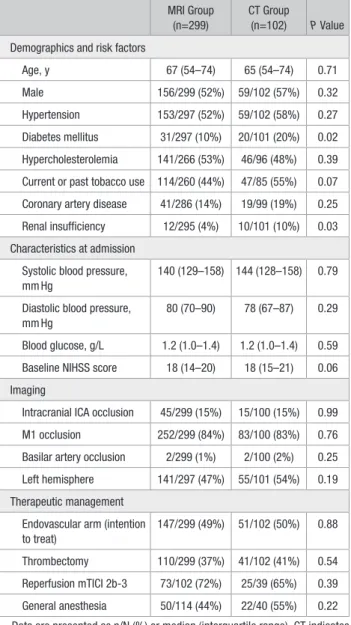
Differences between the MRI and CT groups are given in Table 1. In univariable analysis, MRI-selected patients were less frequently diabetic and had less renal insufficiency than CT-selected patients. There was no statistical difference in baseline National Institutes of Health Stroke Scale scores be-tween groups. MRI and CT were similarly performed during off hours (MRI: 50/299 [17%]; CT: 18/102 [18%]; P=0.83), and weekends (MRI: 55/299 [18%]; CT: 14/102 [14%];
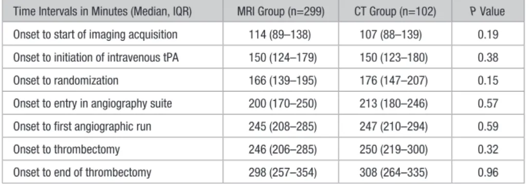
P=0.28). None of the delays from stroke onset differed be-tween the CT and MRI groups (Table 2). The delay bebe-tween clinical examination at hospital arrival and start of imaging did not differ between the MRI and CT groups (MRI: 17 min-utes [11–27]; CT: 12 minmin-utes [7–32]; P=0.13). As shown in the Figure, the mean duration of each discrete step in patient workflow was similar between the CT and MRI groups, ex-cept for scan duration and imaging-to-intravenous tPA time.
MRI scan duration was overall longer (MRI: median 13 minutes [IQR, 10–16]; CT: 9 minutes [7–12]; P<0.001), irre-spective of the imaging protocol (without perfusion imaging [MRI: 12 minutes (10–15); CT: 7 minutes (2–11); P<0.001] or with perfusion imaging [MRI: 16 minutes (15–19); CT: 10 minutes (9–13); P<0.001]). In the MRI group, 68/299 (23%) scans had perfusion imaging, 83/299 (28%) had cervical MR angiography, and 4/299 (1%) had both. Median MRI scan duration was significantly longer with perfusion imaging (16 minutes [15–19]) or with cervical MR angiography (15 minutes [12–18]) than without contrast injection (11 minutes
[9–13]), P<0.001 for both comparisons. In the CT group, 62/102 (61%) patients had CT perfusion in addition to the CTA required to confirm arterial occlusion and 89/102 (87%) had a CTA of the neck. Median CT duration was significantly longer with perfusion imaging (10 minutes [9–13]) than without, that is, simple CT/CTA (7 minutes [2–11]; P<0.001). Similarly, advanced imaging protocols (CT perfusion or MRI) were longer (13 minutes [10–16]; P<0.001) than simple CT/ CTA protocols.
Despite a longer scan duration in the MRI group, time from start of imaging acquisition to initiation of intravenous tPA was shorter (MRI: 31 minutes [24–43]; CT: 42 minutes [34–54]; P<0.001), because of a shorter time from end of im-aging to initiation of intravenous tPA (MRI: 18 minutes [11– 28]; CT: 33 minutes [24–43]; P<0.001; Figure). To disentangle center effect from imaging modality effect on the delay from
imaging to initiation of intravenous tPA, we post hoc analyzed the data using a mixed model, with center as a random effect and imaging modality as a fixed variable. Both imaging mo-dality and center effects were significant (P<0.001). Of note, there was no difference in the time from start of imaging to in-itiation of intravenous tPA between standard and multimodal imaging protocols, using either MRI (30 minutes [22–43] versus 33 [25–42]; P=0.19) or CT (38 minutes [30–60] versus 42 minutes [35–54]; P=0.63).
Favorable outcome was associated with imaging modality in univariable analysis (MRI: 51%, 151 of 294 patients; CT: 38%, 38 of 100 patients; P=0.021). In multivariable analysis with imaging modality forced into the model (n=370/401), favorable outcome was associated with mechanical throm-bectomy group (odds ratio [OR], 1.71; 95% CI, 1.08–2.69;
P=0.021), younger age (OR, 0.97; 95% CI, 0.95–0.98;
P<0.0001), less severe baseline National Institutes of Health Stroke Scale (OR, 0.89; 95% CI, 0.84–0.94; P<0.0001), lower blood glucose (OR, 0.48; 95% CI, 0.25–0.91; P=0.024), prox-imal portion of the middle cerebral artery occlusion (OR, 3.76; 95% CI, 1.86–7.60; P=0.0002). MRI was not associ-ated with outcome (OR, 0.42; 95% CI, 0.27–2.48; P=0.12). Symptomatic hemorrhagic transformation at 24 hours (MRI: 4 of 299 patients; CT: 3 of 102 patients; P=0.45) and mortality at 3 months (MRI: 11%, 33 of 293 patients; CT: 16%, 16 of 100 patients; P=0.22) did not differ between the 2 groups.
Finally, all the above results were similar after exclusion of the 13 patients (3%) who had both imaging modalities be-fore randomization (9 with CT first then MRI and 4 with MRI first then CT).
Discussion
This multicenter study provides a comprehensive picture of the acute stroke workflow in a country where MRI has been recommended as the prime brain imaging technique for almost 10 years.22 In the THRACE trial, the choice of imaging mo-dality for patient inclusion was left to the enrolling center and, as a result, 74% of patients were imaged with MRI. Overall, there was a significant difference of a few minutes between CT and MRI imaging duration, but this had no impact on the global workflow as time from stroke onset-to-intravenous tPA and to endovascular therapy did not depend on the choice of imaging modality. Finally, this choice had no impact on func-tional outcome at 3 months.
There is a prevailing view that stroke MRI scan duration is in the order of 20 minutes and therefore much longer than CT.23 However, in centers that have been using MRI as the prime diagnostic imaging tool for years, standardized stroke MRI protocols last around 10 minutes.14,24,25 This duration could be further reduced to 6 minutes with the use of echo planar imaging at 3 Tesla.23 However, the above scan durations are merely based on the sum of the duration of each sequence, and therefore exclude intersequence time or events that might occur during the MRI examination. The recent endovascular trials provide detailed timings for door-to-imaging and im-aging to the next workflow steps,4 but the precise duration of the complete MRI session is lacking. The 13-minute median MRI scan duration we recorded over a large panel of centers is representative of a real-world setting, since there was no
Table 1. Characteristics of Patients Included in the Study
MRI Group (n=299)
CT Group
(n=102) P Value
Demographics and risk factors
Age, y 67 (54–74) 65 (54–74) 0.71
Male 156/299 (52%) 59/102 (57%) 0.32 Hypertension 153/297 (52%) 59/102 (58%) 0.27 Diabetes mellitus 31/297 (10%) 20/101 (20%) 0.02 Hypercholesterolemia 141/266 (53%) 46/96 (48%) 0.39 Current or past tobacco use 114/260 (44%) 47/85 (55%) 0.07 Coronary artery disease 41/286 (14%) 19/99 (19%) 0.25 Renal insufficiency 12/295 (4%) 10/101 (10%) 0.03 Characteristics at admission
Systolic blood pressure, mm Hg
140 (129–158) 144 (128–158) 0.79 Diastolic blood pressure,
mm Hg
80 (70–90) 78 (67–87) 0.29 Blood glucose, g/L 1.2 (1.0–1.4) 1.2 (1.0–1.4) 0.59 Baseline NIHSS score 18 (14–20) 18 (15–21) 0.06 Imaging
Intracranial ICA occlusion 45/299 (15%) 15/100 (15%) 0.99 M1 occlusion 252/299 (84%) 83/100 (83%) 0.76 Basilar artery occlusion 2/299 (1%) 2/100 (2%) 0.25 Left hemisphere 141/297 (47%) 55/101 (54%) 0.19 Therapeutic management
Endovascular arm (intention to treat)
147/299 (49%) 51/102 (50%) 0.88 Thrombectomy 110/299 (37%) 41/102 (41%) 0.54 Reperfusion mTICI 2b-3 73/102 (72%) 25/39 (65%) 0.39 General anesthesia 50/114 (44%) 22/40 (55%) 0.22 Data are presented as n/N (%) or median (interquartile range). CT indicates computed tomography; ICA, internal carotid artery; M1, proximal portion of the middle cerebral artery; MRI, magnetic resonance imaging; mTICI, modified Treatment in Cerebral Infarction; and NIHSS, National Institutes of Health Stroke Scale.
662 Stroke March 2019
attempt to standardize MRI protocols across centers. In con-trast to MRI, CT is commonly considered as a snapshot in the stroke workflow. Although this was reasonable for noncontrast CT, it is no longer true when CTA is added, a requirement be-fore endovascular treatment decision. As with MRI, the whole CT scan duration is longer than the sum of CT and CTA ac-quisition times. We here provided a measurement of total scan duration (CT/CTA: median, 7 minutes; CT/CTA/CTP: median, 10 minutes), which matches that in the SWIFT PRIME trial (Solitaire With the Intention for Thrombectomy as Primary Endovascular Treatment; CT/CTA: median, 9 minutes).2
Of note, 61% of patients in the CT group had perfusion imaging, as opposed to 23% in the MRI group. In MRI cen-ters, the large amount of information available with standard sequences, such as direct visualization of the infarct core extent on DWI may have obviated the need for additional perfusion. To avoid any loss of time in tailoring the imaging protocol to each individual, standardized imaging stroke protocols (with or without perfusion imaging) are used in some centers,24,26 while others add advanced imaging tech-niques in selected cases. On a large scale, we showed, like others,27 that performing multimodal CT did not delay intra-venous tPA administration, and this was also true for multi-modal MRI, thus complying with the 2018 American Heart Association guidelines.28
Although it did not impact the time from onset-to-intra-venous tPA or to endovascular treatment, time from imaging-to-intravenous tPA was surprisingly shorter in the MRI group. This was related to both an imaging modality effect and a
center effect in the post hoc analysis. About the imaging mo-dality effect, one possible explanation is that the treatment de-cision was made while MRI acquisition was still in progress. In patients with a puzzling clinical presentation, a treatment deci-sion based on straightforward DWI images with clear-cut signal changes might have been faster than one based on subtle early CT ischemic changes. About the center effect, it likely lies in organizational differences between CT and MRI centers. In cen-ters that prioritized round-the-clock immediate access to MRI for stroke patients, the global workflow might have been bet-ter organized and more time efficient, thus reducing time from imaging-to-intravenous tPA when compared with CT centers. Of note, in CT-selected patients, imaging-to-intravenous tPA time was similar to that in the IMS III international multicenter trial (Interventional Management of Stroke III).27
Times from stroke onset and from clinical examination on admission to imaging were similar in the CT and MRI groups. This suggests that none of the following steps are major obsta-cles to the use of MRI, in centers where it is available: access to MRI, including transport to the scanner and immediate access to the MRI room, completion of the MRI safety questionnaire to exclude contraindications, and placement of the patient on the MRI table. The systematic use of MRI in acute stroke implies round-the-clock priority access to MRI with direct admission in the imaging department, and close geographic localization between the stroke center, MRI unit, and angiography suite. The main clinical restrictions to the use of MRI remain con-traindications and restless or unstable ischemic stroke patients, accounting for 9% of patients.14 This acceptable low rate might
Table 2. Workflow Times From Onset of Symptoms
Time Intervals in Minutes (Median, IQR) MRI Group (n=299) CT Group (n=102) P Value
Onset to start of imaging acquisition 114 (89–138) 107 (88–139) 0.19 Onset to initiation of intravenous tPA 150 (124–179) 150 (123–180) 0.38
Onset to randomization 166 (139–195) 176 (147–207) 0.15
Onset to entry in angiography suite 200 (170–250) 213 (180–246) 0.57 Onset to first angiographic run 245 (208–285) 247 (210–294) 0.59
Onset to thrombectomy 246 (206–285) 250 (219–300) 0.32
Onset to end of thrombectomy 298 (257–354) 308 (264–335) 0.96
CT indicates computed tomography; IQR, interquartile range; MRI, magnetic resonance imaging; and tPA, tissue-type plasminogen activator.
Figure. Graph of workflow in patients screened
with magnetic resonance imaging (MRI) or com-puted tomography (CT). IV-tPA indicates intra-venous tissue-type plasminogen activator.
even decrease in the near future, as the vast majority of metal implants are MRI safe, and even concerns related to pacemak-ers may be ovpacemak-erstated.29 Taken together, our findings support the notion that MRI can be accomplished rapidly and within a similar timeframe as CT before endovascular therapy, in line with several studies showing that MRI-based selection is fea-sible and safe.13–15,30 One should however keep in mind that, although there is sufficient evidence that MRI is as reliable as CT for the detection of acute intracerebral hemorrhage, dedi-cated training with interpretation of gradient echo sequence is needed for MRI to be safely used before treatment decision.31
Our study has several limitations. (1) The THRACE trial was not designed for the purpose of the current study and ran-domization was not stratified on imaging modality. As a conse-quence, the choice of imaging modality may have depended on confounding variables. For instance, in centers using MRI as a prime screening imaging modality in stroke patients, the most severely ill patients might be directed towards CT. However, in THRACE, stroke clinical severity did not differ between the CT and MRI groups, likely because the majority of patients medi-cally ineligible for MRI suffer from intracerebral hemorrhage,32 whereas THRACE focused on ischemic stroke. We neverthe-less post hoc verified that all results were similar when we excluded patients imaged with CT in MRI centers, and patients imaged with MRI in CT centers. (2) There was no synchroni-zation across all clocks and imagers, which may have led to some degree of inaccuracy in the times recorded. However, there is no reason why clocks should be more inaccurate in one group. (3) Some of the workflow times were not recorded in the case report form of the THRACE trial (arrival to hospital door and groin puncture), therefore preventing a comprehen-sive evaluation of the workflow, as well as comparisons with others and current guidelines for imaging and door-to-groin puncture times.28,33 However, times from stroke onset and from clinical examination on admission to imaging were not longer in the MRI group, suggesting that access to MRI was not detrimental in terms of delay in patient management. Times of entry in angiography suite and first angiographic run pro-vided an estimation of the groin puncture time. (4) Admission status (direct admission versus transfer to endovascular-capable centers) was not available, preventing comparison of workflow between these 2 groups. However, given the THRACE inclu-sion criteria (initiation of intravenous tPA within 4 hours and thrombectomy within 5 hours of symptom onset), most patients were directly referred to endovascular-capable centers.
Conclusions
In conclusion, although MRI scan duration is a few minutes longer than CT, workflow in THRACE demonstrates that real-world application of MRI for acute stroke evaluation before treatment can be accomplished as rapidly as CT-based selec-tion paradigms. This should help to promote wider use of MRI in acute stroke by counterbalancing concerns about delay and eligibility.
Sources of Funding
The THRACE trial (Thrombectomie des Artères Cérébrales) was supported by the French Ministry of Health.
Disclosures
None.
References
1. Saver JL, Fonarow GC, Smith EE, Reeves MJ, Grau-Sepulveda MV, Pan W, et al. Time to treatment with intravenous tissue plasminogen activator and outcome from acute ischemic stroke. JAMA. 2013;309:2480–2488. doi: 10.1001/jama.2013.6959
2. Goyal M, Jadhav AP, Bonafe A, Diener H, Mendes Pereira V, Levy E, et al; SWIFT PRIME Investigators. Analysis of workflow and time to treatment and the effects on outcome in endovascular treatment of acute ischemic stroke: results from the SWIFT PRIME randomized controlled trial. Radiology. 2016;279:888–897. doi: 10.1148/radiol.2016160204 3. Menon BK, Sajobi TT, Zhang Y, Rempel JL, Shuaib A, Thornton J, et al.
Analysis of workflow and time to treatment on thrombectomy outcome in the Endovascular Treatment for Small Core and Proximal Occlusion Ischemic Stroke (ESCAPE) Randomized, Controlled Trial. Circulation. 2016;133:2279–2286. doi: 10.1161/CIRCULATIONAHA.115.019983 4. Saver JL, Goyal M, van der Lugt A, Menon BK, Majoie CB, Dippel
DW, et al; HERMES Collaborators. Time to treatment with endovascu-lar thrombectomy and outcomes from ischemic stroke: a meta-analysis.
JAMA. 2016;316:1279–1288. doi: 10.1001/jama.2016.13647
5. Chalela JA, Kidwell CS, Nentwich LM, Luby M, Butman JA, Demchuk AM, et al. Magnetic resonance imaging and computed to-mography in emergency assessment of patients with suspected acute stroke: a prospective comparison. Lancet. 2007;369:293–298. doi: 10.1016/S0140-6736(07)60151-2
6. Liu X, Almast J, Ekholm S. Lesions masquerading as acute stroke. J
Magn Reson Imaging. 2013;37:15–34. doi: 10.1002/jmri.23647 7. Fernandes PM, Whiteley WN, Hart SR, Al-Shahi Salman R.
Strokes: mimics and chameleons. Pract Neurol. 2013;13:21–28. doi: 10.1136/practneurol-2012-000465
8. Chernyshev OY, Martin-Schild S, Albright KC, Barreto A, Misra V, Acosta I, et al. Safety of tPA in stroke mimics and neuroimaging-negative cerebral ischemia. Neurology. 2010;74:1340–1345. doi: 10.1212/WNL.0b013e3181dad5a6
9. Leslie-Mazwi TM, Hirsch JA, Falcone GJ, Schaefer PW, Lev MH, Rabinov JD, et al. Endovascular stroke treatment outcomes after patient selection based on magnetic resonance imaging and clinical criteria.
JAMA Neurol. 2016;73:43–49. doi: 10.1001/jamaneurol.2015.3000 10. Nogueira RG, Jadhav AP, Haussen DC, Bonafe A, Budzik RF, Bhuva
P, et al; DAWN Trial Investigators. Thrombectomy 6 to 24 hours after stroke with a mismatch between deficit and infarct. N Engl J Med. 2018;378:11–21. doi: 10.1056/NEJMoa1706442
11. Thomalla G, Simonsen CZ, Boutitie F, Andersen G, Berthezene Y, Cheng B, et al; WAKE-UP Investigators. MRI-guided thrombolysis for stroke with unknown time of onset. N Engl J Med. 2018;379:611–622. doi: 10.1056/NEJMoa1804355
12. Goyal M, Menon BK, van Zwam WH, Dippel DW, Mitchell PJ, Demchuk AM, et al; HERMES Collaborators. Endovascular thrombec-tomy after large-vessel ischaemic stroke: a meta-analysis of individual patient data from five randomised trials. Lancet. 2016;387:1723–1731. doi: 10.1016/S0140-6736(16)00163-X
13. Menjot de Champfleur N, Saver JL, Goyal M, Jahan R, Diener HC, Bonafe A, et al. Efficacy of stent-retriever thrombectomy in magnetic resonance imaging versus computed tomographic perfusion-selected patients in SWIFT PRIME Trial (Solitaire FR With the Intention for Thrombectomy as Primary Endovascular Treatment for Acute Ischemic Stroke). Stroke. 2017;48:1560–1566. doi: 10.1161/STROKEAHA.117.016669 14. Simonsen CZ, Yoo AJ, Rasmussen M, Sørensen KE, Leslie-Mazwi T,
Andersen G, et al. Magnetic resonance imaging selection for endo-vascular stroke therapy: workflow in the GOLIATH Trial. Stroke. 2018;49:1402–1406. doi: 10.1161/STROKEAHA.118.021038 15. Shah S, Luby M, Poole K, Morella T, Keller E, Benson RT, et al. Screening
with MRI for accurate and rapid stroke treatment: SMART. Neurology. 2015;84:2438–2444. doi: 10.1212/WNL.0000000000001678
16. Campbell BC, Mitchell PJ; EXTEND-IA Investigators. Endovascular therapy for ischemic stroke. N Engl J Med. 2015;372:2365–2366. doi: 10.1056/NEJMc1504715
17. Goyal M, Demchuk AM, Menon BK, Eesa M, Rempel JL, Thornton J, et al. Randomized assessment of rapid endovascular treat-ment of ischemic stroke. N Engl J Med. 2015;372:1019–1030. doi: 10.1056/NEJMoa1414905.
664 Stroke March 2019
18. Saver JL, Goyal M, Bonafe A, Diener HC, Levy EI, Pereira VM, et al; SWIFT PRIME Investigators. Stent-retriever thrombectomy after intra-venous t-PA vs. t-PA alone in stroke. N Engl J Med. 2015;372:2285– 2295. doi: 10.1056/NEJMoa1415061
19. Bracard S, Ducrocq X, Mas JL, Soudant M, Oppenheim C, Moulin T, et al; THRACE Investigators. Mechanical thrombectomy after in-travenous alteplase versus alteplase alone after stroke (THRACE): a randomised controlled trial. Lancet Neurol. 2016;15:1138–1147. doi: 10.1016/S1474-4422(16)30177-6
20. Gautheron V, Xie Y, Tisserand M, Raoult H, Soize S, Naggara O, et al. Outcome after reperfusion therapies in patients with large baseline dif-fusion-weighted imaging stroke lesions: a THRACE trial (Mechanical Thrombectomy After Intravenous Alteplase Versus Alteplase Alone After Stroke) subgroup analysis. Stroke. 2018;49:750–753. doi: 10.1161/STROKEAHA.117.020244
21. Xie Y, Oppenheim C, Guillemin F, Gautheron V, Gory B, Raoult H, et al. Pretreatment lesional volume impacts clinical outcome and thrombec-tomy efficacy. Ann Neurol. 2018;83:178–185. doi: 10.1002/ana.25133 22. Haute Autorité de Santé. Stroke: Early Management (Alert, Prehospital
Phase, Initial Hospital Phase, Indications for Thrombolysis). https:// www.has-sante.fr/portail/upload/docs/application/pdf/2010-03/ stroke_early_management_-_guidelines_-_english_version.pdf. Accessed October 1, 2018.
23. Nael K, Khan R, Choudhary G, Meshksar A, Villablanca P, Tay J, et al. Six-minute magnetic resonance imaging protocol for evaluation of acute ischemic stroke: pushing the boundaries. Stroke. 2014;45:1985–1991. doi: 10.1161/STROKEAHA.114.005305
24. Legrand L, Tisserand M, Turc G, Naggara O, Edjlali M, Mellerio C, et al. Do FLAIR vascular hyperintensities beyond the DWI lesion represent the ischemic penumbra? AJNR Am J Neuroradiol. 2015;36:269–274. doi: 10.3174/ajnr.A4088
25. Labeyrie MA, Turc G, Hess A, Hervo P, Mas JL, Meder JF, et al. Diffusion lesion reversal after thrombolysis: a MR correlate of early neurological improvement. Stroke. 2012;43:2986–2991. doi: 10.1161/STROKEAHA.112.661009
26. Emeriau S, Serre I, Toubas O, Pombourcq F, Oppenheim C, Pierot L. Can diffusion-weighted imaging-fluid-attenuated inversion recovery mismatch (positive diffusion-weighted imaging/negative fluid-attenuated inversion recovery) at 3 Tesla identify patients with stroke at <4.5 hours?
Stroke. 2013;44:1647–1651. doi: 10.1161/STROKEAHA.113.001001
27. Vagal A, Foster LD, Menon B, Livorine A, Shi J, Qazi E, et al. Multimodal CT imaging: time to treatment and outcomes in the IMS III Trial. AJNR Am J Neuroradiol. 2016;37:1393–1398. doi: 10.3174/ajnr.A4751
28. Powers WJ, Rabinstein AA, Ackerson T, Adeoye OM, Bambakidis NC, Becker K, et al; American Heart Association Stroke Council. 2018 guidelines for the early management of patients with acute ischemic stroke: a guideline for healthcare professionals from the American Heart Association/American Stroke Association. Stroke. 2018;49:e46–e110. doi: 10.1161/STR.0000000000000158
29. Nazarian S, Hansford R, Rahsepar AA, Weltin V, McVeigh D, Gucuk Ipek E, et al. Safety of magnetic resonance imaging in patients with cardiac devices. N Engl J Med. 2017;377:2555–2564. doi: 10.1056/NEJMoa1604267
30. Schellinger PD, Thomalla G, Fiehler J, Köhrmann M, Molina CA, Neumann-Haefelin T, et al. MRI-based and CT-based thrombolytic therapy in acute stroke within and beyond established time win-dows: an analysis of 1210 patients. Stroke. 2007;38:2640–2645. doi: 10.1161/STROKEAHA.107.483255
31. Copenhaver BR, Shin J, Warach S, Butman JA, Saver JL, Kidwell CS. Gradient echo MRI: implementation of a training tutorial for intra-cranial hemorrhage diagnosis. Neurology. 2009;72:1576–1581. doi: 10.1212/WNL.0b013e3181a411df
32. Singer OC, Sitzer M, du Mesnil de Rochemont R, Neumann-Haefelin T. Practical limitations of acute stroke MRI due to patient-related problems.
Neurology. 2004;62:1848–1849.
33. Sacks D, Baxter B, Campbell BCV, Carpenter JS, Cognard C, Dippel D, et al. Multisociety consensus quality improvement revised consen-sus statement for endovascular therapy of acute ischemic stroke: from the American Association of Neurological Surgeons (AANS), American Society of Neuroradiology (ASNR), Cardiovascular and Interventional Radiology Society of Europe (CIRSE), Canadian Interventional Radiology Association (CIRA), Congress of Neurological Surgeons (CNS), European Society of Minimally Invasive Neurological Therapy (ESMINT), European Society of Neuroradiology (ESNR), European Stroke Organization (ESO), Society for Cardiovascular Angiography and Interventions (SCAI), Society of Interventional Radiology (SIR), Society of NeuroInterventional Surgery (SNIS), and World Stroke Organization (WSO). J Vasc Interv Radiol. 2018;29:441–453. doi: 10.1016/j.jvir.2017.11.026