HAL Id: hal-00672295
https://hal.archives-ouvertes.fr/hal-00672295
Submitted on 21 Feb 2012HAL is a multi-disciplinary open access archive for the deposit and dissemination of sci-entific research documents, whether they are pub-lished or not. The documents may come from teaching and research institutions in France or abroad, or from public or private research centers.
L’archive ouverte pluridisciplinaire HAL, est destinée au dépôt et à la diffusion de documents scientifiques de niveau recherche, publiés ou non, émanant des établissements d’enseignement et de recherche français ou étrangers, des laboratoires publics ou privés.
Phosphoinositide 3-Kinase as a novel functional target
for the regulation of the insulin signaling pathway by
SIRT1
Sara Fröjdö, Christine Durand, Laurent Molin, Andrew L. Carey, Assam
El-Osta, Bronwyn A. Kingwell, Mark A. Febbraio, Florence Solari, Hubert
Vidal, Luciano Pirola
To cite this version:
Sara Fröjdö, Christine Durand, Laurent Molin, Andrew L. Carey, Assam El-Osta, et al.. Phosphoinosi-tide 3-Kinase as a novel functional target for the regulation of the insulin signaling pathway by SIRT1. Molecular and Cellular Endocrinology, Elsevier, 2011, 335 (2), pp.166. �10.1016/j.mce.2011.01.008�. �hal-00672295�
Accepted Manuscript
Title: Phosphoinositide 3-Kinase as a novel functional target for the regulation of the insulin signaling pathway by SIRT1 Authors: Sara Fr¨ojd¨o, Christine Durand, Laurent Molin, Andrew L. Carey, Assam El-Osta, Bronwyn A. Kingwell, Mark A. Febbraio, Florence Solari, Hubert Vidal, Luciano Pirola
PII: S0303-7207(11)00030-X
DOI: doi:10.1016/j.mce.2011.01.008
Reference: MCE 7748
To appear in: Molecular and Cellular Endocrinology Received date: 20-5-2010
Revised date: 10-12-2010 Accepted date: 10-1-2011
Please cite this article as: Fr¨ojd¨o, S., Durand, C., Molin, L., Carey, A.L., El-Osta, A., Kingwell, B.A., Febbraio, M.A., Solari, F., Vidal, H., Pirola, L., Phosphoinositide 3-Kinase as a novel functional target for the regulation of the insulin signaling pathway by SIRT1, Molecular and Cellular Endocrinology (2010), doi:10.1016/j.mce.2011.01.008 This is a PDF file of an unedited manuscript that has been accepted for publication. As a service to our customers we are providing this early version of the manuscript. The manuscript will undergo copyediting, typesetting, and review of the resulting proof before it is published in its final form. Please note that during the production process errors may be discovered which could affect the content, and all legal disclaimers that apply to the journal pertain.
Accepted Manuscript
Phosphoinositide 3-Kinase as a novel functional target for the regulation of the insulinsignaling pathway by SIRT1
Sara Fröjdöa,b,c,d,e, Christine Duranda,b,c,d,e, Laurent Molind,f, Andrew L. Careyg, Assam
El-Ostag, Bronwyn A. Kingwellg, Mark A. Febbraiog, Florence Solarid,f, Hubert Vidala,b,c,d,eand
Luciano Pirola*a,b,c,d,e
aINSERM, U870, IFR62, Oullins, F-69921 ; bINRA, UMR1235, Oullins, F-69921 ; cINSA-Lyon,
RMND, Villeurbanne, F-69621 ; dUniversité Lyon 1, Lyon, F-69008 ; eHospices Civils de Lyon,
Lyon, F-69008, France; fCNRS UMR5201, Lyon, F-69008, France; gBakerIDI Heart and
Diabetes Institute, Melbourne, Australia.
Running title: Interplay between SIRT1 and insulin signaling
*Address correspondence to Luciano Pirola, luciano.pirola@univ-lyon1.fr,
INSERM U870, South Lyon Medical Faculty 165 chemin du Grand Revoyet - BP12
69921 Oullins Cedex, France tel. ++33 4 26 23 59 28
Accepted Manuscript
ABSTRACTThe protein deacetylase SIRT1, and its activator resveratrol, exert beneficial effects on glucose
metabolism. Different SIRT1 targets have been identified, including PTP1B, AMPK, FOXO, PGC-1 and IRS2. The latter may underscore a tight link between SIRT1 and insulin signaling components. However, whether SIRT1 has a direct effect on insulin resistance and whether
resveratrol acts directly or indirectly in this context is still a matter of controversy and this
question has not been addressed in muscle cells.
Here, we show that SIRT1 protein expression is decreased in muscle biopsies and primary
myotubes derived from type 2 diabetic patients, suggesting a contribution of diminished SIRT1 in
the determination of muscle insulin resistance. To investigate the functional impact of SIRT1 on
the insulin pathway, the activation of insulin downstream effector PKB was evaluated after SIRT1
inactivation by RNAi, SIRT1 overexpression, or resveratrol treatments. In muscle cells and
HEK293 cells, downregulation of SIRT1 reduced, while overexpression increased,
insulin-induced PKB activatory phosphorylation. Further molecular characterization revealed that SIRT1
interacts in an insulin-independent manner with the PI3K adapter subunit p85. We then
investigated whether resveratrol may improve insulin signaling in muscle cells via SIRT1, or
alternative targets. Incubation of muscle cells with resveratrol reverted the insulin-resistant state induced by prolonged TNF or insulin treatment. Resveratrol-dependent improvement of insulin-resistance occurred through inhibition of serine phosphorylation of IRS1/2, implicating resveratrol
as a serine kinase inhibitor. Finally, a functional interaction between PI3K and SIRT1 was
demonstrated in C.elegans, where constitutively active PI3K mimicking increased IIS signaling
-lead to shortened lifespan, while removal of sir-2.1 abolished PI3K-induced lifespan shortening.
Accepted Manuscript
Keywords: Insulin signaling, sirtuins, resveratrol, aging, C. elegansAccepted Manuscript
Article Outline1. Introduction
2. Materials and methods
2.1 Materials and cell cultures.
2.2 Western blotting, Immunoprecipitations, glucose uptake and RNA-interference. 2.3 C. elegans studies.
2.4 Statistical analysis.
3. Results
3.1 SIRT1 protein expression is decreased in muscle biopsies and primary myotubes derived from type 2 diabetic subjects.
3.2 SIRT1 modulates proximal insulin signaling by interacting with p85 PI3K
3.3 Resveratrol reverts insulin- and TNFα-induced insulin-resistance by inhibiting IRS protein serine/threonine phosphorylation.
3.4 In C.elegans, PI3K action is dependent on SIR-2.1.
4. Discussion
5. Acknowledgments
References
The abbreviations used are: AMPK, AMP-activated protein kinase; HEK293, human embryo
kidney 293 cells; IIS, insulin/insulin-like signaling; IRS, insulin receptor substrate; MAPK,
mitogen activated protein kinase; PGC-1, PPAR general co-activator 1; PI3K, phosphoinositide 3-kinase; PI(4,5)P2, phosphatidylinositol(4,5)bisphosphate; PKB, protein kinase
Accepted Manuscript
1. INTRODUCTIONThe control of lifespan and glucose homeostasis are tightly linked processes in eukaryotes.
Caloric restriction, an experimental condition that leads to lifespan prolongation, entails an
amelioration of insulin signaling resulting in hypoinsulinaemia and improved glycaemic control.
Ample genetic evidence demonstrates that mild inhibition of insulin signaling components
including the insulin receptor, insulin receptor substrate proteins (IRS), PI3K; or over-activation
of FOXO transcription factors contribute to lifespan extension, which is paralleled by (or
consequential to) improved metabolic profile (Holzenberger et al., 2004). PI3K catalyses the
phosphorylation of PI(4,5)P2 to produce PIP3. The PIP3 phosphatase PTEN downregulates the
insulin signaling pathway. Accordingly, overexpression of the PTEN homolog DAF-18, by
downregulating IIS through termination of PI3K signaling, leads to lifespan extension in
Caenorhabditis elegans (Mihaylova et al., 1999; Solari et al., 2005). Sir2, a NAD+ dependent protein deacetylase (Vaziri et al., 2001), modulates lifespan in lower eukaryotes including
C.elegans (Tissenbaum and Guarente, 2001) and Drosophila melanogaster (Rogina and Helfand, 2004) by acting as calorie-restriction mimetic. It has also been shown that the small polyphenolic
molecule resveratrol, acting via Sir2/SIRT1, induces lifespan extension (Baur et al., 2006; Howitz
et al., 2003; Wood et al., 2004) and improves the glycaemic profile in rodent models (Baur et al.,
2006).
SIRT1 also participates in the control of glucose homeostasis by regulating gluconeogenesis
(Rodgers et al., 2005), insulin secretion (Bordone et al., 2006), lipid mobilization from adipocytes
(Picard et al., 2004) and fatty acid oxidation in skeletal muscle (Gerhart-Hines et al., 2007). By
virtue of these multiple effects on glucose homeostasis, and its capability to protect pancreatic -cells against cytokine toxicity (Lee et al., 2009), SIRT1 is a promising pharmacological
Accepted Manuscript
al., 2009). Several molecular mechanisms account for metabolic control by SIRT1, including i)deacetylation-dependent activation of PGC1(Nemoto et al., 2005), leading to increased mitochondrial function (Rodgers et al., 2008), ii) SIRT1-mediated histone deacetylation on the
PTP1B promoter, resulting in PTP1B gene repression (Sun et al., 2007) and iii)
deacetylation-dependent nuclear retention of FoxO1, promoting expression of gluconeogenesis-related genes
(Frescas et al., 2005). Besides these transcriptionally-related mechanisms, recent research
indicates that SIRT1 modulates IRS2 tyrosine phosphorylation (Zhang, 2007) and improves
insulin sensitivity in adipocytes (Yoshizaki et al., 2009). In view of these findings, we
investigated the effects of SIRT1, and its activator resveratrol, on insulin signaling in human
skeletal myotubes, skeletal muscle being the most relevant tissue for glucose disposal.
Furthermore, we studied the interplay of IIS - elicited by overexpression of a constitutively active
PI3K - and the SIRT1 homolog SIR-2.1 in C.elegans, monitoring lifespan as readout, to
determine whether the interaction between SIRT1 and insulin signaling is a highly conserved
phenomenon.
Here we show that i) SIRT1 protein expression levels are decreased in myotubes obtained from
patients with type 2 diabetes, ii) SIRT1 interacts in an insulin-independent manner with the PI3K
adapter subunit p85, and modulates insulin signaling at physiological insulin concentrations in
skeletal muscle cells, iii) the SIRT1 activator resveratrol protects muscle cells - including human
primary myotubes - from insulin-resistance induced by TNFα or prolonged hyperinsulinaemia
and iv) lifespan shortening in C.elegans, caused by amplification of IIS signaling through
Accepted Manuscript
2. MATERIALS AND METHODS2.1 Materials and cell cultures. Resveratrol and recombinant human insulin were from Sigma.
Recombinant human TNFα was from PeproTech (London). The following antibodies were used:
anti-phospho-Ser-473 PKB, (Cell Signaling Technology,), anti-PKB, IRS1, IRS2, SIRT1, and
anti-pY (Santa Cruz Biotechnology), anti-p85α (Upstate Biotechnology). Flag-SIRT1 and
MYC-SIRT1 plasmids were from Drs. E. Verdin and B.M. Burgering (North et al., 2003; van der Horst
et al., 2004). MYC-SIRT1, Flag-SIRT1 adenovirus were produced as described (Chaussade et al.,
2003).
L6 and C2C12 myoblasts and HEK293 cells were cultured in DMEM, containing 4,5 g/l glucose
and 10% FCS. Cells were starved in the absence of FCS. Human myotubes were derived from
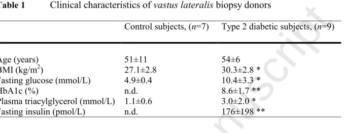
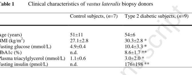
vastus lateralis biopsies and cultured as described (Cozzone et al., 2008). Clinical characteristics of healthy control subjects and patients with type 2 diabetes, from whom vastus lateralis biopsies
were obtained, are reported in table 1. Experimental protocols handling human biopsies were
approved by the Ethical Committees of “Hospices Civils de Lyon” and the Alfred Hospital
(Melbourne, Australia).
Insulin-resistance was induced by 24h insulin treatment (1 µM for L6, C2C12, 100 nM for human
myotubes) (Pirola et al., 2003) in the presence or absence of 100 µM resveratrol. After 24h,
insulin was removed for 1h by replacing DMEM, followed by 10-min stimulation with 1 µM
insulin. As TNFα induces insulin-resistance in cultured cells (de Alvaro et al., 2004), we also
treated L6 cells with 2 nM TNFα for 48h, followed by medium replacement and insulin
stimulation. TNFα treatment was done alone or in parallel with 100 µM resveratrol.
2.2 Western blotting, Immunoprecipitations, glucose uptake and RNA-interference. Cells
Accepted Manuscript
cell lysates were incubated with protein A-Sepharose and anti-SIRT1, IRS1, IRS2 antibodies.Standard immunoblotting procedures and ECL detection were employed. Glucose uptake assays
were performed as previously described (Frojdo et al., 2007).
siRNAs targeting SIRT1 and IRS2 were from Qiagen. Transfection with siRNAs was performed
by calcium phosphate precipitation. Sense sequences of siRNAs were as follows: SIRT1:
5’-CCCUGUAAAGCUUUCAGAAdTdT; IRS2: 5’-CACUCGGACAGCUUCUUCUdTdT.
2.3 C. elegans studies. The strains wild-type N2-bristol and VC199 sir-2.1 mutant
(Caenorhabditis Genetic Center) were used. sir-2.1 mutants were outcrossed three times with N2.
Constructs and transgenic lines. Vectors driving the expression of PI3K-p110 catalytic subunit under the control of the daf-18 promoter were obtained by inserting p110α-wt/KD-CAAX cDNA
fragments from pcDNA3-p110α-CAAX (Khwaja et al., 1998) into pPD95.75-daf-18p (Solari et
al., 2005). p110α-wt/KD-CAAX were also expressed as EGFP-fusion plasmid to verify
expression and activity in 293 cells. Wild-type worms were injected with 20 ng/μl of
daf-18p-p110α-wt-CAAX or daf-18p-p110α-KD-CAAX vectors. The injection marker plasmid
pRF4(rol-6) was co-injected at 100 ng/μl, causing a rolling phenotype.
Lifespan assays. C.elegans were maintained on nematode growth medium plates and fed with E.coli strain OP50. Lifespan assays were performed at 20°C. Animals were grown at 20°C until the L4 stage and then shifted to plates containing 10 µM 5-fluorodeoxyuracile to prevent growth
of progeny. The shift day was counted as day 0 in the lifespan assay. Animals were scored as
dead when they ceased moving and responding to prodding.
Accepted Manuscript
Student’s t test and Kaplan Meier log-rank for C. elegans lifespan comparisons) are indicated inAccepted Manuscript
3. RESULTS3.1 SIRT1 protein expression is decreased in muscle biopsies and primary myotubes derived from type 2 diabetic subjects.
Research on cultured cell lines indicated that SIRT1 is required to achieve full insulin-induced
IRS2 tyrosine phosphorylation in hepatocytes (Zhang, 2007). As peripheral insulin-resistance in
type 2 diabetes is characterised by decreased insulin action in skeletal muscle (Fig. 1A and
(Cozzone et al., 2008)), the tissue prominently contributing to glucose disposal, we investigated
whether alterations of SIRT1 expression occur in muscle biopsies and primary myotubes derived
from patients with type 2 diabetes. A significant reduction in SIRT1 protein content in both
primary skeletal muscle cells (Fig. 1B) and muscle biopsy samples (Fig. 1C) was observed in the
patient cohort relative to control subjects. This difference was likely due to post-transcriptional
modifications, as no differences in SIRT1 mRNA levels were observed between controls and type
2 diabetic patients (data not shown). Consistent with these data, a recent study demonstrated that
SIRT1 protein stability can be promoted by phosphorylation-dependent regulation (Ford et al.,
2008).
3.2 SIRT1 modulates proximal insulin signaling by interacting with p85 PI3K
The observation that SIRT1 protein expression is diminished in muscle biopsies and primary
myotubes from insulin-resistant type 2 diabetic subjects prompted us to study the impact of
SIRT1 on downstream insulin signaling skeletal muscle-derived cell lines, and HEK293 cells.
We hypothesized that, since skeletal muscle contributes prominently to glucose disposal,
functional interaction between SIRT1 and insulin signaling in these tissue is relevant for the
Accepted Manuscript
Firstly, we evaluated whether modulation of SIRT1 expression levels had an impact on theinsulin responsiveness of cells. Downregulation of SIRT1 by RNAi resulted in a decrease of
insulin-induced PKB-Ser473 phosphorylation after 10-minutes, 10 nM insulin treatment in
HEK293 cells (Fig. 2A), while no difference was seen at saturating insulin concentrations
(100-1000 nM), suggesting a modulatory function for SIRT1 at submaximal insulin concentrations.
Similarly, in L6 myoblasts treated with siRNAs to SIRT1, insulin-induced phosphorylation of
PKB was reduced, with the most prominent effect at 10 and 100 nM insulin (Fig. 2B). In L6
cells, RNAi to IRS2 was used as a positive control for insulin signaling downregulation (Pirola et
al., 2003). Next, we investigated whether SIRT1 overexpression could improve insulin
responsiveness. Overexpression in 293 cells of SIRT1 by calcium phosphate transfection resulted
in increased phosphorylation of PKB in response to 10 nM insulin stimulation for 10 minutes as
compared to untransfected or GFP-transfected cells (Fig. 3A). Adenoviral-mediated
overexpression of SIRT1 in L6 myoblasts (Fig. 3B) and primary human myotubes (Fig. 3C) also
resulted in increased phosphorylation of PKB, in response to stimulation with 1 M (in L6) or 100 nM (in primary myotubes) insulin. Thus, increased SIRT1 expression level improved the
insulin-induced phosphorylation of PKB in multiple cell types.
Collectively, these results show that modulation of SIRT1 levels have a direct impact on insulin
action, with increased levels of SIRT1 giving a stronger response to insulin and decreased SIRT1
levels reducing the response. These data suggest the existence of a direct interplay between
SIRT1 and the insulin signaling pathway. To further explore the molecular mechanisms involved,
HEK293 cells were transfected with either IRS2 or SIRT1 and stimulated with insulin. Cell
lysates were immunoprecipitated with antibodies to IRS2 or SIRT1 and probed with
anti-phosphotyrosine antibodies (Fig. 4A, upper panel). SIRT1 immunoprecipitates from
Accepted Manuscript
representing tyrosine phosphorylated IRS1/2, as the same signal was also present in IRS2immunoprecipitates (Figure 4A, upper panel, lanes 2,4). Furthermore, we detected
tyrosine-phosphorylated proteins between the 72-95 kDa molecular weight markers. Therefore, we
performed an anti-PI3K-p85 western blot (Fig. 4A, lower panel), showing that p85 co-immunoprecipitates with IRS2 and SIRT1. These results suggest the putative existence of
interactions between SIRT1, the p85-PI3K adapter and IRS proteins. To further demonstrate such
interaction, SIRT1, IRS1 and IRS2 were co-overexpressed in HEK293 followed by
immunoprecipitation of SIRT1. Western-blotting to p85-PI3K (Fig. 4B,C) indicated that p85 immunoprecipitates with SIRT1, but not with control IgG (Fig. 4D). SIRT1-p85 co-immunoprecipitation occurred independently from insulin stimulation, pointing to the existence
of a constitutive complex between SIRT1 and PI3K that could be observed both upon SIRT1 (or
IRS and SIRT1) overexpression (Fig. 4B,C, lanes 3 to 6), as well as on endogenous proteins (Fig.
4B,C, lanes 1-2). Further western blotting to IRS1 and IRS2 (Fig. 4E,F) on SIRT1
immunoprecipitates confirmed, as previously reported (Zhang, 2007), an insulin-induced
interaction between SIRT1 and IRS1 (Fig 4E), and between SIRT1 and IRS2 (Fig. 4F). Overall,
considering that SIRT1 co-immunoprecipitates tyrosine phosphorylated proteins and the PI3K
adapter p85, it is likely that the IRS-PI3K-SIRT1 complex built upon insulin stimulation, from a pre-existing SIRT1-p85 complex, potentiates the insulin signaling cascade.
3.3 Resveratrol reverts insulin- and TNFα-induced insulin-resistance by inhibiting IRS protein serine/threonine phosphorylation
Resveratrol has been shown to activate SIRT1 (Borra et al., 2005; Howitz et al., 2003) and has
subsequently been used to demonstrate the beneficial effects of SIRT1 on longevity and
Accepted Manuscript
resveratrol has also been shown to inhibit short term insulin action independently from SIRT1(Zhang 2006), likely through PI3K inhibition (Frojdo et al., 2007). Therefore, we investigated the
effects of resveratrol on insulin signaling in muscle cell lines in the light that resveratrol may act
either via SIRT1 activation or PI3K inhibition.
Firstly, we studied the impact of resveratrol treatment on IRS1 tyrosine phosphorylation and
IRS1Ser307/IRS2Thr348 (equivalent to IRS1Ser307) phosphorylation (Solinas et al., 2006),
which mirror the proteins’ activatory potential. L6 myoblasts overexpressing IRS1 or IRS2 were
pre-treated with resveratrol (5-100 M) prior to insulin stimulation. Serine/threonine and tyrosine phosphorylation of immunoprecipitated IRS1/2 and associated PI3K-p85 were monitored with the respective antibodies. We observed that resveratrol pre-treatment did not affect tyrosine
phosphorylation levels of either IRS1 or IRS2, nor the interaction of IRS proteins with
PI3K-p85 (Fig. 5A). However, resveratrol reduced the phosphorylation of IRS1Ser307 and IRS2Thr348 (Fig. 5A), which are known to be markers of insulin resistance (Draznin, 2006).
Decreased IRS1/2 serine/threonine phosphorylation also correlated to inhibition of JNK (Fig. 5A)
and, as previously reported (Frojdo et al., 2007), PKB phosphorylations. As JNK is responsible
for IRS1Ser307 and IRS2Thr348 phosphorylations, we propose that resveratrol affects
IRS-Ser/Thr phosphorylation status via inhibition of a protein kinases cascade, with PKB being
putatively upstream of JNK. That resveratrol acts as a protein kinase inhibitor, rather than SIRT1
activator, in this experimental setup, is further confirmed by the observation that SIRT1
overexpression – expected to mimic resveratrol activation - did not decrease insulin-induced
IRS1 phosphorylation on Serine307 (Fig. 5B) and SIRT1 knock-down did not affect the
inhibitory action of resveratrol on insulin-stimulated PKB phosphorylation (Fig. 5C).
Accepted Manuscript
independently from changes in IRS proteins tyrosine phosphorylation (Fig. 5B) further confirmsthat the relevant molecular event resides in the p85-SIRT1 interaction, which occurs downstream
of IRS1/2 tyrosine phosphorylation events.
We next asked whether resveratrol-dependent inhibition of IRS1/2 serine phosphorylation could,
over the long-term, improve the insulin sensitivity of cells subjected to chronic treatment with
insulin or TNFα, which both lead to insulin-resistance. In the three muscle cell lines tested (L6,
C2C12 and primary myotubes), the 24h chronic insulin treatment induced marked
insulin-resistance, as monitored by reduced PKB-Ser473 phosphorylation following acute insulin
stimulation (Fig. 6A). Within this experimental system, we observed that resveratrol
co-administration during chronic hyperinsulinemia alleviated the induction of insulin-resistance in
the three cell lines used, leading to increased phosphorylation of PKB upon acute insulin
stimulation (Fig. 6A). However, 24h simultaneous treatment with resveratrol slightly inhibited
glucose transport, in L6 myotubes (Fig. 6E), indicating that amelioration of a distal insulin
signaling response such as glucose uptake by resveratrol may require longer incubation times.
Interestingly, resveratrol also prevented insulin-resistance as induced by TNF (Fig. 6C), indicating a beneficial action in multiple models of induction of insulin resistance. Improvement
of PKB responsiveness upon prolonged resveratrol treatment was independent from changes in
SIRT1 expression, as assessed in L6 cells in which SIRT1 protein levels were unaltered after 24
hours exposure to insulin or insulin and resveratrol (Fig. 6B). Furthermore, the independency
from SIRT1 of cell responses to resveratrol have also been verified in SIRT1 knock-down L6
cells, in which the alleviating effect of resveratrol on insulin-induced insulin resistance was
retained (Fig. 6D). Collectively, these results indicate that multiple inducers of insulin-resistance
Accepted Manuscript
IRS2Thr348) including PI3K-mTOR (Frojdo et al., 2007; Ozes et al., 2001) and JNK (Fig. 5A),rather than SIRT1 activation.
3.4 In C.elegans, PI3K action is dependent on SIR-2.1
To study the interplay between insulin signaling and SIRT1 in an entire organism, we used
C.elegans as a model system in which IIS and SIRT1 actions can be monitored by assessing lifespan. To fully induce IIS we generated transgenic worms expressing a membrane-targeted
constitutively active PI3K(wt) and, as a control, worms expressing a membrane-targeted, but
catalytically inactive PI3K (herein referred to as kinase dead, KD). PI3K(wt) worms displayed
body size defects, in that they appeared shorter but larger than wild-type worms (Fig. 7A), while
PI3K(KD) and wild-type controls were indistinguishable (data not shown). Furthermore,
expression of PI3K(wt) reduced the average lifespan by 15% in 3 independent transgenic lines
compared with wild-type N2 and PI3K(KD) worms (Fig. 7B). Thus, constitutive IIS activation
via PI3K expression decreases C.elegans lifespan, in agreement with previous genetic reports
showing that mild downregulation of IIS by mutations in the PI3K homologue age-1
(Ayyadevara et al., 2008) or overexpression of the PTEN homologue DAF-18 (Solari et al., 2005)
conversely prolong lifespan.
Having demonstrated that PI3K(wt) expression shortens lifespan, we crossed PI3K(wt) transgenic
worms with worms mutated for the SIRT1 homolog sir-2.1, with the rationale that if SIRT1
directly contributes to IIS signaling, its ablation may affect the short-lived phenotype induced by
PI3K(wt) expression. Indeed, while sir-2.1 null worms had lifespan comparable to N2 wild-type
worms (Figure 7C), the sir-2.1 null allele crossed within the constitutively active PI3K(wt) strain
showed normal body morphology and reverted the reduced lifespan phenotype (Fig. 7D). Thus,
Accepted Manuscript
likely by exerting a permissive function in driving 3`-phosphoinositides lipid messengerproduction by PI3K.
Overall, these data indicate that, in a whole organism, SIRT1 mediates the effects of constitutive
activation of PI3K. The data also demonstrate that the functional interaction between SIRT1 and
insulin signaling is highly conserved between human cells and invertebrate species and
Accepted Manuscript
4. DISCUSSIONAs SIRT1 exerts positive metabolic effects through several mechanisms (reviewed in (Chaudhary
and Pfluger, 2009), and in view of the decreased SIRT1 protein expression observed by us
(Figure 1) and others (de Kreutzenberg et al., 2010) in muscle and peripheral blood mononuclear
cells from insulin resistant patients, we sought to investigate the role of SIRT1, and its activator
resveratrol, on the insulin signaling cascade in muscle cells, since in this cell type insulin
signaling is quantitatively relevant for the disposal of circulating glucose. Furthermore, we
studied the interplay of IIS and the SIRT1 homolog SIR-2.1 in the model organism C. elegans,
monitoring lifespan as readout, to define the interaction between IIS and SIRT1 in an entire
organism.
Downregulation of SIRT1 expression levels by RNAi diminished insulin-stimulated PKB
phosphorylation when sub-maximal insulin concentrations were used (10 nM in 293 cells and
10-100 nM in L6). Conversely, at saturating insulin concentrations (1 in L6 and 10-100 nM in 293 cells), PKB responsiveness was unaltered. This may indicate that SIRT1 plays a modulatory role
within the insulin signaling cascade at submaximal insulin concentrations. Reciprocally, we
observed increased insulin-stimulated PKB phosphorylation upon overexpression of SIRT1 in
muscle-derived cell lines, including human primary myotubes, and HEK293. Thus, our data
suggest that SIRT1 positively modulates the activity of upstream components in the insulin
pathway. In contrast to our observations, Yoshizaki and colleagues reported that SIRT1
overexpression in 3T3-L1 adipocytes was not sufficient to increase insulin-stimulated PKB and
MAPK phosphorylations (Yoshizaki et al., 2009). These contrasting data may be explained on
the basis of the different cell lines analyzed, the different adenoviral constructs and multiplicities
of infection employed to achieve SIRT1 overexpression and the different insulin concentrations
Accepted Manuscript
concentration in adenoviral-infected myotubes to overcome a higher basal PKB phosphorylationinduced by adenoviral overexpression .
To investigate whether the SIRT1 modulation of insulin-stimulated PKB phosphorylation
depends on a direct interaction of SIRT1 with the insulin signaling pathway, we performed
co-immunoprecipitation experiments which showed that SIRT1 interacts with tyrosine
phosphorylated proteins, and with the PI3K-p85 adapter in a constitutive manner, leading to the formation of a SIRT1-p85-IRS1/2 ternary complex upon insulin stimulation in an overexpression
system (Fig. 4, E,F). The existence of a modulatory role of SIRT1 in receptor tyrosine kinase
dependent signaling pathways has previously been shown in neurons (Li et al., 2008) and human
cancer cell lines (Ota et al., 2006). In these studies, inhibition of SIRT1 by sirtinol or
nicotinamide decreased IGF-1 and EGF induced Ras-MAPK signaling, leading to
neuroprotection and senescence-like growth arrest, respectively (Li et al., 2008; Ota et al., 2006).
In contrast, numerous studies indicate that SIRT1 activation exerts a beneficial role in the control
of metabolism (reviewed in (Elliott and Jirousek, 2008)). In accordance with these previous
studies, the SIRT1 knock-down and overexpression experiments in muscle cells, demonstrating
that SIRT1 positively participates in the activation of insulin signaling, underscore the
importance of SIRT1 action in increasing insulin signaling via its constitutive association to
PI3K-p85 adapter. Our observations add a further mechanism of direct action of SIRT1 on the insulin signaling pathway that complements the previously reported mechanism by Sun et al.
implicating transcriptional downregulation of PTP1B (Sun et al., 2007). As pointed out above,
the observation that SIRT1 protein expression is diminished in insulin-resistant primary
myotubes derived from type 2 diabetic patients and in vastus lateralis biopsies from patients with
Accepted Manuscript
SIRT1 downregulation in T2D subjects is likely dependent on accelerated degradation, asdecreased protein amounts were observed in spite of unaltered SIRT1 mRNA levels. This
observation is reminiscent of increased fasting-induced SIRT1 protein levels in liver in the
absence of variation of the corresponding mRNA (Rodgers et al., 2005).
Besides it action on SIRT1 (Howitz et al., 2003) - which is controversial (Kaeberlein et al. 2005;
Pacholec et al., 2010) - resveratrol has been shown to act on a plethora of different molecular
targets (reviewed in (Harikumar and Aggarwal, 2008)), including insulin signaling enzymes
(Zhang, 2006). Since several of the biological effects mediated by resveratrol, such as increased
insulin sensitivity and prolonged lifespan, could also be explained by modulation of insulin
signaling, we sought to define more precisely the implication of the insulin signaling pathway in
the beneficial metabolic effects of resveratrol. In L6 myoblasts, short pre-treatment with
resveratrol did not affect insulin induced tyrosine phosphorylation of IRS1/2, nor their interaction
with p85 (Fig. 5A). In contrast, IRS1Ser307, and the homologous IRS2Thr348, phosphorylations were totally blocked by >50 M resveratrol. We suggest that inhibition of IRS1/2 Ser/Thr phosphorylation depends on direct inhibition of protein kinases by resveratrol
rather than an indirect action initiated by SIRT1 activation, inasmuch SIRT1 overexpression, as
opposed to resveratrol treatment, did not affect insulin-stimulated IRS1Ser307 phosphorylation
(Fig. 5B).
Given that phosphorylations on IRS1Ser307 and IRS2Thr348 relay a feedback inhibition of the
insulin signaling pathway (Lee et al., 2003; Solinas et al., 2006) and are known to be augmented
in insulin resistance (Morino et al., 2005), we evaluated whether induction of insulin-resistance in
Accepted Manuscript
by resveratrol administration (Fig. 6). In all the muscle cells studied, we observed thatco-incubation of resveratrol during chronic insulin treatment resulted in improved insulin
responsiveness. Similarly, resveratrol diminished the extent of insulin-resistance induced by
TNFα treatment in L6 myoblasts. A previous study by Sun et al. (Sun et al., 2007) demonstrated
that palmitate- and glucosamine-induced insulin-resistance was accompanied by decreased
SIRT1 protein levels in hepatocyte-derived cell lines. In our experiments, insulin-resistance
induced by TNFα in L6 or insulin in L6, C2C12 and human primary myotubes did not coincide
with decreased SIRT1 protein expression as observed in other studies (Sun et al., 2007;
Yoshizaki et al., 2009). This discrepancy is likely explained by the different treatment used to
induce insulin-resistance (insulin/TNF versus palmitate/glucosamine) and by the different cell types used. In spite of these differences, our results showing that resveratrol treatment
ameliorates PKB phosphorylation in insulin resistant cells support the data by Sun et al. (Sun et
al., 2007) and are in line with a recent report showing SIRT1 depletion in peripheral blood
monocytes cells from insulin resistant subjects but lack of SIRT1 downregulation in
insulin-treated THP-1 monocytes (de Kreutzenberg et al., 2010).
Overall, the above findings suggest that direct interaction between SIRT1 and insulin signaling
does exist in cell culture models. To obtain insight on the occurrence of IIS and SIRT1 interplay
in a whole organism we used C. elegans as model system. We constructed C.elegans transgenic
worms overexpressing constitutively active PI3K(wt). Since mild inhibition of key components
of IIS signaling in C. elegans - i.e. DAF-2, and AGE-1; homologous to insulin-receptor and PI3K
respectively - leads to lifespan prolongation (Tatar et al., 2003), we reasoned that chronic
activation of IIS signaling by expression of constitutively active PI3K would shorten the worm’s
Accepted Manuscript
the interaction between IIS and SIRT1 we crossed PI3K(wt) transgenic worms with sir-2.1 nullworms, to define whether SIR-2 was required for lifespan shortening induced by constitutively
active PI3K. Interestingly, while lifespan of sir-2.1(-) worms was comparable to that of N2
wild-type C. elegans (Figure 7C), lifespan of PI3K(wt);sir-2.1(-) worms was reverted to normal
(Figure 7D), indicating that SIR-2.1 mediates the life-shortening action of PI3K(wt) and
supporting the existence of a specific regulatory network encompassing SIRT1 and PI3K in the
determination of lifespan. These observations are unexpected in light of data reported by
Tissenbaum et al. showing that SIR-2.1 overexpression prolongs C.elegans lifespan in a
DAF-16/FOXO dependent manner (Tissenbaum and Guarente, 2001). We, therefore, expected that
SIR-2.1 inactivation should have been deleterious rather than beneficial for lifespan.
Furthermore, recent results from Berdichervsky et al. (Berdichevsky et al., 2006) showed that
SIR-2.1 activates DAF-16 via deacetylation under stress conditions. However, they also
demonstrated that SIR-2.1/DAF-16 interaction is unrelated to lifespan regulation by the IIS
pathway. In this latter context, our results show that when PI3K is constitutively activated,
SIR-2.1 potentiates - rather than antagonizes – the PI3K role in lifespan control. This functional
interaction involves – rather than a FOXO/DAF-16 pathway – a SIR-2.1 interaction with other
upstream components of the IIS pathway. The recovery of normal lifespan in PI3K(wt); sir-2.1(-)
worms can be explained by postulating that SIR-2.1 acts either downstream, or - as our
experiments in mammalian cells suggest - in parallel to PI3K (Fig. 8). Overall, lifespan data in
C.elegans suggest that the results obtained in cell culture, demonstrating a functional interaction between SIRT1 and insulin signaling, are transposable to an entire organism.
In conclusion, our experiments support the existence of a functional interplay between insulin
signaling/IIS and SIRT1 both in muscle derived cell lines and C.elegans, leading to positive
Accepted Manuscript
how SIRT1 regulates insulin action and IIS. Furthermore, we provide evidence that resveratrolcan reverse insulin resistance through targets other than SIRT1, as demonstrated by the inhibition
of protein serine kinases that result in decreased phosphorylation of IRS1/2, indicating that the
pleiotropic actions of resveratrol should be taken into account when studying its effects on
Accepted Manuscript
5. ACKNOWLEDGEMENTSWe thank Drs. B.M. Burgering (University Medical Centre Utrecht, Utrecht, The Netherlands)
and E. Verdin (Gladstone Institute of Virology and Immunology, University of California,) for
donation of SIRT1 plasmids. We acknowledge Cyrille Debard and Graeme Lancaster for
preparation and maintenance of human myotubes and discussion respectively. Nematode strains
were provided by the Caenorhabditis Genetic Center. This work was supported by INSERM (To
LP, Programme National de Recherche sur le Diabète) and “Association pour la Recherche contre
le Cancer” (to FS). SF was supported by a French University Ministry pre-doctoral fellowship. . LP was partly supported by the French-Australian INSERM-NHMRC programme. AE-O, MAF
Accepted Manuscript
REFERENCESAyyadevara, S., Alla, R., Thaden, J.J., and Shmookler Reis, R.J. (2008). Remarkable longevity and stress resistance of nematode PI3K-null mutants. Aging Cell 7, 13-22.
Baur, J.A., Pearson, K.J., Price, N.L., Jamieson, H.A., Lerin, C., Kalra, A., Prabhu, V.V., Allard, J.S., Lopez-Lluch, G., Lewis, K., et al. (2006). Resveratrol improves health and survival of mice on a high-calorie diet. Nature 444, 337-342.
Berdichevsky, A., Viswanathan, M., Horvitz, H.R., and Guarente, L. (2006). C. elegans SIR-2.1 interacts with 14-3-3 proteins to activate DAF-16 and extend life span. Cell 125, 1165-1177. Bordone, L., Motta, M.C., Picard, F., Robinson, A., Jhala, U.S., Apfeld, J., McDonagh, T., Lemieux, M., McBurney, M., Szilvasi, A., et al. (2006). Sirt1 regulates insulin secretion by repressing UCP2 in pancreatic beta cells. PLoS Biol 4, e31.
Borra, M.T., Smith, B.C., and Denu, J.M. (2005). Mechanism of human SIRT1 activation by resveratrol. J Biol Chem 280, 17187-17195.
Chaudhary, N., and Pfluger, P.T. (2009). Metabolic benefits from Sirt1 and Sirt1 activators. Curr Opin Clin Nutr Metab Care 12, 431-437.
Chaussade, C., Pirola, L., Bonnafous, S., Blondeau, F., Brenz-Verca, S., Tronchere, H., Portis, F., Rusconi, S., Payrastre, B., Laporte, J., et al. (2003). Expression of myotubularin by an adenoviral vector demonstrates its function as a phosphatidylinositol 3-phosphate [PtdIns(3)P] phosphatase in muscle cell lines: involvement of PtdIns(3)P in insulin-stimulated glucose transport. Mol Endocrinol 17, 2448-2460.
Cozzone, D., Frojdo, S., Disse, E., Debard, C., Laville, M., Pirola, L., and Vidal, H. (2008). Isoform-specific defects of insulin stimulation of Akt/protein kinase B (PKB) in skeletal muscle cells from type 2 diabetic patients. Diabetologia 51, 512-521.
de Alvaro, C., Teruel, T., Hernandez, R., and Lorenzo, M. (2004). Tumor necrosis factor alpha produces insulin resistance in skeletal muscle by activation of inhibitor kappaB kinase in a p38 MAPK-dependent manner. J Biol Chem 279, 17070-17078.
de Kreutzenberg, S.V., Ceolotto, G., Papparella, I., Bortoluzzi, A., Semplicini, A., Dalla Man, C., Cobelli, C., Fadini, G.P., and Avogaro, A. (2010). Downregulation of the longevity-associated protein sirtuin 1 in insulin resistance and metabolic syndrome: potential biochemical mechanisms. Diabetes 59, 1006-1015.
Draznin, B. (2006). Molecular mechanisms of insulin resistance: serine phosphorylation of insulin receptor substrate-1 and increased expression of p85alpha: the two sides of a coin. Diabetes 55, 2392-2397.
Elliott, P.J., and Jirousek, M. (2008). Sirtuins: novel targets for metabolic disease. Curr Opin Investig Drugs 9, 371-378.
Ford, J., Ahmed, S., Allison, S., Jiang, M., and Milner, J. (2008). JNK2-dependent regulation of SIRT1 protein stability. Cell Cycle 7, 3091-3097.
Frescas, D., Valenti, L., and Accili, D. (2005). Nuclear trapping of the forkhead transcription factor FoxO1 via Sirt-dependent deacetylation promotes expression of glucogenetic genes. J Biol Chem 280, 20589-20595.
Frojdo, S., Cozzone, D., Vidal, H., and Pirola, L. (2007). Resveratrol is a class IA phosphoinositide 3-kinase inhibitor. Biochem J 406, 511-518.
Gerhart-Hines, Z., Rodgers, J.T., Bare, O., Lerin, C., Kim, S.H., Mostoslavsky, R., Alt, F.W., Wu, Z., and Puigserver, P. (2007). Metabolic control of muscle mitochondrial function and fatty acid oxidation through SIRT1/PGC-1alpha. Embo J 26, 1913-1923.
Accepted Manuscript
Harikumar, K.B., and Aggarwal, B.B. (2008). Resveratrol: a multitargeted agent for age-associated chronic diseases. Cell Cycle 7, 1020-1035.Holzenberger, M., Kappeler, L., and De Magalhaes Filho, C. (2004). IGF-1 signaling and aging. Exp Gerontol 39, 1761-1764.
Howitz, K.T., Bitterman, K.J., Cohen, H.Y., Lamming, D.W., Lavu, S., Wood, J.G., Zipkin, R.E., Chung, P., Kisielewski, A., Zhang, L.L., et al. (2003). Small molecule activators of sirtuins extend Saccharomyces cerevisiae lifespan. Nature 425, 191-196.
Kaeberlein, M., McDonagh, T., Heltweg, B., Hixon, J., Westman, E.A., Caldwell, S.D., Napper, A., Curtis, R., DiStefano, P.S., Fields, S., et al. (2005). Substrate-specific activation of sirtuins by resveratrol. J Biol Chem 280, 17038-17045.
Khwaja, A., Lehmann, K., Marte, B.M., and Downward, J. (1998). Phosphoinositide 3-kinase induces scattering and tubulogenesis in epithelial cells through a novel pathway. J Biol Chem 273, 18793-18801.
Lagouge, M., Argmann, C., Gerhart-Hines, Z., Meziane, H., Lerin, C., Daussin, F., Messadeq, N., Milne, J., Lambert, P., Elliott, P., et al. (2006). Resveratrol Improves Mitochondrial Function and Protects against Metabolic Disease by Activating SIRT1 and PGC-1alpha. Cell 127, 1109-1122. Lee, J.H., Song, M.Y., Song, E.K., Kim, E.K., Moon, W.S., Han, M.K., Park, J.W., Kwon, K.B., and Park, B.H. (2009). Overexpression of SIRT1 protects pancreatic beta-cells against cytokine toxicity by suppressing the nuclear factor-kappaB signaling pathway. Diabetes 58, 344-351.
Lee, Y.H., Giraud, J., Davis, R.J., and White, M.F. (2003). c-Jun N-terminal kinase (JNK) mediates feedback inhibition of the insulin signaling cascade. J Biol Chem 278, 2896-2902. Li, Y., Xu, W., McBurney, M.W., and Longo, V.D. (2008). SirT1 Inhibition Reduces IGF-I/IRS-2/Ras/ERK1/2 Signaling and Protects Neurons. Cell Metab 8, 38-48.
Liang, F., Kume, S., and Koya, D. (2009). SIRT1 and insulin resistance. Nat Rev Endocrinol 19, 19.
Mihaylova, V.T., Borland, C.Z., Manjarrez, L., Stern, M.J., and Sun, H. (1999). The PTEN tumor suppressor homolog in Caenorhabditis elegans regulates longevity and dauer formation in an insulin receptor-like signaling pathway. Proc Natl Acad Sci U S A 96, 7427-7432.
Morino, K., Petersen, K.F., Dufour, S., Befroy, D., Frattini, J., Shatzkes, N., Neschen, S., White, M.F., Bilz, S., Sono, S., et al. (2005). Reduced mitochondrial density and increased IRS-1 serine phosphorylation in muscle of insulin-resistant offspring of type 2 diabetic parents. J Clin Invest 115, 3587-3593.
Nemoto, S., Fergusson, M.M., and Finkel, T. (2005). SIRT1 functionally interacts with the metabolic regulator and transcriptional coactivator PGC-1{alpha}. J Biol Chem 280, 16456-16460.
North, B.J., Marshall, B.L., Borra, M.T., Denu, J.M., and Verdin, E. (2003). The human Sir2 ortholog, SIRT2, is an NAD+-dependent tubulin deacetylase. Mol Cell 11, 437-444.
Ota, H., Tokunaga, E., Chang, K., Hikasa, M., Iijima, K., Eto, M., Kozaki, K., Akishita, M., Ouchi, Y., and Kaneki, M. (2006). Sirt1 inhibitor, Sirtinol, induces senescence-like growth arrest with attenuated Ras-MAPK signaling in human cancer cells. Oncogene 25, 176-185.
Ozes, O.N., Akca, H., Mayo, L.D., Gustin, J.A., Maehama, T., Dixon, J.E., and Donner, D.B. (2001). A phosphatidylinositol 3-kinase/Akt/mTOR pathway mediates and PTEN antagonizes tumor necrosis factor inhibition of insulin signaling through insulin receptor substrate-1. Proc Natl Acad Sci U S A 98, 4640-4645.
Pacholec, M., Bleasdale, J.E., Chrunyk, B., Cunningham, D., Flynn, D., Garofalo, R.S., Griffith, D., Griffor, M., Loulakis, P., Pabst, B., et al. SRT1720, SRT2183, SRT1460, and resveratrol are not direct activators of SIRT1. J Biol Chem 285, 8340-8351.
Accepted Manuscript
Picard, F., Kurtev, M., Chung, N., Topark-Ngarm, A., Senawong, T., Machado De Oliveira, R., Leid, M., McBurney, M.W., and Guarente, L. (2004). Sirt1 promotes fat mobilization in white adipocytes by repressing PPAR-gamma. Nature 429, 771-776.Pirola, L., Bonnafous, S., Johnston, A.M., Chaussade, C., Portis, F., and Van Obberghen, E. (2003). Phosphoinositide 3-kinase-mediated reduction of insulin receptor substrate-1/2 protein expression via different mechanisms contributes to the insulin-induced desensitization of its signaling pathways in L6 muscle cells. J Biol Chem 278, 15641-15651.
Rodgers, J.T., Lerin, C., Gerhart-Hines, Z., and Puigserver, P. (2008). Metabolic adaptations through the PGC-1alpha and SIRT1 pathways. FEBS Lett 582, 46-53.
Rodgers, J.T., Lerin, C., Haas, W., Gygi, S.P., Spiegelman, B.M., and Puigserver, P. (2005). Nutrient control of glucose homeostasis through a complex of PGC-1alpha and SIRT1. Nature 434, 113-118.
Rogina, B., and Helfand, S.L. (2004). Sir2 mediates longevity in the fly through a pathway related to calorie restriction. Proc Natl Acad Sci U S A 101, 15998-16003.
Solari, F., Bourbon-Piffaut, A., Masse, I., Payrastre, B., Chan, A.M., and Billaud, M. (2005). The human tumour suppressor PTEN regulates longevity and dauer formation in Caenorhabditis elegans. Oncogene 24, 20-27.
Solinas, G., Naugler, W., Galimi, F., Lee, M.S., and Karin, M. (2006). Saturated fatty acids inhibit induction of insulin gene transcription by JNK-mediated phosphorylation of insulin-receptor substrates. Proc Natl Acad Sci U S A 103, 16454-16459.
Sun, C., Zhang, F., Ge, X., Yan, T., Chen, X., Shi, X., and Zhai, Q. (2007). SIRT1 Improves Insulin Sensitivity under Insulin-Resistant Conditions by Repressing PTP1B. Cell Metab 6, 307-319.
Tatar, M., Bartke, A., and Antebi, A. (2003). The endocrine regulation of aging by insulin-like signals. Science 299, 1346-1351.
Tissenbaum, H.A., and Guarente, L. (2001). Increased dosage of a sir-2 gene extends lifespan in Caenorhabditis elegans. Nature 410, 227-230.
van der Horst, A., Tertoolen, L.G., de Vries-Smits, L.M., Frye, R.A., Medema, R.H., and Burgering, B.M. (2004). FOXO4 is acetylated upon peroxide stress and deacetylated by the longevity protein hSir2(SIRT1). J Biol Chem 279, 28873-28879.
Vaziri, H., Dessain, S.K., Ng Eaton, E., Imai, S.I., Frye, R.A., Pandita, T.K., Guarente, L., and Weinberg, R.A. (2001). hSIR2(SIRT1) functions as an NAD-dependent p53 deacetylase. Cell 107, 149-159.
Viswanathan, M., Kim, S.K., Berdichevsky, A., and Guarente, L. (2005). A role for SIR-2.1 regulation of ER stress response genes in determining C. elegans life span. Dev Cell 9, 605-615. Wood, J.G., Rogina, B., Lavu, S., Howitz, K., Helfand, S.L., Tatar, M., and Sinclair, D. (2004). Sirtuin activators mimic caloric restriction and delay ageing in metazoans. Nature 430, 686-689. Yoshizaki, T., Milne, J.C., Imamura, T., Schenk, S., Sonoda, N., Babendure, J.L., Lu, J.C., Smith, J.J., Jirousek, M.R., and Olefsky, J.M. (2009). SIRT1 exerts anti-inflammatory effects and improves insulin sensitivity in adipocytes. Mol Cell Biol 29, 1363-1374.
Zhang, J. (2006). Resveratrol inhibits insulin responses in a SirT1-independent pathway. Biochem J 397:519-527.
Zhang, J. (2007). The direct involvement of SirT1 in insulin-induced insulin receptor substrate-2 tyrosine phosphorylation. J Biol Chem 282, 34356-34364.
Accepted Manuscript
FIGURE LEGENDSFigure 1: SIRT1 protein levels are lower in myotubes and muscle biopsies from type 2
diabetic patients compared to control subjects. (A) Myotubes derived from control subjects
(control) or type 2 diabetic patients (T2D) were starved overnight and stimulated 10 minutes with
100 nM insulin. PKB pS473 phosphorylation was measured as a readout for the insulin resistant
state observed in primary myotubes derived from type 2 diabetic patients, as previously reported
(Cozzone et al., 2008). (B, Upper part) SIRT1 protein expression levels were evaluated by
immunoblotting in myotubes derived from controls and T2D patients. Separation bars indicate
non-contiguous lanes. Tubulin loading controls were as previously published (Cozzone et al.,
2008). As no difference in SIRT1 expression level was seen after the 10 minutes insulin
stimulation, both conditions were analysed together to quantify SIRT1 expression level (B, lower
part). Quantification is based on 10 samples. Controls, filled bar; T2D patients, empty bar. *p <
0.05 versus SIRT1 protein levels in controls. (C, Upper part) SIRT1 protein expression levels in
muscle biopsies from controls (C) and T2D patients (T2D). (B, lower part). Quantification is
based on 7 controls biopsies and 9 T2D biopsies and was adjusted to tubulin expression levels.
Controls, filled bar; T2D patients, empty bar. Data in (B,C) are expressed as means ± SEM. *p <
0.05 versus SIRT1 protein levels in controls.
Figure 2. SIRT1 modulates insulin signaling in 293 cells and L6 myoblasts. (A) 293 cells
were transfected with increasing concentrations (0 nM, 16.6 nM and 50 nM) of siRNA directed to
SIRT1 (as depicted graphically). 48 hours post-transfection cells were starved overnight and
Accepted Manuscript
separated proteins were immunoblotted with antibodies to IRS2 and PKB, to monitor proteinloading, SIRT1 and PKB pS473 as indicated. Lower panel : quantification of PKB pS473 ; results
are expressed as means ± SEM (n=4). * PKB pS473 levels in SIRT1 knock-down cells upon 10
nM insulin stimulation are significantly different from the respective control (p<0.05 at Student’s
t test). (B) L6 myoblasts were transfected with 50 nM siRNA to SIRT1 and IRS2 as indicated. 48
hours post-transfection myoblasts were starved overnight and stimulated for 10 min with the
indicated concentrations of insulin. SDS-PAGE separated proteins were immunoblotted with
antibodies to IRS2, SIRT1, PKB pS473, and PKB (to monitor protein loading), as indicated. **
Non-specific band.
Figure 3. SIRT1 overexpression increases insulin action in 293 cells, L6 myoblasts and
primary human myotubes. (A) 293 cells were transfected with increasing amounts of
pCDNA3<MYC-SIRT1> or pEGFP-C2 (1 to 5 g plasmid DNA). 48 hours post-transfection cells were starved overnight and stimulated for 10 min with the indicated concentrations of
insulin. Separated proteins were immunoblotted with antibodies to SIRT1 and PKB pS473 as
indicated. L6 myoblasts (B) and differentiated human myotubes (C) were infected with
adenoviruses coding for GFP, Flag-SIRT1 and MYC-SIRT1. 48 hours post-infection cells were
starved overnight and stimulated for 10 min with 1000 nM (L6) or 100 nM insulin (primary
myotubes). Separated proteins were immunoblotted with antibodies to SIRT1, PKB pS473 and
total PKB as indicated. Lanes of the pPKB blot from primary myotubes, from the same PVDF
membrane, were rearranged to maintain loading consistency with L6 cells.
SIRT1-Accepted Manuscript
transfected with expression plasmids for MYC-SIRT1 and IRS2 as indicated. 48 hourspost-transfection cells were starved overnight and stimulated or not for 10 min with 1000 nM insulin.
Cell lysates were immunoprecipitated with antibodies directed to IRS2 or SIRT1.
Immunoprecipitates were probed for phosphotyrosine content with anti-PY antibodies (upper
panel) and associated PI3K with anti p85 antibody (lower panel). * indicates a non specific (or unidentified) band. (B,C) 293 cells were co-transfected with expression plasmids for
MYC-SIRT1, IRS1 and IRS2 as indicated. 48 hours post-transfection cells were starved overnight and
stimulated or not for 10 min with 1000 nM insulin. p85, IRS1, IRS2 and SIRT1 expression levels
were verified on total lysates. Cell lysates were then immunoprecipitated with antibodies directed
to SIRT1 and immunoprecipitates were probed with antibodies directed to p85. The constitutive SIRT1-p85 association pattern has been observed in two independent experiments. (D) To monitor immunoprecipitation specificity, lysates from unstimulated and insulin-stimulated
SIRT1-overexpressing 293 cells were immunoprecipitated with antibodies directed to SIRT1 or
IgG. p85 and SIRT1 expression levels were verified on total lysates (upper blots) and SIRT1-associated p85was visualized by immunoblotting with anti-p85 antibodies (lower blot). (E,F) 293 cells were co-transfected with expression plasmids for MYC-SIRT1 and IRS1 or IRS2. 48
hours post-transfection cells were starved overnight and stimulated or not for 10 min with 1000
nM insulin. The expression of each protein was verified with the relevant antibody on total
lysates. Cell lysates were then immunoprecipitated with antibodies directed to SIRT1. SIRT1
immunoprecipitates were probed with antibodies directed to IRS1 (D) and IRS2 (E). IP,
Accepted Manuscript
Figure 5: Resveratrol inhibits insulin-stimulated serine phosphorylation of IRS proteinsand PKB/JNK phosphorylation independently from SIRT1 (A, upper part) L6 myoblasts
were infected with adenoviruses expressing IRS1 (left panels) and IRS2 (right panels). 48 hours
post-infection, myoblasts were serum-starved overnight, pre-treated for 20 min with the indicated
concentrations of resveratrol and subsequently stimulated 10 min with 1000 nM insulin. IRS1
and IRS2 were immunoprecipitated with the respective antibodies and immunoprecipitates were
immunoblotted with anti-PY, anti IRS-1 pS307 (that cross react with the homologous
phosphorylation site in IRS2) and anti p85 antibodies. (A, lower part) Phospho-JNK, PKB pS473 and SIRT1 were detected in total lysates. (B) L6 myoblasts were infected with IRS1 and
SIRT1 adenoviruses as indicated and transgene expression levels as well as PKB pS473 and total
PKB as loading control were visualized by immunoblotting (upper blots on total lysates). 48
hours post-infection, myoblasts were serum-starved overnight and subsequently stimulated with
insulin. IRS1 was immunoprecipitated and immunoblotted with anti phospho-tyrosine and anti
IRS-1 phosphoS307 (Lower blots). (C) 293 cells were transfected with siRNA directed to SIRT1
as indicated. 48 hours post-transfection cells were starved overnight, pretreated with 100 M RSV for 20 min and stimulated for 10 min with the indicated concentrations of insulin. Total cell
lysates were immunoblotted with antibodies to GAPDH, to monitor protein loading, SIRT1, total
PKB and PKB pS473 as indicated.
Figure 6: Resveratrol prevents insulin- and TNFα- induced insulin-resistance in muscle
cells without interfering on SIRT1 expression. Insulin-resistance was induced by incubation
for 24 hours with 1000 nM insulin in L6, C2C12 myoblasts and with 100 nM in primary
Accepted Manuscript
Simultaneously to the insulin-resistance inducing stimulus, cells were co-incubated with 100 M resveratrol where indicated. At the end of the 24/48 hours incubation period, cells were placed infresh DMEM medium for 1 hour and subsequently stimulated for 10 min with 1000 nM (L6,
C2C12) or 100 nM (primary myotubes) insulin as indicated. (A), Resveratrol-mediated protection
from insulin-induced insulin-resistance in L6 myoblasts, primary human myotubes and C2C12
cells. Upper panel, evaluation of Ser473 phosphorylation of PKB together with total PKB and/or
α-tubulin as loading controls in the three muscle cell lines. Lower panel, quantification of PKB
pS473 levels, adjusted to total PKB, in L6 from 6 independent experiments, results are means ±
SEM. PKB pS473 levels are expressed as arbitrary units. * PKB pS473 levels are significantly
diminished in the insulin-resistant condition versus non-insulin resistant cells p<0.05. ** PKB
pS473 levels are significantly increased in insulin resistant-cells co-treated with resveratrol,
p<0.05, Student’s t-test. (B) SIRT1 protein expression levels in L6 were quantified by western
blot and normalized for the tubulin loading control. ns: not statistically significant versus the
control condition. (C), Resveratrol-mediated protection from TNF- induced insulin-resistance in L6 myoblasts. Upper panel, evaluation of pPKB S473 and pMAPK levels. Lower panel,
quantification of PKB pS473 levels. Each bar represents the ratio between the basal and the
insulin-stimulated condition. Results are means ± SEM from 3 independent experiments. * PKB
pS473 fold activation is significantly diminished in the insulin-resistant condition versus
non-insulin resistant cells p<0.05. ** PKB pS473 fold activation is significantly increased in non-insulin
resistant-cells co-treated with resveratrol. p<0.05, Student’s t-test. (D) L6 myoblasts transfected
with siRNA to GFP or SIRT1 were treated and analyzed as in panel A. Resveratrol-induced
amelioration from insulin resistance was observed also in SIRT1 knock-down cells. (E)
Accepted Manuscript
adipocytes. L6 myoblasts were left untreated or treated for 24 hours with 1000 nM insulin and100 M resveratrol as indicated. Prior to performing glucose uptake, cells were switched to glucose free DMEM medium for 60 minutes. * Glucose uptake significantly increased by insulin
treatment versus untreated cells p<0.05. ** Glucose uptake significantly decreased by
insulin/RSV treated cells as compared to insulin-treated cells. p<0.05, Student’s t-test.
Figure 7: Constitutively active PI3K decreases C. elegans lifespan in a SIR-2 dependent
manner (A) Light microscopy pictures (20x magnification) of C. elegans wild-type N2 strain
and PI3K(wt) overexpressing strain, the latter displaying shortened and enlarged body shape. (B)
Expression of constitutively active PI3K in N2 worms (PI3K(wt)) reduced the lifespan of
wild-type worms (mean lifespan 13.3 ± 0.7, log-rank p<0.05 versus N2) compared to wild-wild-type N2.
Worms expressing the kinase dead PI3K (PI3K(KD)) had a lifespan comparable to wild-type
(mean lifespan 15.3 ± 0.7 and 15.6 ± 0.9, respectively, log-rank p=NS versus N2). Lifespan tests
have been repeated four times with three independent transgenic strains and gave similar results.
The survival curve show results obtained for one representative experiment. (C) wild-type N2
and sir-2.1 null C. elegans display comparable lifespan (mean lifespan 16.96 ± 0.63 and 16.90 ±
0.40, respectively, log-rank p=0.334) (D) sir-2.1 mediates reduction of lifespan induced by
constitutive activation of PI3K. PI3K(wt);sir-2.1(-) worms show an extension of lifespan
compared to wild-type worms expressing constitutively active PI3K (PI3K(wt)) (mean lifespan
20.0 ± 0.6 and 14.5 ± 0.7, respectively, log-rank p<0.0001). The average lifespans of sir-2.1(-)
mutants and PI3K(wt);sir2.1(-) worms are similar (19.7±0.6 and 20.0 ± 0.6, respectively,
Accepted Manuscript
Figure 8: Model for functional interaction between SIRT1 and the insulin pathway inmammalian muscle cells and in C.elegans. (A) SIRT1 is required for physiological activation
of the insulin pathway; (B) Downregulation of SIRT1 expression lowers insulin-induced PKB
Ser473 phosphorylation and may contribute to insulin-resistance; (C) Constitutive activation of
PI3K shortens lifespan, through PKB-AKT activation and inhibition of DAF-16/FOXO
transcription factor; (D) Absence of SIR-2.1 suppresses the effect of constitutive activation of
Accepted Manuscript
Table 1 Clinical characteristics of vastus lateralis biopsy donorsControl subjects, (n=7) Type 2 diabetic subjects, (n=9)
Age (years) 51±11 54±6
BMI (kg/m2) 27.1±2.8 30.3±2.8 * Fasting glucose (mmol/L) 4.9±0.4 10.4±3.3 *
HbA1c (%) n.d. 8.6±1.7 **
Plasma triacylglycerol (mmol/L) 1.1±0.6 3.0±2.0 * Fasting insulin (pmol/L) n.d. 176±198 **
Data are mean values ± SD. * denotes p<0.05 for type 2 diabetic patients versus control subjects, unpaired t-test. ** denotes clinically elevated parameters. High SD for fasting insulin is due to 2 study participants with high values (369 and 633 pmol/L).
Accepted Manuscript
Table 1 Clinical characteristics of vastus lateralis biopsy donorsControl subjects, (n=7) Type 2 diabetic subjects, (n=9)
Age (years) 51±11 54±6 BMI (kg/m2) 27.1±2.8 30.3±2.8 * Fasting glucose (mmol/L) 4.9±0.4 10.4±3.3 * HbA1c (%) n.d. 8.6±1.7 ** Plasma triacylglycerol (mmol/L) 1.1±0.6 3.0±2.0 * Fasting insulin (pmol/L) n.d. 176±198 **
Data are mean values ± SD. * denotes p<0.05 for type 2 diabetic patients versus control subjects, unpaired t-test. ** denotes clinically elevated parameters. High SD for fasting insulin is due to 2 study participants with high values (369 and 633 pmol/L).
Accepted Manuscript
Figure 1
PKB pS473 Controls T2DA
Insulin 10min 100nM - + - + - + - +SI
R
T
1
ex
pres
s
ion
lev
el,
arbit
rary
unit
s
Controls T2DB
SIRT1*
Insulin 10min 100nM - + - + - + - + - + - +C
SI
R
T
1
ex
pres
s
ion
lev
el,
arb
it
rary
units
*
Controls T2D0
1
2
3
SIRT1 C C Tubulin T2D T2D Figure 1Accepted Manuscript
Figure 2
A
IRS2 SIRT1 PKB pS473 siRNA to SIRT1 Insulin, nM 0 10 102 103 0 200 400 600 800 1000*
P K B p S 4 7 3 , a rbitrary u n itsB
IRS2 SIRT1 PKB pS473**
siRNA to - SIRT1 IRS2Insulin, nM 0 10 102103 0 10 1021030 10 102103 293 L6 PKB PKB Figure 2
Accepted Manuscript
Figure 3
FLA G -S IR T 1 GFP MY C -S IR T 1 103nM Insulin - + - + - + - + PKB pS473 SIRT1 AdenovirusB
C
L6 PKBA
PKB pS473 Insulin, nM 0 10 103 10 SIRT1 Overexpression - SIRT1 GFP 293 Figure 3Accepted Manuscript
Figure 4
IgG MW, kDa 170 130 95 72 p85 103nM insulin, 10 min - + - + - + IgGOverexpression - IRS2 SIRT1
IP to IRS2 SIRT1
A
p85a IRS1 SIRT1B
C
Total lysates IgG p85 IRS1/2 WB pTyr 103nM insulin, 10 min - + - + - + WB PI3K p85 Overexpression IRS1 - SIRT1 SIRT1 p85a 95 72 56 p85a IRS2 SIRT1 Total lysates IgG Overexpression IRS2 - SIRT1 SIRT1 p85a 95 72 56 IP: SIRT1, WB: p85a MW, kDa 10 nM insulin, 10 min - + - + - + 130 95 72 170 56*
IP: SIRT1,WB: p85a Figure 4Accepted Manuscript
Figure 4
Overexpression - IRS2 SIRT1 IP: SIRT1 WB: IRS2 IRS2 IRS2F
Total lysates Insulin - + - +Figure 4
Overexpression - SIRT1 SIRT1 p85a Overexpression - IRS1 SIRT1 IRS1 Myc-SIRT1 IRS1 IP: SIRT1 WB: IRS1E
IgG Insulin - + - +IP to: SIRT1 IgG
WB: p85a
Insulin - + - +