Growth Cone Motility and Axon Guidance
The MIT Faculty has made this article openly available.
Please share
how this access benefits you. Your story matters.
Citation
Dent, Erik W, and Frank B Gertler. “Cytoskeletal Dynamics and
Transport in Growth Cone Motility and Axon Guidance.” Neuron 40.2
(2003): 209–227. Copyright © 2003 Cell Press.
As Published
http://dx.doi.org/10.1016/S0896-6273(03)00633-0
Publisher
Elsevier
Version
Final published version
Citable link
http://hdl.handle.net/1721.1/83929
Terms of Use
Article is made available in accordance with the publisher's
policy and may be subject to US copyright law. Please refer to the
publisher's site for terms of use.
Transport in Growth Cone Motility
and Axon Guidance
ganism, by isolating neurons in a relatively simple, easily manipulated, two-dimensional environment, we have learned much about the cytoskeleton and its role in motility and guidance. Our focus in this review will be on the final common target of signaling cascades in the Erik W. Dent* and Frank B. Gertler
Biology Department 68-270
Massachusetts Institute of Technology Cambridge, Massachusetts 02139
growth cone, the actin, and the microtubule cytoskele-ton and the proteins to which they bind directly. There are several aspects of cytoskeletal signaling which, due Recent studies indicate the actin and microtubule
cy-toskeletons are a final common target of many signal- to the scope of this piece, we will not emphasize. First, there has been much effort to elucidate the signaling ing cascades that influence the developing neuron.
Regulation of polymer dynamics and transport are cru- cascades downstream of specific axon guidance recep-tors, and especially prominent in these studies is the cial for the proper growth cone motility. This review
addresses how actin filaments, microtubules, and importance of Rho GTPases, which are known to play an important role in axon outgrowth and pathfinding their associated proteins play crucial roles in growth
cone motility, axon outgrowth, and guidance. We pres- in vivo and in culture. This work has been thoroughly covered in a number of recent reviews and so will not ent a working model for cytoskeletal regulation of
di-rected axon outgrowth. An important goal for the fu- be discussed here (Dickson, 2002; Huber et al., 2003; Lee and Van Vactor, 2003; Luo, 2000, 2002; Meyer and ture will be to understand the coordinated response
of the cytoskeleton to signaling cascades induced by Feldman, 2002; Mueller, 1999; Song and Poo, 2001). Second, a number of reviews have focused on the simi-guidance receptor activation.
larities between cytoskeletal reorganization in growth cone guidance and those associated with neuronal mi-Introduction
Ramo´n y Cajal first described the growth cone in 1890 gration and polarization of axons and dendrites (see reviews by da Silva and Dotti, 2002; Feng and Walsh, from sections of embryonic spinal cord stained with
silver chromate (Cajal, 1890). Amazingly, Cajal correctly 2001; Lambert de Rouvroit and Goffinet, 2001); these topics will also not be discussed in detail here. By focus-described the growth cone from his fixed specimens as
“a concentration of protoplasm of conical form, en- ing on the cytoskeleton in relation to axon outgrowth and guidance, we hope to provide a framework for those dowed with amoeboid movements.” However, it wasn’t
until 1907 that Harrison, using newly devised tissue cul- studying guidance cues, their receptors, and down-stream signaling cascades in their efforts to connect ture techniques in conjunction with long-term
time-lapse observation, provided definitive proof that the these pathways to the molecules that ultimately control morphological and mechanical responses of neurons to growth cone was indeed a motile structure. In doing so
he demonstrated unambiguously that axons extended their environment.
Throughout this review we will refer to several mor-from a single neuronal cell body, rather than mor-from a
merging of cells into the elongated cylindrical shape of phological features of the growth cone. Starting at the distal extent of the growth cone, filopodia or microspikes the mature neuron (Harrison, 1907). Importantly, Speidel
(1933) confirmed Harrison’s observation in a living em- are narrow cylindrical extensions capable of extending tens of microns from the periphery of the growth cone bryo. In his descriptions of isolated neural tissue from
R. pipiens embryos, Harrison wrote, “…[these] experi- (Figure 1). Lamellipodia are flattened, veil-like exten-sions at the periphery of the growth cone. We will also ments show that two elementary phenomena are
in-volved in nerve development: (a) the formation of the refer to different regions of the growth cone following the operational definitions of previous work (Bridgman primitive nerve fiber through extension of the
neuro-blastic protoplasm into a filament—protoplasmic move- and Dailey, 1989; Forscher and Smith, 1988; Smith, 1988). These regions include the peripheral (P) domain, ment; (b) the formation of the neurofibrillae within the
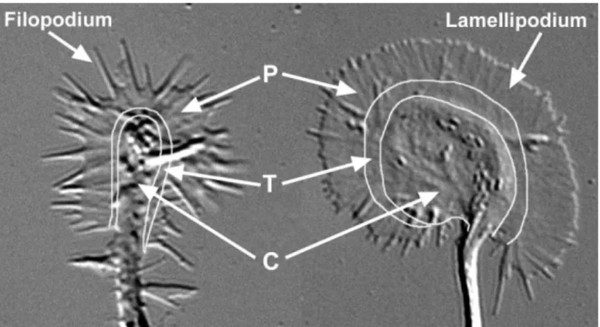
filament—tissue differentiation…” (Harrison, 1910). It is composed primarily of lamellipodia and filopodia; the this protoplasmic movement of the growth cone and the transitional (T) domain, a band of the growth cone at underlying dynamic cytoskeletal structures (neurofi- the interface of the P domain and the central (C) domain; brillae) that are the subject of this review. and the C domain, composed of thicker regions invested In this review, we focus in particular on the cytoskele- by organelles and vesicles of varying sizes. Generally, tal dynamics of actin and microtubules in neurons that growth cones from all species examined to date contain have been studied in culture. The high-resolution optical filopodia and lamellipodia, as well as P, T, and C do-methods available to image cultured neurons have per- mains, although the shapes and sizes of the domains mitted detailed study of cytoskeletal dynamics at a sub- and number of filopodia and lamellipodia vary dramati-cellular level, which have been technically impossible cally. Before describing what is currently known about for the most part in neurons of living animals. Although growth cone cytoskeletal dynamics and how these dy-clearly it will be important to expand these studies into namic structures play an essential role in motility and the complex three-dimensional environment of the or- guidance, it is necessary to delineate the morphological changes that occur as a growth cone is converted into a stable axon shaft.
Figure 1. Growth Cones Vary in Shape and Size
Differential interference contrast images of two hippocampal growth cones in culture. Growth cones can exhibit a wide variety of shapes and sizes. An example of a “filopod-ial” growth cone is shown at left and a “lamel-lipodial” growth cone is shown at right. Gen-erally, growth cones contain both filopodia and lamellipodia and peripheral (P), transition (T), and central (C) regions, although these vary dramatically in shape and size. Both growth cones are shown at the same magnifi-cation.
Stages of Axon Outgrowth thus extrapolations of the phases outlined a year earlier for chicken sensory neurons. Subsequent studies dem-Early phase contrast studies of growth cone dynamics
showed that the rate of advance and the shape of the onstrated that PC12 cells, rodent sympathetic (Aletta and Greene, 1988), and cortical neurons (Kalil, 1996) growth cone were linked. Studies in sympathetic
neu-rons demonstrated that the rate of growth cone advance progressed through the same phases of development during formation of new axons. All of the above studies directly correlated with the size and dynamics of
lamelli-podia and filolamelli-podia (Argiro et al., 1984, 1985). Quantita- documented growth cone motility and random out-growth in cell culture; however, out-growth cone motility tive analysis of filopodia from dorsal root ganglion
neu-rons demonstrated that filopodial movement and growth and axon outgrowth in the developing embryo also is known to occur through protrusion, engorgement, and cone advance were directly correlated as well (Bray and
Chapman, 1985). This group showed that growth cones consolidation (Godement et al., 1994; Halloran and Kalil, 1994; Harris et al., 1987). However, in vivo outgrowth exhibited retrograde flow of material from the peripheral
to the central region and into the axon shaft itself. Impor- is not random, but directed to specific postsynaptic targets. We propose that the difference between ran-tantly, they also demonstrated that the growth cone
undergoes a systematic maturation that is continuously dom or induced outgrowth in culture and directed out-growth in vivo is likely to be that gradients of guidance repeated during elaboration of the axon. This maturation
process consists of the following series of events: filo- cues bias one side of the growth cone to progress through these stages toward (by an attractant) or away podia and lamellipodia form at the leading edge of the
growth cone, followed by flow of the filopodia around from (by a repellent) the guidance cue more rapidly than the other side of the growth cone. Thus, axon guidance the lateral aspects of the growth cone and subsequent
retraction of filopodia at the base of the growth cone. can be thought of as directed protrusion, engorgement, and consolidation.
These investigators also proposed that filopodial
move-ments were driven by the flow of actin filamove-ments and In the mammalian central nervous system (CNS), ax-ons often elongate past their eventual targets and only associated proteins, which make up the filopodia and
lamellipodia (see below). later branch into these targets through a process of delayed interstitial axon branching (O’Leary et al., 1990; Nevertheless, these phase-contrast images were
in-capable of discerning fine details of growth cone struc- Kalil et al., 2000). This process of forming a growth cone from an axon shaft also occurs through protrusion, en-ture. With the invention of video-enhanced contrast
dif-ferential interference contrast (VEC-DIC) microscopy gorgement, and consolidation, with the new axon branching off of the parent axon (Figure 2). Therefore, (Allen, 1985), it was possible to determine that growth
cones from the mollusk Aplysia californica progressed axon guidance, through directed turning or branching, occurs through a conserved mechanism. The rest of the through three morphologically distinct stages to form
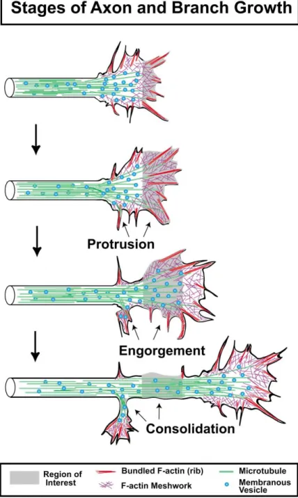
new axon segments (Goldberg and Burmeister, 1986). review will elaborate the underlying cytoskeletal mecha-nisms that regulate protrusion, engorgement, and con-These stages are termed protrusion, engorgement, and
consolidation (Figure 2). Protrusion occurs by the elon- solidation, leading to directed outgrowth. gation of filopodia and lamellipodia (often between two
filopodia), apparently through the polymerization of ac- Pioneering Studies in Growth Cone Cytoskeleton Although microtubules (MTs), microfilaments (filamen-tin filaments. Engorgement occurs when veils become
invested with vesicles and organelles, likely through tous actin or F-actin), and neurofilaments were observed in sections of nervous system tissue under the electron both Brownian motion and directed microtubule-based
transport. Consolidation occurs as the proximal part microscope throughout the 1950’s and 1960’s (Hughes, 1953; Nakai, 1956), it was not until the experimental of the growth cone assumes a cylindrical shape and
transport of organelles becomes bidirectional, thus add- studies of Wessels’ group that MTs and F-actin poly-mers were shown to be necessary for neurite outgrowth ing a new distal segment of axon. These three stages,
iterated many times, give rise to the elongated axon. (Yamada et al., 1970, 1971). Yamada and colleagues incubated chick embryo dorsal root ganglia cultures in The three stages outlined for Aplysia neurons were
Figure 2. Stages of Axon and Branch Growth Three stages of axon outgrowth have been termed protrusion, engorgement, and con-solidation (Goldberg and Burmeister, 1986). Protrusion occurs by the rapid extensions of filopodia and thin lamellar protrusions, often between filopodia. These extensions are pri-marily composed of bundled and mesh-like F-actin networks. Engorgement occurs when microtubules invade protrusions bringing membranous vesicles and organelles (mito-chondria, endoplasmic reticulum). Consoli-dation occurs when the majority of F-actin depolymerizes in the neck of the growth cone, allowing the membrane to shrink around the bundle of microtubules, forming a cylindrical axon shaft. This process also occurs during the formation of collateral branches off the growth cone or axon shaft.
cytochalasin B (CB), which at high concentrations re- MTs did not immediately affect the filopodia but eventu-ally resulted in retraction of the axons (Yamada et al., sults in F-actin depolymerization, and colchicine, which
at high concentrations causes MT depolymerization. Ex- 1971). From these studies it can be concluded that F-actin is the primary cytoskeletal element that main-posure to CB induced retraction of growth cone
filo-podia, which made the growth cone adopt a club-like tains the growth cone shape and is essential for proper axon guidance, whereas MTs are essential for giving appearance and resulted in cessation of axon
out-growth. However, subsequent experiments in dissoci- the axon structure and serve an important function in axon elongation.
ated culture (Dent and Kalil, 2001; Lafont et al., 1993;
Marsh and Letourneau, 1984), in the grasshopper limb Subsequent studies suggested that there was actually an interaction between the F-actin cytoskeleton and (Bentley and Toroian-Raymond, 1986), in the Xenopus
retinotectal system (Chien et al., 1993), and in the Dro- MTs. Bray and colleagues demonstrated that after addi-tion of colchicine, neurites retracted, as demonstrated sophila peripheral nervous system (Kaufmann et al.,
1998) demonstrated that CB-induced F-actin depoly- by others (Yamada et al., 1970), but that depolymer-ization of MTs also caused new growth cone-like merization actually resulted in continued extension but
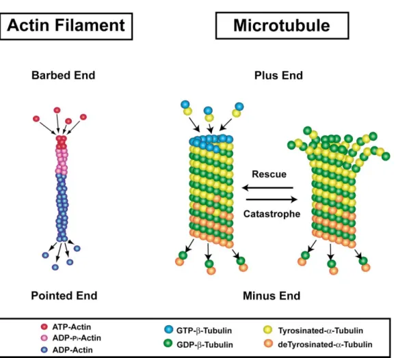
Figure 3. Actin Filaments and Microtubules Are Polarized Polymers
Actin filaments in vitro are capable of adding and removing ATP-actin and ADP-actin from both the barbed and pointed ends. However, the equilibrium constant for ATP dissociation is greater at the pointed end. Consequently, at steady-state, actin filaments devoid of actin-associated proteins undergo slow treadmilling through the addition of ATP-actin to the barbed end and release of ADP-actin from the pointed end (Pollard and Borisy, 2003). Actin filaments also exhibit aging, in which ATP-actin is hydrolyzed rapidly to ADP-pi-actin, followed by a slow dissociation of the␥-phosphate, giving ADP-actin. Microtubules are also polarized structures with ␣/GTP--tubulin dimers adding to the plus or growing end and␣/GDP--tubulin dimers dissociating from the minus end. Microtubules also contain an internal mechanism of GTP hydrolysis that occurs rapidly, giving a “GTP-cap” to the polymers. They also exhibit posttranslational modifications (detyrosination shown here) that correlate with the age of the polymer.
quiescent axon shaft (Bray et al., 1978). The authors 1981; Tennyson, 1970), but not during the initial axon outgrowth of hippocampal (Shaw et al., 1985) and corti-concluded that MTs suppress actin polymerization in
the axon shaft, which maintains the axon in a cylindrical cal (E.W.D., unpublished data) neurons in culture. Trans-genic mice lacking axonal neurofilaments are viable, form. Another group, using EM after stabilization of the
cytoskeleton, documented that MTs oftentimes abut or with few abnormalities in their neural connections (Eyer and Peterson, 1994). Furthermore, Drosophila develop run parallel to F-actin bundles in peripheral regions
of the growth cone (Letourneau, 1983). Thus, we have fully functional nervous systems without neurofilaments. Although neurofilaments are clearly an important cy-known for more than twenty years that there is likely to
be a dynamic interplay between cytoskeletal elements. toskeletal component during development of the verte-brate nervous system (Lin and Szaro, 1995), their func-To understand this interplay, it is necessary to identify
what cytoskeletal components are present in growth tion in growth cone motility and axon guidance is unknown. Therefore, this review will focus on actin fila-cones and what higher-order forms they take.
ments and microtubules. Actin Filaments
The Growth Cone Cytoskeleton Is Composed
of Polarized Polymers Actin filaments are helical polymers composed of actin monomers, often referred to as globular actin (G-actin) The two principle cytoskeletal components in growth
cones are actin filaments and microtubules (Figure 3). (Figure 3). Neurons contain approximately equal amounts of nonmuscle isotypes of actin, -actin and ␥-actin However, in developing peripheral nervous system
neu-rons, neurofilaments are also present in the axon and (Choo and Bray, 1978), although most research has fo-cused on the-actin isotype. It is not known if these C domain of the growth cone (Bunge, 1973; Shaw et al.,
two isotypes of actin have distinctive functions in neu- mers are assembled from one␣-tubulin subunit and one
-tubulin subunit, resulting in an ␣/ dimer. In mammals
rons and growth cones, although both their mRNA and
protein show differential regulation in neurons (Bassell there are six known␣-tubulin genes and seven known
-tubulin genes. Of these, three ␣-tubulins (␣1, ␣2, and
et al., 1998; Gunning et al., 1998). It is also not known
whether the composition of individual actin filaments ␣4) and five -tubulins (I, II, III, IVa, and IVb) are found in brain (reviewed in Luduena, 1998). These␣-tubulin in growth cones is homogeneous, composed of either
-actin or ␥-actin, or heterogeneous, containing both heterodimers are arranged in a linear array of alternating
␣- and -tubulin subunits, which forms a protofilament -actin and ␥-actin.
Globular actin can exist as ATP-actin, ADP-pi-actin, (Figure 3). Between 11 and 15 protofilaments constitute the wall of the microtubule (usually 13 in mammalian and ADP-actin (Figure 3). Although ATP- and ADP-actin
can associate and dissociate from both barbed and cells), giving rise to a tubular structure approximately 25 nm in diameter (Luduena, 1998). Because these␣ pointed ends in vitro, ADP-actin dissociation from the
pointed ends is kinetically favored (Pollard and Borisy, dimers are arranged in a head-to-tail configuration, the MT is inherently polarized, with one end termed the 2003). This results in slow addition of monomers at the
barbed end and slow dissociation of monomers at “plus” end and the other the “minus” end. In most cells the plus end of the MT grows and shrinks, while the the pointed end. In growth cones, the barbed end of the
actin filament generally faces the distal membrane and minus end of the MT is inherently unstable and shrinks unless it is stabilized, presumably by minus end capping the pointed end faces the T region. Actin is also modified
as it “ages.” G-actin polymerizes onto actin filaments proteins. Neuronal MTs can be extremely stable and long lived (Li and Black, 1996). Therefore, nervous sys-as ATP-actin and is hydrolyzed first to ADP-pi-actin and
then finally into ADP-actin upon phosphate release. In- tem tissue is likely to be an excellent source of MT minus end capping proteins.
terestingly, several actin-associated proteins have been
found to bind preferentially to these different forms of Any MT present in a neuron is likely to be a heteroge-neous polymer composed of several combinations of actin (Gungabissoon and Bamburg, 2003).
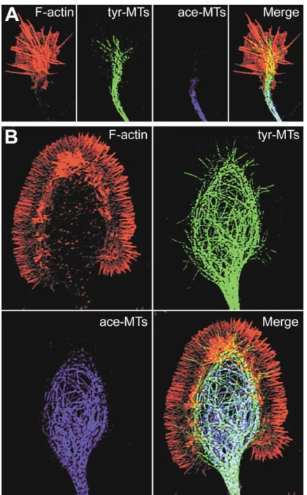
In growth cones, F-actin content is highest in the P ␣/ dimer isotypes. However, the actual isotypic makeup of individual MTs in neurons is not known. Nevertheless, and T regions of the growth cone and diminishes to
varying levels in the C region of the growth cone (Figure it appears that different isotypes may confer distinct properties to the MT. For example, in vitro studies have 4). The bulk of F-actin forms two types of arrays. A
polarized bundled array of F-actin composes the core shown that MTs composed exclusively of␣III-tubulin are more stable than those assembled from␣II-tubulin of filopodia and often extends proximally into the T
re-gion of the growth cone (Figure 4). These bundled (Schwarz et al., 1998) and less sensitive to vinblastine and taxol, well-known microtubule destabilizing and F-actin structures are generally termed F-actin ribs or
actin ribs if they are present within the lamellipodial P stabilizing drugs, respectively (Derry et al., 1997; Khan and Luduena, 2003). Other studies indicate that different and T regions of the growth cone. F-actin can also adopt
a meshwork-like array. It is this meshwork array of ␣- and -tubulin isotypes can change microtubule plus end dynamics (Bode et al., 2003; Panda et al., 1994). III-F-actin that forms the bulk of the lamellipodia in the P
region of the growth cone (Bridgman and Dailey, 1989; tubulin is generally found only in postmitotic neurons. Interestingly, the upregulation of this isotype in gliomas Forscher and Smith, 1988; Lewis and Bridgman, 1992;
Smith, 1988). and in lung and prostate cancer correlates with the grade of malignancy and resistance to taxol (Katsetos F-actin can also take the form of other dynamic and
stable structures in the growth cone. Dynamic comet- et al., 2003; Ranganathan et al., 1998; Verdier-Pinard et al., 2003).
like structures that emanate from the T region and
ex-tend into the P region are termed intrapodia (Dent and A major way in which tubulin and MTs are functionally modified is by several forms of posttranslational modifi-Kalil, 2001; Katoh et al., 1999; Rochlin et al., 1999). The
actin structure of intrapodia forms an elongated mesh- cation including tyrosination/detyrosination, acetyla-tion, phosphorylaacetyla-tion, polyglutamylaacetyla-tion, and polyglycy-work array, similar to the tails of bacterial and viral
pathogens, about 1–2m wide and up to 5–10 m in lation (reviewed in Luduena, 1998). Free ␣/-tubulin dimers generally contain a C-terminal tyrosine residue length (Rochlin et al., 1999). Thus, intrapodia are a hybrid
of the bundled and meshwork arrays that compose the on the␣ subunit. However, after assembly into microtu-bules, this tyrosine residue can be cleaved by tubulin bulk of growth cone F-actin. Actin can also adopt an
arc-like structure in the T region of the growth cone that carboxypeptidase, yielding detyrosinated tubulin (re-viewed in Barra et al., 1988; MacRae, 1997). The penulti-has distinct kinetics from lamellipodial and filopodial
actin (Schaefer et al., 2002). Other forms of F-actin in- mate amino acid, glutamate, can also be cleaved, giving delta2-tubulin (Lafanechere and Job, 2000). Most of clude puncta, located within the central region of the
growth cone and axon shaft, and a thin subplasmalem- these modifications occur at the highly divergent C ter-minus of both␣- and -tubulins, which is exposed on mal cortical meshwork in the axon shaft (Letourneau,
1983; Schnapp and Reese, 1982). These actin puncta the outside of the MT. However, acetylation occurs at lysine-40 of␣-tubulin, which faces the MT lumen (No-and subplasmalemmal network appear to be quite
sta-ble because they are insensitive to long-term incubation gales et al., 1999).
It is well known that MTs are heterogeneous polymers with the F-actin capping or G-actin sequestering drugs
CB and latrunculin A, respectively (Dent and Kalil, 2001). in terms of posttranslational modifications. As an exam-ple, individual neuronal MTs have been shown to be Microtubules
Microtubules are polarized structures composed of tu- highly acetylated and sparsely tyrosinated at their minus ends, with a fairly abrupt transition to highly tyrosinated bulin dimers assembled into linear arrays. Tubulin
di-Figure 4. Distribution of Actin Filaments and Microtubules in Growth Cones
(A) A small, rapidly extending hippocampal neuron growth cone was fixed and simultane-ously labeled for F-actin (with phalloidin), ty-rosinated MTs (tyr-MTs), and acetylated MTs (ace-MTs) with specific antibodies. Note the prominent F-actin bundles and the splaying of tyr-MTs into the actin-rich region, often along the F-actin bundles. Acetylated MTs are much farther back in the C region and axon shaft and do not colocalize with F-actin. (B) A large, paused hippocampal growth cone fixed and labeled as above. Note the halo of F-actin around the prominent looped micro-tubules in the C region. Tyr-MTs also extend into the actin-rich P region, but ace-MTs are limited to the central region and do not show colocalization with F-actin. Both growth cones are shown at the same magnification.
and sparsely acetylated at their plus ends (Figure 3; ner, 1991). This splaying is thought to result from MT dynamics (Rochlin et al., 1996). MTs can also adopt a Brown et al., 1993). Although degree of acetylation and
detyrosination of microtubules correlates directly with looped morphology when growth cones are in a paused state (Figure 4B; Dent and Kalil, 2001; Dent et al., 1999; the age of the microtubule, these modifications do not
confer stability to the microtubule (Khawaja et al., 1988). Morfini et al., 1994; Tanaka and Kirschner, 1991; Tsui et al., 1984). Interestingly, this looped morphology was However, it is likely that such posttranslational changes,
like the hydrolysis of ATP-actin to ADP-actin, affect recently discovered to be associated with synapse for-mation in the Drosophila neuromuscular junction (Roos binding of associated proteins and thus interactions
with other cytoskeletal components and intracellular et al., 2000). To sprout new synaptic boutons, the MT loop would transiently break down, allowing MT poly-signaling pathways (Bonnet et al., 2001; Boucher et al.,
1994; Gurland and Gundersen, 1995; Kreitzer et al., 1999; merization and transport, followed by reformation of the loop at both the old and newly formed synaptic bouton. Larcher et al., 1996; Liao and Gundersen, 1998).
MTs form a dense parallel array in the axon shaft. It was thought that MTs rarely extended into the pe-riphery of the growth cone and were inhibited by the When they enter the growth cone, they generally splay
apart (Figure 4A; Dailey and Bridgman, 1991; Forscher dense F-actin meshwork present in the P region (Forscher and Smith, 1988). However, others had found and Smith, 1988; Letourneau, 1983; Tanaka and
Kirsch-that MTs were often present in the P region of fixed and istry, functional studies (i.e., knockout and overexpres-sion phenotypes, GFP-labeling and dynamic localiza-stained growth cones, sometimes extending well into
tion, immunoelectron localization, and determination of filopodia (Dailey and Bridgman, 1991; Gordon-Weeks,
temporal and spatial activation patterns) have yet to be 1991; Letourneau, 1983). Once fluorescently labeled
performed on many of these proteins. Furthermore, very MTs were observed with time-lapse fluorescent
micros-few studies have directly placed ABPs downstream of copy, it became obvious that they could rapidly extend
specific guidance receptors. Examples include studies into and retract from the peripheral actin-rich regions
demonstrating that Ena/VASP proteins function down-of growth cones. In fact, over a period down-of tens down-of minutes,
stream of both Netrin/DCC and Slit/Robo pathways (Ba-MTs can explore almost the entire P region of the growth
shaw et al., 2000; Colavita and Culotti, 1998; Gitai et al., cone through dynamic polymerization/depolymerization
2003), Cofilin functions in the Semaphorin3A/Neuropilin and transport (Dent and Kalil, 2001; Dent et al., 1999;
pathway (Fritsche et al., 1999; Aizawa et al., 2001), Pro-Kabir et al., 2001; Schaefer et al., 2002; Tanaka and
filin functions in the Dlar pathway (Wills et al., 1999), Kirschner, 1991, 1995).
AbLIM functions in the Netrin/DCC pathway (Gitai et al., 2003), Capulet functions in the Slit/Robo pathway (Wills Polymer Dynamics
et al., 2002), and Abl functions in the Slit/Robo and Dlar Actin filaments and MTs are in a constant state of flux.
pathways (reviewed in Moresco and Koleske, 2003). In An essential feature of both polymers is that they are
general, the role of ABPs in nonneuronal cells have been required by the cell to exist sometimes in a stable state
explored more extensively (reviewed in Pollard and Bo-and other times as dynamic structures. For neurons
risy, 2003). Nevertheless, the complement of ABPs differ to extend long axons and dendrites and steer these
between neuronal and nonneuronal cells. Therefore, it processes to their eventual synaptic partners, they must
will be important to determine how the expression and exert precise control over the dynamics of both actin
activities of these neural-specific ABPs are orchestrated filaments and MTs. To accomplish these tasks, neurons
in growth cones during outgrowth and pathfinding. For contain a complex set of actin- and MT-associated
pro-example, are they localized to analogous structures in teins in addition to the variety of isoforms and
posttrans-nonneuronal cells and growth cones (i.e., leading edge, lational modifications mentioned above (Gordon-Weeks,
filopodia, actin bundles)? What protein complexes exist 2000). Furthermore, some cytoskeletal-associated
pro-between actin and ABPs in the growth cone and how teins are able to influence both actin filaments and MTs
are these regulated? One important area of future study (Rodriguez et al., 2003).
will be to determine how these ABPs are involved in Actin Filament Dynamics
signaling cascades from guidance cues, such as Ne-The dynamics of actin filaments are regulated by both
trins, Semaphorins, Slits, and Ephrins, to the actin cy-their intrinsic polarity and a cornucopia of
actin-associ-toskeleton. ated proteins. More than twenty proteins bind directly
Microtubule Dynamics to F- and/or G-actin and have been localized
immunocy-Like F-actin, MT dynamics are influenced by their asso-tochemically to the growth cone (Table 1). We will refer
ciated proteins and by their intrinsic properties. In neu-to these proteins throughout the text as actin binding
rons, microtubules generally assume a plus end distal proteins (ABPs). It is beyond the scope of this review
distribution in the axon and a mixed polarity distribution, to discuss in detail how each of these proteins is thought
with both plus and minus end distal MTs in dendrites to function. Furthermore, most of the functional studies
(Baas et al., 1989). The plus ends of MTs exhibit a prop-of these proteins have been done in nonneuronal cells
erty termed dynamic instability, where they cycle and may not always translate directly to neurons. Later through periods of growth and shrinkage, punctuated in the review we will discuss how some of these proteins by occasional pauses (Mitchison and Kirschner, 1984). may function in axon guidance. The transition from growth to shrinkage is termed catas-Generally, ABPs can be divided into several catego- trophe, and the transition from shrinkage to growth is ries based upon their function. These categories include termed rescue (Figure 3). One consequence of this intrin-proteins that (1) bind and/or sequester actin monomers, sic dynamic instability of MTs is that they are capable (2) nucleate actin filaments, (3) cap the barbed or (4) of efficiently probing the intracellular space (Holy and pointed ends of actin filaments, (5) act as barbed end Leibler, 1994; Mitchison and Kirschner, 1984).
anticapping proteins, (6) sever F-actin, (7) bundle, cross- The dynamic instability of microtubules was first di-link, or otherwise stabilize F-actin, and (8) anchor F-actin rectly observed in vivo in fibroblasts (Sammak and Bo-to membrane adhesions or specific regions of the mem- risy, 1988; Schulze and Kirschner, 1988; Walker et al., brane. However, many of these proteins are probably 1988) and later in Xenopus spinal neurons (Tanaka and capable of functioning in several of these categories Kirschner, 1991; Tanaka et al., 1995). However, it was depending on the internal state of the growth cone, the only recently that microtubule dynamic parameters such area in which they are localized, and whether or not they as time spent extending, retracting, and pausing as well are phosphorylated. Nevertheless, it is interesting to as catastrophe and rescue frequencies were measured note that the sheer number and complexity of interac- in living neurons (Kabir et al., 2001; Schaefer et al., 2002). tions of these ABPs indicates that actin filaments are In these studies Forscher and colleagues used large probably always well decorated and almost certainly paused growth cones from Aplysia, which are particu-never exist in the “naked” state shown in the cartoon larly advantageous for imaging cytoskeletal dynamics in Figure 3. due to their large size and very slow growth rates. In Although most of the proteins listed in Table 1 have the future it will be important to demonstrate directly how MT dynamics change as growth cones turn toward been localized to the growth cone by
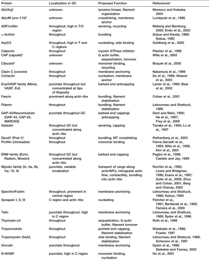
immunocytochem-Table 1. Actin Binding Proteins in the Growth Cone
Protein Localization in GC Proposed Function Referencesa
Abl/Argb unknown tyrosine kinase, filament Moresco and Koleske,
organization 2003
AbLIM (unc-115)b unknown crosslinking, membrane Lundquist et al., 1998
anchor
ADF/cofilin throughout, high in T/C severing, recycling Meberg and Bamburg,
region 2000; Endo et al., 2003
␣-Actinin throughout bundling Sobue and Kanda, 1989;
Sobue, 1993 Arp2/3 throughout, high in T and nucleating, side binding Goldberg et al., 2000
C regions
Calponin throughout myosin ATPase inhibitor Plantier et al., 1999
CAP (capulet)b unknown G actin buffer, Wills et al., 2002
sequestration, turnover
Ciboulotb unknown monomer binding, Boquet et al., 2000
recycling
Clipin C (coronin) throughout membrane anchoring Nakamura et al., 1999
Cortactin throughout nucleation, membrane Du et al., 1998; Weaver
anchor et al., 2003
Ena/VASP family (Mena, punctate throughout but barbed end anticapping Lanier et al., 1999; Bear
VASP, Evl) concentrated at tips et al., 2002
of filopodia
Fascin prominent along actin ribs bundling, filament Cohan et al., 2001
stabilization
Filamin throughout bundling, filament Letourneau and Shattuck,
stabilization 1989
GAP-43/Neuromodulin punctate throughout GC barbed end capping/ Dent and Meiri, 1992;
(GAP-43, CAP-23, anticapping He et al., 1997;
MARCKS) Frey et al., 2000
Gelsolin throughout GC but severing, capping Tanaka et al., 1993; Lu et
concentrated along al., 1997
actin ribs
Dpod1 (Pod-1)c throughout bundling, MT crosslinking Rothenberg et al., 2003
Profilin (chickadee) throughout monomer binding Faivre-Sarrailh et al.,
1993; Wills et al., 1999; Kim et al., 2001 ERM family (Ezrin, throughout GC but barbed end capping Paglini et al., 1998;
Radixin, Moesin) concentrated along Castelo and Jay, 1999
actin ribs
Myosin family (Ic, IIa, IIb, punctate, variable transport of cargo along Rochlin et al., 1995;
Va,cVI, X) localization actin/MTs, retrograde actin Lewis and Bridgman,
flow, contractility, bundling 1996; Evans et al., 1997; into actin ribs Suter et al., 2000; Zhou
and Cohan, 2001; Berg and Cheney, 2002 Spectrin/Fodrin throughout, prominent in membrane anchoring Letourneau and Shattuck,
central region 1989; Sobue, 1993
Synapsin I, II, III C region and actin ribs nucleating Fletcher et al.,
1991; Benfenati et al., 1992; Ferreira et al., 2000
Talin punctate throughout, high membrane anchoring Letourneau and Shattuck,
in C region 1989; Sydor et al., 1996
Thymosin-4 throughout sequestration, G-actin Roth et al., 1999
buffer, filament turnover
Tropomodulin throughout pointed end capping, Watakabe et al., 1996;
filament stabilization Fowler, 1997
Tropomyosin (5a/b) throughout side binding, filament Letourneau and Shattuck, 1989;
stabilization Schevzov et al., 1997
Vinculin punctate throughout membrane anchoring Sydor et al., 1996;
Steketee and Tosney, 2002
N-WASP punctate, high in C region monomer binding, Ho et al., 2001
nucleation
aThese are only representative articles; many more have generally been published on each protein.
bThese proteins have not been specifically localized to the growth cone but bind actin and play an important role in axon guidance. (There
are several proteins that have been found in nonneuronal cells that may be found in the growth cone but have yet to be localized there, and a whole host of proteins that are known to bind to these actin-binding proteins but do not bind actin directly. These proteins have not been included in the table.)
attractive or away from repulsive axon guidance mole- polymers. This movement occurs through the action of molecular motors. Molecular motors are well known for cules.
There have been a number of indirect studies, plus a transporting vesicles and organelles throughout the cy-toplasm on F-actin and MTs, but they are also capable few studies in which MTs have been directly observed
in the growth cone, that implicate MT dynamics as a of directed movement of the cytoskeletal polymers themselves. Movement of F-actin and MTs has a number key event in axon outgrowth, guidance, and branching.
The first direct demonstration of microtubule dynamics of implications for axon outgrowth and guidance. Retrograde F-Actin Flow
in living neurons was performed in both Xenopus neural
tube cultures and in grasshopper limb in situ (Sabry et A well-documented phenomenon in growth cones is termed retrograde actin flow (Forscher and Smith, 1988). al., 1991; Tanaka et al., 1993). These authors showed
that MTs were capable of dynamic exploration of the This constitutive phenomenon occurs as ATP-actin is assembled into filaments near the membrane in the dis-entire growth cone and interconverted between splayed,
looped, and bundled arrays on the order of minutes. tal P region of the growth cone and is transported rear-ward into the T region of the growth cone as polymeric Furthermore, they demonstrated that orientation of MTs
toward the future direction of outgrowth was an early F-actin (Lin and Forscher, 1995; Suter and Forscher, 2000). In the T region the F-actin, now composed of step in pathfinding, both in culture and in situ (Sabry et
al., 1991; Tanaka and Kirschner, 1995; Tanaka et al., ADP-actin monomers, is likely severed and depolymer-ized by several proteins including gelsolin and ADF/ 1995). Other work, in which neurons were fixed and
stained after visualization of growth cone motility and cofilin (see Table 1). The ADP-actin becomes recycled into ATP-actin and the cycle is repeated (Gungabissoon turning, confirmed and extended these observations by
showing that the dynamic (tyrosinated) pool of MTs was and Bamburg, 2003).
The rearward transport of F-actin in the P region of instrumental in both axon outgrowth toward a cellular
target and the turning away of growth cones from inhibi- the growth cone occurs in both filopodia and lamelli-podia (Dent and Kalil, 2001; Lin and Forscher, 1993, tory substrate bound proteins (Challacombe et al., 1997;
Lin and Forscher, 1993, 1995; Rochlin et al., 1996; Ta- 1995; Mallavarapu and Mitchison, 1999; Schaefer et al., 2002; Welnhofer et al., 1997, 1999) and is a myosin mo-naka and Kirschner, 1995; Williamson et al., 1996).
Re-cent studies demonstrate directly that MTs are trans- tor-driven process (Brown and Bridgman, 2003a; Diefen-bach et al., 2002; Lin et al., 1996). This is sometimes ported in the axon and growth cones and how MTs
dynamically reorganize when forming axon branches referred to as actin treadmilling but actually is motor-driven transport. The difference is that if a polymer tread-(Dent and Kalil, 2001; Dent et al., 1999; Kabir et al., 2001;
Schaefer et al., 2002; Wang and Brown, 2002). Another mills, the monomers within the polymer do not move; rather, they polymerize from one end and depolymerize study has recently shown that MTs can serve an
instruc-tive role for growth cone turning toward guidance cues from the other. This appears as movement because the polymer as a whole changes position, but the monomers (Buck and Zheng, 2002).
Like actin filaments, MT dynamics are regulated by a within the polymer do not. Transport of F-actin has been number of MT-associated proteins (MAPs) (reviewed in demonstrated directly by either following photoactiva-Cassimeris and Spittle, 2001). For this review we will tion/photobleaching of a small segment of the polymer limit ourselves to those microtubule binding proteins (Mallavarapu and Mitchison, 1999; Okabe and Hirokawa, (MBPs) that have been shown to bind tubulin/MTs di- 1991) or by tracking speckled filaments and following rectly and have been localized to the growth cone. There them over time (Ponti et al., 2003). If F-actin treadmilled, are fewer MBPs than ABPs in growth cones, but the list a small labeled region would remain stationary within is growing quickly (Table 2). Many MBPs that were first the P domain of the growth cone. This does not appear discovered in neurons were ascribed the role of stabiliz- to be the case. All live cell imaging of the growth cone ers of MTs (MAP1B, MAP2, tau) and termed structural after labeling actin filaments indicates that marked or MAPs (Matus, 1991). Other MBPs were grouped as MT speckled filaments move retrogradely at rates of 1–7 motors (dynein and kinesins) (Sheetz et al., 1989). These m/min, depending on the neuronal cell type analyzed two distinctions generally still hold, although the diver- (Dent and Kalil, 2001; Mallavarapu and Mitchison, 1999; sity of MBPs has increased greatly. Furthermore, there Schaefer et al., 2002). Interestingly, when two growth are a number of interesting MBPs that copolymerize cones interact, turning toward one another, retrograde with MTs, termed plus end tracking proteins (⫹TIPs) F-actin flow decreases along the axis of contact (Lin (Carvalho et al., 2003; Schuyler and Pellman, 2001). and Forscher, 1995). This decrease in F-actin flow is These proteins have been implicated in plus end MT thought to be induced by the engagement of a clutch dynamics and linking MTs with actin-associated pro- mechanism that bridges the F-actin cytoskeleton with teins in the cell cortex (Galjart and Perez, 2003). How- cell adhesion molecules (Suter and Forscher, 2000). The ever, few of these proteins have been studied in the engagement of this “clutch,” through an unknown pro-growth cone (Morrison et al., 2002; Stepanova et al., tein complex, attenuates retrograde flow, allowing pro-2003). The functions of these proteins in axon outgrowth trusion to occur in front of the adhesion point and fa-and guidance will undoubtedly yield many insights into voring MT invasion behind the adhesion point (Suter et how MTs respond to guidance cues and how they inter- al., 1998).
act with F-actin and other intracellular targets. Currently, the exact nature of the myosin motor(s) that drive retrograde flow remain somewhat controversial. One group, using microchromaphore-assisted laser in-Polymer Transport
In addition to polymer dynamics, F-actin and MTs also activation (micro-CALI) on chick dorsal root ganglion growth cones, showed that inhibition of myosin Ic undergo directed movement through the cytoplasm as
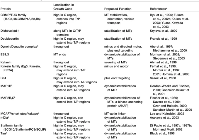
Table 2. Microtubule Binding Proteins in the Growth Cone Localization in
Protein Growth Cone Proposed Function Referencesa
CRMP/TUC family high in C region, MT stabilization, Byk et al., 1996; Fukata
(TUC4,4b,CRMP1A,2A,Bs) extends into T/P orientation, vesicle et al., 2002b; Quinn et al.,
regions transport 2003; Yuasa-Kawada
et al., 2003
Dishevelled-1 along MTs in C/T/P stabilization of MTs Krylova et al., 2000
domains
Doublecortin high in C region, may stabilization of MTs Francis et al., 1999
extend into T/P regions
Dynein/Dynactin complexb throughout minus end directed motor, Abe et al., 1997;
plus end targeting Niethammer et al., 2000
EB1,3 MT ends dynamics/stabilization of Morrison et al., 2002;
MTs Stepanova et al., 2003
Katanin throughout severing of MTs Ahmad et al., 1999
Kinesin family (Eg5, Kinesin, high in C region, minus end motor Ferhat et al., 1998;
KIF2A) may extend into T/P Morfini et al., 1997,
regions 2001; Homma et al., 2003
Lis1 high in C region, plus end targeting Sasaki et al., 2000
may extend into T/P regions
MAP1Bb high in C region, may dynamics/stabilization Gordon-Weeks and Fischer,
extend into T/P regions of MTs 2000; Gonzalez-Billault et
al., 2001 MAP2B,Cb high in C region, can dynamics/stabilization of Fischer et al., 1986;
extend into T/P regions MTs, a-kinase anchoring Davare et al., 1999; protein (AKAP) Ozer and Halpain, 2000;
Sanchez-Martin et al., 2000
MCAF7/short stop/kakapob throughout MT-actin linker Lee and Kolodziej, 2002
mDiab high in C region, can dynamics/stabilization Arakawa et al., 2003
extend into T/P regions of MTs
Stathmin family high in C region, may dynamics/stabilization Di Paolo et al., 1997a, 1997b; (SCG10/Stathmin/RC3/SCLIP) extend into T/P regions of MTs Mori and Morii, 2002
Taub high in C region, can dynamics/stabilization Black et al., 1996
extend into T/P regions of MTs
aThese are only representative articles; many more have generally been published on each protein. Several other MT-binding proteins, such
as APC, BPAG-1, CLIP-170, and CLASPs, found to play important roles in nonneuronal cells are also likely to exist in growth cones but have yet to be localized there.
bAlso known to bind actin or act as an actin-associated protein.
caused a marked decrease in retrograde flow, whereas reduce actin ribs without noticeable actin depolymeriza-tion. These data are consistent with a study that showed inhibition of myosin IIB slightly increased retrograde
flow rates (Diefenbach et al., 2002). Another group, growth cones from myosin IIB knockout mice also had decreased numbers of actin ribs and a smaller lamellar working with myosin IIB knockout mice, showed that
retrograde flow was increased, similar to the aforemen- area (Bridgman et al., 2001). Furthermore, localized ap-plication of collapsing agents to one side of the growth tioned study (Brown and Bridgman, 2003b). However,
this group did not find any evidence of myosin Ic staining cone is capable of inducing repulsive turning through actin rib loss (Zhou et al., 2002). Actin rib loss also in the T or P regions of the growth cone, implicating a
different myosin, possibly myosin IIA, as the primary causes decreases in the number of MTs on the side of the growth cone nearest the source of collapsing factor, motor behind retrograde flow. Future experiments using
selective inhibition of each myosin family member alone implicating signaling between F-actin bundles and MTs. Interestingly, a recent study has shown that myosin II and in combination will likely sort out this enigma.
Interestingly, myosin II has also been implicated as activity, by overexpressing myosin light chain kinase, is important in both attractive and repulsive guidance an important factor for F-actin bundling in growth cones.
Cohan and colleagues have recently shown that phar- pathways (Kim et al., 2002). Thus, in addition to actin bundling/stabilizing proteins, such as␣-actinin, fascin, macological inhibition of myosin light chain kinase,
which effectively inhibits myosin II, decreases the num- filamin, and tropomyosin, myosin II plays an important role in regulating actin rib dynamics, growth cone ber of actin ribs in growth cones by merging adjacent
actin ribs, resulting in growth cone collapse (Zhou and spreading, and axon guidance. Microtubule Transport Cohan, 2001). Thus, myosin II somehow functions to
keep bundled F-actin arrays separated in the growth Microtubule transport has generated much controversy in recent years, mainly centering on the form that MTs cone, maintaining the growth cone in a spread state.
The way in which myosin II accomplishes this feat is take when they are transported. However, recent ad-vances in imaging cytoskeletal dynamics show that a currently not known. Nevertheless, both physiological
and nonphysiological agents that induce growth cone number of dynamic processes are involved in con-structing and maintaining the microtubule array in axons collapse, some presumably acting through myosin II,
and growth cones during development (reviewed in Tanaka et al., 1995; Williamson et al., 1996). All of these studies specifically targeted actin and/or MT dynamics. Baas, 2002; Black, 1994; Brown, 2003). We will
concen-Therefore, it is likely that outgrowth, guidance, and trate on the studies pertinent to axon outgrowth and
branching are all linked to the level of activity of F-actin pathfinding.
and MTs, which are probably controlled by the activity MTs are assembled at the centrosome, which is
lo-of upstream components, such as Rac. Interestingly, in cated in the neuronal cell body. However, they do not
fibroblasts, polymerizing MTs are thought to stimulate remain attached to the centrosome, as occurs in several
Rac activity (Waterman-Storer et al., 1999; Wittmann other cell types, but are severed by katanin and rapidly
and Waterman-Storer, 2001; Wittmann et al., 2003). transported away from the cell body (anterogradely) into
Therefore, there is probably bidirectional signaling be-axons, with their plus end leading, by dynein-driven
tween cytoskeletal elements and proteins with which transport (Ahmad et al., 1998, 1999; Dent et al., 1999;
they are associated. Gallo and Letourneau, 1999; Slaughter et al., 1997; Wang
On the cytoskeletal level, the process of axon guid-and Brown, 2002; Yu et al., 1996). During this transport,
ance shares many similarities with the process of polar-MTs also polymerize and depolymerize (Dent and Kalil,
ization and chemotaxis in nonneuronal cells (Song and 2001; Wang and Brown, 2002), consistent with the fact
Poo, 2001; Rodriguez et al., 2003). In this sense the that the axon contains high levels of tyrosinated MTs,
growth cone too is a polarized structure. A recent model a posttranslational modification associated with highly
that has emerged to explain the underlying microtubule dynamic MTs (Baas and Black, 1990). Surprisingly, MTs
reorganization that accompanies polarization and chemo-are also capable of rapid retrograde movement as well
taxis has been termed microtubule capture (Gundersen, (Dent et al., 1999; Wang and Brown, 2002). Thus,
retro-2002). This phenomenon has been most thoroughly grade movement, depolymerization, and severing are
studied in budding yeast and polarizing fibroblasts and all possible mechanisms for redistributing MTs during
may take place in the neuronal growth cone as follows. axon retraction and branch pruning (Ahmad et al., 2000;
Dynamic microtubules probe the intracellular environ-Dent et al., 1999; Gallo and Letourneau, 1999; Wang and
ment. During this process, the plus ends of microtubules Brown, 2002).
come into contact with F-actin-rich cortical and/or adhe-However, the bulk of MTs in axons are stationary at
sion sites (Fukata et al., 2002a; Krylyshkina et al., 2003). any given time, probably attached to the axonal
mem-When microtubules contact these cortical sites along brane skeleton, neurofilaments, and other MTs
(re-the inner membrane, microtubule tip-associated pro-viewed in Brown, 2003). If most MTs are stationary, how
teins act as “ligands” for “receptors” in these actin-are new MT arrays constructed in rapidly elongating
rich regions. Such a ligand/receptor complex has been axons? As random or directed axon outgrowth occurs, a
documented in fibroblasts in which activated Rac1/ combination of MT polymer transport, tubulin transport,
Cdc42 demarcate active regions along the cell cortex MT dynamics, MT severing/breaking, and possibly local
and act as “receptors,” along with IQGAP1, to transiently tubulin synthesis contributes to a readily accessible pool
capture the plus end microtubule tip binding protein of tyrosinated tubulin. This polymerization-competent
CLIP-170 (Fukata et al., 2002b). This interaction is be-tubulin exists throughout the axon but at a higher
con-lieved to convey signals between the cell cortex and the centration in areas undergoing active growth. These
ac-cytoskeleton that allow for continued actin dynamics tive areas include the growth cone of the parent axon
and membrane insertion needed for polarization and and dynamic areas along the axon shaft, some of which
chemotaxis (Gundersen, 2002). develop into axon branches. A possible scenario for
Can this model for fibroblast polarization be applied how F-actin/MT dynamics and transport in the growth
to the polarization and turning of a growth cone toward cone regulates directed protrusion, engorgement, and
or away from guidance cues? Many of the same ABPs consolidation follows.
and MBPs found to be instrumental in fibroblast polar-ization exist in neurons as well (Tables 1 and 2). Thus, A Model for Cytoskeletal Regulation of Axon it is likely that microtubules will be found to interact Outgrowth and Guidance transiently with cortical structures in the growth cone, As mentioned above, we have considered axon guid- which may allow localized insertion of membrane and ance as the process of favored axon outgrowth toward signaling proteins essential for growth in a preferred or away from a particular region. Thus, axon guidance direction (Zakharenko and Popov, 1998). Nevertheless, can be thought of as a process of biasing the extension/ this process of transient MT capture is unlikely to be retraction of one side of the growth cone or axon shaft, sufficient for directed growth to continue. If so, there as in the case of collateral branching, compared to the should be some mechanism operating in tandem that other side (reviewed in Tanaka and Sabry, 1995). How allows MT stabilization and recruitment of more MTs can this model be reconciled with the convincing evi- in the favored direction of growth. When microtubule dence that exists in Drosophila that axon branching, dynamics have been recorded in living growth cones, turning, and outgrowth are distinct processes, de- microtubules have never been shown to attach to the pending on the level of Rac activity in the neuron (Ng actin-rich cortex for more than a few seconds (Dent and et al., 2002)? We propose that the key to resolving this Kalil, 2001; Dent et al., 1999; Kabir et al., 2001; Schaefer apparent paradox is to keep in mind that many studies et al., 2002). Thus, it is unlikely that growth cone turning have shown that both guidance and branching can be involves “pioneer” microtubule capture on one side of selectively inhibited without affecting axon outgrowth the growth cone, followed by recruitment of other micro-per se (Buck and Zheng, 2002; Challacombe et al., 1996, tubules along the pioneer MT.
If transient interactions are not sufficient, then what 1997; Dent and Kalil, 2001; Marsh and Letourneau, 1984;
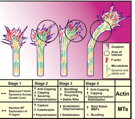
Figure 5. Hypothetical Model for the Cy-toskeletal Reorganization Underlying Growth Cone Turning
Stages of growth cone turning in an attractive gradient (pink shading). All stages are in refer-ence to the area of interest (black circle). Stage 1: F-actin takes the form of dynamic bundled filaments (in filopodia) and mesh-work array (in lamellipodia). We assume that there are balanced F-actin dynamics across the growth cone. MTs form a thick bundle in the axon shaft and splay apart in the growth cone, stochastically exploring the P region. Stage 2: When a chemoattractant is detected, F-actin selectively polymerizes on the side of the growth cone toward the attractant, form-ing increased numbers of filopodia and lamel-lipodia. This bias of actin polymerization to one side of the growth cone causes less poly-merization on the opposite side, favoring shrinkage/retraction of lamellipodia and filo-podia. Microtubules continue stochastically probing the P region but may be transiently captured at actin-rich regions and undergo fewer catastrophes when they interact with F-actin bundles on the right side of the growth cone. Stage 3: Numbers and size of F-actin bundles (ribs) increase and are stabilized by actin-bundling proteins and linkages to the substrate. Because MTs exploring the actin-rich side of the growth cone are maintained in the polymerized state longer than the MTs on the opposite side, they may be selectively posttranslationally modified. Stage 4: F-actin is depolymerized, capped, and stabilized into puncta and a subplasmalemmal network. MTs are subsequently stabilized by recruitment of stabilizing MBPs, causing them to bundle in the newly oriented axon shaft.
process would be required? It is likely that proximal (Bentley and O’Connor, 1994; Lin et al., 1994). This pref-erential protrusion can take the form of filopodia, lamelli-stabilization of MTs is important (Mack et al., 2000).
Indeed, when microtubules have been pharmacologi- podia/ruffles, and intrapodia (Figure 5, stage 2). Thus, actin anticapping and leaky capping proteins, such as cally stabilized on one side of the growth cone by focal
uncaging of taxol, growth cones turn toward the side in Ena/VASP proteins and GAP-43, respectively, are likely to play important roles in this initial protrusion phase which MTs have been stabilized (Buck and Zheng, 2002).
Conversely, when the microtubule-destabilizing drug because these proteins are known to enhance actin polymerization (Bear et al., 2002; Dent and Meiri, 1998; nocodazole is locally applied to one side of the growth
cone, turning to the opposite side occurs (Buck and He et al., 1997; Lanier et al., 1999). Additionally, actin-capping proteins may be inactivated in these regions Zheng, 2002). If local stabilization/destabilization of the
microtubule array is instrumental in directional guid- of protrusion.
These newly formed protrusions must then be stabi-ance, then there must be an intrinsic mechanism for this
to occur. As noted in Table 2, there are many microtubule lized against retraction. Any number of F-actin bundling/ stabilizing proteins are likely to be essential for the con-stabilizing/destabilizing proteins found in growth cones.
Furthermore, neurons are one of the few cell types that tinued protrusion of filopodia and lamellipodia (Table 1). Also, gelsolin has been found to destabilize filopodia, contain high levels of posttranslationally modified
tu-bulin (acetylated, detyrosinated, delta2-tutu-bulin) that possibly through its capping ability, allowing retraction to take place (Lu et al., 1997). Thus, gelsolin and other correlate with microtubule stability (Challacombe et al.,
1996; Dent and Kalil, 2001; Paturle-Lafanechere et al., capping proteins may be inactivated locally. Further-more, it is not sufficient to simply stabilize these F-actin 1994; Williamson et al., 1996).
One possible scenario for the underlying cytoskeletal structures because they are subject to myosin-based retrograde transport, resulting in growth cone collapse involvement in directed protrusion, engorgement, and
consolidation in response to an attractive cue is as fol- (Ahmad et al., 2000; Gallo et al., 2002). Instead, the bundled and crosslinked actin filaments must be stabi-lows (Figure 5). Obviously, similar mechanisms are
in-volved when a growth cone turns in response to a re- lized through a mechanism that involves adhesion to the substrate (Suter and Forscher, 2000). In addition, pulsive cue. We assume that there is balanced actin
polymerization and depolymerization across the growth severing/recycling proteins such as cofilin and profilin may be activated to keep the level of free G-actin and cone over time, when the growth cone is involved in
random locomotion. This would result in balanced pro- barbed ends high for continued polymerization (Endo et al., 2003; Meberg and Bamburg, 2000).
trusion/retraction so that the growth cone would
main-tain a straight trajectory. When a chemoattractant is Because MTs stochastically explore the growth cone P region, certain MTs may be transiently captured at detected on one side of the growth cone, F-actin-driven