HAL Id: hal-01875950
https://hal.sorbonne-universite.fr/hal-01875950
Submitted on 18 Sep 2018
HAL is a multi-disciplinary open access
archive for the deposit and dissemination of
sci-entific research documents, whether they are
pub-lished or not. The documents may come from
teaching and research institutions in France or
abroad, or from public or private research centers.
L’archive ouverte pluridisciplinaire HAL, est
destinée au dépôt et à la diffusion de documents
scientifiques de niveau recherche, publiés ou non,
émanant des établissements d’enseignement et de
recherche français ou étrangers, des laboratoires
publics ou privés.
Distributed under a Creative Commons Attribution| 4.0 International License
Bruno Sovran, Julien Planchais, Sarah Jegou, Marjolene Straube, Bruno
Lamas, Jane Mea Natividad, Allison Agus, Louise Dupraz, Jeremy Glodt,
Grégory da Costa, et al.
To cite this version:
Bruno Sovran, Julien Planchais, Sarah Jegou, Marjolene Straube, Bruno Lamas, et al..
Enterobac-teriaceae are essential for the modulation of colitis severity by fungi. Microbiome, BioMed Central,
2018, 6, pp.152. �10.1186/s40168-018-0538-9�. �hal-01875950�
R E S E A R C H
Open Access
Enterobacteriaceae are essential for the
modulation of colitis severity by fungi
Bruno Sovran
1, Julien Planchais
1, Sarah Jegou
2,3, Marjolene Straube
2,3, Bruno Lamas
1, Jane Mea Natividad
1,
Allison Agus
1, Louise Dupraz
1, Jérémy Glodt
1, Grégory Da Costa
1, Marie-Laure Michel
1, Philippe Langella
1,
Mathias L. Richard
1*and Harry Sokol
1,2,3*Abstract
Background: Host-microbe balance maintains intestinal homeostasis and strongly influences inflammatory conditions such as inflammatory bowel diseases (IBD). Here we focused on bacteria-fungi interactions and their implications on intestinal inflammation, a poorly understood area.
Methods: Dextran sodium sulfate (DSS)-induced colitis was assessed in mice treated with vancomycin (targeting gram-positive bacteria) or colistin (targeting Enterobacteriaceae) and supplemented with either Saccharomyces boulardii CNCM I-745 or Candida albicans. Inflammation severity as well as bacterial and fungal microbiota compositions was monitored.
Results: While S. boulardii improved DSS-induced colitis and C. albicans worsened it in untreated settings, antibiotic treatment strongly modified DSS susceptibility and effects of fungi on colitis. Vancomycin-treated mice were fully protected from colitis, while colistin-treated mice retained colitis phenotype but were not affected anymore by administration of fungi. Antibacterial treatments not only influenced bacterial populations but also had indirect effects on fungal microbiota. Correlations between bacterial and fungal relative abundance were dramatically decreased in colistin-treated mice compared to vancomycin-treated and control mice, suggesting that colistin-sensitive bacteria are involved in interactions with fungi. Restoration of the Enterobacteriaceae population by administrating colistin-resistant Escherichia coli reestablished both beneficial effects of S. boulardii and pathogenic effects of C. albicans on colitis severity. This effect was at least partly mediated by an improved gut colonization by fungi.
Conclusions: Fungal colonization of the gut is affected by the Enterobacteriaceae population, indirectly modifying effects of mycobiome on the host. This finding provides new insights into the role of inter-kingdom functional interactions in intestinal physiopathology and potentially in IBD.
Keywords: Enterobacteriaceae, S. boulardii CNCM I-745, C. albicans, Colitis, Microbiota, Fungi
Background
The gastrointestinal tract (GIT) is composed of a com-plex association of epithelial cells, immune cells, food antigens, and microorganisms. The mammalian GIT is colonized by diverse commensal microbial communities consisting of bacteria, fungi, and viruses [1]. Alteration of the composition of function of the gut microbiota, i.e., dysbiosis, has been associated with several human diseases and particularly with inflammatory bowel dis-ease (IBD). The bacterial microbiota in IBD has been
thoroughly investigated; several groups worldwide have observed a reduction in biodiversity, a decreased relative abundance of bacteria belonging to the phylum Firmicutes (such as Faecalibacterium prausnitzii), and an increased relative abundance of bacteria belonging to the phylum Proteobacteria (such as Escherichia coli) [2– 6]. Recently, we showed that the fungal microbiota is similarly skewed in IBD, characterized by an increased Basidiomycota/Ascomycota ratio, a decreased propor-tion of Saccharomyces cerevisiae, and an increased pro-portion of Candida albicans in IBD patients compared with healthy subjects [7]. Interestingly, these two yeasts were reported to have opposite impacts on intestinal
* Correspondence:mathias.lavie-richard@inra.fr;harry.sokol@aphp.fr
1Micalis Institute, INRA, AgroParisTech, Université Paris-Saclay, Domaine de
Vilvert, Jouy-en-Josas, France
Full list of author information is available at the end of the article
© The Author(s). 2018 Open Access This article is distributed under the terms of the Creative Commons Attribution 4.0 International License (http://creativecommons.org/licenses/by/4.0/), which permits unrestricted use, distribution, and reproduction in any medium, provided you give appropriate credit to the original author(s) and the source, provide a link to the Creative Commons license, and indicate if changes were made. The Creative Commons Public Domain Dedication waiver (http://creativecommons.org/publicdomain/zero/1.0/) applies to the data made available in this article, unless otherwise stated.
inflammation. The probiotic strain S. boulardii CNCM I-745, which belongs to the species S. cerevisiae [8], pro-tects against pathogen-associated diarrhea and colitis in both murine models and humans [9–11] and has a bene-ficial effect on IBD in murine models [12, 13]. On the other hand, C. albicans, which is the most prevalent fun-gus in the human intestinal microbiota [14, 15], shows improved colonization in inflammatory context and sim-ultaneously worsens intestinal inflammation in a murine colitis model [16].
Antibiotics used in the treatment of bacterial infections indiscriminately kill pathogens and commensal microbes in the gut, leading to dysbiosis [17]. Moreover, it has been shown for decades that broad-spectrum antibiotics and antibiotics targeting anaerobic bacteria induce differential effects on susceptibility to fungal infection and, notably, predispose patients to vaginal mucosal infections by C. albicans [18]. Broad-spectrum antibiotic treatment trig-gers a strong increase in gut fungi loads in a mouse model [19], which has commonly been interpreted as resulting mainly from the liberation of ecological niches for fungi proliferation. However, fungal-bacterial interactions and their role in a physiopathological context have been poorly studied to date. We have previously showed that both the bacterial and fungal microbiota are dysbiotic in IBD and that bacterifungal inter-kingdom interactions are al-tered in ulcerative colitis and Crohn’s disease compared to healthy conditions [7].
Here, we used C. albicans and S. boulardii CNCM I-745, as model fungi, to explore the potential role of bacterial components in mediating the effects of fungi in intestinal inflammation. We showed that the presence of bacterial species belonging to the Enterobacteriaceae family greatly influences the fitness of C. albicans and S. boulardii in the gastrointestinal tract and further deter-mines how these fungi modulate the outcome of intes-tinal inflammation. Overall, the results demonstrate the crucial role of inter-kingdom functional interactions in intestinal physiopathology.
Methods
Mice
For the conventional experiment, female C57BL/6J mice were purchased from Janvier (France) and used 1 week after delivery. All experiments were done with appropriate control groups (H2O), from the same batch of mice. Mice
were kept 1 week in the same room prior starting treat-ments. Ten-week-old mice were fed ad libitum with Ssniff control diet V112x M-Z (Ssniff Spezialdiaten, Germany). All experiments were performed in accordance with the Comite d’Ethique en Experimentation Animale (COMETHEA C2EA – 45, Jouy en Josas, France). Every experiment was repeated two or three times.
Antibiotic treatment and induction of colitis with dextran sodium sulfate (DSS)
A broad-spectrum antibiotic cocktail was prepared by mixing ampicillin (2 g/L), neomycin (2 g/L), metronida-zole (2 g/L), and vancomycin (1 g/L) in drinking water. To selectively deplete specific bacteria, we treated the mice until sacrifice with vancomycin (1 g/L in drinking water) or colistin (600 μg/gavage; Sigma). One week after starting the antibiotic treatment, mice were given 2% (wt/vol) DSS (molecular weight, 36,000–50,000; MP Biomedicals, Solon, OH) dissolved in sterile drinking water ad libitum for 7 days, followed by a recovery period (water only) of 5 days. Animals were monitored daily for weight loss and disease activity index (including three parameters: weight loss, stool consistency, and presence of blood in feces).
Gavage with fungi and E. coli strains
S. boulardii CNCM I-745 (syn. HANSEN CBS 5926, Biocodex Laboratories, Gentilly, France) and C. albicans SC5314 (ATCC, Molsheim, France) were used in this study. Both yeasts were grown on yeast extract peptone dextrose (YEPD) medium overnight at 37 °C. Aliquots containing a yeast suspension of 5 × 10⁷ CFU/mL were prepared and stored at − 80 °C. A yeast suspension (107CFU in 200 μL) or control medium was adminis-tered daily to 10-week-old mice by intragastric gavage.
The E. coli MCR1 strain, which is rendered resistant to colistin by the mobilized colistin resistance (MCR) gene, was used in this study. This strain is a veterinary isolate commensal [20] and was provided by Thomas Guillard (CHU Reims, Hôpital Robert Debré, Laboratoire de Bactériologie-Virologie-Hygiène, F-51092 Reims, France). The bacteria were grown in LB at 37 °C overnight and then aliquoted at 5 × 10⁸CFU/mL. A bacterial suspension (108CFU in 200μL) or control medium was administered daily to 10-week-old mice by intragastric gavage.
Histology
Colon samples for histological studies were fixed in 4% paraformaldehyde (Electron Microscopy Sciences, Hatfield, PA, USA), embedded in paraffin, and then stained with hematoxylin and eosin (Sigma-Aldrich, Saint Louis, USA) for histological scoring. Histological scoring was performed blinded according to the method previously described [21].
Gene expression analysis using quantitative reverse-transcription PCR (qRT-PCR)
Total RNA was isolated from colon samples using an RNeasy Mini Kit (Qiagen, Hilden, Germany), including a DNAse treatment step, according to the manufacturer’s instructions. Quantitative RT-PCR was performed using SuperScript II Reverse Transcriptase (Life Technologies,
Saint Aubin, France) followed by a Takyon SYBR Green PCR kit (Eurogentec, Liège, Belgium) in a StepOnePlus ap-paratus (Applied Biosystems, Foster City, CA, USA) with specific mouse oligonucleotides. The oligonucleotides used were as follows: GAPDH—sense: 5′-AACTTTGGCATTG TGGAAGG-3′; antisense: 5′-ACACATTGGGGGTAGGA ACA-3′, Reg3g—sense: 5′-TTCCTGTCCTCCATGATCA AAA-3′; antisense: 5′-CATCCACCTCTGTTGGGTTCA -3′. We used the 2−ΔΔCtquantification method with mouse
GAPDH as an endogenous control and calibrated the assay to the wild type.
Cytokine quantification
The colonic explants were cultured (37 °C, 10% CO2)
overnight in 24-well tissue culture plates (Costar, Corn-ing, Amsterdam, the Netherlands) in l mL of complete RPMI 1640 medium. The culture supernatants were col-lected and stored at− 80 °C until processing. ELISA was performed on the supernatants according to the manufacturer’s instructions in order to quantify IFN-γ (Mabtech, Nacka Strand, Sweden). For the colonic ex-plants, cytokine concentrations were normalized accord-ing to the weight of each colonic explant.
Quantification of fecal lipocalin (LCN2) levels
Frozen fecal samples were weighed and suspended in cold PBS. Samples were then agitated on a FastPrep (MP Bio-medicals, Santa Ana, USA) bead beating machine for 40 s on setting 6 using 4.5-mm glass beads to obtain a homogenous fecal suspension. Samples were then centri-fuged for 5 min at 10,000g (4 °C), and clear supernatants were collected and stored at− 20 °C until analysis. LCN2 levels were estimated using a DuoSet murine LCN2 ELISA kit (R&D Systems, Minneapolis, USA) as per the manufac-turer’s instructions and expressed as pg/mg of stool.
Quantification of fungi and bacteria in fresh stools
Fresh stools were collected over the course of the study to determine the quantities of yeast and bacteria remaining after intragastric gavage. Fresh stools were weighed and suspended in cold PBS (3μL/mg of feces). Tenfold serial dilutions were performed until the desired concentrations were reached. For fungi quantification, diluted feces were plated on YEPD agar plates supplemented with ampicillin (100 mg/mL) and incubated at 30 °C. After 2 days of growth, fungi were counted, and the absolute quantities of yeast were determined according to the corresponding di-lutions. For E. coli MCR1 (resistant to colistin) quantifica-tion, diluted feces were plated on LB agar plates supplemented with colistin (4 mg/L) and incubated at 37 °C. After 1 day of growth, bacteria were counted, and the ab-solute quantities of E. coli were determined according to the corresponding dilutions.
Fecal DNA extraction and fungal quantification via quantitative PCR (qPCR)
Fecal DNA was extracted from weighed stool samples as previously described [22]. More precisely, the fecal sam-ples were weighed and then resuspended for 10 min at room temperature in 250μL of 4 M guanidine thiocyanate in 0.1 M Tris (pH 7.5) (Sigma-Aldrich, Saint Louis, USA) and 40 μL of 10% N-lauroyl sarcosine (Sigma-Aldrich, Saint Louis, USA). After the addition of 500 μL of 5% N-lauroyl sarcosine in 0.1 M phosphate buffer (pH 8.0), the 2-mL tubes were incubated at 70 °C for 1 h. One vol-ume (750 mL) of a mixture of 0.1- and 0.6-mm-diameter silica beads (Sigma-Aldrich, Saint Louis, USA) (previously sterilized by autoclaving) was added, and the tube was shaken at 6.5 m/s in three bouts of 30 s each in a FastPrep (MP Biomedicals, Santa Ana, USA) apparatus. Polyvinyl-polypyrrolidone (15 mg) (Sigma-Aldrich, Saint Louis, USA) was added to the tube, which was then vortexed and centrifuged for 5 min at 20,000g. After recovery of the supernatant, the pellets were washed with 500 μL of TENP (50 mM Tris (pH 8), 20 mM EDTA (pH 8), 100 mM NaCl, 1% polyvinylpolypyrrolidone) and centri-fuged for 5 min at 20,000g, and the new supernatant was added to the first supernatant. The washing step was re-peated two times. The pooled supernatant (approximately 2 mL) was briefly centrifuged to remove particulate matter and then split into two 2-mL tubes. Nucleic acids were precipitated by the addition of 1 volume of isopropanol for 10 min at room temperature and centrifugation for 10 min at 20,000g. The pellets were resuspended and pooled in 450μL of 100 mM phosphate buffer, pH 8, and 50 mL of 5 M potassium acetate. The tube was placed on ice overnight and centrifuged at 20,000g for 30 min. The supernatant was then transferred to a new tube containing 20 μL of RNase (1 mg/mL) and incubated at 37 °C for 30 min. Nucleic acids were precipitated by the addition of 50μL of 3 M sodium acetate and 1 mL of absolute etha-nol. The tube was incubated for 10 min at room temperature, and the nucleic acids were recovered by cen-trifugation at 20,000g for 15 min. The DNA pellet was fi-nally washed with 70% ethanol, dried, and resuspended in 100μL of Tris–EDTA (TE) buffer. The DNA suspensions were stored at− 20 °C for real-time qPCR analysis of the 16S rDNA or ITS2 sequences. DNA was then subjected to qPCR by using a Takyon SYBR Green PCR kit (Eurogen-tec, Maastricht, The Netherlands) for quantification of all fungal sequences or by using TaqMan Gene Expression Assays (Life Technologies) for quantification of all bacter-ial sequences. The probes and primers for the bacterbacter-ial 16S DNA genes and primers for the fungal 18S DNA genes were as described previously [23]. Reference stand-ard (with quantified C. albicans DNA) was used in the same qPCR, allowing the quantification of the CFU in each sample. The threshold cycle for each sample was
determined for each gene and was normalized to the CT value of the all-bacteria 16S ribosomal RNA gene. Data were calculated using the 2−ΔΔCtmethod.
16S DNA gene and ITS2 sequencing
DNA was isolated from the feces of mice before and after DSS treatment using the protocol described above. Bacter-ial diversity was determined for each sample by targeting a portion of the ribosomal genes. A 16S DNA gene fragment comprising the V3 and V4 hypervariable regions (16S (sense) 5′-TACGGRAGGCAGCAG-3′ and (anti-sense) 5′-CTACCNGGGTATCTAAT-3′) was amplified using an optimized and standardized 16S-amplicon-library preparation protocol (Metabiote, GenoScreen, Lille, France). Briefly, 16S DNA gene PCR was performed using 5 ng of genomic DNA according to the manufacturer’s protocol (Metabiote) using 192 bar-coded primers (Meta-biote MiSeq Primers) at final concentrations of 0.2 μM and an annealing temperature of 50 °C for 30 cycles. The PCR products were purified using an Agencourt AMPure XP-PCR Purification system (Beckman Coulter, Brea, CA, USA), quantified according to the manufacturer’s proto-col, and multiplexed at equal concentrations. Sequencing was performed using a 300-bp paired-end sequencing protocol on an Illumina MiSeq platform (Illumina, San Diego, CA, USA) at GenoScreen, Lille, France. Raw paired-end reads were subjected to the following pro-cesses: (1) quality-filtering using the PRINSEQ-lite PERL script [24] by truncating from the 3′ end those bases that did not exhibit a quality < 30 based on the Phred algo-rithm; (2) paired-end read assembly using FLASH [25] (fast length adjustment of short reads to improve gen-ome assemblies) with a minimum overlap of 30 bases and a 97% overlap identity; and (3) searching for and removing both forward and reverse primer sequences using CutAdapt, with no mismatches allowed in the primer sequences. Assembled sequences for which perfect forward and reverse primers were not found were eliminated. A similar approach was used for fungi microbiota using the primers ITS2 (sense) 5′-GTGARTCATCGAATCTTT-3′ and (antisense) 5′-GAT ATGCTTAAGTTCAGCGGGT-3′ and the optimized and standardized ITS2-amplicon-library preparation protocol (Metabiote, GenoScreen).
16S and ITS2 sequence analysis
The sequences were demultiplexed and quality filtered using the QIIME version 1.8.0 software package [26]. The sequences were then assigned to OTUs using the UCLUST algorithm [27] with a 97% pairwise identity threshold and classified taxonomically using the Green-genes reference database (version 13.5) for bacteria and the UNITE ITS database (alpha version 12_11) for fungi [28]. Rarefaction was performed (20,000 and 250
sequences per sample for 16S and ITS2, respectively) and used to compare the relative abundance of OTUs across samples. Alpha diversity was estimated using the Shannon diversity index or the number of observed species. Beta di-versity was measured by a Bray–Curtis distance matrix and was used to build principal coordinates analysis (PCoA) plots. The linear discriminant analysis (LDA) ef-fect size (LEfSe) algorithm was used to identify taxa that were specific to diet and/or treatment. Deposition of the raw sequence data in the European Nucleotide Archive is in process; the accession number is pending.
Statistics
GraphPad Prism version 6.0 (San Diego, CA, USA) was used for all analyses and preparation of graphs. For all data displayed in graphs, the results are expressed as the mean ± s.e.m. (n = 5 to 12 per group). For comparisons between two groups, a two-tailed Student’s t test for un-paired data or a nonparametric Mann–Whitney test was used. For comparisons among more than two groups, one-way analysis of variance (ANOVA) and a post hoc Tukey test or a nonparametric Kruskal–Wallis test followed by a post hoc Dunn’s test was used. For all stat-istical tests, differences with a P value less than 0.05 were considered significant.
Correlation between bacterial and fungal taxon relative abundance was measured by distance correlation [29] and as described previously [7]. Only taxa present in at least 50% of the samples of each group were included. In addition to the distance correlation, the sign of Spear-man’s correlation was computed to describe heuristically the direction of association between microbial taxa. The distance correlation was computed in R-3.2.3 using the package energy v1.6.2. The P values were corrected using the Benjamini–Hochberg procedure to control the false discovery rate (P < 0.25).
Results
C. albicans and S. boulardii have opposite effects on DSS-induced colitis in untreated mice but lose their effects after broad-spectrum antibiotic treatment
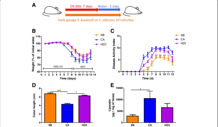
We first evaluated the effect of daily administration of C. albicans and Saccharomyces boulardii CNCM I-745 on dextran sodium sulfate (DSS)-induced colitis (Fig.1a). As previously described [16], administration of C. albicans in-duced an increased severity of colitis at D7, characterized by significant weight loss (Fig. 1b), higher disease activity index (DAI) (Fig. 1c), colon shortening (Fig. 1d), and increased lipocalin levels (Fig.1e). On the other hand, S. boulardii showed an anti-inflammatory effect, reducing most symptoms of colitis evaluated by the DAI (Fig.1b–d), with reduced levels of lipocalin (Fig.1e).
To explore the role of the bacterial microbiota in modulating the effects of fungi on colitis, we treated
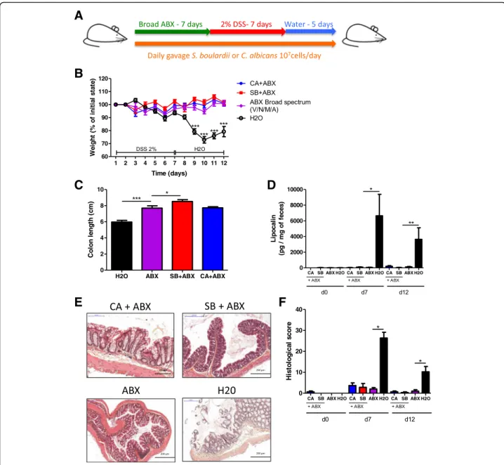
mice with either C. albicans or S. boulardii daily by ga-vages and administered broad-spectrum antibiotics (ABX) for 7 days prior to colitis induction with DSS (Fig. 2a). ABX treatment completely protected the mice from colitis and abrogated the impact of fungi on colitis. Only untreated mice exhibited a strong colitis phenotype with significant weight loss (Fig. 2b), reduced colon length (Fig.2c), high lipocalin levels (Fig.2d), and histo-logical damage (Fig.2e, f ). Analysis of the microbiota by 16S RNA sequencing showed a strong effect of ABX on the biodiversity and composition of the bacterial micro-biota, characterized by a significant reduction of ob-served species (Additional file1: Figure S1A and B) and dramatically reduced levels of Firmicutes and Bacteroi-detes, along with a dramatic increase in Proteobacteria, which is majorly composed of bacteria from the family Enterobacteriaceae(Additional file1: Figure S1C and D). These results showed that some components of the bac-terial microbiota are required for the development of DSS-induced colitis. Consequently, in the absence of these bacteria, colitis does not transpire and pro- or anti-inflammatory fungi challenges do not further bring significant changes in the mice phenotype.
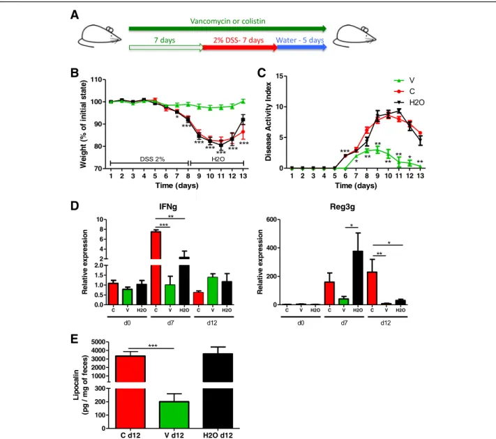
Vancomycin but not colistin has a protective effect on DSS-induced colitis
To evaluate which specific bacterial population in the microbiota affects DSS-induced colitis, we used vanco-mycin or colistin, which target either gram-positive bac-teria or Enterobacbac-teriaceae, respectively (Fig. 3a). First, we confirmed the observations made by Nakanishi et al. [29] showing that vancomycin treatment alone recapitu-lated the strong protective effects of broad-spectrum antibiotics, whereas colistin did not modify the severity of colitis in the treated group compared to the control group. Vancomycin but not colistin reduced DSS-in-duced pathological changes, such as body weight loss (Fig. 3b), disease activity index (Fig. 3c), colonic tran-scripts (IFN-g and Reg3g) and lipocalin levels (Fig.3d, e), and colon histological scores (Additional file 1: Figure S2A and B), in treated mice compared with control mice. Collectively, these results confirm that certain gram-positive bacteria contribute to the development of DSS-induced colitis.
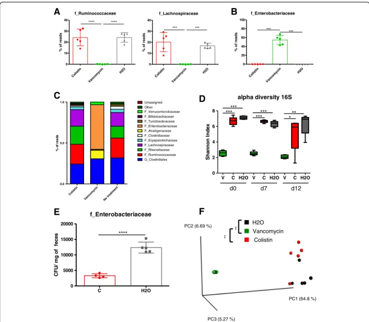
We then investigated the effects of vancomycin and colistin on the intestinal microbiota. First, we confirmed using 16S RNA sequencing that vancomycin treatment
A
B
C
D
E
Fig. 1 In untreated mice, S. boulardii and C. albicans have opposite effects on DSS-induced colitis. a Experimental design for the administration of fungi (C. albicans and S. boulardii) and dextran sulfate sodium (DSS) administration in conventional mice. b Weight of DSS-exposed mice. For statistical
comparisons, an asterisk (*) indicates C. albicans (CA) versus H2O. c Disease activity index (DAI) of DSS-exposed mice. For statistical comparisons, an
asterisk (*) indicates S. boulardii versus H2O, and (&) indicates C. albicans versus H2O. d Length of the colons of mice treated with C. albicans (CA),
S. boulardii (SB), and vehicle (H2O). e Secreted lipocalin (pg/mg of feces) in the feces of mice treated with C. albicans (CA), S. boulardii (SB), and
untreated controls (H2O). Throughout, data are given as the mean ± s.e.m. *P < 0.05, **P < 0.01, and
&&&
P < 0.001 by one-way ANOVA with a post hoc
induced a decrease in gram-positive bacteria such as those from the families Lachnospiraceae and Ruminococcaceae (Fig.4a). Similarly to broad-spectrum-treated mice, vanco-mycin treatment dramatically increases Enterobacteriaceae (Fig. 4b, c, Additional file1: Figure S2C) and significantly reduced bacterial alpha diversity (Shannon index) com-pared to the colistin and control treatments (Fig. 4d). Colistin showed little effect on the diversity or the global composition of the bacterial microbiota except for the level of Enterobacteriaceae (Fig. 4c–e, Additional file 1:
Figure S2C). A beta diversity analysis confirmed a relatively weak effect of colistin treatment on the global bacterial microbiota compared to that of the control, while the micro-biota after vancomycin treatment formed a cluster signifi-cantly separated (P value < 0.01) from the microbiota of the two other groups (Fig. 4f). Collectively, these findings showed that vancomycin-sensitive bacteria (consisting essen-tially of the gram-positive species), but not colistin-sensitive bacteria (consisting essentially of the Enterobacteriaceae), are required for the development of DSS-induced colitis.
A
B
C
D
E
F
Fig. 2 Broad-spectrum ABX protect against DSS-induced colitis, and fungi have limited effects on DSS-induced colitis in their presence. a Experimental design for the administration of broad-spectrum antibiotics (ABX) and dextran sulfate sodium (DSS). b Weight of DSS-exposed mice
(n = 12). For statistical comparisons, asterisk (*) indicates ABX versus H2O. c Length of the colons of mice treated with ABX + C. albicans, ABX + S.
boulardii, or ABX and H2O on day 12 (n = 12). d Secreted lipocalin (pg/mg of feces) at d0, d7, and d12 after DSS (n = 6). e Representative
H&E-stained images of proximal colon cross sections on day 12 after initial DSS exposure. f Histological scores on day 12 (n = 6). Throughout, data are presented as the mean ± s.e.m. *P < 0.05, **P < 0.01, and ***P < 0.001 by one-way ANOVA with a post hoc Tukey test
Antibiotic treatments alter fungal composition and diversity during colitis
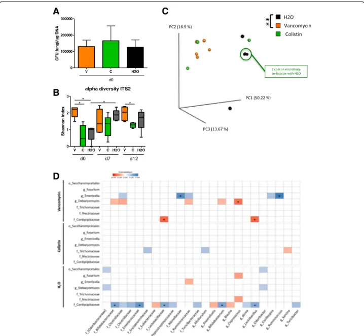
To evaluate the effects of specific antibacterial treatment on the fungal populations during DSS-induced colitis, we treated mice with vancomycin or colistin and compared their fungal microbiota to that of the untreated control mice. Fungal colonization was homogenous among groups at D0, after antibacterial therapy but before DSS treat-ment, confirming that in our settings colistin or vanco-mycin had no direct antifungal effect (Fig. 5a); we also tested that it did not change the global bacterial loads for each condition (Additional file 1: Figure S3A). A detailed
examination of the fungal microbiota showed strong dif-ferences between groups. The alpha diversity, illustrated by the Shannon index, was significantly increased after vancomycin treatment compared to colistin treatment or no treatment (Fig. 5b). Additionally, beta diversity, illustrating the inter-sample differences, showed a significant modification of the fungal microbiota in vancomycin-treated mice but not colistin-treated mice compared to the control group (Fig.5c).
Interestingly, these differences, along with the modi-fied bacterial microbiota, had a strong influence on the evolution of the fungal microbiota during DSS treatment
A
B
D
E
C
Fig. 3 Vancomycin and colistin have different effects on DSS-induced colitis. a Experimental design for the administration of vancomycin or colistin antibiotics, and dextran sulfate sodium (DSS). b Weight of DSS-exposed mice. For statistical comparisons, an asterisk (*) indicates vancomycin
versus colistin/H2O. c Disease activity index (DAI) of DSS-exposed mice. For statistical comparisons, an asterisk (*) indicates vancomycin versus H2O.
d IFN-γ (left) and Reg3g (right) transcript expression in the colon after initiation of DSS treatment. e Secreted lipocalin (pg/mg of feces) in the feces of
mice treated with vancomycin (V), colistin (C), and vehicle (H2O). Throughout, data are presented as the mean ± s.e.m. *P < 0.05, **P < 0.01, and
(Additional file1: Figure S3). The equilibrium between fun-gal phyla also differed between groups: while colistin-treated mice showed a strong decrease in Ascomy-cotaat D7 and D12 (Additional file1: Figure S3b and c), the exact opposite was observed in vancomycin-treated mice, with a nearly complete disappearance of all the Basid-iomycota (Additional file1: Figure S3D). Altogether, these data illustrate the presence of strong functional connections between the bacterial and fungal populations in the gut, with an impact on host response in the context of colitis.
Colistin treatment induces altered correlation between fungal and bacterial relative abundance
In an attempt to elucidate further the interactions be-tween the bacterial and fungal microbiotas, we measured the correlations between the relative abundance of bac-terial and fungal taxa at the genus and family levels under antibiotic treatment and compared them to the correlations in untreated mice. We observed an antibiotic-specific pattern with a reduced number of cor-relations under colistin treatment compared to
C
D
A
Col istin Vancom ycin No trea tmen t 0.0 0.5 1.0 % o f rea d s O_Clostridiales F_Ruminococcaceae F_Rikenellaceae F_Lachnospiraceae F_Erysipelotrichaceae F_Clostridiaceae F_Turicibacteraceae F_Alcaligenaceae F_Bifidobactriaceae Unassigned Other F_Enterobacteriaceae F_VerrucomicrobiaceaeV C H2O V C H2O V C H2O
0 2 4 6 8 d0 d7 d12 ****** ****** *** alpha diversity 16S x e d nI n o n n a h S
B
E
Vancomycin H2O Colistin ** ** PC3 (5.27 %) PC2 (6.69 %) PC1 (64.8 %)F
Colistin Vanco myc in H2O 0 10 20 30 40 f_Ruminococcaceae **** **** s d a er f o % Col istin Vanco myc in H2O 0 10 20 30 40 f_Lachnospiraceae *** *** s d a er f o % Co listin Vanc om ycin H2O 0 20 40 60 80 100 f_Enterobacteriaceae *** *** s d a er f o % C H2O 0 5000 10000 15000 20000 f_Enterobacteriaceae **** s e c ef f o g m / U F CFig. 4 Assessment of microbiota composition after vancomycin or colistin antibiotic treatment. a Relative abundance (% reads) of gram-positive Ruminococcaceae and Lachnospiraceae in the feces of mice treated with colistin, vancomycin, or vehicle at day 0 prior DSS. b Relative abundance (% reads) of Enterobacteriaceae in the feces of mice treated with colistin compared to untreated mice at day 0. c Bacterial-taxon-based analysis at the phylum level in the feces of mice treated with colistin, vancomycin, and vehicle. d Shannon index, describing the alpha diversity of the bacterial
microbiota (16S) in the fecal microbiota of mice treated with vancomycin (V), colistin (C), and vehicle (H2O). e Enterobacteriaceae levels (CFU/mg of feces) in
the feces of mice treated with colistin compared to those of untreated mice. f Beta diversity. Principal coordinate analysis of Bray–Curtis distance with each
sample colored according to the disease phenotype. PC1, PC2, and PC3 represent the top three principal coordinates that captured most of the diversity. The fraction of diversity captured by the coordinate is given as a percentage. Groups were compared using PERMANOVA. Throughout, data are presented as the mean ± s.e.m. *P < 0.05, **P < 0.01, and ***P < 0.001 by one-way ANOVA with a post hoc Tukey test; n = 5 mice per group
vancomycin and control; however, none of them showed statistically significant correlation after correction for the false discovery rate (Fig. 5d). In contrast, controls
and vancomycin-treated mice showed larger number of correlations, and the strength of those correlations was higher under vancomycin.
A
C
B
D
Fig. 5 Antibiotic treatments alter fungal microbiota before and during colitis. a Fungal levels on day 0 in the fecal microbiota of mice treated with vancomycin, colistin, and vehicle were quantified using 18S rRNA qRT-PCR and were normalized to the bacterial population and quantity of
DNA (μg of DNA). Data are presented as the mean ± s.e.m. b Shannon index describing the alpha diversity of the fungal microbiota (ITS2) in the
fecal microbiota of mice treated with vancomycin (V), colistin (C), and vehicle (H2O). c Beta diversity. Principal coordinate analysis of Bray–Curtis
distance with each sample colored according to the disease phenotype. PC1, PC2, and PC3 represent the top three principal coordinates that, together, captured most of the diversity. The fraction of diversity captured by the coordinate is given as a percentage. Groups were compared using PERMANOVA. d Specific bacterial-fungal correlation pattern in mice treated with vancomycin, colistin, and vehicle. Distance correlation plots of the relative abundance of fungal and bacterial families and genera. Statistical significance was determined for all pairwise comparisons; only
significant correlations (P value < 0.05) are displayed and“*” means that correlations remained statistically significant after correction for false
discovery rate. Positive values (blue squares) indicate positive correlations, and negative values (red squares) indicate negative correlations. The shading of the square indicates the magnitude of the association; darker shades are more strongly associated than lighter shades. The sign of the
correlation was determined using Spearman’s method. Throughout, data are mean ± s.e.m. *P < 0.05 and **P < 0.01 by one-way ANOVA with a
Altogether, these data suggest that the presence of colistin-sensitive bacteria strongly influence the bacterial-fungal interactions within the intestine.
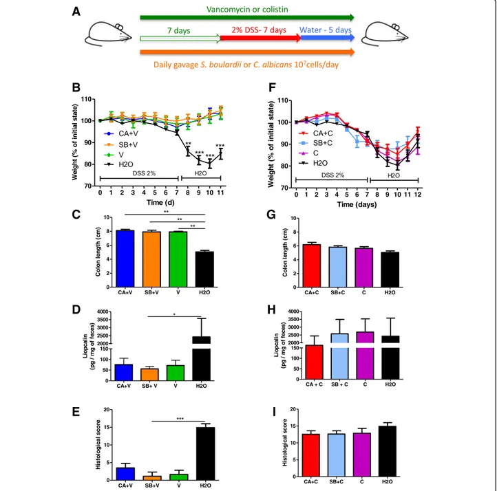
A specific bacterial environment is necessary for the effects of fungi in colitis
To assess whether specific bacterial components of the gut microbiota have an impact on the effects of fungi on gut inflammation, mice gavaged daily with C. albicans or S. boulardii were treated with either vancomycin or co-listin for 7 days before colitis was induced with DSS (Fig.6a). Under vancomycin treatment, all the mice were protected from colitis, showing that gram-positive bacteria are necessary for the onset of colitis, irrespective of the presence of specific fungi (Fig. 6b–e) and
Additional file 1: Figure S4A-C). Under colistin treat-ment, neither C. albicans nor S. boulardii had any impact on colitis severity, whether at a macroscopic (colon length, weight loss, and histological score) or molecular (lipocalin concentration) level (Fig. 6f–i).
Altogether, these results show that the detrimental effects of C. albicans and the beneficial effects of S. boulardii in colitis depend on the presence of colistin-sensitive bacteria, most likely Enterobacteriaceae, within the gut microbiota.
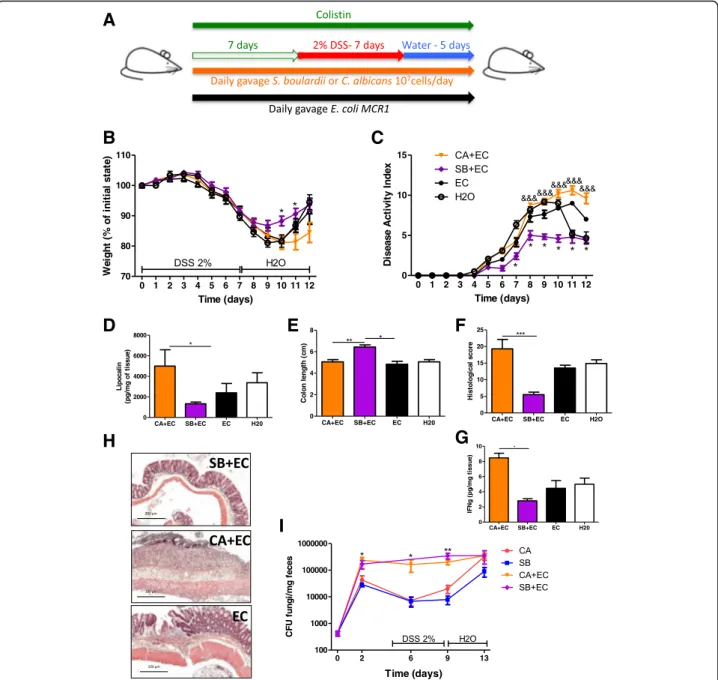
Supplementation with E. coli restores the effects of fungi on colitis in colistin-treated mice
To test whether Enterobacteriaceae are necessary for fungi to modulate the severity of colitis, we supple-mented colistin-treated mice with a colistin-resistant E. colistrain (E. coli MCR1) in order to repopulate the gut ecosystem with Enterobacteriaceae (Fig. 7a). E. coli MCR1 administration alone restored both the beneficial effects of S. boulardii and the detrimental effects of C. albicans on the severity of colitis as assessed by weight and disease activity index (Fig. 7b, c). On day 7, mice supplemented with C. albicans and E. coli had higher lipocalin levels than did mice supplemented with S. bou-lardiiand E. coli (Fig. 7d). Colon length measurements, together with histological scores, also showed a clear im-provement when the mice were co-colonized with S. boulardii and E. coli (Fig. 7e, f, and h). These results were confirmed at a molecular level, showing an in-crease in pro-inflammatory cytokines such as IFN-γ in the colon of mice treated with C. albicans and E. coli compared to those treated with S. boulardii and E. coli (Fig. 7g). To assess the potential effects of E. coli on the level of colonization of C. albicans and S. boulardii in vivo, we quantified each fungus with and without E. coli during the course of the experiment. We observed that E. colisupplementation in colistin-treated mice was suf-ficient to promote colonization of C. albicans and S. boulardii, particularly during the DSS exposition phase (Fig. 7i). Taken together, these results show that
Enterobacteriaceae are required for the positive and negative effects of fungi in the context of intestinal in-flammation. Moreover, the mechanisms involve, at least partly, an ecological effect by which Enterobacteriaceae favor intestinal colonization by fungi such as S. boulardii and C. albicans.
Enterobacteriaceae allow maintenance of fungal loads in the intestine
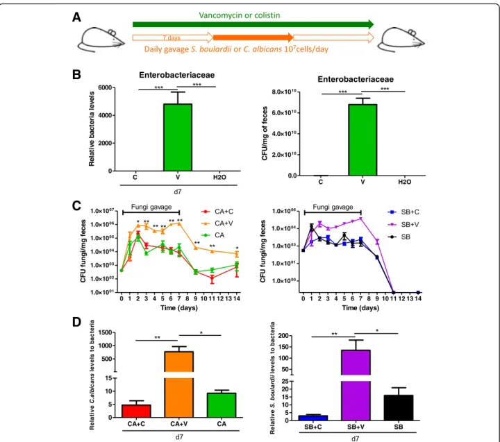
To evaluate the role of bacteria from the family Entero-bacteriaceae in the ecological fitness of fungi in the gut in a general context, we administered both C. albicans and S. boulardii to mice treated with vancomycin or co-listin but without colitis (Fig.8a). Under vancomycin, an increased Enterobacteriaceae level (both relative and ab-solute) was observed (Fig. 8b), and both C. albicans and S. boulardii achieved higher intestinal loads during the daily gavage period in the vancomycin group than in the untreated or colistin-treated group. After the daily gav-age was stopped, higher CFU levels of C. albicans were still present in vancomycin-treated mice compared to colistin-treated and untreated mice (Fig.8c). In contrast, S. boulardii was rapidly eliminated from the gut after the last gavage in all groups, supporting its reduced adaptation to the gut environment compared to C. albi-cans. The results obtained by culture were confirmed by a specific real-time qPCR approach (Fig. 8d). Taken to-gether, these results suggest that, in a non-colitis con-text, bacteria from the Enterobacteriaceae family cooperate with fungi to favor their gut colonization.
Discussion
Crosstalk between the gut microbiota and the host is crucial for the creation and maintenance of intestinal homeostasis and is similarly involved in the pathogenesis of diseases such as IBD. Patients with IBD show a strong alteration of their gut microbiota, with a decrease in bacterial diversity and heavy modifications of the equi-librium not only within bacterial and fungal phyla but also possibly in terms of bacterial-fungal inter-kingdom interactions [7]. However, how bacteria and fungi influ-ence each other and whether their interaction can im-pact microbiota-host homeostasis have been poorly explored to date.
In this study, we investigated the impact of C. albicans and S. boulardii, two model yeasts, for their effects on gut inflammation, focusing on their interaction with the bacterial microbiota. We showed that the presence of specific bacteria (Enterobacteriaceae) is essential for the effect of fungi on gut inflammation, at least partly through their positive influence on fungal colonization.
Although an increase in C. albicans and a decrease in S. cerevisiae were observed in IBD patients, associated with a bacterial dysbiosis [7, 15, 30, 31], data regarding
functional inter-kingdom interactions in inflammatory settings are scarce.
Fungi and bacteria coexist in the environment of the animal gastrointestinal tract; they most likely co-evolved
with the hosts over millions of years of animal evolution. The same is true in any other niche, but the gut environ-ment represents a very specific niche with a high con-centration of microorganisms that interact extensively
A
B
C
D
E
F
G
H
I
Fig. 6 Fungi lose their effects under specific anti-Enterobacteriaceae treatment (colistin). a Experimental design for the administration of vancomycin or colistin, yeasts (C. albicans and S. boulardii), and dextran sulfate sodium (DSS). b Weight of DSS-exposed mice treated with C. albicans + vancomycin, S. boulardii + vancomycin, and vancomycin alone (n = 12). c Length of the colons of mice treated with C. albicans + vancomycin (CA + V), S. boulardii + vancomycin (SB + V), and vancomycin alone (V) (n = 6). d Secreted lipocalin (pg/mg of feces) in the feces of mice treated with C. albicans + vancomycin, S. boulardii + vancomycin, and vancomycin alone (n = 6). e Histological scores of mice treated with C. albicans + vancomycin (CA + V), S. boulardii + vancomycin (SB + V), and vancomycin (V) alone on day 12 after DSS (n = 6). f Weight of DSS-exposed mice treated with C. albicans + colistin (CA + C), S. boulardii + colistin (SB + C), and colistin alone. g Length of the colons of mice treated with C. albicans + colistin (CA + C) (n = 11), S. boulardii + colistin (SB + C) (n = 8), and colistin alone (C) (n = 7). h Secreted lipocalin (pg/mg of feces) in the feces of mice treated with C. albicans + colistin (n = 12), S. boulardii + colistin (n = 8), and colistin alone (n = 7). i Histological scores of mice treated with C. albicans + colistin (n = 9), S. boulardii + colistin (n = 9), and colistin alone (n = 7) on day 12 after DSS
with host tissue and immune cells. Our recent sequen-cing study on a large IBD cohort of 235 patients revealed that positive or negative correlations between fungi and bacteria differ strongly between healthy subjects, Crohn
disease (CD) patients, and ulcerative colitis (UC) pa-tients [7]. Specifically, positive and negative correlations between bacteria and fungi in UC patients were higher and statistically stronger than healthy subjects or CD
A
B
D
E
F
H
G
I
C
Fig. 7 Restoration of the effect of fungi on colitis with E. coli supplementation. a Experimental design for the administration of E. coli MCR1, yeasts (C. albicans and S. boulardii), colistin, and DSS. b Weight of DSS-exposed mice (n = 12 per group). For statistical comparisons, an asterisk (*) indicates C. albicans + E. coli + colistin versus S. boulardii + E. coli + colistin and E. coli. c Disease activity index (DAI) of DSS-exposed mice (n = 12 per group). For statistical comparisons, asterisk (*) indicates S. boulardii + E. coli + colistin versus E. coli + colistin, and (&) indicates C. albicans + E. coli + colistin versus S. boulardii + E. coli + colistin. d Secreted lipocalin (pg/mg of feces) in the feces of mice treated with C. albicans + E. coli + colistin (n = 6), S. boulardii + E. coli + colistin (n = 7), and E. coli + colistin (n = 7). e Length of the colons of mice treated with C. albicans + E. coli + colistin (n = 9), S. boulardii + E. coli + colistin (n = 8), and E. coli + colistin (n = 8). f Histological scores of mice treated with C. albicans + E. coli + colistin (CA + EC) (n = 7), S. boulardii + E. coli +
colistin (SB + EC) (n = 7), and E. coli + colistin (EC) (n = 4) on day 12 after DSS. g Amounts (pg/mg tissue) of IFN-γ (left) secreted by colon explants from
mice treated with C. albicans + E. coli + colistin, S. boulardii + E. coli + colistin, and E. coli + colistin. h Representative H&E-stained images of proximal colon cross sections on day 12 after initial DSS exposure. i C. albicans (CA) and S. boulardii (SB) counts in the feces of mice treated with colistin or with colistin and E. coli (EC) and administered CA and SB (n = 5 per group and time point) by gavage. Throughout, data are presented as the mean ± s.e.m. *P < 0.05,
patients displayed. These data suggest that, depending on the localization and the type of inflammation, fungi might not have the same impact. Here, we used antibi-otics and yeast challenges to produce disequilibrium be-tween the fungal and bacterial microbiota in order to follow the influence of such an imbalance on the severity of colitis. The initial experiment with broad-spectrum antibiotics highlighted that, in conventional mice, the bacterial microbiota is essential for the development of
colitis, since large-scale depletion of the diversity and quantity of bacteria resulted in resistance to DSS colitis. Although early studies reported that broad-spectrum an-tibiotics worsened DSS-induced colitis [32], our result is in accordance with more recent data by Nakanishi et al. [33]. This controversy illustrates the complexity under-lying the initiation and progression of gut inflammation and that antimicrobial drugs might have opposite effects depending on parameters that are not fully understood
A
B
C
D
Fig. 8 Enterobacteriaceae overgrowth increased fungal fitness during gut colonization. a Experimental design for the administration of colistin and vancomycin antibiotics and fungal gavages in conventional mice without DSS treatment. b Relative (left) and absolute (right) Enterobacteriaceae levels in the fecal microbiota of mice treated for 7 days with colistin, vancomycin, or vehicle (PBS) were quantified using 16S RNA qRT-PCR and were normalized to the vehicle group (PBS), day 0. Data are presented as the mean ± s.e.m. (n = 5 per group). c C. albicans (CA) and S. boulardii (SB) counts in the feces of mice treated with colistin (C), vancomycin (V), or vehicle (PBS) and administered CA and SB by gavage. For statistical comparisons, an asterisk (*) indicates C. albicans + vancomycin versus C. albicans + colistin, and (&) indicates S. boulardii + vancomycin vs. C. albicans + vancomycin (n = 5 per group). d C. albicans levels (left) and S. boulardii levels (right) in the fecal microbiota were quantified using specific qRT-PCR primers and were
normalized to the bacterial population (n = 5 per group). Throughout, data are presented as the mean ± s.e.m. *P < 0.05, **P < 0.01, and&&&P < 0.001 by
yet. A simple hypothesis with the current knowledge would be that the basal microbiota composition, which is very likely to be different from one study to another, could influence the effects of antibiotics on the severity of inflammation. Interestingly, in this study, we showed that a pro-inflammatory yeast has negligible inflamma-tory effects when added in an imbalanced microbiota due to antibiotic treatments but has a potent effect on inflammation if added to the normal microbiota. Thus, C. albicans, a well-described opportunistic fungal pathogen and a human gut commensal, requires the presence of specific bacteria that trigger intestinal in-flammation in order to increase the intensity. Specific antibiotic treatment with vancomycin or colistin further corroborated these results. Vancomycin was sufficient to recapitulate the effect of broad-spectrum antibiotics, whereas colistin had no visible effect on susceptibility to inflammation. Bacterial microbiota analysis showed that vancomycin profoundly reduced the bacterial alpha diversity and modified the composition of the micro-biota, with depletion of gram-positive bacteria and overgrowth of Enterobacteriaceae. After vancomycin treatment, mice are completely protected from DSS colitis, and therefore, the protective effects of S. boular-dii are difficult to assess. On the other hand, colistin only decreased the relative abundance of Enterobacteri-aceae without vital changes in the microbial diversity. Altogether, these data demonstrated that specific bac-teria, most likely gram-positive bacteria of the families Ruminococcaceae and Lachnospiraceae, are required to trigger inflammation using DSS. On the other hand, colistin-treated mice, with their Enterobacteriaceae de-pleted, exhibited normal susceptibility to colitis but C. albicans and S. boulardii no longer showed colitis-modulating properties in this setting. Studies of fungal microbiota showed strong disparities between the treated and untreated groups, confirming the indir-ect effindir-ect of bacteria on the fungal population in nor-mal or inflammatory settings. Colistin-treated mice, still sensitive to DSS, showed a strong decrease in Asco-mycota at D7 and D12 after DSS, a phenotype equally observed during flare-ups in IBD patients [7]. In con-trast, the exact opposite was observed in vancomycin treated mice, with the nearly complete disappearance of the Basidiomycota. Analysis of specific bacterial-fungal interactions showed an antibiotic-specific pattern with a greatly reduced number of correlations under colistin treatment, which is in accordance with a potential a role of colistin-sensitive Enterobacteriaceae in the global interactions between bacteria and fungi within the gut.
Following the reintroduction of Enterobacteriaceae (colistin-resistant E. coli), C. albicans and S. boulardii recovered their respective negative and positive effects
on the severity of colitis in colistin-treated mice, sug-gesting that Enterobacteriaceae are necessary for the modulatory effect of fungi on gut inflammation. The mechanisms involved might consist, at least in part, of increased fitness of the fungal population in the pres-ence of these bacteria. Our results are, to our know-ledge, the first in vivo data documenting an ecological effect of a specific bacterial population on fungal growth and colonization in the gut, with important consequences for gut physiology. Enterobacteriaceae have a specific status in the study of gut inflammation and colitis, with publications reporting their role in in-ducing intestinal inflammation [34]. In our study, a clinical isolate of one colistin-resistant E. coli strain was sufficient to strongly modify the fungal population and thus influence the gut ecosystem. These results correl-ate very well with a recent work by Hoarau et al. study-ing the microbiota of CD patients and comparstudy-ing it to that of their non-diseased first-degree relatives [35]; in that study, the authors identified a positive correlation between the presence of C. tropicalis and two Entero-bacteriaceae, E. coli and Serratia marcescens. In vitro data demonstrated an increase in C. tropicalis biofilm formation only with both bacteria and, through micro-scopic imaging, suggested possible interactions at the cellular level. Other studies have also explored fungal-bacterial interactions in other ecosystems, not-ably in the lungs of patients with cystic fibrosis, where Pseudomonas aeruginosa and C. albicans are often identified together [36]. In this particular case, several studies described a detrimental effect of P. aeruginosa on C. albicans growth through cell-cell interaction and effector secretion [37], pointing out another type of interaction between Enterobacteriaceae and fungi.
Conclusions
In conclusion, our results show a link between the bac-terial composition of the microbiota and the gastro-intestinal fitness of fungi, as well as their positive or negative effects on inflammation. By manipulating the bacterial composition of the mouse gut microbiota with specific antibiotics, we showed that Enterobacteriaceae such as E. coli cooperate with yeast to favor their colonization and their active role in intestinal inflam-mation. These results are a first step toward under-standing of the functional connections between fungi and bacteria in the gut. These results also suggest po-tential therapeutic applications such as potentiating the protective action of a yeast probiotic strain, e.g., S. bou-lardii CNCM I-745, or fighting C. albicans infections. For instance, specific targeting of Enterobacteriaceae together with anti-fungal drugs could be a promising strategy in patients suffering C. albicans overgrowth.
Additional file
Additional file 1:Figure S1. Broad-spectrum antibiotics disrupt microbiota with selection of Enterobacteriaceae. Figure S2. Vancomycin and colistin have different effects on DSS-induced colitis. Figure S3. Effects of antibiotics and DSS treatment on fungal microbiota. Figure S4. Gram-positive bacteria are necessary to trigger DSS-induced colitis. (DOCX 1685 kb)
Abbreviations
ABX:Antibiotics; C. albicans: Candida albicans; CFU: Colony-forming unit;
DAI: Disease activity index; DSS: Dextran sodium sulfate; GIT: Gastrointestinal tract; IBD: Inflammatory bowel disease; IFN: Interferon; IL17a: Interleukin 17a; MCR1: Mobilized colistin resistance 1; PBS: Phosphate-buffered saline; Reg3g: Regenerating islet-derived protein 3 gamma; RPMI: Roswell Park Memorial Institute; S. boulardii: Saccharomyces boulardii; YEPD: Yeast extract peptone dextrose
Acknowledgements
The authors thank the employees of the animal facility at INRA and the histology facility at UMR 1313 GABI (Jouy-en-Josas, France) for their technical help. E. coli MCR1 was kindly provided by Thomas Guillard (CHU Reims, Hôpital Robert Debré, Laboratoire de Bactériologie-Virologie-Hygiène, F-51092 Reims, France).
Funding
This study was funded by Biocodex (Gentilly, France). Availability of data and materials
All data generated or analyzed during this study are included in this published article (and its supplementary information files).
Deposition of the raw sequence data in the European Nucleotide Archive is in process; the accession number is pending.
Authors’ contributions
BS, MLR, and HS designed the study, collected, analyzed, and interpreted the data. JP, SJ, MS, BL, JMN, AA, LD, JG, GDC, and MLM helped in collecting, analyzing, and interpreting the data. BS, PL, MLR, and HS wrote the manuscript. All authors read and approved the final manuscript.
Ethics approval
All animal experiments were performed in accordance with the Comite
d’Ethique en Experimentation Animale (COMETHEA C2EA – 45, Jouy en
Josas, France).
Consent for publication Not applicable Competing interests
The authors declare that they have no competing interests.
Publisher’s Note
Springer Nature remains neutral with regard to jurisdictional claims in published maps and institutional affiliations.
Author details
1Micalis Institute, INRA, AgroParisTech, Université Paris-Saclay, Domaine de
Vilvert, Jouy-en-Josas, France.2Sorbonne Universités, École Normale
Supérieure, CNRS, INSERM, APHP Laboratoire des Biomolécules (LBM), 27 rue de Chaligny, Paris, France.3Department of Gastroenterology, Saint Antoine
Hospital, 184 rue du Faubourg Saint-Antoine, Paris, France.
Received: 22 June 2018 Accepted: 24 August 2018
References
1. Qin J, Li R, Raes J, Arumugam M, Burgdorf KS, Manichanh C, Nielsen T, Pons
N, Levenez F, Yamada T, et al. A human gut microbial gene catalogue
established by metagenomic sequencing. Nature. 2010;464(7285):59–65.
2. Morgan XC, Tickle TL, Sokol H, Gevers D, Devaney KL, Ward DV, Reyes JA,
Shah SA, LeLeiko N, Snapper SB, et al. Dysfunction of the intestinal microbiome in inflammatory bowel disease and treatment. Genome Biol. 2012;13(9):R79.
3. Sokol H, Pigneur B, Watterlot L, Lakhdari O, Bermudez-Humaran LG,
Gratadoux JJ, Blugeon S, Bridonneau C, Furet JP, Corthier G, et al. Faecalibacterium prausnitzii is an anti-inflammatory commensal bacterium identified by gut microbiota analysis of Crohn disease patients. Proc Natl
Acad Sci U S A. 2008;105(43):16731–6.
4. Sokol H, Seksik P, Furet JP, Firmesse O, Nion-Larmurier I, Beaugerie L, Cosnes
J, Corthier G, Marteau P, Dore J. Low counts of Faecalibacterium prausnitzii
in colitis microbiota. Inflamm Bowel Dis. 2009;15(8):1183–9.
5. Willing BP, Dicksved J, Halfvarson J, Andersson AF, Lucio M, Zheng Z,
Jarnerot G, Tysk C, Jansson JK, Engstrand L. A pyrosequencing study in twins shows that gastrointestinal microbial profiles vary with inflammatory
bowel disease phenotypes. Gastroenterology. 2010;139(6):1844–54. e1841
6. Gevers D, Kugathasan S, Denson LA, Vazquez-Baeza Y, Van Treuren W, Ren
B, Schwager E, Knights D, Song SJ, Yassour M, et al. The treatment-naive
microbiome in new-onset Crohn’s disease. Cell Host Microbe. 2014;15(3):
382–92.
7. Sokol H, Leducq V, Aschard H, Pham HP, Jegou S, Landman C, Cohen D,
Liguori G, Bourrier A, Nion-Larmurier I, et al. Fungal microbiota dysbiosis in
IBD. Gut. 2017;66(6):1039–48.
8. Khatri I, Tomar R, Ganesan K, Prasad GS, Subramanian S. Complete genome
sequence and comparative genomics of the probiotic yeast Saccharomyces boulardii. Sci Rep. 2017;7(1):371.
9. McFarland LV, Surawicz CM, Greenberg RN, Fekety R, Elmer GW, Moyer KA,
Melcher SA, Bowen KE, Cox JL, Noorani Z, et al. A randomized placebo-controlled trial of Saccharomyces boulardii in combination with standard
antibiotics for Clostridium difficile disease. JAMA. 1994;271(24):1913–8.
10. Kirchhelle A, Fruhwein N, Toburen D. Treatment of persistent diarrhea with
S. boulardii in returning travelers. Results of a prospective study. Fortschr
Med. 1996;114(11):136–40.
11. Pothoulakis C. Review article: anti-inflammatory mechanisms of action of
Saccharomyces boulardii. Aliment Pharmacol Ther. 2009;30(8):826–33.
12. Jawhara S, Poulain D. Saccharomyces boulardii decreases inflammation and
intestinal colonization by Candida albicans in a mouse model of
chemically-induced colitis. Med Mycol. 2007;45(8):691–700.
13. Lee SK, Kim YW, Chi SG, Joo YS, Kim HJ. The effect of Saccharomyces
boulardii on human colon cells and inflammation in rats with
trinitrobenzene sulfonic acid-induced colitis. Dig Dis Sci. 2009;54(2):255–63.
14. Standaert-Vitse A, Sendid B, Joossens M, Francois N, Vandewalle-El Khoury P,
Branche J, Van Kruiningen H, Jouault T, Rutgeerts P, Gower-Rousseau C, et al. Candida albicans colonization and ASCA in familial Crohn's disease. Am J
Gastroenterol. 2009;104(7):1745–53.
15. Richard ML, Lamas B, Liguori G, Hoffmann TW, Sokol H. Gut fungal
microbiota: the Yin and Yang of inflammatory bowel disease. Inflamm
Bowel Dis. 2015;21(3):656–65.
16. Jawhara S, Thuru X, Standaert-Vitse A, Jouault T, Mordon S, Sendid B,
Desreumaux P, Poulain D. Colonization of mice by Candida albicans is promoted by chemically induced colitis and augments inflammatory
responses through galectin-3. J Infect Dis. 2008;197(7):972–80.
17. Ferrer M, Mendez-Garcia C, Rojo D, Barbas C, Moya A. Antibiotic use and
microbiome function. Biochem Pharmacol. 2017;134:114–26.
18. Spinillo A, Capuzzo E, Acciano S, De Santolo A, Zara F. Effect of antibiotic
use on the prevalence of symptomatic vulvovaginal candidiasis. Am J
Obstet Gynecol. 1999;180(1 Pt 1):14–7.
19. Dollive S, Chen YY, Grunberg S, Bittinger K, Hoffmann C, Vandivier L, Cuff C,
Lewis JD, Wu GD, Bushman FD. Fungi of the murine gut: episodic variation and proliferation during antibiotic treatment. PLoS One. 2013;8(8):e71806.
20. Haenni M, Poirel L, Kieffer N, Châtre P, Saras E, Métayer V, Dumoulin R,
Nordmann P, Madec JY. Co-occurrence of extended spectrumβ lactamase
and MCR-1 encoding genes on plasmids. Lancet Infect Dis. 2016;16(3):281–2.
21. Lamas B, Richard ML, Leducq V, Pham HP, Michel ML, Da Costa G,
Bridonneau C, Jegou S, Hoffmann TW, Natividad JM, et al. CARD9 impacts colitis by altering gut microbiota metabolism of tryptophan into aryl
hydrocarbon receptor ligands. Nat Med. 2016;22(6):598–605.
22. Tomas J, Wrzosek L, Bouznad N, Bouet S, Mayeur C, Noordine ML,
Honvo-Houeto E, Langella P, Thomas M, Cherbuy C. Primocolonization is associated with colonic epithelial maturation during conventionalization. FASEB J. 2013;
23. Iliev ID, Funari VA, Taylor KD, Nguyen Q, Reyes CN, Strom SP, Brown J, Becker CA, Fleshner PR, Dubinsky M, et al. Interactions between commensal fungi and the C-type lectin receptor Dectin-1 influence colitis. Science.
2012;336(6086):1314–7.
24. Schmieder R, Edwards R. Quality control and preprocessing of
metagenomic datasets. Bioinformatics. 2011;27(6):863–4.
25. Magoc T, Salzberg SL. FLASH: fast length adjustment of short reads to
improve genome assemblies. Bioinformatics. 2011;27(21):2957–63.
26. Caporaso JG, Kuczynski J, Stombaugh J, Bittinger K, Bushman FD, Costello EK,
Fierer N, Pena AG, Goodrich JK, Gordon JI, et al. QIIME allows analysis of
high-throughput community sequencing data. Nat Methods. 2010;7(5):335–6.
27. Edgar RC. Search and clustering orders of magnitude faster than BLAST.
Bioinformatics. 2010;26(19):2460–1.
28. Koljalg U, Nilsson RH, Abarenkov K, Tedersoo L, Taylor AF, Bahram M, Bates
ST, Bruns TD, Bengtsson-Palme J, Callaghan TM, et al. Towards a unified paradigm for sequence-based identification of fungi. Mol Ecol. 2013;22(21):
5271–7.
29. Kosorok MR. On Brownian distance covariance and high dimensional data.
Ann Appl Stat. 2009;3(4):1266–9.
30. Chehoud C, Albenberg LG, Judge C, Hoffmann C, Grunberg S, Bittinger K,
Baldassano RN, Lewis JD, Bushman FD, Wu GD. Fungal signature in the gut microbiota of pediatric patients with inflammatory bowel disease. Inflamm
Bowel Dis. 2015;21(8):1948–56.
31. Mukherjee PK, Sendid B, Hoarau G, Colombel JF, Poulain D, Ghannoum MA.
Mycobiota in gastrointestinal diseases. Nat Rev Gastroenterol Hepatol. 2015;
12(2):77–87.
32. Rakoff-Nahoum S, Paglino J, Eslami-Varzaneh F, Edberg S, Medzhitov R.
Recognition of commensal microflora by toll-like receptors is required for
intestinal homeostasis. Cell. 2004;118(2):229–41.
33. Nakanishi Y, Sato T, Ohteki T. Commensal gram-positive bacteria initiates
colitis by inducing monocyte/macrophage mobilization. Mucosal Immunol.
2015;8(1):152–60.
34. Garrett WS, Gallini CA, Yatsunenko T, Michaud M, DuBois A, Delaney ML,
Punit S, Karlsson M, Bry L, Glickman JN, et al. Enterobacteriaceae act in concert with the gut microbiota to induce spontaneous and maternally
transmitted colitis. Cell Host Microbe. 2010;8(3):292–300.
35. Hoarau G, Mukherjee PK, Gower-Rousseau C, Hager C, Chandra J, Retuerto
MA, Neut C, Vermeire S, Clemente J, Colombel JF, et al. Bacteriome and Mycobiome interactions underscore microbial Dysbiosis in familial Crohn's
disease. MBio. 2016;7(5).https://doi.org/10.1128/mBio.01250-16.
36. Holcombe LJ, McAlester G, Munro CA, Enjalbert B, Brown AJP, Gow NAR,
Ding C, Butler G, O'Gara F, Morrissey JP. Pseudomonas aeruginosa secreted factors impair biofilm development in Candida albicans. Microbiology. 2010;
156(Pt 5):1476–86.
37. Fourie R, Ells R, Swart CW, Sebolai OM, Albertyn J, Pohl CH. Candida albicans
and Pseudomonas aeruginosa interaction, with focus on the role of eicosanoids. Front Physiol. 2016;7:64.