HAL Id: hal-02307162
https://hal-univ-rennes1.archives-ouvertes.fr/hal-02307162
Submitted on 18 May 2020HAL is a multi-disciplinary open access
archive for the deposit and dissemination of sci-entific research documents, whether they are pub-lished or not. The documents may come from teaching and research institutions in France or abroad, or from public or private research centers.
L’archive ouverte pluridisciplinaire HAL, est destinée au dépôt et à la diffusion de documents scientifiques de niveau recherche, publiés ou non, émanant des établissements d’enseignement et de recherche français ou étrangers, des laboratoires publics ou privés.
Validated Nomogram Predicting 6-Month Survival in
Pancreatic Cancer Patients Receiving First-Line
5-Fluorouracil, Oxaliplatin, and Irinotecan
Lorenzo Fornaro, Francesco Leone, Angélique Vienot, Andrea
Casadei-Gardini, Caterina Vivaldi, Astrid Lièvre, Pasquale Lombardi,
Emmanuele de Luca, Dewi Vernerey, Elisa Sperti, et al.
To cite this version:
Lorenzo Fornaro, Francesco Leone, Angélique Vienot, Andrea Casadei-Gardini, Caterina Vivaldi, et al.. Validated Nomogram Predicting 6-Month Survival in Pancreatic Cancer Patients Receiving First-Line 5-Fluorouracil, Oxaliplatin, and Irinotecan. Clinical Colorectal Cancer, Elsevier, 2019, 18 (4), pp.e394-e401. �10.1016/j.clcc.2019.08.004�. �hal-02307162�
Original article
Title:
Validated nomogram predicting 6-month survival in pancreatic cancer patients receiving first-line 5-fluorouracil, oxaliplatin and irinotecan
Authors:
Lorenzo Fornaro1*, Francesco Leone2,3°, Angélique Vienot4, Andrea Casadei Gardini5, Caterina Vivaldi1, Astrid Lièvre6, Pasquale Lombardi2,3, Emmanuele De Luca2,7, Dewi Vernerey8, Elisa Sperti7, Gianna Musettini1, Maria Antonietta Satolli2,9, Julien Edeline10, Rosella Spadi9, Cindy Neuzillet11, Alfredo Falcone1,12, Giulia Pasquini1, Mario Clerico13, Alessandro Passardi14, Paola Buscaglia15, Aurélia Meurisse8, Massimo Aglietta2,3, Clémence Brac16, Enrico Vasile1, Francesco Montagnani13
Institutions:
1Unit of Medical Oncology 2, Azienda Ospedaliero-Universitaria Pisana, Pisa, Italy 2Department of Medical Oncology, University of Turin, Turin, Italy
3
Medical Oncology, Candiolo Cancer Institute, FPO, IRCCS, Candiolo, Italy
4
Department of Medical Oncology, Besancon University Hospital, Besançon, France
5Department of Oncology, University Hospital of Modena and Reggio Emilia, Modena, Italy
6Department of Gastroenterology, Rennes University Hospital; Rennes 1 University; INSERM, Rennes 1
University, COSS (Chemistry Oncogenesis Stress Signaling), UMR_S 1242, Rennes, France
7
S.C.D.U. Oncologia, A.O. Ordine Mauriziano, Ospedale Umberto I, Turin, Italy
8
Methodological and Quality of Life in Oncology Unit, EA 3181; Besançon University Hospital, Besançon, France
9
Medical Oncology 1 Division, Città della Salute e della Scienza, Turin, Italy
10Oncology Department, Cancer Institute Eugène Marquis; Rennes 1 University; INSERM, INRA, Rennes 1
University, Nutrition Metabolism and Cancer (NuMeCan), Rennes, France
11
Department of Medical Oncology, Curie Institute, Saint Cloud, France
12
Department of Translational Research and New Technologies in Medicine, University of Pisa, Pisa, Italy
13
S.C. Oncologia, Department of Oncology, ASL BI, Biella, Italy
14Department of Medical Oncology, Istituto Scientifico Romagnolo per lo Studio e la Cura dei Tumori (IRST)
IRCCS, Meldola, Italy
15Oncology, ASL VCO Verbano Cusio Ossola, Verbania, Italy 16
Oncology Department, Cancer Institute Eugène Marquis, Rennes, France ° on behalf of the Rete Oncologica del Piemonte e d ella Valla d'Aosta
Correspondence:
*Lorenzo Fornaro, MD
Unit of Medical Oncology 2, Azienda Ospedaliero-Universitaria Pisana, Via Roma 67, 56126 Pisa, Italy
DISCLOSURE OF POTENTIAL CONFLICTS OF INTEREST
MICROABSTRACT
FOLFIRINOX is an accepted standard in metastatic and locally advanced pancreatic cancer but long term prognosis is still poor. Indeed, no criteria reliably identify patients with limited, if any, chances of long-term benefit. We therefore developed and externally validated a prognostic nomogram predicting the risk of early death in pancreatic cancer patients treated with first-line triplet chemotherapy.
ABSTRACT
Background: FOLFIRINOX is an option for fit patients with metastatic (MPC) and locally
advanced unresectable (LAPC) pancreatic cancer. However, no criteria reliably identify patients with better outcome.
Patients and Methods: We investigated putative prognostic factors among 137 MPC/LAPC
patients treated with triplet chemotherapy. Association with 6-month survival status (primary endpoint) was assessed by multivariate logistic regression models. A nomogram predicting the risk of death at 6 months was built by assigning a numeric score to each identified variable, weighted on its level of association with survival. External validation was performed in an independent dataset of 206 patients.
Results: Four variables (performance status, liver metastases, baseline CA19.9 and
neutrophil-to-lymphocyte ratio) were found associated with 6-month survival by multivariate analysis or had sufficient clinical plausibility to be included in the nomogram. Accuracy was confirmed in the validation cohort (C-index 0.762; 95%CI 0.713–0.825). After grouping all cases, four subsets with different outcomes were identified by none, 1, 2 or >2 poor prognostic features (P<0.0001).
Conclusion: Our nomogram accurately predicts the risk of death in the first 6 months after
initiation of FOLFIRINOX in MPC/LAPC patients. This tool could be useful to guide communication about prognosis and inform the design and interpretation of clinical trials.
Clinical Trial Registration: The study is registered on ClinicalTrials.gov (NCT03590275).
Keywords:
INTRODUCTION
Pancreatic cancer (PC) represents a major challenge, as it actually stands fourth among the leading causes of cancer death and is expected to rise up to become the second most lethal malignancy by 2030.1-3 Despite recent advances in systemic treatment, prognosis of patients with metastatic (MPC) or locally advanced, unresectable (LAPC) disease remains poor, with 5-year overall survival (OS) of less than 5%.4 Phase 3 trials established FOLFIRINOX (5-fluorouracil/leucovorin, oxaliplatin and irinotecan) and gemcitabine plus nab-paclitaxel (Gem-Nab) as current standards in the first-line treatment of fit patients with MPC.5,6 Both regimens proved promising efficacy also in patients with LAPC.7-9 In particular, FOLFIRINOX is now regarded as a suitable option in LAPC cases10,11 and has been recently established as the new reference also in the adjuvant setting.12
Nonetheless, triplet chemotherapy is burdened by potentially severe adverse events (mainly digestive and haematological toxicities, with grade 3-4 neutropenia occurring in 46% of patients treated with FOLFIRINOX, including 5.4% febrile neutropenia), and median OS barely exceeds 11 months even in selected patients enrolled in randomized studies (i.e. performance status [PS] 0-1, bilirubin level <1.5 times the upper limit of normal and age ≤75 years).5 In routine clinical practice, only about 25% of patients with MPC would be eligible for FOLFIRINOX.13 Different strategies (comprehensively known as modified FOLFIRINOX) aiming at improving tolerability have been tested, and are mostly based on removing 5-fluorouracil bolus and/or decreasing irinotecan dose, or on the upfront administration of growth factors support.14 This approach seems to reduce the rate of grade 3-4 gastrointestinal or hematologic events, with comparable results in terms of OS with the PRODIGE4-ACCORD11 trial.14,15
The Gem-Nab combination represents an accepted alternative option in first-line6. Despite being associated with an overall similar incidence of hematologic toxicities compared to FOLFIRINOX (grade 3-4 neutropenia: 38%; febrile neutropenia: 3%), Gem-Nab results in a higher rate of grade 3 or more peripheral neuropathy (17% vs. 9%) and a lower rate of severe diarrhoea (6% vs. 12.7%)5,6 and is therefore generally regarded as a suitable option for a greater percentage of MPC patients in everyday practice.13 With the intent to improve risk stratification and patient selection for routine clinical decision making and future trials, several authors investigated clinical and laboratory factors putatively linked with patient outcome.16,17
Goldstein and colleagues recently queried the MPACT study dataset, identified several variables associated with OS and developed a nomogram able to predict patient survival probability at different time points when treated with gemcitabine with or without nab-paclitaxel.18 Predictive algorithms are recently gaining momentum in clinical practice: among them, nomograms are the most frequently used tools thanks to their accuracy and ease of use.19 Previous studies with modified FOLFIRINOX reported that liver metastases, PS and neutrophil-to-lymphocyte ratio (NLR) are independently associated with OS.14 However, no tool is available to predict single patient prognosis with the triplet regimen.
As different treatment options are available and no head-to-head comparison has been conducted so far, discussing the relative benefits and risks of FOLFIRINOX and Gem-Nab with patients is challenging. Based on these considerations, we aimed at developing and validating a simple nomogram able to predict 6-month survival probability in MPC and LAPC patients treated with first-line triplet chemotherapy (FOLFIRINOX, as per classic or modified schedule).
MATERIALS AND METHODS
Patient selection and data collection
The developing set (DS) was constituted by consecutive MPC and LAPC patients treated at a single Institution (Azienda Ospedaliero-Universitaria Pisana, Pisa, Italy) from January 2008 to December 2014 and discussed by dedicated multidisciplinary team dealing with pancreatic malignancies. Eligible patients were identified as follows: age >18 years; cytologically or histologically confirmed pancreatic carcinoma; non resectable, stage III or IV disease according to the American Joint Committee on Cancer (AJCC) staging system; access to clinical data collected before beginning of first-line chemotherapy; availability of laboratory information before treatment initiation, objective tumour response evaluation and survival data. The FOLFOXIRI schedule used in Pisa represents an alternative to standard FOLFIRINOX, derived from the experience in colorectal cancer20 with apparently super imposable efficacy compared with FOLFIRINOX in MPC/LAPC: details about the modified regimen have been described elsewhere.14
The putative predictors investigated were the following: age; gender; Eastern Cooperative Group (ECOG) PS (0 vs. 1); AJCC stage (III vs. IV); tumour location (head vs. body-tail); prior surgery of primary tumour (yes vs. no); previous adjuvant chemotherapy (yes vs. no); presence of biliary drainage (yes vs. no); number of disease sites; presence (yes vs. no) of metastases at specific sites, such as liver, lung, peritoneal or bone; neutrophil, lymphocyte and platelet counts, as well as NLR and platelet-to-lymphocyte ratio (PLR) before the first cycle of treatment; pre-treatment lactate dehydrogenase (LDH), carcinoembryonic antigen (CEA) and carbohydrate antigen 19.9 (CA19.9) serum levels. Age, number of disease sites and laboratory parameters were recorded and analyzed as continuous variables.
The external validation cohort involved MPC/LAPC patients treated at different Italian and French Institutions from January 2011 to June 2017. Inclusion criteria for the validating set (VS) were the same used in the DS, as were the variables collected for analysis. All patients included in the VS received FOLFIRINOX as per PRODIGE4-ACCORD11 schedule.5
The analyses included in this study were performed in accordance with the Declaration of Helsinki and were approved by the Ethics Committee of the Coordinating Centre (Azienda Ospedaliero-Universitaria Pisana, Pisa, Italy). Written informed consent from the patients for research use of data was obtained before the investigation. The protocol is registered on ClinicalTrials.gov (NCT03590275).
Statistical analyses
An early death binary variable indicating 6-month survival status was calculated from survival times, with values “1” if an event of death occurred in the first 180 days and 0 otherwise. Association of different covariates with 6-month survival status was evaluated by building univariate unconditional logistic regressions, modelling each variable with 6-month survival status. Wald test was used to assess statistical significance, defined as a two-tailed P-value <0.05. Considering the high variability of CA19.9, this covariate was logarithmically transformed before the analyses. Statistically significant covariates were used to developed different multivariate logistic regression models. Forward and backward methods were used. Wald test was used to assess the significance of each covariate in the multivariate model. Global fit was evaluated with Nagelkerke's R2, Somer's D and model log-likelihood ratio chi-square. Collinearity was addressed using t-test, Mann-Whitney, Fisher’s exact test, ANOVA,
linear regressions and Variance Inflation Factors (VIF), depending on the nature of the covariates and their characteristics (binary, categorical or continuous). The same tests were also used to assess differences in clinical characteristics between patients included in the VS and DS. Decision regarding inclusion of a specific variable into the final model was addressed taking into consideration their statistical significance, the percentage of models in which it remained significant, the global fit of the model and the clinical plausibility of covariates. Predicted probabilities were tested against the observed probabilities in the VS. Somer's D, C-index, Spiegelhalter Z-test and Brier score were used to evaluate the discrimination of the model. 95% confidence intervals (95%CIs) of the C-index were calculated with bootstrap. Calibration plot was assessed visually. Survival analyses were performed using the Kaplan-Meier method with log-rank test and by building Cox regression models. Median follow-up times were calculated with reverse Kaplan-Meier method.
Response rate (RR) was evaluated according to RECIST v.1.1 criteria. Progression-free survival (PFS) was defined as time from start of FOLFIRINOX to clinical or radiological progression or death from any cause, whichever occurred first, or until the date of the last follow-up, at which point data were censored. OS was defined as time from start of FOLFIRINOX to death from any cause. Survival data were censored at the last follow-up. ROC curves were used to assess the best cut-off values for categorization of continuous variables. Packages “Survival” and “rms” of R were used for all the analyses.
RESULTS
Patient characteristics and treatment outcome
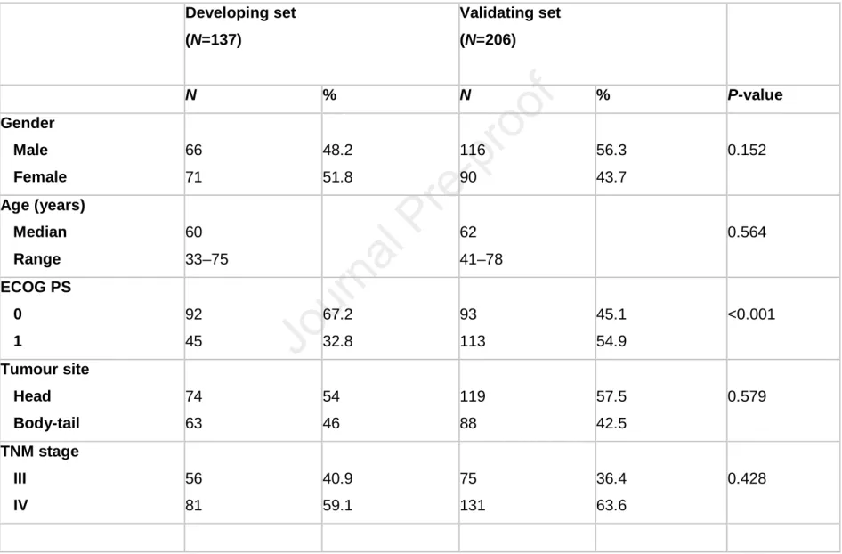
Characteristics of the patients in the DS and VS are presented in Table 1. A total of 343 patients were analyzed, with 137 and 206 cases included in the DS and the VS, respectively. More patients in the VS had an ECOG PS of 1 compared to the DS (54.9% vs. 32.8%; P<0.001). NLR was also significantly higher among the patients in the VS (median, 3.2 vs. 2.3; P<0.001). No significant differences in number and location of metastases, basal CA19.9 serum level or other known prognostic factors were observed (all P-values >0.1).
Median follow-up was 30 months for the DS and 35 months for the VS. Outcomes achieved in the two cohorts were similar. RR was 38.6% and 31.4%, while median PFS was 8.0 (95%CI 6.7–9.2) and 7.2 (95%CI 5.6–8.2) months in the DS and VS, respectively. Median OS was 11.6 (95%CI 10.5–13.9) months in the DS and 10.5 (95%CI 9.2–12.1) months in the VS. Death events were observed in the majority of patients, with only 8.8% and 9.7% of patients censored for OS in the DS and VS, respectively. Notably, there were no censored observations in the first 180 days.
Prognostic nomogram: development
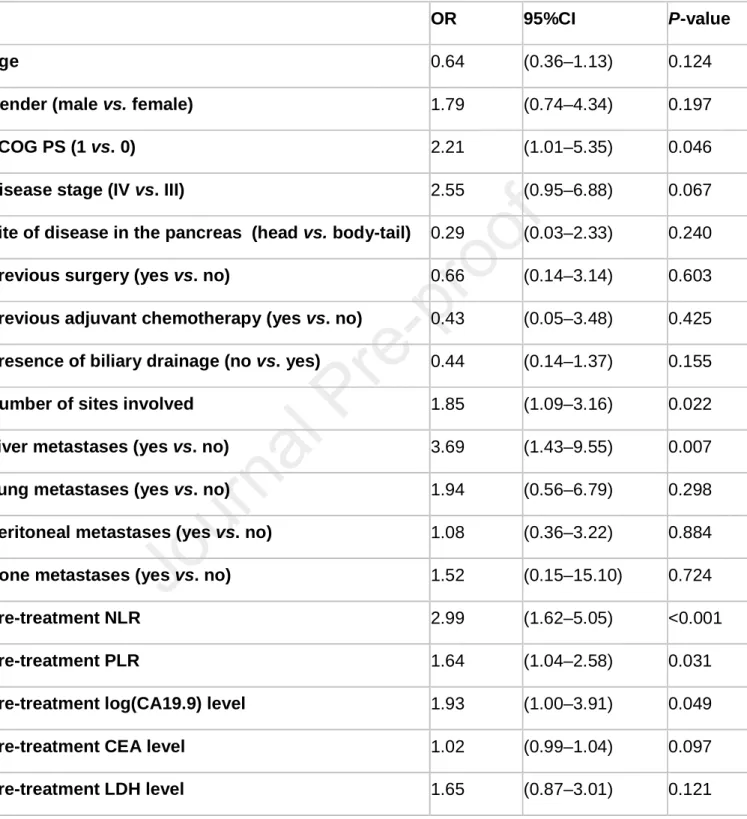
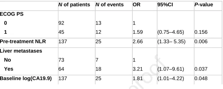
All the collected variables were analyzed for association with 6-month survival (Table 2). Four out of the considered variables were selected in the final multivariable model: ECOG PS, pre-treatment NLR, liver metastases and basal serum CA19.9 (Table 3). Collinearity analyses revealed a slight correlation between CA19.9 and presence of liver metastases and between ECOG PS and NLR. However VIF was always lower than 2, so we decided to keep the model without further modifications. On the contrary, pre-treatment PLR, number of sites involved and disease stage, although significant or borderline significant at univariate analysis, were not retained due to an excessive amount of collinearity with NLR and liver metastases. Global fit was evaluated with Nagelkerke's R2, Somer's D and Area Under the Curve (AUC). The model showed a good global fit with a Nagelkerke's R2 of 0.283, Somer's D of 0.592, C-index of 0.796 and a highly significant log-likelihood (P<0.0001). The resulting nomogram is showed in Figure 1.
Prognostic nomogram: validation
Probabilities predicted by the nomogram were tested against those observed in the VS. The nomogram discriminative ability was satisfying with a Somer's D of 0.524, corresponding to a C-index of 0.762 (95%CI 0.713–0.825). Brier score resulted 0.16 and the Spiegelhalter Z-test was not significant (P=0.087). Visual inspection of the calibration plot showed a good overlap between predicted and observed probabilities, even if there was a slight underestimation for patients at very high risk of early death (Figure 2).
Survival analysis based on prognostic factors
As an ancillary analysis, we performed a categorization of the variables included in the model to assess if they could be used to stratify patients into different risk groups. In order to do that, we combined the patients in the TS and VS and designed 4 different risk categories on the basis of the number of poor prognostic features present, i.e. ECOG PS 1, presence of liver metastases, log(CA19.9) and NLR above a threshold value. ROC curves were developed for the continuous variables log(CA19.9) and NLR, and returned an AUC of 0.641 and 0.676, respectively. We therefore set a threshold of 6.75 for log(CA19.9) (which corresponds to a basal value of 845 U/mL), obtaining a sensitivity of 0.64 and a specificity of 0.62. For NLR, the threshold was set at 2.46, with a sensitivity of 0.78 and a specificity of 0.52. Median OS significantly differed among the four subgroups identified, ranging from 7.2 (95%CI 5.6–8.7) months, through 10.8 (95%CI 9.4–12.9) and 13.9 (95%CI 12.5–16.6) months and up to 18.3 (95%CI 14.5–23.5) months for patients with >2, 2, 1 and 0 risk factors, respectively (P<0.0001 for overall comparison) (Figure 3).
DISCUSSION
Life expectancy of unresectable PC patients treated with first-line chemotherapy remains poor despite the recent introduction of more active chemotherapy regimens.4-6 In light of the toxicity profile of an intensive triplet schedule such as classic or modified FOLFIRINOX, the ability to anticipate single patient prognosis is of high value. Indeed, it allows discussing the benefit-to-risk ratio of this regimen and taking a more informative decision about different first-line therapeutic options. Recently, authoritative experts advocate the need for alternative measures to understand and communicate the impact of treatment on OS.21 Moreover, literature evidence demonstrates that discussion about prognosis during clinical encounters strengthens the patient-oncologist relationship,22 prompting the need for validated and easy-to-use instruments to clearly communicate risks at defined time points during the course of the disease. Similar instruments have been recently proposed for second-line therapy in MPC,23 but are currently lacking for the triplet chemotherapy regimen used in the first-line setting. Moreover, in case of clinical trials, prognostic nomograms could be useful tools for a
better stratification of the enrolled patients and interpretation of the results in different subgroups.24
Our study identified easily available and measurable parameters as major determinants of prognosis in this population, such as ECOG PS, NLR, liver metastases and CA19.9 levels, making the nomogram accessible in the routine clinical setting. A large body of literature supports the prognostic importance of these variables in PC patients treated with gemcitabine-based chemotherapy, particularly for what regards PS and CA19.9 values.25,26 Nonetheless, to the best of our knowledge, this is the first attempt to include them in a validated model able to predict early deaths of MPC/LAPC patients treated with a more modern regimen such as FOLFIRINOX. Of interest, stratifying patients for the presence of each single determinant identified different populations with distinct survival outcomes. In particular, in the most favourable risk subgroup (i.e. no poor prognostic features present) median OS was almost three-times longer than that observed for the worst-risk category (i.e. >2 poor prognostic features present), making the information retrieved by the nomogram useful for both practice and research. Currently, validated predictive biomarkers are lacking in this setting,27 and prognostic stratification is thus essential to discuss alternative treatment options in single cases. Therefore, our nomogram could represent a suitable tool for the identification of different patient subgroups and prompts research on the biological basis explaining the influence of these clinical variables on survival outcome.
Variables included in the nomogram were either confirmed as independent prognostic determinants at multivariate analysis or retained due to the robust evidence of their prognostic value from available literature. Notably, tumour stage has been already demonstrated as a prognostic determinant in previous studies.4,7 However, significance was not formally demonstrated in our datasets (P=0.067 at univariate analysis). This could be possibly due to the relatively low number of LAPC cases in our study, as well as the potential presence of other poor prognostic features in the LAPC cohort. We then decided not to retain it in the final nomogram. This decision was also supported by the evidence of high collinearity between disease stage and presence of liver metastases, as previously described.
Three out of four factors included in our nomogram were also included in the nomogram developed from the MPACT database (comprising PS, NLR, liver metastases, serum albumin, sum of the largest lesions, analgesic use and treatment arm),18 further underlining the external validity of our work. Notably, the relative contribution of CA19.9 to the performance of
the MPACT nomogram when added to PS, NLR, liver lesions and serum albumin was limited. On the contrary, in our study CA19.9 was the strongest predictor of OS among the analysed variables. This is in line with previous literature, which convincingly established this serum tumour marker as a main confirmed determinant of patient outcome in this population.17,25,26 Moreover, some limitations of the MPACT nomogram make it of relatively low immediate utility in routine practice. The lack of external validation, the few points assigned to several variables (such as analgesic use and treatment arm) and the inclusion of highly selected patients from a registrative phase 3 trial might prevent the applicability of these results to other populations and/or other treatment regimens. On the other hand, in light of the partly overlapping factors included in the two nomograms and the easy-of-use of our prognostic variables, it could be of interest to test the performance of this FOLFIRINOX nomogram among patients treated with other regimens, namely Gem-Nab. In this regard, a recently published study confirmed the role of PS, NLR and CA19.9 in a prognostic nomogram developed from a retrospective series of 210 patients treated with first-line Gem-Nab.28
When planning the study design, we decided to build an instrument specifically addressing the risk of early death instead of general OS. This decision was taken in light of the risks of toxicity associated with FOLFIRINOX and other potential issues impacting on patient daily life, such as the need to implant a central venous catheter for prolonged infusions. Indeed, it is of little doubt that FOLFIRINOX (whichever the schedule used) remains a challenging treatment option, ideally to be used in patients able to experience the greatest benefit in terms of OS while sparing those who are likely to get little or no advantage due to very short OS probability. The choice of the 6-month period as primary outcome measure was based on the results of the PRODIGE4-ACCORD11 trial, which reported a median PFS of 6.4 months and a median OS of 11.1 months in the experimental arm.5 In our opinion, the probability of experiencing early death in the first 6 months after treatment initiation (i.e. less than the median PFS expected with FOLFIRINOX) can be considered an acceptable criterion to discuss with the patient treatment options alternative to triplet chemotherapy.
The main criticism of our study relies in its retrospective design and non-exhaustive nature of data collection about other potentially prognostic parameters. However, patient characteristics were generally well balanced in the DS and VS. Furthermore, when treatment activity and efficacy were investigated, no difference was reported in terms of RR, PFS and OS between the two cohorts (and so between FOLFIRINOX and FOLFOXIRI) and results
were comparable with literature data. As discussed, we did not evaluate the outcome of the different risk categories with other treatment options. Therefore, it should be kept in mind that our study was not designed to validate the developed nomogram as a predictive tool to anticipate the benefit from a specific regimen (i.e. FOLFIRINOX) when compared to other options (such as Gem-Nab, single-agent chemotherapy or supportive care only). The information retrieved from the nomogram is a more detailed prognostic assessment of the single cases, and FOLFIRINOX (or modified schedules) remains a valid option for fit MPC/LAPC patients.
CONCLUSION
To conclude, it is possible to accurately predict the risk of death in the first 6 months after starting FOLFIRINOX for MPC/LAPC by few easily available, reproducible and cheap clinical and laboratory parameters. Our nomogram as well as different risk categories allow immediate prognostic stratification and provide an easy-to-interpret tool for both clinicians and patients. This instrument could facilitate patient-physician communication in clinical practice and improve prognostic stratification in clinical research. Validation of this tool for other treatment regimens such as Gem-Nab is warranted.
CLINICAL PRACTICE POINTS
• Despite recent advances, prognosis of patients with metastatic and locally advanced pancreatic cancer remains poor.
• FOLFIRINOX and gemcitabine plus nab-paclitaxel are both standards in the first-line treatment of fit patients and no head-to-head comparison has been conducted so far. • Toxicities of triplet chemotherapy may be relevant and many patients derive limited
benefit from intensive treatment: indeed, no tool is currently available to individualize the therapeutic approach in single cases.
• We therefore developed (from a single Institution experience) and validated (by external collection of cases from Italian and French referral centres) a simple nomogram able to predict 6-month survival probability in pancreatic cancer patients receiving first-line FOLFIRINOX (as per classic or modified schedule).
• Easily available, reproducible and cheap clinical and laboratory parameters confirmed their prognostic value in this population and were finally included in the model: performance status, neutrophil-to-lymphocyte ratio, liver metastases and CA19.9 levels before treatment.
• The presence of these variables also stratified our series into four risk categories with significantly different survival outcome.
• Our nomogram can be immediately implemented in clinical practice in order to improve communication about prognosis with pancreatic cancer patients.
• Moreover, this tool could be of help in clinical research, as it demonstrated to improve patient stratification.
• Validation of the nomogram in series treated with other treatment regimens such as gemcitabine plus nab-paclitaxel is warranted.
ACKNOWLEDGEMENTS
None.
FUNDING
This work was supported by Fondazione ARCO Onlus (Azioni Ricerche e Cure in Oncologia) (no specific grant number applies). The funding source had no involvement in the study design; in the collection, analysis and interpretation of data; in the writing of the report; and in the decision to submit the article for publication.
REFERENCES
1. Siegel RL, Miller KD, Jemal A. Cancer Statistics, 2017. CA Cancer J Clin 2017; 67: 7-30. http://doi.org/10.3322/caac.21387.
2. Rahib L, Smith BD, Aizenberg R et al. Projecting cancer incidence and deaths to 2030: the unexpected burden of thyroid, liver, and pancreas cancers in the United States. Cancer Res 2014; 74: 2913-2921. http://doi.org/10.1158/0008-5472.CAN-14-0155.
3. Ferlay J, Partensky C, Bray F. More deaths from pancreatic cancer than breast cancer in the EU by 2017. Acta Oncol 2016; 55: 1158-1160. http://doi.org/ 10.1080/0284186X.2016.1197419.
4. Kamisawa T, Wood LD, Itoi T, Takaori K. Pancreatic cancer. Lancet 2016; 10039: 73-85. http://doi.org/10.1016/S0140-6736(16)00141-0.
5. Conroy T, Desseigne F, Ychou M et al. FOLFIRINOX versus gemcitabine for metastatic
pancreatic cancer. N Engl J Med 2011; 364: 1817-1825.
http://doi.org/10.1056/NEJMoa1011923.
6. Von Hoff DD, Ervin T, Arena FP et al. Increased survival in pancreatic cancer with nab-paclitaxel plus gemcitabine. N Engl J Med 2013; 369: 1691-1703. http://doi.org/10.1056/NEJMoa1304369.
7. Suker M, Beumer BR, Sadot E et al. FOLFIRINOX for locally advanced pancreatic cancer: a systematic review and patient-level meta-analysis. Lancet Oncol 2016; 17: 801-810. http://doi.org/10.1016/S1470-2045(16)00172-8.
8. Hammel P, Lacy J, Portales F et al. Phase II LAPACT trial of nab-paclitaxel (nab-P) plus gemcitabine (G) for patients with locally advanced pancreatic cancer (LAPC). J Clin Oncol 2018; 36 (Suppl 4S; abstr 204). http://doi.org/10.1200/JCO.2018.36.4_suppl.204
9. Marthey L, Sa-Cunha A, Blanc JF et al. FOLFIRINOX for locally advanced pancreatic adenocarcinoma: results of an AGEO multicenter prospective observational cohort. Ann Surg Oncol 2015; 22: 295-301. http://doi.org/10.1245/s10434-014-3898-9.
10. NCCN Clinical Practice Guidelines in Oncology. Pancreatic Adenocarcinoma. Version
1.2019- November 8, 2018.
https://www.nccn.org/professionals/physician_gls/PDF/pancreatic.pdf. Accessed on: January 04,2019).
11. A randomized phase III trial comparing Folfirinox to gemcitabine in locally advanced pancreatic carcinoma (NEOPAN). https://clinicaltrials.gov/ct2/show/NCT02539537. Accessed on: January 04, 2019.
12. Conroy T, Hammel P, Hebbar M et al. FOLFIRINOX or gemcitabine as adjuvant therapy for pancreatic cancer. N Engl J Med 2018; 379: 2395-2406. http://doi.org/10.1056/NEJMoa1809775.
13. Peixoto RD, Ho M, Renouf DJ et al. Eligibility of metastatic pancreatic cancer patients for first-line palliative intent nab-paclitaxel plus gemcitabine versus FOLFIRINOX. Am J Clin Oncol 2017; 40: 507-511. http://doi.org/10.1097/COC.0000000000000193.
14. Vivaldi C, Caparello C, Musettini G et al. First-line treatment with FOLFOXIRI for advanced pancreatic cancer in clinical practice: Patients' outcome and analysis of prognostic factors. Int J Cancer 2016; 139: 938-945. http://doi.org/10.1002/ijc.30125. 15. Mahaseth H, Brutcher E, Kauh J et al. Modified FOLFIRINOX regimen with improved
safety and maintained efficacy in pancreatic adenocarcinoma. Pancreas 2013; 42: 1311-1315. http://doi.org/10.1097/MPA.0b013e31829e2006.
16. Tabernero J, Chiorean EG, Infante JR et al. Prognostic factors of survival in a randomized phase III trial (MPACT) of weekly nab-paclitaxel plus gemcitabine versus gemcitabine alone in patients with metastatic pancreatic cancer. Oncologist 2015; 20: 143-150. http://doi.org/10.1634/theoncologist/2014-0394.
17. Vernerey D, Huguet F, Vienot A et al. Prognostic nomogram and score to predict overall survival in locally advanced untreated pancreatic cancer (PROLAP). Br J Cancer 2016; 115: 281-289. http://doi.org/10.1038/bjc.2016.212.
18. Goldstein D, Von Hoff D, Chiorean E et al. Relative contribution of baseline variables in a nomogram to predict survival in patients treated with nab-paclitaxel plus gemcitabine or gemcitabine alone for metastatic pancreatic cancer. Ann Oncol 2017; 28 (Suppl_3): iii137-49. https://doi.org/10.1093/annonc/mdx262.016.
19. Shariat SF, Capitanio U, Jeldres C, Karakiewicz PI. Can nomograms be superior to other prediction tools? BJU Int 2009; 103: 492-495. https://doi.org/10.1111/j.1464-410X.2008.08073.x.
20. Masi G, Vasile E, Loupakis F et al. Randomized trial of two induction chemotherapy regimens in metastatic colorectal cancer: an updated analysis. J Natl Cancer Inst 2011; 103: 21-30. https://doi.org/10.1093/jnci/djq456.
21. Saad ED, Zalcberg JR, Péron J et al. Understanding and communicating measures of treatment effect on survival: can we do better? J Natl Cancer Inst 2018; 110: 232-240. https://doi.org/10.1093/jnci/djx179.
22. Fenton JJ, Duberstein PR, Kravitz RL et al. Impact of prognostic discussions on the patient-physician relationship: prospective cohort study. J Clin Oncol 2018; 36: 225-230. https://doi.org/10.1200/JCO.2017.75.6288.
23. Vienot A, Beinse G, Louvet C et al. Overall survival prediction and usefulness of second-line chemotherapy in advanced pancreatic adenocarcinoma. J Natl Cancer Inst 2017; 109 (10). https://doi.org/10.1093/jnci/djx037.
24. Mariotto AB, Noone AM, Howlader N et al. Cancer survival: an overview of measures, uses, and interpretation. J Natl Cancer Inst Monogr 2014; 2014: 145-186. Erratum in: J Natl Cancer Inst Monogr 2015; 2015: 97. https://doi.org/10.1093/jncimonographs/lgu024. 25. Reni M, Zanon S, Balzano G et al. Selecting patients for resection after primary
chemotherapy for non-metastatic pancreatic adenocarcinoma. Ann Oncol 2017; 28: 2786-2792. https://doi.org/10.1093/annonc/mdx495.
26. Kou T, Kanai M, Yamamoto M et al. Prognostic model for survival based on readily available pretreatment factors in patients with advanced pancreatic cancer receiving palliative chemotherapy. Int J Clin Oncol 2016; 21: 118-125. https://doi.org/10.1007/s10147-015-0864-x.
27. Caparello C, Meijer LL, Garajova I et al. FOLFIRINOX and translational studies: towards personalized therapy in pancreatic cancer. World J Gastroenterol 2016; 22: 6987-6700. https://doi.org/10.3748/wjg.v22.i31.6987.
28. Fernández A, Salgado M, García A et al. Prognostic factors for survival with nab-paclitaxel plus gemcitabine in metastatic pancreatic cancer in real-life practice: the ANICE-PaC study. BMC Cancer 2018; 18: 1185. https://doi.org/10.1186/s12885-018-5101-3.
Table 1. Patient characteristics Developing set (N=137) Validating set (N=206) N % N % P-value Gender Male Female 66 71 48.2 51.8 116 90 56.3 43.7 0.152 Age (years) Median Range 60 33–75 62 41–78 0.564 ECOG PS 0 1 92 45 67.2 32.8 93 113 45.1 54.9 <0.001 Tumour site Head Body-tail 74 63 54 46 119 88 57.5 42.5 0.579 TNM stage III IV 56 81 40.9 59.1 75 131 36.4 63.6 0.428
Previous surgery Yes No 15 122 10.9 89.1 34 172 16.5 83.5 0.160 Adjuvant CT Yes No 11 126 8 92 24 182 11.7 88.3 0.363 N. of involved sites 1 2 ≥3 60 62 15 43.8 45.3 10.9 75 104 28 36.2 50.2 13.6 0.356 Liver metastases Yes No 64 73 46.7 53.3 108 98 52.4 47.6 0.322 Lung metastases Yes No 14 123 10.2 89.8 25 181 12.1 87.9 0.608 Peritoneal metastases Yes No 26 111 19 81 35 171 17 83 0.667 Bone metastases Yes No 4 133 2.9 97.1 4 203 1.5 98.5 0.718
CA19.9 (U/mL) Median Range IQR 470 1–75000 91–2001.5 570 0.1–181300 77–3713 0.183 NLR Median Range 2.3 0.6–9.1 3.2 0.3–9.3 <0.001 PLR Median Range 48 17–261 52 15–302 0.276
Abbreviations: CT, chemotherapy; ECOG PS, Eastern Cooperative Oncology Group performance status; IQR, interquartile range; N, number; NLR, neutrophil-to-lymphocyte ratio; PLR, platelet-to-lymphocyte ratio.
Table 2. Results of the univariate models
OR 95%CI P-value
Age 0.64 (0.36–1.13) 0.124
Gender (male vs. female) 1.79 (0.74–4.34) 0.197
ECOG PS (1 vs. 0) 2.21 (1.01–5.35) 0.046
Disease stage (IV vs. III) 2.55 (0.95–6.88) 0.067
Site of disease in the pancreas (head vs. body-tail) 0.29 (0.03–2.33) 0.240
Previous surgery (yes vs. no) 0.66 (0.14–3.14) 0.603
Previous adjuvant chemotherapy (yes vs. no) 0.43 (0.05–3.48) 0.425
Presence of biliary drainage (no vs. yes) 0.44 (0.14–1.37) 0.155
Number of sites involved 1.85 (1.09–3.16) 0.022
Liver metastases (yes vs. no) 3.69 (1.43–9.55) 0.007
Lung metastases (yes vs. no) 1.94 (0.56–6.79) 0.298
Peritoneal metastases (yes vs. no) 1.08 (0.36–3.22) 0.884
Bone metastases (yes vs. no) 1.52 (0.15–15.10) 0.724
Pre-treatment NLR 2.99 (1.62–5.05) <0.001
Pre-treatment PLR 1.64 (1.04–2.58) 0.031
Pre-treatment log(CA19.9) level 1.93 (1.00–3.91) 0.049
Pre-treatment CEA level 1.02 (0.99–1.04) 0.097
Pre-treatment LDH level 1.65 (0.87–3.01) 0.121
Abbreviations: 95%CI, 95% confidence interval; CA19.9, carbohydrate antigen 19.9; CEA,
status; LDH, lactate dehydrogenase; NLR, neutrophil-to-lymphocyte ratio; OR, odds ratio; PLR, platelet-to-lymphocyte ratio.
Table 3. Results of the multivariable model
N of patients N of events OR 95%CI P-value
ECOG PS 0 1 92 45 13 12 1 1.59 (0.75–4.65) 0.156 Pre-treatment NLR 137 25 2.66 (1.33– 5.35) 0.006 Liver metastases No Yes 73 64 7 18 1 3.21 (1.07–9.61) 0.037 Baseline log(CA19.9) 137 25 1.81 (1.01–4.22) 0.048
Abbreviations: CA19.9, carbohydrate antigen 19.9; ECOG PS, Eastern Cooperative
Oncology Group performance status; N, number; NLR, neutrophil-to-lymphocyte ratio; OR, odds ratio; 95%CI, 95% confidence interval.
FIGURE LEGENDS
Figure 1. Nomogram predicting the risk of death at 6 months after initiation of FOLFIRINOX.
Legend: To calculate the score for the single variable, locate the appropriate point in each
axis and draw a line up to the ‘”POINTS” axis. Then, sum the scores for each variable, locate the total score on the “TOTAL POINTS” and draw a line downwards to the “PROBABILITY OF DEATH IN 6 MONTHS” axis. The identified value represents the probability of death at 6 months after starting treatment.
Abbreviations: ECOG PS, Eastern Cooperative Oncology Group performance status; NL
Ratio, neutrophil-to-lymphocyte ratio.
Figure 2. Calibration plot for external validation of the nomogram.
Legend: Calibration plot of observed vs. predicted probabilities. Gray line represents an ideal
perfect model. Dotted line represents the results of the model.
Abbreviations: Brier, Brier score; C (AUC), C-index; Dxy, Somer's D; S:p, P-value of
Spiegelhalter z-test; S:z, z-value of Spiegelhalter z-test.
Figure 3. Survival curves for different risk categories.
Legend: Details are shown in the figure. Risk factors were the following: i) presence of liver
metastases; ii) ECOG performance status 1; iii) baseline neutrophil-to-lymphocyte ratio >6.75; iv) baseline CA19.9 >845 U/mL (see text for details).