HAL Id: tel-03004761
https://tel.archives-ouvertes.fr/tel-03004761
Submitted on 13 Nov 2020
HAL is a multi-disciplinary open access
archive for the deposit and dissemination of sci-entific research documents, whether they are pub-lished or not. The documents may come from teaching and research institutions in France or abroad, or from public or private research centers.
L’archive ouverte pluridisciplinaire HAL, est destinée au dépôt et à la diffusion de documents scientifiques de niveau recherche, publiés ou non, émanant des établissements d’enseignement et de recherche français ou étrangers, des laboratoires publics ou privés.
Synthesis and biological evaluation of ganglioside GM3
analogues
Changping Zheng
To cite this version:
Changping Zheng. Synthesis and biological evaluation of ganglioside GM3 analogues. Cancer. Sor-bonne Université, 2019. English. �NNT : 2019SORUS432�. �tel-03004761�
THESE
Présentée à
SORBONNE UNIVERSITÉ
Ecole doctorale de Chimie Moléculaire de Paris Centre –ED 406
ParM Changping ZHENG
Pour obtenir le grade de
DOCTEUR de SORBONNE UNIVERSITÉ
Spécialité:Chimie Organique
Synthesis and biological evaluation of
ganglioside GM3 analogues
Directeur de thèse:Dr. Yongmin ZHANGSoutenance prévue le 12/07/2019 Devant le jury composé de :
Monsieur ZHANG Yongmin Monsieur SOLLOGOUB Matthieu Madame MULARD Laurence Monsieur VAUZEILLES Boris Madame SALMAIN Michèle
Directeur de Recherche Professeur Directeur de Recherche Directeur de Recherche Directeur de Recherche Directeur de thèse Invited Rapporteur Rapporteur Examinateur
Contents
Acknowledgements...5 List of abbreviations...7 Résumé... 9 Abstract... 11 Chapter I Introduction... 13 1.1 Glycosphingolipids...16 1.1.1 Structures of GSLs... 16 1.1.2 Biological functions of GSLs...17 1.2 Gangliosides... 22 1.2.1 Sialic acid... 221.2.2 Gangliosides and tumors... 23
1.2.3 Gangliosides and nerves...26
1.3 Ganglioside GM3 and cancers... 30
1.3.1 Expression on tumor cells... 32
1.3.2 Effect on tumor EGFR... 33
1.3.3 Effect on tumor angiogenesis...35
1.3.4 Effect on membrane microdomain...37
1.3.5 Effect on tumor cell movement...38
1.4 Synthetic strategies towards gangliosides... 40
1.4.1 Synthesis of sialosides...41
1.4.2 Glycosylation of azidosphingosines...46
1.5 Previous studies on ganglioside GM3 analogues...49
1.6 Objective of this project... 53
Chapter II Synthesis of galactose- and mannose- containing ganglioside GM3 analogues... 55
2.1 Introduction... 57
2.2 Preparation of sialic acid block... 58
2.3 Preparation of azidosphingosine block...59
2.4 Galactose-containing analogues...61
2.4.1 Preparation of galactose with a free 6-OH block... 61
2.4.2 Synthesis of galactose-containing analogues... 63
2.5 Mannose-containing analogues... 69
2.5.1 Preparation of mannose with a free 6-OH block...69
2.5.2 Synthesis of mannose-containing analogues...70
3.1 Introduction... 77
3.2 Retrosynthetic analysis...77
3.3 Preparation of lactal block with a free 6-OH at galactose...78
3.4 Synthesis of lactose-containing analogues...80
Chapter IV Synthesis of glucosamine-containing ganglioside GM3 analogues by developing new chemoenzymatic method... 85
4.1 Introduction... 87
4.2 Regioselective hydrolysis of 2-amino pyranose derivatives by enzyme...88
4.2.1 Preparation of different substrates for enzymatic hydrolysis...88
4.2.2 Enzymatic hydrolysis... 90
4.3 Synthesis of sialylated glucosamine or galactosamine derivatives...94
4.4 Synthesis of glucosamine-containing GM3 analogues... 97
Chapter V Biological evaluation of synthetic ganglioside GM3 analogues...101
5.1 Introduction... 103
5.2 Inhibitory effects on tumor cell growth and migration... 103
5.2.1 Experimental methods...104
5.2.2 Results and discussion...105
5.2.2.1 Galactose- and mannose- containing analogues...105
5.2.2.2 Lactose- and glucosamine- containing analogues... 111
5.3 Inducing neurite outgrowth activity... 116
5.3.1 Experimental methods...117
5.3.2 Results and discussion...118
Chapter Ⅵ Experimental section... 123
6.1 General methods...125
6.2 Molecule numbering...126
6.3 Preparation of sialic acid block... 127
6.4 Preparation of azidosphingosine block...131
6.5 Synthesis of galactose-containing analogues... 139
6.6 Synthesis of mannose-containing analogues...151
6.7 Synthesis of lactose-containing analogues...163
6.8 Chemoenzymatic synthesis of sialylated glucosamine or galactosamine derivatives... 172
6.9 Synthesis of glucosamine-containing analogues...199
5
Acknowledgements
I would like to appreciate all the people who helped and supported me during my PhD study.
Firstly, I would like to thank my supervisor, and I express my sincere gratitude to Dr. Yongmin ZHANG. Four years ago, I wanted to pursue my PhD study, and thanks for Dr. Yongmin ZHANG providing the opportunity in the lab. In particular, I gratefully acknowledge the guidance and help of Dr. Yongmin ZHANG, not only for scientific working, but also for personal development.
Then I would like to thank the thesis reviewers and jury members of the defense for their great efforts.
I also would like to appreciate Prof. Marco TERRENI from University of Pavia for his instruction on enzymatic hydrolysis study. I also would like to appreciate Prof. Matthieu SOLLOGOUB for his help and kindness.
I am grateful to Prof. Jianhua XU from Fujian Medical University and Dr. Huanhuan QU from Shanghai Institute of Materia Medica in China for biological tests.
Many members in the GOBS group have contributed to my personal and professional time. At first, I am grateful to all the permanent staffs in our lab. Thanks for their kindness and suggestions. Then I am grateful to Mlle Lorien BENDA, Mlle Sawsen CHERRABEN, M Jorge MEIJIDE and M Martin WARLOP. They are very nice. I don’t speak French, and they help me a lot in both personal and professional aspects. I enjoy my time in France for these four years, and I would like to thank all the Chinese colleagues in our lab. I think working with them is the precious memory in my life. Thanks for Mme Xiaolei ZHU, M Zhihao LI, M Guangcan XU, Mlle Wenting HU, M Jiang LIU, M Haipeng LIU and M Zhonghang WEN.
I sincerely acknowledge the China Scholarship Council (CSC) for supporting me with a PhD scholarship. I also acknowledge the financial supports from the Centre
6
National de la Recherche Scientifique (CNRS) and the Sorbonne Université.
At last, I would like to express my sincere thanks to my family for their supporting and love in these years.
Changping ZHENG May 2019
7
List of abbreviations
Ac acetyl
AgOTf silver trifluoromethanesulfonate BF3•Et2O boron trifluoride diethyl etherate
Bz benzoyl
calcd calculated value
Cy cyclohexane
d doublet
DBU 1,8-diazabicyclo[5.4.0]undec-7-ene
dd doublet of doublet
DMF N,N-dimethylformamide
DMSO dimethyl sulfoxide
DTBP 2,6-di(tert-butyl)pyridine
EDC 1-ethyl-3-(3-dimethylaminopropyl)carbodiimide
eq. equivalent
Et ethyl
GFR growth factor receptor
h hour
HRMS high-resolution mass spectrometry
Hz hertz
J coupling constant (in Hz)
m-CPBA meta-chloroperoxybenzoic acid
min minute
NIS N-iodosuccinimide
NMR nuclear magnetic resonance
Me methyl
PDMP l-phenyl-2-decanolyamlno-3-morpholino-l-propanol
Ph phenyl
PhSCl benzenesulfenyl chloride
Phth phthalimido
ppm parts per million
PTSA p-toluenesulfonic acid
8 quant. quantitative r.t. room temperature s singlet t triplet TBDMSCl tert-butyldimethylsilyl chloride TE 2-(trimethylsilyl)ethyl
Tf2O trifluoromethanesulfonic anhydride or triflic anhydride
TfOH trifluoromethanesulfonic acid or triflic acid
THF tetrahydrofuran
TLC thin layer chromatography
9
Synthèse et évaluation biologique d’analogues
du ganglioside GM3
Résumé
Glycosphingolipides (GSLs) sont des composants omniprésents des membranes cellulaires animales et impliqués dans de nombreuses fonctions cellulaires, y compris la prolifération, l’adhérence, la motilité et la différenciation. Ganglioside GM3 (NeuAcα3Galβ4Glcβ1Cer), le premier et le plus simple membre de la série métabolique d’une famille de GSLs, contenant de l’acide sialique, du lactose et du céramide. Il a été rapporté que GM3 est impliqué dans certaines maladies graves humaines, telles que les cancers et les maladies nerveuses. La recherche au cours des dernières décennies a démontré que GM3 et ses analogues jouent des rôles importants dans de nombreux processus physiologiques et pathologiques. Il est bien connu que GM3 est un antigène glucide associé à la tumeur sur plusieurs types de tumeur. Dans la présente étude, une série d’analogues avec des sialosides α-2, 6 contenant des sucres différents, y compris le galactose, le mannose, le lactose et la glucosamine, ont été entièrement synthétisés sur la base d’un protocole concis provenant de substrats et de réactifs disponibles dans le commerce. Et les blocs de construction de sucre avec un 6-OH libre ont été préparés par hydrolyse enzymatique, qui peut éviter de multiples procédures chimiques. En particulier, nous avons développé une nouvelle méthode chimioenzymatique pour synthétiser les dérivés glucosamine sialylée ou galactosamine sialylée, puis en utilisant cette approche, des analogues GM3 contenant de la glucosamine ont été construits avec succès. Tous les nouveaux composés ont été purifiés par chromatographie et ont été caractérisés par les spectres RMN et HRMS.
Les activités biologiques de ces analogues synthétiques de GM3 ont été évaluées. Par des essais de cytotoxicité, les analogues GM3 ont été déterminés contre la cellule du cancer du côlon humain HCT116 et la cellule de leucémie K562. Il est intéressant de noter que, par le
10
test de guérison des plaies, certains analogues ont montré des effets significatifs sur le mouvement des cellules tumorales, qui est censé représenter l’un des facteurs pertinents dans l’évaluation de la malignité des cancers. En outre, nous avons employé le modèle de cellule PC12 pour évaluer les effets de certains analogues de GM3 sur l’excroissance de neurites. Cette étude peut fournir des informations précieuses pour synthétiser divers analogues de GM3 et leurs études biologiques.
Mots-clés: glycosphingolipide, ganglioside GM3, acide sialique, synthèse, tumeur,
11
Synthesis and biological evaluation of
ganglioside GM3 analogues
Abstract
Glycosphingolipids (GSLs) are ubiquitous components on animal cell membranes and involved in many cellular functions, such as cell proliferation, adhesion, motility and differentiation. Ganglioside GM3 (NeuAcα3Galβ4Glcβ1Cer), the first and simplest member in the metabolic series of a GSLs family, containing sialic acid, lactose and ceramide. It has been reported that GM3 is implicated in some human serious diseases, such as cancers and nervous diseases. Studies over the past decades have demonstrated that GM3 and its analogues play important roles in many physiological and pathological processes. It is well known that GM3 is a tumor-associated carbohydrate antigen on several types of cancer. In the present study, a series of GM3 analogues with α-2,6 sialosides containing different sugars, including galactose, mannose, lactose and glucosamine, were totally synthesized based on a concise protocol from commercially available substrates and reagents. And the sugar building blocks with a free 6-OH were prepared by regioselective enzymatic hydrolysis, which can avoid multiple chemical procedures. In particular, we developed a new chemoenzymatic method to synthesize sialylated glucosamine or galactosamine derivatives, then using this approach, glucosamine-containing GM3 analogues were constructed successfully. All the new compounds were purified by chromatography and characterized by NMR and HRMS spectra.
Furthermore, the biological activities of these synthetic GM3 analogues were evaluated. By cytotoxicity assays, synthesized GM3 analogues were determined against human colon cancer cell HCT116 and leukemia cell K562. Interestingly, by wound healing test, some analogues showed significant effects on tumor cell movement, which is believed to represent one of the relevant factors in assessing the malignancy of cancers. In addition, we employed the PC12 cell model to evaluate the effects of some GM3 analogues on neurite outgrowth.
12
This study can provide valuable information for synthesizing various GM3 analogues and their biological studies.
Keywords: glycosphingolipid, ganglioside GM3, sialic acid, synthesis, tumor, neurite
13
15
Carbohydrates, along with proteins, nucleic acids and lipids, are the most important organic natural products on earth. It has been demonstrated that carbohydrates and their derivatives play important roles in many biological processes, such as protein structure modulation or cell-cell recognition. The biological implications of carbohydrates are strongly related with serious human diseases, such as cancers or diabetes. Carbohydrates are becoming highly attractive targets for biological study, including drug discovery 1.
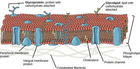
Carbohydrates usually expose on the outer surface of cell membranes, and exist as glycoconjugates, including glycolipids and glycoproteins as shown inFigure I-1. The
glycolipids are composed of hydrophobic lipid moieties with carbohydrate attached, including acylglycerol, ceramide and prenyl phosphate, and the glycoproteins contain proteins with carbohydrate attached.
Figure I-1 Carbohydrates exist as glycoconjugates on the cell membranes.
Moreover, glycolipids can be divided into two categories, glycosphingolipids (GSLs) and glycoglycerolipids, depending on the different structures of lipids as shown in
Figure I-2. The former has an alkaline-stable sphingoid base backbone. However, the
later has an alkaline-labile glycerol backbone. Ceramide is the most important lipid anchor for animal glycolipids, it is composed of sphingosine that is substituted at its
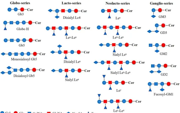
17
Figure I-3 Structures of typical GSLs.
1.1.2 Biological functions of GSLs
In various tissues, GSLs exhibit different distribution and the specific pattern which indicate that they may play important and specific functions in these tissues. At the cellular level, the organizing potential of GSLs plays important roles in the lateral build-up of membranes. In addition, the self-organizing principles of GSLs in combination with their structural specificity have been confirmed to play crucial roles in signaling pathways at the cell surface. Studies over the past decades have demonstrated that GSLs are involved in many physiological and pathological processes 3,4. Figure I-4 describes some of the biological functions that are
associated with GSLs.
3 S. Roseman, J. Biol. Chem.,2001, 276, 41527-41542. 4 T. Yamakawa, Glycoconj. J.,1996, 13, 123-126.
18
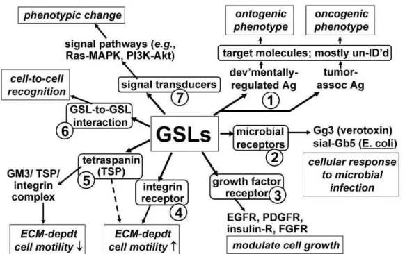
Figure I-4 Some of the biological functions that are associated with GSLs. GSLs associate with
oncogenesis (process1). GSLs interact with bacterial receptors (process 2), with GFRs (process 3),
with integrin receptor (process4), or with integrin/TSP complex (process 5). Cells recognition is
mediated by GSL-to-GSL interaction in microdomains (process 6). GSLs interact with signal
transducers in signal pathways (process7).
Due to the tightly packed backbone of GSLs, they are able to interact with themselves or with sphingomyelin (SM) and cholesterol in a phospholipid environment, therefore forming rafts (Figure I-5) 5,6. It is believed that raft formation is necessary for many
properties of GSLs. They are thought to be enriched in both specific proteins in their lumenal leaflet 7,8 and peripheral proteins carrying myristoyl and palmitoyl chains in
their cytoplasmic leaflet, such as Src-family kinases related to signal pathway 9,10.
Then the concept of glycosynapse has emerged, defined as a microdomain involved in glycosylation-dependent adhesion, recognition and signaling at the cell surface.
5 K. Simons, D. Toomre, Nat. Rev. Mol. Cell Biol.,2000, 1, 31-39. 6 K. Simons, E. Ikonen, Nature,1997, 387, 569-572.
7 D. A. Brown, J. K. Rose, Cell,1992, 68, 533-544.
8 W. Rodgers, B. Crise, J. K. Rose, Mol. Cell Biol.,1994, 14, 5384-5391.
9 S. Hakomori, K. Handa, K, Iwabuchi, S. Yamamura, A. Prinetti, Glycobiology,1998, 8, xi–xix. 10 S. Hakomori, K. Handa, FEBS Lett.,2002, 531, 88–92.
19
Figure I-5 Scheme of a sphingolipid/cholesterol raft. GSLs can form a hydrogen-bonded network
with sphingomyelin and cholesterol in a phospholipid environment, forming rafts on the noncytosolic side of the plasma membrane. GPI-anchored proteins (G) are enriched in these rafts.
Acylated proteins (A) are thought to associate with the cytosolic leaflet of rafts and prenylated
proteins (P) are excluded.
Cell-cell interactions are involved in many biological functions, and the results displayed that carbohydrates play an important role as recognition molecules in such processes. Cell adhesion can be mediated by carbohydrate-to-carbohydrate interaction (CCI). CCI can mediate cell adhesion based on both interfacing glycosyl groups carried by clustered GSLs and GSLs interaction with other glycosyl residues presenting in the same membrane microdomain, as illustrated inFigure I-6 11.
20
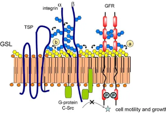
Figure I-6 Carbohydrate-to-carbohydrate interactions controlling glycosylation-dependent cell
motility and growth. GFR, TSP and integrin subunits α and β can be mediated by N-linked glycans and surrounding GSLs. GSLs associate with Src family kinases and other signal transducers. (a) GFR and its interaction with surrounding GSLs. (b) TSP interaction with GSLs.
GSLs are ubiquitous components on animal cell membranes. Some studies have revealed that they are involved in tumor cells proliferation, adhesion, invasion and apoptosis 12,13,14. Usually, the specific types of GSLs are expressed more highly in
tumors than in normal tissues, which are known as tumor-associated antigens. The expression of GSLs on some kinds of human cancers is shown in Table I-1. It has
been concluded that most cancer cells exhibit altered GSLs patterns on their surface, especially the aberrant sialylation of GSLs, abnormal GSLs signaling and biosynthesis, which together play a major role in tumor progression 15,16,17.
12 M. S. Toledo, E. Suzuki, K. Handa, S. Hakomori, J. Bio. Chem.,2004, 279 (33), 34655-34664. 13 M. Ono, K. Handa, S. Sonnino, D. A. Withers, H. Nagai, S. Hakomori, Biochem., 2001, 40,
6414-6421.
14 D. Zhuo, X. Li, F. Guan, Front. Physiol.,2018, 9, 466.
15 J. L. Daniotti, A. A. Vilcaes, V. T. Demichelis, F. M. Ruggiero, M. Rodriguez-Walker, Front. Oncol.,
2013, 3, 306.
16 B. Ogretmen, Y. A. Hannun, Nat. Rev. Cancer,2004, 4, 604-616. 17 S. Hakomori, Cancer Res.,1996, 56, 5309-5318.
21
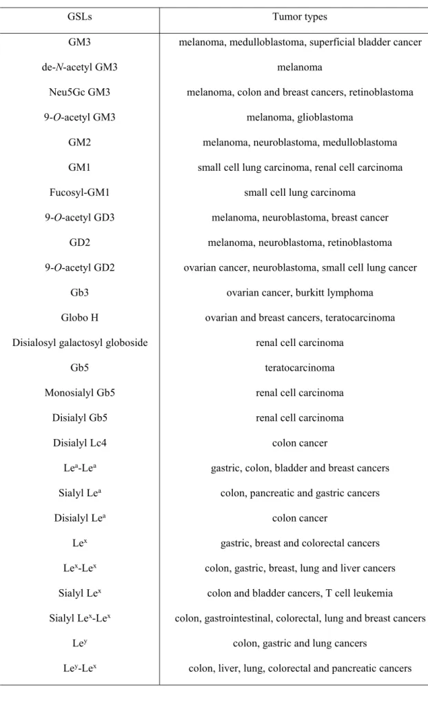
Table I-1 The expression of GSLs on some types of cancers
GSLs Tumor types
GM3 melanoma, medulloblastoma, superficial bladder cancer
de-N-acetyl GM3 melanoma
Neu5Gc GM3 melanoma, colon and breast cancers, retinoblastoma 9-O-acetyl GM3 melanoma, glioblastoma
GM2 melanoma, neuroblastoma, medulloblastoma GM1 small cell lung carcinoma, renal cell carcinoma Fucosyl-GM1 small cell lung carcinoma
9-O-acetyl GD3 melanoma, neuroblastoma, breast cancer GD2 melanoma, neuroblastoma, retinoblastoma 9-O-acetyl GD2 ovarian cancer, neuroblastoma, small cell lung cancer
Gb3 ovarian cancer, burkitt lymphoma Globo H ovarian and breast cancers, teratocarcinoma Disialosyl galactosyl globoside renal cell carcinoma
Gb5 teratocarcinoma
Monosialyl Gb5 renal cell carcinoma Disialyl Gb5 renal cell carcinoma Disialyl Lc4 colon cancer
Lea-Lea gastric, colon, bladder and breast cancers
Sialyl Lea colon, pancreatic and gastric cancers
Disialyl Lea colon cancer
Lex gastric, breast and colorectal cancers
Lex-Lex colon, gastric, breast, lung and liver cancers
Sialyl Lex colon and bladder cancers, T cell leukemia
Sialyl Lex-Lex colon, gastrointestinal, colorectal, lung and breast cancers
Ley colon, gastric and lung cancers
23
(α2→6)- linked to N-acetyl galactosaminides. In addition, the disialosyl structures (α2→8) and (α2→9) have been found as constituents of glycoconjugates. Polysialic acids of Neu5Ac and Neu5Gc, having (α2→8), (α2→9), or alternating (α2→8)/(α2→9) glycosidic linkages, have also been found. Other sialosides, such as (α2→4)-linked homopolymer and (α2→5)-linked derivatives, have also been reported
18,19,20.
At the terminal of natural glycoconjugates on the cell surface, sialic acids can mediate carbohydrate-protein interactions in cell-cell recognition processes. For example, sialic acids are involved in the sialyl Lewisx-selectin binding which occurs in the
recruitment of leukocytes during the inflammation process. In addition, sialic acids can act as receptors for some toxins, viruses and bacteria, such as the interaction between heamaglutinin and Neu5Ac that is the first stage of infection by influenza virus. Sialic acids are also implicated in preventing biological recognition, and the cell interaction can be interfered by modifications of sialic acids. For example,
O-acetylation or N-acetyl hydroxylation can result in a longer lifespan for rat
erythrocytes due to hindering the action of sialidases 21,22,23.
1.2.2 Gangliosides and tumors
Some evidences have already demonstrated that tumor cellular function and phenotype are strongly influenced by the presence of gangliosides. The research results have indicated that tumor-associated gangliosides play a key role in invasion and metastasis of cancer development. The role of gangliosides in tumor development
18 K. Yamada, Y. Harada, Y. Nagaregawa, T. Miyamoto, R. Isobe, R. Higuchi, Eur. J. Org. Chem.,
1998, 2519-2525.
19 S. Kitazume, K. Kitajima, S. Inoue, S. M. Haslam, H. R. Morris, A. Dell, W. J. Lennarz, Y. Inoue, J.
Biol. Chem.,1996, 271, 6694-6701.
20 M. Inagaki, R. Isobe, R. Higuchi, Eur. J. Org. Chem.,1999, 771-774. 21 A. Varki, Glycobiology,1993, 3, 97-130.
22 G. Herrler, J. Hausmann, H. D. Klenk, in Biology of the sialic acids (Ed. A. Rosenberg), Plenum
Press, New York and London,1995, 315-336.
23 W. Reutter, E. Kottgen, C. Bauer, W. Gerok, in Sialic acids. chemistry, metabolism and function (Ed.
24
is crucially important for cancer research 24.
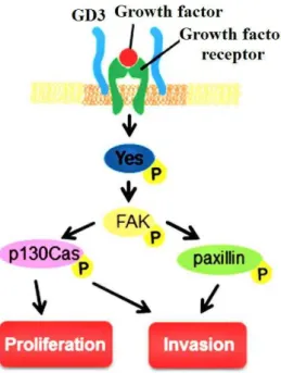
Figure I-8 Malignancy enhancement by ganglioside GD3 in melanomas. GD3 can activate FAK,
p130Cas and paxillin, thus promoting cells proliferation and invasion.
For example, in human melanomas, ganglioside GD3 is the predominant ganglioside component 25,26. Activation of natural killer T (NKT) cells produces cytokines to
influence immune responses against cancers, and GD3 can induce NKT cells response in melanoma. In addition, some studies showed that GD3 can enhance malignancy of melanomas as shown in Figure I-8. The molecules involved in GD3-mediated
signaling pathway include focal adhesion kinase (FAK), p130Cas and paxillin. Further, siRNA can block p130Cas and paxillin, which can obviously suppress melanoma growth 26. In GD3-positive, not GD3-negative, human melanoma SK-MEL-28-N1
cells, the proliferation and apoptosis resistance were enhanced through hepatocyte growth factor (HGF) or adhesion to collagen type I via MAPK (Mitogen-activated protein kinases) and Akt (Protein kinase B) signaling pathways. Increased GD3 expression can promote melanoma cells adhesion to surrounding tissues and
24 S. Birklé, G. Zeng, L. Gao, R. K. Yu, J. Aubry, Biochim.,2003, 85, 455-463.
25 T. Tsuchida, R. E. Saxton, D. L. Morton, R. F. Irie, J. Natl. Cancer Inst.,1987, 78, 45-54.
26 Y. Makino, K. Hamamura, Y. Takei, R. H. Bhuiyan, Y. Ohkawa, Y. Ohmi, H. Nakashima,
25
susceptibility to HGF presented in the tumor microenvironment 27,28. Other results
further showed that the melanoma malignancy is enhanced by GD3 due to g-secretase recruiting to rafts and facilitating efficient cleavage of neogenin 29.
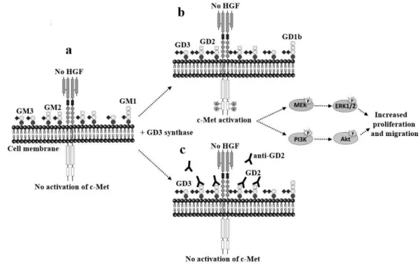
Figure I-9 Activation of c-Met by ganglioside GD2. (a) Breast cancer cell line MDA-MB-231
mainly expresses GM3 and GM2. (b) GD3 synthase induces b- and c- series gangliosides accumulation, mainly GD2, which can activate c-Met in the absence of HGF, then increase proliferation and migration via PI3K/Akt and MEK/Erk pathways. (c) In the competition assays, anti-GD2 mAb was used, which can inhibit c-Met phosphorylation and cells proliferation.
In a MDA-MB-231 breast cancer cell line, it was reported that ganglioside GD2 can enhance proliferation and tumorigenicity by ganglioside-mediated activation of c-Met receptor 30 as shown in Figure I-9. GD2 has been considered as a breast cancer
marker 31. The expression of GSLs in CD44hi/CD24lobreast cancer stem cells (CSCs)
27 K. Furukawa, M. Kambe, M. Miyata, Y. Ohkawa, O. Tajima, K. Furukawa, Cancer Sci.,2014, 105,
52-63.
28 K. Hamamura, K. Furukawa, Cancer Transl. Med.,2017, 3 (6), 209-213.
29 K. Kaneko, Y. Ohkawa, N. Hashimoto, Y. Ohmi, N. Kotani, K. Honke, M. Ogawa, T. Okajima,
K. Furukawa, K. Furukawa, J. Biol. Chem.,2016, 291, 16630-16643.
30 S. Groux-Degroote, Y. Guérardel, S. Julien, P. Delannoy, Biochemistry (Mosc), 2015, 80 (7),
808-819.
31 V. L. Battula, Y. Shi, K. W. Evans, R. Y. Wang, E. L. Spaeth, R. O. Jacamo, R. Guerra, A. A. Sahin,
26
and non-CSCs was also studied by flow cytometry, and up-regulation of GD2, GD3, GM2, and GD1a in CSCs were detected 32.
It has been demonstrated that gangliosides are shed from tumor to their microenvironment in the form of micelles, monomers, or membrane vesicles 33,34.
Shed gangliosides can bind and interact with a wide variety of proteins, including signaling molecules presenting in the tumor microenvironment 35. In addition, shed
gangliosides can be incorporated into the membrane of neighboring host cells, and it is possible that they can modulate tumor–host cell interactions 36,37.
1.2.3 Gangliosides and nerves
1.2.3.1 Distribution of gangliosides in brain and nervous tissues
It is well known that gangliosides are widely expressed in mammalian tissues, particularly in brain and nervous tissues 38. Gangliosides express differently at
different time period during neurodevelopment, brain maturation or aging, and their expression patterns alter obviously during these processes 39,40,41.
There is a specific regional distribution about gangliosides in the adult human brain. In general, a-series gangliosides, such as GD1a and GM1, were found in parietal, frontal and temporal cortical areas, while b-series gangliosides, including GQ1b, GT1b, and GD1b, were found in occipital cortex or structures related to visual system. In addition, in cerebellum, b-series gangliosides were found as the main gangliosides,
32 Y. J. Liang, Y. Ding, S. B. Levery, M. Lobaton, K. Handa, S. Hakomori, Proc. Natl. Acad. Sci. USA,
2013, 110, 4968-4973.
33 Y. Kong, R. Li, S. Ladisch, Biochim. Biophys. Acta,1998, 1394, 43–56.
34 V. Dolo, R. Li, M. Dillinger, S. Flati, J. Manel, B. J. Taylor, A. Pavan, S. Ladisch, Biochim. Biophys.
Acta,2000, 1486, 265–274.
35 M. Rusnati, E. Tanghetti, C. Urbinati, G. Tulipano, S. Marchesini, M. Ziche, M. Presta, Mol. Biol.
Cell,1999, 10, 313–327.
36 F. Chang, R. Li, S. Ladisch, Exp. Cell Res.,1997, 234, 341–346. 37 R. X. Li, S. Ladisch, Biochim. Biophys. Acta,1991, 1083, 57–64. 38 Y. T. Li, S. C. Li, Adv. Carbohydr. Chem. Biochem.,1982, 40, 235-288.
39 L. Svennerholm, K. Bostrom, B. Jungbjer, L. Olsson, J. Neurochem.,1994, 63, 1802-1811. 40 H. Rosner, Prog. Mol. Subcell. Biol.,2003, 32, 49-73.
41 I. Kracun, H. Rosner, V. Drnovsek, Z. Vukelic, C. Cosovic, M. Trbojevic-Cepe, M. Kubat,
27
and in hippocampal archicortex and the amygdala 42, a-series gangliosides are the
predominant species. The c-series gangliosides are biosynthesized during the early neurodevelopment, and found in modest quantities in adult brain. GM1, GD1b and GT1b are abundant during neurogenesis, neuronal migration and neuritogenesis. GD1a is in much higher proportion during synaptogenesis. It was reported that gangliosides are enriched in terminal axons and synaptic endings 43.
Gangliosides expression differed in the brain samples of Alzheimer's disease from the parietal, frontal and temporal cortices, displaying decreased ganglioside GD1a, GD1b and GT1b, and increased ganglioside GM2, GM3 and GM4. The expression of gangliosides in brain is highly region-specific and strongly associated with neurodevelopment, including neural tube formation, neuritogenesis, synaptogenesis, axonogenesis and myelination 44,45,46,47.
1.2.3.2 Effect of gangliosides on nerve growth
The effects of gangliosides on nerve growth and development have already been studied for a long time. It has been revealed that ganglioside GD3 plays a key role in maintaining the self-renewal capacity for neural stem cells, and hence neurogenesis, through sustaining EFG-induced EFGR signaling 48. The study also displayed that
GM1a clustering in lipid rafts is involved in neurite outgrowth 49. In addition, in the
developing human brain, GM1a is implicated in glia-neuronal contacts during neuroblast migration 50. Gangliosides also have been confirmed to be neuroprotective
against toxic nutrients insult 51. Anti-apoptotic during neural cell differentiation 52
42 I. Kracun, H. Rosner, C. Cosovic, A. Stavljenic, J. Neurochem.,1984, 43, 979-989.
43 K. Palmano, A. Rowan, R. Guillermo, J. Guan, P. McJarrow, Nutrients,2015, 7, 3891-3913. 44 P. B. Crino, M. D. Ullman, B. A. Vogt, E. D. Bird, L. Volicer, Arch. Neurol.,1989, 46, 398-401. 45 I. Kracun, S. Kalanj, J. Talan-Hranilovic, C. Cosovic, Neurochem. Int.,1992, 20, 433-438. 46 S. Kalanj, I. Kracun, H. Rosner, C. Cosovic, Neurol. Croat.,1991, 40, 269-281.
47 L. Svennerholm, Prog. Brain Res.,1994, 101, 391-404.
48 J. Wang, R. K. Yu, Proc. Nat. Acad. Sci. USA,2013, 110, 19137-19142.
49 N. Ichikawa, K. Iwabuchi, H. Kurihara, K. Ishii, T. Kobayashi, T. Sasaki, N. Hattori, Y. Mizuno, K.
Hozumi, Y. Yamada, E. Arikawa-Hirakawa, J. Cell Sci.,2009, 122, 289-299.
50 M. Stojiljkovic, T. Blagojevic, S. Vukosavic, N. Zvezdina, S. Pekovic, G. Nikezic, L. Rakic, Int. J.
Dev. Neurosci.,1996, 14, 35-44.
28
and pro-apoptotic during neural cell proliferation 53 were also observed, respectively.
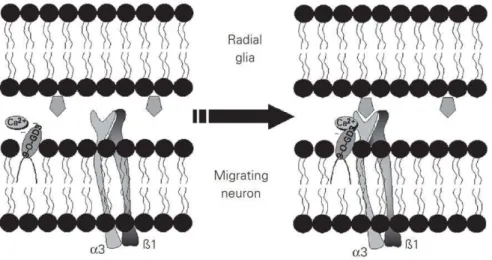
The 9-O-acetyl GD3 is implicated in glial-guided neuronal migration and neurite outgrowth for both the developing and adult rat nervous system. Additionally, during the development and regeneration of mouse peripheral nerve, 9-O-acetyl GD3 is also involved in facilitation of axonal growth and Schwann cell-induced myelination 54,55.
The 9-O-acetyl GD3 involved in neuronal motility has also been studied as shown in
Figure I-10 56.
Figure I-10 Mechanism for 9-O-acetyl GD3 on neuronal motility. The 9-O-acetyl GD3 may
modulate the integrin receptor through a Ca2+-dependent mechanism or by a direct interaction
between the ceramide moiety and the integrin transmembrane helix. Gangliosides in the adjacent membrane are laterally packed around the integrin receptor, affording an optimal Ca2+-enriched
microenvironment for the activation and recognition of extracellular matrix proteins by this receptor.
Despite exogenously added gangliosides are known to promote neurite outgrowth in a variety of cell lines, including some neuroblastoma cell lines, a study in SH-SY5Y human neuroblastoma cell line demonstrated that gangliosides can inhibit neurite outgrowth. Further results revealed that gangliosides have inhibitory effects on cell
52 E. Bieberich, S. MacKinnon, J. Silva, R. K. Yu, J. Biol. Chem.,2001, 276, 44396-44404. 53 Y. Nakatsuji, R. H. Miller, Exp. Neurol.,2001, 168, 290-299.
54 M. F. Santiago, S. S. Liour, R. Mendez-Otero, R. K. Yu, J. Neurosci. Res.,2005, 81, 9-20.
55 V. T. Ribeiro-Resende, T. A. Gomes, S. de Lima, M. Nascimento-Lima, M. Bargas-Rega, M. F.
Santiago, R. A. de Melo Reis, F. G. de Mello, PLos One,2014, 9, e108919.
29
growth through the regulation of lipid rafts 57,58,59.
Myelin-associated glycoprotein (MAG) can bind to the nerve cell surface resulting in inhibiting nerve regeneration. Gangliosides GD1a and GT1b can be as ligands for MAG-mediated inhibition of neurite outgrowth from primary rat cerebellar granule neurons. Other results showed that MAG–gangliosides binding can not only ensure optimal axon–myelin cell–cell interactions, but also enhance long-term axon–myelin stability, and further inhibit axon outgrowth after injury. It is encouraging that studying brain gangliosides interactions can provide new chances to enhance recovery after nerve injury 60,61.
1.2.3.3 Neural tumors
It has been observed that anti-GD2 mAbs addition can significantly improve event-free survival and overall survival in patients with high-risk neuroblastoma 62.
The GSLs composition in an F-11 neuroblastoma cell line has also been studied, and the results showed that major neutral GSLs are Gb4, Gb3, LacCer, and GlcCer, and major gangliosides are GM3, GD3, O-acetylated GD3, and GD1a with trace amounts of GD2. Further studies indicated that the alteration of gangliosides, especially GD3 and O-acetylated GD3, is associated with neuroblastoma behavior, and the specific ganglioside expression may be as a neural cancer marker 63,64. A ratio between GD3
and 9-O-acetylated GD3 on glioblastoma promotes tumor survival, and restoring GD3 through removing acetyl group can reduce tumor cell viability through inducing
57 K. Kabayama, T. Sato, K. Saito, N. Loberto, A. Prinetti, S. Sonnino, M. Kinjo, Y. Igarashi, J.
Inokuchiet, Proc. Natl. Acad. Sci. USA,2007, 4, 13678-13683.
58 M. S. Toledo, E. Suzuki, K. Handa, S. Hakomori, J. Biol. Chem.,2005, 280, 16227-16234.
59 F. Koichi, O. Yuhsuke, Y. Ohkawa, T. Noriyo, K. Yuji, T. Orie, F. Keiko, Neurochem Res.,2011, 36,
1578-1586.
60 G. Mukhopadhyay, P. Doherty, F. S. Walsh, P. R. Crocker, M. T. Filbin, Neuron, 1994, 13,
757-767.
61 A. A. Vyas, H. V. Patel, S. E. Fromholt, M. Heffer-Lauc, K. A. Vyas, J. Dang, M. Schachner, R. L.
Schnaar, Proc. Natl. Acad. Sci. USA,2002, 19, 8412-8417.
62 S. Sait, S. I. Modak, Expert Rev. Anticancer Ther.,2017, 17 (10), 889-904.
63 T. Ariga, G. M. Blaine, H. Yoshino, G. Dawson, T. Kanda, G. C. Zeng, T. Kasama, Y. Kushi, R. K.
Yu, Biochem.,1995, 34, 11500-11507.
64 T. Kaneko, H. Okita, H. Nakajima, K. Iijima, N. Ogasawara, Y. Miyagawa, Y. U. Katagiri,
31
also inhibit tumor cell migration. In particular, GM3 can modulate functions of other molecules co-localized in membrane microdomain.
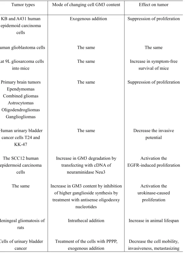
Table I-2 Cancers related with ganglioside GM3
Tumor types Mode of changing cell GM3 content Effect on tumor KB and A431 human
epidemoid carcinoma cells
Exogenous addition Suppression of proliferation
Human glioblastoma cells The same The same Rat 9L gliosarcoma cells
into mice
The same Increase in symptom-free survival of mice Primary brain tumors
Ependymomas Combined gliomas
Astrocytomas Oligodendrogliomas
Gangliogliomas
The same Suppression of proliferation
Human urinary bladder cancer cells T24 and
KK-47
The same Decrease the invasive potential The SCC12 human
epidermoid carcinoma cells
Increase in GM3 degradation by transfecting with cDNA of
neuraminidase Neu3
Activation the EGFR-induced proliferation The same Increase in GM3 content by inhibition
of higher ganglioside synthesis by treatment with antisense oligodeoxy
nucleotides Activation the urokinase-caused proliferation Meningeal gliomatosis of rats
Intrathecal addition Increase in animal lifespan Cells of urinary bladder
cancer
Treatment of the cells with PPPP, exogenous addition
Decrease the cell mobility, invasiveness, metastasizing
32
1.3.1 Expression on tumor cells
GM3 expression is strongly related to human cancers, and change of GM3 expression is associated with the growth and invasion of tumor cells 71,72. It has been reported
that GM3 is expressed with a much higher proportion in the primary and metastatic melanomas than GD2, and that GM3 and GD3 expressions are strongly related to each other. GM3 can not be found in normal skin or on skin neighboring primary melanoma, which is in contrast with GD3 73,74. GM3 is more exposed in the strongly
metastatic line than the weakly metastatic line on the melanomas. It is clear that GM3 expression on the tumor cell surface is an important factor in determining the metastatic phenotype in cancer cells 75.
Cancer cell growth can be stimulated with GM3 depletion, whereas increasing GM3 can lead to suppression. In the murine MBT-2 bladder tumors, the exogenous GM3 can inhibit tumor growth and invasion. It is obvious that GM3 has a significant antitumor effect on bladder cancer 76,77. And a large quantity of GM3 is accumulated
in nonmuscle-invasive bladder cancer, but a small quantity of GM3 is accumulated in muscle-invasive bladder tumor.
The expression of GM3 on the invasive and metastatic properties in four mouse melanoma Bl6 variants has been studied. The results showed that GM3 expression is the highest in B16, and it is the most invasive and metastatic type, followed by F10, F1 and WA4. It has been reported that GM3 can be regarded as an adhesion molecule for another two GSLs, gangliotriaosylceramide (Gg3Cer) and lactosylceramide (LacCer). The metastasis of Bl6 is triggered through the recognition between GM3 and Gg3Cer or LacCer on endothelial cells, which is GSL-GSL interaction, and the
71 Y. Gu, J. Zhang, W. Mi, J. Yang, F. Han, X. Lu, W. Yu, Breast Cancer Res.,2008, 10 (1), 1-12. 72 P. Chefalo, Y. Pan, N. Nagy, Z. Guo, C. Harding, Biochem.,2006, 45, 3733-3739.
73 P. Hersey, O. Jamal, C. Henderson, I. Zardawi, G. D'Alessandro, Int. J. Cancer,1988, 41, 336-343. 74 S. Ichikawa, N. Nakajo, H. Sakiyama, Y. Hirabayashi, Biochem.,1994, 91, 2703-2707.
75 C. Tringali1, I. Silvestri1, F. Testa, P. Baldassari, L. Anastasia, R. Mortarini, A. Anichini, A.
López-Requena, G. Tettamanti, B. Venerando, BMC Cancer,2014, 14, 560.
76 W. Ryuji, O. Chikara, A. Hiroshi, T. Toshiko, S. Makoto, S. Seiichi, H. Senji, I. Atsushi, S. Masaki,
A. Yoichi, Cancer Res.,2002, 62, 3850-3854.
77 S. Kawamura, C. Ohyama, R. Watanabe, M. Satoh, S. Saito, S. Hoshi, S. Gasa, S. Orikasa, Int. J.
33
interaction initiates the signaling pathway 78,79.
N-glycolyl-GM3 contains N-glycolylneuraminic acid instead of N-acetylneuraminic acid, and it is widely expressed in most mammals 80. However, it has been detected in
many human cancers, such as colon carcinomas and breast cancers, not in normal human cells, and it can be considered as one of potential target molecules for cancer therapy 81.
1.3.2 Effect on tumor EGFR
Over activation of epidermal growth factor receptor (EGFR) is well known to be associated with cancer development and progression. Many studies have demonstrated that GM3 has an inhibitory effect on EGFR. In human epidermoid carcinoma A431 cells, exogenous addition of GM3 inhibited EGF-induced autophosphorylation of EGFR 82. GM3 has been known to inhibit EGFR tyrosine
kinase since 1986 83,84. Further research results showed that GM3 inhibits EGFR
tyrosine kinase through binding to N-linked glycan having multivalent GlcNAc termini on EGFR. It is important to realize that GM3 susceptibility for EGFR is strongly dependent on the presence of N-glycosylation, and it is carbohydrate to carbohydrate interaction in the same membrane plane with presumably within the same glyco synaptic microdomain 85,86.
The studies also indicated that receptor dimer formation is not strongly blocked by GM3, but tyrosine phosphorylation on both monomeric and dimeric EGFR is
78 N. Kojima, M. Shiota. Y. Sadahira, K. Handa, S. Hakomori, J. Bio. Chem.,1992, 267, 17264-17270. 79 E. Otsuji, Y. S. Park, K. Tashira, N. Kojima, T. Toyakuni, S. Hakomori, Int. J. Oncol., 1995, 6,
319-327.
80 A. N. Samraj, H. Laubli, N. Varki, A. Varki, Front. Oncol.,2014, 4, 33. 81 R. Blanco, J. Mol. Bio. Diagnosis,2016, 07 (06), 1000e124.
82 E. G. Bremer, J. Schlessinger, S. Hakomori, J. Biol. Chem.,1986, 261, 2434-2440.
83 E. G. Bremer, S. Hakomori, D. F. Bowen-Pope, E. Raines, R. Ross, J. Biol. Chem., 1984, 259,
6818-6825.
84 R. Abdelhadi, H. Jay, Y. Hirotaka, S. K. Donna, G. B. Eric, Glycobiology,1996, 6, 399-406.
85 S. J. Yoon, K. Nakayama, T. Hikita, K. Handa, S. Hakomori, Proc. Natl. Acad. Sci. USA,2006, 103
(50), 18987-18991.
34
inhibited by GM3 87. Besides, other studies further determined the effect of GM3 on
the Tyr-1173 and Tyr-1086 residues phosphorylation of EGFR. The results indicated that GM3 suppressed the phosphorylation of Tyr-1173 residue, and enhanced the phosphorylation of Tyr-1086 residue. Both Tyr-1173 and Tyr-1086 residues, which located at EGFR’s C-terminal, are important EGFR autophosphorylation sites 88,89.
The importance of sialosyl group in GM3 for its EGFR inhibitory effect was also studied. In A431 cells, they were transfected with a gene that encodes for a soluble Mr 42,000 sialidase. Compared with control cells, despite having similar level of EGF binding, the transfected cells could decrease sialic acid expression, promote cells growth, and enhance EGFR autophosphorylation 90. Miyagi’s group demonstrated
that the enhanced NEU3 expression can lead to suppression of apoptosis and decreased NEU3 expression can inhibit Ras activation. The involvement of NEU3 up regulation in cancer development can also be confirmed from increased EGFR phosphorylation detected in NEU3 transgenic mice 91,92,93,94.
Previous study revealed the order of binding intensity for some gangliosides, and it is clear that GM3 exhibits the strongest effect on inhibiting EGFR among ganglioside family 95.
GM3 is the main ganglioside for Hepa1-6 cells. Research found that GM3 can suppress the EGF-induced autophosphorylation of EGFR at tyr1173, and subsequently,
87 X. Q. Wang, P. Sun, A. S. Paller, J. Biol. Chem.,2003, 278 (49), 48770-48778.
88 E. Kovacs, R. Das, Q. Wang, T. S. Collier, A. Cantor, Y. Huang, K. Wong, A. Mirza, T. Barros, P.
Grob, N. Jura, R. Bose, J. Kuriyan, Mol. Cell. Biol.,2015, 35 (17), 3083-3102.
89 J. Pourazar, A. Blomberg, F. J. Kelly, D. E. Davies, S. J. Wilson, S. T. Holgate, T. Sandstrom, Part.
Fibre Toxicol.,2008, 5, 8.
90 E. J. Meuillet, R. Kroes, H. Yamamoto, T. G. Warner, J. Ferrari, B. Mania-Farnell, D. George, A.
Rebbaa, J. R. Moskal, E. G. Bremer, Cancer Res.,1999, 59, 234-240.
91 C. Tringali, B. Lupo, I. Silvestri, N. Papini, L. Anastasia, G. Tettamanti, B. Venerando, J. Bio.
Chem.,2012, 287 (51), 42835-42845.
92 S. Ueno, S. Saito, T. Wada, K. Yamaguchi, M. Satoh, Y. Arai, T. Miyagi, J. Bio. Chem.,2006, 281
(12), 7756-7764.
93 K. Shiozaki, K. Yamaguchi, I. Sato, T. Miyagi, Cancer Sci.,2009, 100 (4), 588-594.
94 M. Miyata, M. Kambe, O. Tajima, S. Moriya, H. Sawaki, H. Hotta, Y. Kondo, H. Narimatsu, T.
Miyagi, K. Furukawa, Cancer Sci.,2011, 102 (12), 2139-2149.
95 E. A. Miljan, E. J. Meuillet, B. Mania-Farnell, D. George, H. Yamamoto, H. G. Simon, E. G.
35
down regulates the PI-3K/Akt signaling pathway 96. However, GM3 can enhance the
HGF-induced autophosphorylation of HGFR at Tyr-1313 and Tyr-1365, and subsequently, up regulates the PI-3K/Akt signaling pathway 97. It is demonstrated that
GM3 has an opposite effect on the EGF and HGF-induced Hepa1-6 cells motility and migration 98. Actually, GM3 is implicated in tumor progression through different
ways, especially for EGFR activity. Firstly, GM3 can directly influence the EGFR activity by interaction with N-linked GlcNAc termini on the receptor. Secondly, GM3 can indirectly regulate EGFR activity not only through the interaction between EGFR and membrane proteins such as integrins and CD9, but also influencing the intracellular Src kinase and protein tyrosine phosphatase activity. Therefore, it seems that the mechanism of GM3 modulating EGFR activity is rather complex.
The co-expression of EGFR and ganglioside Neu5Gc GM3 in different human tumors has been reported 99. Neu5Gc GM3 and EGFR can coordinately contribute to the
tumor progression. Combinations against these two targets will provide a valuable insight for cancer therapy.
1.3.3 Effect on tumor angiogenesis
The ratio of GM3 to a variety of gangliosides, such as GM1, GD3, GD1a and GT1b, can influence the angiogenic properties on many brain tumor types. The ratio of GM3 to the proangiogenic gangliosides GD3 and GD1a (GM3/GD3; GM3/GD1a) is lower in more metastatic and aggressive tumors than that in less metastatic tumors 100,101,102.
In mouse or most cultured human brain tumor cells, GM3 expression serves to regulate or counteract the proangiogenic action of other gangliosides 103. It has been
96 X. Huang, Y. Zhang, J. Xu, Y. Tian, K. Ma, J. Cell. Biochem.,2013, 114 (7), 1616-1624. 97 Y. Li, X. Huang, W. Zhong, J. Zhang, K. Ma, Mol. Cell. Biochem.,2013, 382 (1-2), 83-92. 98 Y. Li, X. Huang, C. Wang, Y. Li, M. Luan, K. Ma, Mol. Med. Rep.,2015, 11 (4), 2959-2966. 99 A. G. Palomo, R. B. Santana, X. E. Pérez, D. B. Santana, M. R. Gabri, K. L. Monzon, A. C. Pérez,
Clin. Exp. Metas.,2016, 33 (7), 717-725.
100 L. E. Abate, P. Mukherjee, T. N. Seyfried, J. Neurochem.,2006, 98 (6), 1973-1984.
101 A. Giulio, F. Stefania, S. Paola, M. Francoise, P. Pietro, S. Federico, C. P. Mario, M. G. Pietro,
Cancer Res.,1987, 47, 4243-4247.
102 M. H. Ravindranath, T. Tsuchida, D. L. Morton, R. F. Irie, Cancer,1991, 67, 3029-3035. 103 T. N. Seyfried, P. Mukherjee, J. Oncol.,2010, 961243.
36
reported that brain tumors with high GM3 expression are less angiogenic. Other studies showed that removing GM3 expression gene in the ependymoblastoma tumor that contains GM3 as the only ganglioside, increased angiogenesis was observed
104,105. And in the highly angiogenic CT-2A astrocytoma, decreased angiogenesis was
observed when GM3 expression was up regulated 106.
It has been reported that urokinase plasminogen activator receptor (μPAR) localization in caveolar lipid-rafts (LRs) is necessary for an efficient angiogenesis program in endothelial progenitor cells or endothelial colony forming cells, EPCs/ECFCs, usually considered as the main source of vasculogenesis in human cancers 107,108. Further studies found that μPAR is present on the ECFC surface in at
least three compartments, one associated to GM1, another associated to GM3, a third one related to caveolar-LRs. Following the exogenous GM1 addition, μPAR is depleted in the GM3 compartment which is recruited within caveolar-LRs compartment. Therefore, angiogenesis-related signaling pathways are triggered, which result in enhanced ECFC invasion and capillary morphogenesis 109.
Some evidences also indicated that GM3 can reduce the ability of endothelial cell membrane to transfer growth signals present in the microenvironment into the endothelial cell itself, leading to block the growth stimulation of vascular endothelium
110. Some cancer cells, such as melanomas, shed GM3 or other gangliosides into the
microenvironment. Then gangliosides can act as modulators of the angiogenic response by promoting the angiogenic capacity of PGEl (Prostaglandin E1) and bFGF (Basic fibroblast growth factor), normally present in the tissue microenvironment.
104 T. N. Seyfried, M. el-Abbadi, M. L. Roy, Mol. Chem. Neuropathol.,1992, 17, 147-167. 105 J. A. Ecsedy, K. A. Holthaus, H. C. Yohe, T. N. Seyfried, J .Neurochem.,1999, 73, 254-259. 106 M. G. Manfredi, S. Lim, K. P. Claffey, T. N. Seyfried, Cancer Res.,1999, 59, 5392-5397.
107 F. Margheri, A. Chillà, A. Laurenzana, S. Serratì, B. Mazzanti, R. Saccardi, M. Santosuosso,
G. Danza, N. Sturli, F. Rosati, L. Magnelli, L. Papucci, L. Calorini, F. Bianchini, M. Del Rosso, G. Fibbi, Blood,2011, 118, 3743-55.
108 A. Chillà, F. Magherini, F. Margheri, A. Laurenzana, T. Gamberi, L. Bini, L. Bianchi, G. Danza, B.
Mazzanti, S. Serratì, A. Modesti, M. Del Rosso, G. Fibbi, Mol. Cell Proteomics, 2013, 12,
1926-1938.
109 F. Margheri, L. Papucci, N. Schiavone, R. D’Agostino, S. Trigari, S. Serrat, A. Laurenzana, A.
Biagioni, C. Luciani, A. Chill, E. Andreucci, T. Del Rosso, G. Margheri, M. Del Rosso, G. Fibbi, J.
Cell Mol. Med.,2015, 19, 113-123.
37
Perhaps their modulatory effects on angiogenesis play roles in metastatic or primary tumor growth 111.
Vascular endothelial growth factor receptor-2 (VEGFR-2) is also involved in tumor angiogenesis. The various downstream signaling pathways can be regulated by GM3 through the inhibitory effect on VEGF-induced VEGFR-2 activation 112,113. GM3 can
inhibit the VEGF-induced activation of PI-3K/Akt, extracellular signal–regulated kinases (ERK), vascular endothelial cadherin and FAK/paxillin, then suppress proliferation, migration and the tube formation caused by VEGF.
1.3.4 Effect on membrane microdomain
The concept of membrane microdomains is that GSLs (GM3), growth factor receptors (GFRs), and other signaling molecules, such as TSs and integrins, are localized and functionalized as clusters in the membrane. Further studies indicated that membrane microdomains are involved in tumor cells interaction, adhesion or other related signal transduction.
Studies of melanoma B16 cells revealed the presence of “GSL signaling domains”, which is different from caveolin-containing membrane domains 114. In mouse
neuroblastoma Neuro2a cells, studies found that GM3 enriched microdomains were involved in signal transduction during neurite formation 115. Moreover, Hakomori’s
group indicated that clustered GSLs associated with c-Src, small G-protein (Rho A) and FAK are involved in GSL-dependent signaling pathways 116,117. A typical
example is GM3 clustering at the cell surface of B16, forming a “glycosignaling
111 P. M. Gullino, Acta Oncol.,1995, 34, 439-441.
112 T. W. Chung, S. J. Kim, H. J. Choi, K. J. Kim, M. J. Kim, S. H. Kim, H. J. Lee, J. H. Ko, Y. C. Lee,
A. Suzuki, C. H. Kim, Glycobiology,2009, 19 (3), 229-239.
113 P. Mukherjee, A. C. Faber, L. M. Shelton, R. C. Baek, T. C. Chiles, T. N. Seyfried, J. Lipid Res.,
2008, 49 (5), 929-938.
114 K. Iwabuchi, K. Handa, S. Hakomori, J. Biol. Chem.,1998, 273, 33766-33773. 115 A. Prinetti, K. Iwabuchi, S. Hakomori, J. Biol. Chem.,1999, 274, 20916-20924.
116 K. Iwabuchi, S. Yamamura, A. Prinetti, K. Handa, S. Hakomori, J. Biol. Chem., 1998, 273,
9130-9138.
117 K. Iwabuchi, Y. Zhang, K. Handa, D. A. Withers, P. Sinay, S. Hakomori, J. Biol. Chem.,2000, 275,
38
domain” (GSD) 118. GM3 attributes to initiate the adhesion of melanoma to
endothelial cells, which is the first step in metastasis 119. Antigenicity, adhesion
mediation and signaling initiation by GM3 are believed to be maintained by GM3 clustering in GSD.
The “glycosynapse” is highly enriched in TSs, hydrophobic proteolipids, integrins, and a wider range of signaling molecules and GFRs 120,121. The studies demonstrated
that glycosynapses are involved in various cellular functions, such as cell growth and motility, which are enhanced during tumor progression 122. Studies also showed that
increased GM3 expression can enhance the anti-metastatic functions of TSs, CD82 and CD9 123. In a human colon cancer cell line HRT18 that highly expresses CD9, the
increased GM3 expression resulted in increased interaction between CD9 and integrin α3 and decreased cell migration through laminin-5-coated membranes 124.
1.3.5 Effect on tumor cell movement
Tumor cell adhesion, motility and migration are very important in the development and progression of cancers. Studies have demonstrated that GM3 has a clear effect on cell adhesion, motility and migration, and change of GM3 expression plays multiple roles in the tumor cell motility, invasiveness and survival 125.
On ovarian cancer, the effect of GM3 synthase over expression on cell motility is due to the change of ganglioside content. The cell motility can be reduced by the exogenous addition of GM3, GM2 or GM1 (not GD1a) as well as pharmacologically
118 Y. Zhang, K. Iwabuchi, S. Nunomura, S. Hakomori, Biochem.,2000, 39, 2459-2468.
119 N. Kojima, M. Shiota, Y. Sadahira, K. Handa, S. Hakomori, J. Biol. Chem., 1992, 267,
17264-17270.
120 S. K. Bromley, W. R. Burack, K. G. Johnson, K. Somersalo, T. N. Sims, C. Sumen, M. M. Davis, A.
S. Shaw, P. M. Allen, M. L. Dustin, Ann. Rev. Immun.,2001, 19, 375-396.
121 A. Viola, S. Schroeder, Y. Sakakibara, A. Lanzavecchia, Sci.,1999, 283, 680-682. 122 S. Hakomori, Proc. Natl. Acad. Sci. USA,2002, 99 (16), 10231-10233.
123 M. Ono, K. Handa, D. A. Withers, S. Hakomori, Cancer Res.,1999, 59, 2335-2339.
124 Y. Kawakami, K. Kawakami, W. F. Steelant, M. Ono, R. C. Baek, K. Handa, D. A. Withers, S.
Hakomori, J. Biol. Chem.,2002, 277 (37), 34349-34358.
125 S. Ueno, S. Saito, T. Wada, K. Yamaguchi, M. Satoh, Y. Arai, T. Miyagi, J. Bio. Chem.,2006, 281
39
achieved higher ganglioside levels in human ovarian cancer cell line A2780 126,127.
Further results demonstrated that the reduced cell motility is associated with modulating caveolin expression and regulating c-Src kinase activity in the glycolipid-enriched microdomains.
GM3 has already been confirmed as a cofactor of CD9, which is a motility-regulatory membrane receptor. In regulating cancer cell motility, CD9 complexed with GM3 can inhibit motility. The enhanced synthesis of GM3 caused by brefeldin A can lead to decreased invasiveness in some tumors, including bladder cancer and colonic cancer, due to the capability of GM3 to interconnect integrin with CD9 128.
GM2/GM3 complex fixed on silica nanospheres strongly inhibits cell motility through CD82/c-Met-mediated pathway has been reported 129. Other report also explained
that HGFR/c-Met is involved in the regulation of cell motility and migration 47.
Further studies showed that GM3 and GM2, independently or through the formation of GM3/GM2 dimers, can negatively regulate the motility through inhibiting c-Src or c-Met tyrosine kinase signaling pathways in the tetraspanin-expressing normal and tumor cells 130.
GM3-enriched microdomain plays an important role in modulating tumor cell motility, and three kinds of bladder cancer cell lines have been studied, and show different cell motility and GM3 expression 131. Through the study, the scheme of the inhibitory or
suppressive effects for GM3 on cell motility and growth was established (Figure I-12). Firstly, GM3 can enhance interaction between CD9 and integrin α3β1 and
suppresses c-Src activation by the Ln-induced integrin activation. Secondly, GM2 alone or GM2/GM3 complex binds with CD82. However, the former binding is
126 A. Prinetti, T. Cao, G. Illuzzi, S. Prioni, M. Aureli, N. Gagliano, G. Tredici, V.
Rodriguez-Menendez, V. Chigorno, S. Sonnino, J. Biol. Chem.,2011, 286 (47), 40900-40910.
127 A. Prinetti, M. Aureli, G. Illuzzi, S. Prioni, V. Nocco, F. Scandroglio, N. Gagliano, G. Tredici, V.
Rodriguez-Menendez, V. Chigorno, S. Sonnino, Glycobiology,2010, 20 (1), 62-77.
128 M. Ono, S. Sonnino, D. A. Withers, H. Nagai, S. Hakomori, Biochem.,2001, 40, 6414-6421. 129 A. R. Todeschini, J. N. Dos Santos, K. Handa, S. Hakomori, Proc. Natl. Acad. Sci. USA,2008, 105
(6), 1925-1930.
130 K. Mitsuzuka, K. Handa, M. Satoh, Y. Arai, S. Hakomori, J. Bio. Chem., 2005, 280 (42),
35545-35553.
40
weaker, and the complex can inhibit HGF-induced activation of c-Met and the cross-talk between c-Met and integrin α3β1. Further, various signal transducer molecules, such as FAK, Src, Ras, Raf and MAPK (mitogen-activated protein kinase), are implicated. Finally, the cell adhesion, motility and growth are mediated 132. These
results demonstrated that GM3 inhibits cell motility through modulating functions of other molecules co-localized in membrane microdomains.
Figure I-12 Scheme of the inhibitory or suppressive effects for GM3 on cell motility and growth
through modulating functions of other molecules co-localized in membrane microdomains.
1.4 Synthetic strategies towards gangliosides
Several important concepts can be applied to the synthesis of almost all gangliosides. At first, the lipid moiety should be incorporated into the oligosaccharide chain after complete construction of the sugar chain. For example, to synthesize ganglioside GM3, it has been found that when the lipid precursor is coupled with glucose that is the reducing terminal of the oligosaccharide chain, due to the steric hindrance, the reactivity of the resulting glycolipid intermediate is too low to serve as a glycosyl acceptor for further sugar chain elongation. In addition, it is popular that azidosphingosine is employed as a ceramide precursor, a sphingosine equivalent.
132 A. R. Todeschini, J. N. Dos Santos, K. Handa, S. Hakomori, J. Biol. Chem., 2007, 282 (11),
43
be obtained. This approach is principally effective for the sialylation of primary acceptors 142,143.
In particular, thioglycosides have been used as versatile building blocks for sugar synthesis, and for its activation, many different conditions have been developed 144,145.
When synthesizing Neu5Acα2→3Gal derivative, in the presence of promoter dimethyl(methylthio)sulfonium trifluoromethanesulfonate (DMTST, Scheme I-3),
good yield with high α-anomeric selectivity was achieved. Especially, the best results were obtained when glycosyl acceptor was only protected at C-1 and C-6 positions
146,147,148,149. For this sialylation, the complete regioselectivity is attributed to the
greater reactivity of the 3-OH and a considerable steric bulkiness of the sialosyl donor.
When still using DMTST as promoter for the sialylation with a lactosyl acceptor bearing a free secondary 3′,4′-diol, the yield accompanied with the stereoselectivity was reduced, 38% yield of a mixture α/β - 4/1 (Scheme I-3). However, the yield and
the stereoselectivity (69% yield of a mixture α/β - 6/1) were improved using highly reactive promoter system NIS/TfOH. It has been confirmed that NIS/TfOH promoter is especially useful for glycosylations of sterically more hindered hydroxyls 150.
142 H. Paulsen, H. Tietz, Angew. Chem. Int. Ed. Engl.,1982, 21, 927-928.
143 G. Pazynina, A. Tuzikov, A. Chinarev, P. Obukhova, N. Bovin, Tetrahedron Lett., 2002, 43,
8011-8013.
144 S. Oscarson, in Carbohydrates in chemistry and biology, Vol. 1 (Eds. B. Ernst, G. W. Hart, P.
Sinay), Wiley-VCH, Weinheim, New York,2000, 93-116.
145 S. Oscarson, in Glycoscience: chemistry and chemical biology, Vol. 1 (Eds. B. Fraser-Reid, K.
Tatsuta, J. Thiem), Springer, Berlin-Heidelberg-New York,2001, 643-671.
146 O. Kanie, M. Kiso, A. Hasegawa, J. Carbohydr. Chem.,1988, 7, 501-506. 147 T. Murase, H. Ishida, M. Kiso, A. Hasegawa, Carbohydr. Res.,1988, 184, c1-c4. 148 T. Murase, H. Ishida, M. Kiso, A. Hasegawa, Carbohydr. Res.,1989, 188, 71-80.
149 T. Murase, A. Kameyama, K. P. R. Kartha, H. Ishida, M. Kiso, A. Hasegawa, J. Carbohydr. Chem.,
1989, 8, 265-283.
150 A. Hasegawa, T. Nagahama, H. Ohki, K. Hotta, H. Ishida, M. Kiso, J. Carbohydr. Chem.,1991, 10,
46
the C-1 and C-3 positions 153,154,155,156,157. In addition, C-5 acetamido group
modifications have also been reported that have effects on glycosylation reactions. Furthermore, it has been shown that the nature of the protecting group at the C-5 position plays an important role in influencing the reactivity of both sialosyl donors and acceptors 158,159,160,161,162,163.
1.4.2 Glycosylation of azidosphingosines
Usually, β-glucosyl- or β-galactosyl- ceramides are synthesized by coupling between azidosphingosine 164 and glucose or galactose trichloroacetimidates protected with
participating groups, including acetyl, benzoyl or pivaloyl esters. Then the obtained glycosyl azidosphingosines can be further converted into the amino derivatives to construct the ceramide moieties. Finally, goal products are synthesized. As shown in
Scheme I-6, glucosyl azidosphingosine L-4 was prepared in 56% yield through
reaction between L-1 and lipid precursor L-3 using BF3•Et2O as promoter 165.
Galactosyl azidosphingosine L-5a was synthesized in 55% yield by reaction of L-2a
withL-3 using triethylsilyl trifluoromethanesulfonate as promoter 166. From galactose
trichloroacetimidate L-2b using BF3•Et2O as promoter, L-5b was obtained in an
excellent 88% yield 167.
153 T. Takahashi, H. Tsukamoto, H. Yamada, Tetrahedron Lett.,1997, 38, 8223-8226. 154 K. Okamoto, T. Kondo, T. Goto, Chem. Lett.,1986, 9, 1449-1452.
155 Y. Ito, T. Ogawa, Tetrahedron,1990, 46, 89-102.
156 J. M. Haberman, D. Y. Gin, Org. Lett.,2001, 3, 1665-1668. 157 A. Ishiwata, Y. Ito, Synlett,2003, 9, 1339-1343.
158 A. V. Demchenko, G. J. Boons, Tetrahedron Lett.,1998, 39, 3065-3068. 159 A. V. Demchenko, G. J. Boons, Chem. Eur. J.,1999, 5, 1278-1283. 160 E. Schreiner, E. Zbiral, Liebigs Ann. Chem.,1990, 581-586.
161 C. S. Yu, K. Niikura, C. C. Lin, C. H. Wong, Angew. Chem. Int. Ed.,2001, 40, 2900-2903. 162 C. De Meo, A. V. Demchenko, G. J. Boons, J. Org. Chem.,2001, 66, 5490-5497.
163 H. Ando, Y. Koike, S. Koizumi, H. Ishida, M. Kiso, Angew. Chem. Int. Ed.,2005, 44, 2-7. 164 R. R. Schmidt, P. Zimmermann, Angew. Chem. Int. Ed. Engl.,1986, 25, 725-726.
165 R. I. Duclos, Chem. Phys. Lipids,2001, 111, 111-138.
166 L. Franchini, F. Compostella, A. Donda, L. Mori, D. Colombo, G. De Libero, P. Matto, F.
Ronchetti, L. Panza, Eur. J. Org. Chem.,2004, 4755-4761.
167 F. Compostella, L. Franchini, G. De Libero, G. Palmisano, F. Ronchetti, L. Panza, Tetrahedron,
54
activities of these analogues will be studied.
In addition, the important role of ganglioside in neuritogenesis has been revealed. Neurite outgrowth is a key process in the development of functional neuronal circuits and regeneration of the nervous system after injury. However, the study of ganglioside GM3 on neurite outgrowth is very limited. In the present study, the effect of GM3 analogues on neurite outgrowth will also be studied.
55