Adipogenesis and Angiogenesis: Roles in Tissue Engineering and
Glucose Metabolism
by MAS HUS OF TECHIJoshua Tam
B.S. Biomedical EngineeringJohns Hopkins University (2001)
LIBRA
SUBMITTED TO THE HARVARD-MIT DIVISION OF HEALTH SCIENCES AND TECHNOLOGY IN PARTIAL FULLFILLMENT OF THE REQUIREMENTS FOR THE
DEGREE OF
DOCTOR OF PHILOSOPHY IN BIOMEDICAL ENGINEERING AT THE
MASSACHUSETTS INSTITUTE OF TECHNOLOGY
© 2008 Massachusetts Institute of Technology. All rights reserved.
Signature of Author:
Certified by:
Accepted by:
iai rvarac T Division of Health Sciences and Technology
Rakesh K. Jain, Ph.D. Andrew Werk Cook Professor of Tumor Biology, Harvard Medical School Thesis Supervisor
(,V
Ram Sasisekharan, Ph.D.Edward Hood Taplin Professor of Biological Engineering and Health Sciences & Technology Director, Harvard-MIT Division of Health Sciences and Technology
rr IST TE
NOLOGYRIES2009
rRIES
Adipogenesis and Angiogenesis: Roles in Tissue Engineering and
Glucose Metabolism
Joshua Tam
Submitted to the Harvard-MIT Division of Health Sciences and Technology on December 24,
2008, in partial fulfillment of the requirements for the degree of Doctor of Philosophy in
Biomedical Engineering
ABSTRACT
Adipose tissue serves two main functions in the body: (1) it is the body's primary energy depot;
and (2) it also serves as an important endocrine organ, producing and secreting various enzymes,
growth factors, cytokines, and hormones. Both of these functions require ample access to
circulating blood. Many aspects of angiogenesis during adipose tissue expansion remain poorly
understood. Adipocytes produce a large variety of molecules involved in angiogenesis, and
obesity is associated with elevated circulating levels of Vascular Endothelial Growh Factor
(VEGF). Our lab has previously shown that angiogenesis and adipogenesis are mutually
dependent via a VEGF receptor 2 (VEGFR2)-mediated mechanism. Since then several other
studies have reinforced a role for the VEGF-VEGFR system in energy metabolism. For example,
genetically obese mice treated with anti-VEGF antibody had lower fat pad weights, but the
VEGF receptor responsible for this observation is not known. There is also disagreement on the
cell type(s) responsible for fat tissue's angiogenic capability, with some studies supporting a
dominant role for adipocytes, while others attribute most of the angiogenic capacity to the
adipose tissue stromal cells (ASC).
This thesis project aimed to fill some of these gaps by examining the angiogenic capacity of
adipose tissue relative to other tissues, the effects of VEGFR-1 and R-2 blockade in mouse
models of adipogenesis and diet-induced obesity, the respective angiogenic capabilities of
adipocytes and ASC, and the possibility of harnessing the angiogenic potential of adipose tissue
for vascular tissue engineering. In addition, a physiologically-based mathematical model was
developed to simulate the regulatory effects of the leptin pathway on murine energy homeostasis.
Thesis Supervisor: Rakesh K. Jain
Andrew Werk Cook Professor of Tumor Biology
Biographical Sketch for Joshua Tam Education
2001 Johns Hopkins University, Biomedical Engineering, B.S.
Professional Experience
2002-present Research Fellow, Massachusetts General Hospital and Harvard Medical School, Boston, MA. Advisor: Rakesh K. Jain, PhD.
2001-present Doctoral Candidate, Biomedical Engineering, Harvard-Massachusetts Institute of Technology, Division of Health Sciences and Technology, Cambridge, MA
1999-2001 Research Assistant, Department of Chemical Engineering, Johns Hopkins University, Baltimore, MD. Advisor: Justin S. Hanes, PhD.
Honors and Awards
The Whitaker Foundation Graduate Fellowship in Biomedical Engineering (2001-2005), Whiting School of Engineering Dean's Research Awards for Biomedical Engineering Students (2001), Barry M. Goldwater Scholarship (2000), General Electric Faculty for the Future Fund, summer research scholarship (2000), Tau Beta Pi (National Engineering Honor Society), Alpha Eta Mu Beta (National Biomedical Engineering Honor Society)
Publications
Tam J, Fukumura D, Jain RK. A mathematical model of murine metabolic regulation by
leptin: energy balance and defense of a stable body weight. Cell Metabolism, accepted.
Tam J, Fukumura D, Shibuya M, Jain RK. VEGFR1 regulates glucose metabolism during
diet-induced obesity. Submitted.
Tam J*, Duda DG*, Perentes JY, Quadri RS, Fukumura D, Jain RK, Blockade of VEGFR2
and not VEGFR1 can limit diet-induced fat tissue expansion: Role of local versus bone-marrow derived endothelial cells. Submitted. *Equal contributors.
Au P*, Tam J*, Kwok J, Fukumura D, Jain RK. Dynamic measurement of hepatic tissue function by multiphoton microscopy. Submitted. *Equal contributors.
Luong MX*, Tam J*, Lin Q*, Hagendoorn J, Moore KJ, Padera TP, Seed B, Fukumura D, Kucherlapati R, Jain RK. Lack of lymphatic vessel phenotype in LYVE-1/CD44 double knock out mice. J Cell Physiol. Accepted. *Equal contributors.
Au P, Tam J, Duda DG, Fukumura D, Jain RK. PDGF-BB overexpression in endothelial cells leads to rapid regression of engineered blood vessels in vivo. Submitted.
Au P, Tam J, Fukumura D, Jain RK. Bone marrow derived mesenchymal stem cells facilitate engineering of long-lasting functional vasculature. Blood. 2008; 111(9): 4551-4558.
Fukumura D, Ushiyama A, Duda DG, Xu L, Tam J, Krishna V, Chatterjee K, Garkavtsev I, Jain RK. Paracrine regulation of angiogenesis and adipocyte differentiation during in vivo adipogenesis. Circ Res. 2003 Oct 31; 93(9):e88-97.
Reviews and Commentaries
Au P, Tam J, Fukumura D, Jain RK. Small blood vessel engineering. Methods Mol Med. 2007; 140:183-95.
Jain RK, Au P, Tam J, Duda DG, Fukumura D. Engineering vascularized tissue. Nat Biotechnol. 2005 Jul; 23(7):821-3.
Presentations and Conference Proceedings
Tam J, Fukumura D, Jain RK. "A mathematical model of murine metabolic regulation by
leptin: energy balance and defense of a stable body weight." 3rdAnnual MGHResearch Symposium (2008).
Tam J, Jain RK. "VEGFR2-Mediated Reciprocal Regulation of Angiogenesis and
Adipogenesis". The Whitaker Foundation Biomedical Engineering Research Conference (2004).
Tam J, Jain RK. "Paracrine regulation of angiogenesis and adipocyte differentiation during in
vivo adipogenesis". HST Forum (2004).
Tam J, Fiegel J, Hanes J. "Protein crystals for pulmonary controlled drug delivery". 28th
International Symposium on Controlled Release ofBioactive Materials (2001). Abstract # 6209.
~I
Acknowledgements
I am indebted to many for the work described in this thesis. I would like to begin by thanking my
thesis advisor, Dr. Rakesh Jain, for his unwavering support and encouragement throughout the
years, and for giving me the freedom to explore areas of research that are outside the traditional
focus of our laboratory. His dedication to science and his genuine caring for those under his
mentorship has been truly inspiring. I can only hope to live up to his example some day.
I would like to thank the members of my thesis committee, Drs. Jeffrey Flier (HMS), Eleftheria
Maratos-Flier (HMS), Brian Seed (HMS), and Myron Spector (MIT), for being extremely
generous with their time and advice. Some of the key experiments that are crucial parts of my
thesis would not have been possible without their help and guidance. I would also like to thank
Drs. Michael Badman (HMS) and Hwijin Kim (HMS) for giving me much needed help with
various technical issues. The Whitaker Foundation provided funding for parts of my thesis
project, for which I am very grateful.
Many thanks must go to my friends and colleagues in the Steele Lab. It has been a privilege
indeed to be able to work with and learn from such a talented and gracious group. Dai Fukumura
and Dan Duda have been my mentors since the beginning of my stay at the Steele Lab, and have
taught me much both in technical skills and in scientific reasoning. Ed Brown, Emmanuelle di
Tomaso, Michael Dupin, Igor Garkavtsev, Johanna Lahdenranta, Gregory Nelson, Aaron
Mulivor, Timothy Padera, Lance Munn, Triantafyllos Stylianopoulos, Lei Xu, Michelle Dawson, Sung Suk Chae, and Gang Cheng have all given me invaluable help and mentorship on various
Roberge, Michele Riley, Mingtau Lee, Carolyn Smith, Chelsea Swandal, and Pei-Chun Lin for
their outstanding technical support, and Phyllis McNally for keeping the lab running.
I would like to thank my fellow graduate students in the Steele Lab: Ryan Lanning, Ricky Tong, David Cochran, Trevor McKee, Wilson Mok, Janet Tse, Benjamin Diop, and Vikash Chauhan, as well as my friends and colleagues at MIT and Harvard, for being great sources of support and
camaraderie through the thick and thin of graduate school. Most especially I would like to thank
Patrick Au - Patrick and I have known each other since college, and we ended up in the same
graduate program and working in the same lab. We have shared many experiences over the years, and I have benefited tremendously from both his intellect and his friendship.
I would also like to thank my undergraduate research mentor, Dr. Justin Hanes. I joined the
Hanes lab out of curiosity to see what biomedical research was like. I was so inspired and excited
by the experience that I decided to pursue a career in research.
Most of all I would like to thank my family: my wife Sara for her love and support, and for being
my best friend; my parents for always doing everything in their power to give their children the
best education and opportunities; my brother Daniel and sister Joyce for always keeping me in
Table of Contents
Chapter 1 : Introduction and Thesis Aims ... 8
Chapter 2 : Angiogenic Capacity of Adipose Tissue... 13
Chapter 3 : Adipose Stromal Cells for Blood Vessel Engineering ... 27
Chapter 4 : Dynamic measurement of hepatocyte function by two-photon microscopy.. 36
Chapter 5 : Metabolic consequences of disrupted VEGFR1 pathway signaling ... 51
Chapter 6 : Mathematical modeling of energy balance regulation in mice by the leptin pathway
... ... 7 6
R eferences ... ... 118
~l~r ---
-ii-
--- ---- i~~
~ ~
-r--~ ~*l_____ii~~~rrr--r;1 .
Obesity as a medical problem
Obesity is a major worldwide public health problem, and its incidence is increasing at an
alarming rate. The prevalence of obese and overweight people in the United States has been
rising continuously since the late 1970's. Over 30% of the US population was obese and over
60% was overweight by year 20001. Far from being a solely cosmetic issue, obesity is associated
with a multitude of diseases, including type 2 diabetes mellitus, coronary heart disease,
endothelial dysfunction, respiratory complications, hypertension, osteoarthritis, and certain types
of cancers2-4. Recent studies have established that overweight and obese individuals have
significantly decreased life expectancies5. Obesity is currently the second leading cause of death
in the US, accounting for an estimated 400,000 deaths per year6. In year 2000, the direct (e.g.
doctor visits, medications, hospitalizations) and indirect (e.g. lost wages and productivity)
obesity-related health costs combined to a staggering $117 billion7.
Obesity is notoriously obstinate: treatment directed towards long-term reduction in body weight
is seldom effective, with 90-95% of patients treated for obesity eventually regaining the weight
they lost8. This is in part because energy homeostasis is under very rigid regulation by both
endocrine and nervous feedback systems8' 9. Studies in energy expenditure suggest that in each
person there may be a certain "stable" weight, and that any abrupt reduction or elevation of body
weight away from this stable weight is vigorously opposed by compensatory changes in energy
expenditurel0, 11. In addition, although obesity is generally considered to be caused by unhealthy
dietary habits and lack of exercise, it actually has a significant genetic component. The
heritability of obesity has been estimated to be between 50 and 90%12, which is roughly
The few currently approved drugs for treating obesity are limited by side effects and modest
efficacy, and are only effective while treatment remains active
14. The disappointing lack of
progress in the development of anti-obesity drugs prompted one author to remark that "since the
introduction of thyroid hormone to treat obesity in 1893, almost every drug that has been tried in
obese patients has led to undesirable outcomes that have resulted in their termination"". The
need for novel therapies to treat obesity is obvious.
Adipogenesis and angiogenesis
Adipose tissue serves two main functions in the body: (1) it is the body's primary energy depot;
and (2) it also serves as an important endocrine organ, producing and secreting various enzymes,
growth factors, cytokines, and hormones'
6. Both of these functions require ample access to the
blood stream. Indeed it has been shown that, on a per protoplasm basis, fat tissue is more richly
perfused by capillaries than skeletal muscles
17. During embryonic development, fat tissue and its
corresponding vasculature develop in close spatial and temporal synchrony; and in some
experimental animals, lean and obese fetuses can be distinguished by adipose tissue blood
vessels well before major differences in adipocyte size or number is observed'
8.
It has long been known that adipose tissue is highly angiogenic. Surgeons have been using
adipose tissue grafts to promote wound healing and to revascularize ischemic tissue for centuries,
a practice dating back to the early 17
thcentury, when, after one of the many battles between the
Spaniards and the Dutch at the siege of Ostend, Dutch surgeons "sallied forth in strength... and
brought in great bags filled with human fat, esteemed the sovereignest remedy in the world for
wounds and disease"
19. To this day surgeons continue to use omental tissue transplants to
promote wound healing and revascularization
20. Adipocytes produce a large variety of molecules
involved in angiogenesis, including vascular endothelial growth factor (VEGF), basic fibroblast
growth factor (bFGF), leptin, and matrix metalloproteinases (MMPs)
21. Obesity is associated
with elevated serum VEGF in both mice (unpublished data by our lab) and humans
22.
The
potential of the adipose tissue vasculature as a therapeutic target is demonstrated by two recent
studies showing that anti-angiogenic treatment significantly reduced body fat in several obese
mouse models without overt side effects23' 24The goal of this project was to investigate the relationship between angiogenesis and
adipogenesis, and to apply such knowledge on two fronts: 1) identify potential therapeutic targets
for the treatment of obesity; and 2) harness the angiogenic potential of adipose tissue for vascular
tissue engineering.
Specific Aims
Hypothesis 1: Fat tissue has enhanced angiogenic capacity.
Specific Aim 1 a: Verify the angiogenic capability of fat tissue compared to other tissues.
Specific Aim lb: Examine the cell source responsible for fat tissue's angiogenic capacity.
Hypothesis 2: Engineered microvascular networks can support survival and function of
parenchymal cells
Specific Aim 2a: Utilize the cell source identified in Aim lb to produce stable engineered blood
vessels
----Specific Aim 2b: Incorporate parenchymal cells in tissue engineering constructs to determine
whether engineered vessels produced using our system could support the survival and function of
parenchymal cells.
Hypothesis 3: The VEGF-VEGFR system is involved in fat tissue angiogenesis
Specific Aim 3a: Examine the effect of pharmaceutical VEGFR1 and VEGFR2 inhibition on diet
induced obesity
Specific Aim 3b: Examine the effect of genetic disruption of the VEGFR1 pathway on diet
induced obesity
Specific Aim 3c: Characterize the metabolic defects in mice with genetic disruption of the
VEGFR1 pathway
~-Chapter 2
:
Angiogenic Capacity of Adipose Tissue
Portions of this chapter have been taken from:
Fukumura D, Ushiyama A, Duda DG, Xu L, Tam J, Krishna V, Chatterjee K, Garkavtsev I, Jain
RK. Paracrine regulation of angiogenesis and adipocyte differentiation during in vivo
adipogenesis. Circ Res. 2003 Oct 31; 93(9):e88-97.
Tam J*, Duda DG*, Perentes JY, Quadri RS, Fukumura D, Jain RK, Blockade of VEGFR2
and not VEGFR1 can limit diet-induced fat tissue expansion: Role of local versus bone-marrow
Introduction
Adipose tissue serves two main functions in the body: (1) it is the body's primary energy depot;
and (2) it also serves as an important endocrine organ, producing and secreting various enzymes,
growth factors, cytokines, and hormones
16. Both of these functions require ample access to
circulating blood. Indeed it has been shown that, on a per protoplasm basis, fat tissue is more
richly perfused by capillaries than skeletal muscles'
7. During embryonic development, fat tissue
and its corresponding vasculature develop in close spatial and temporal synchrony; and in some
experimental animals, lean and obese fetuses can be distinguished by adipose tissue blood
vessels well before major differences in adipocyte size or number is observed
18.
It has long been known that adipose tissue is highly angiogenic. Surgeons have been using
adipose tissue grafts to promote wound healing and to revascularize ischemic tissue for centuries,
a practice dating back to the early
17
thcentury, when, after one of the many battles between the
Spaniards and the Dutch at the siege of Ostend, Dutch surgeons "sallied forth in strength... and
brought in great bags filled with human fat, esteemed the sovereignest remedy in the world for
wounds and disease"
9.
Vascular endothelial growth factor (VEGF) is believed to be responsible for most of adipose
tissue's angiogenic capacity
25. VEGF is a master regulator of angiogenesis, in both physiologic
and pathologic conditions. VEGF binds to two tyrosine kinase receptors
-
VEGFR1 and
VEGFR2. VEGFR2 is responsible for the bulk of VEGF's angiogenic properties, including
promoting endothelial cell growth, survival, and migration, and increasing vascular
permeability
26. VEGFR1 was previously thought to be a non-signaling "decoy" receptor, but
recent studies have demonstrated its involvement in pathologic angiogenesis
27and recruitment of
bone-marrow derived cells28. Whereas VEGFR2 is mostly expressed by endothelial cells,VEGFR1 is expressed by various cell types, including cells of the monocyte/macrophage
lineage
27, which have recently been recognized as a significant contributor to adipose tissue
composition and function
2 9' 30Many aspects of angiogenesis during adipose tissue expansion remain poorly understood. It is
generally accepted that adipose tissue is highly angiogenic, but whether adipose tissue is truly
more angiogenic than other tissues has never been demonstrated. VEGF is up-regulated during
adipogenesis
31, but there are conflicting reports regarding both local and systemic VEGF levels
during obesity 2 2, 32-34. Our lab has previously shown that angiogenesis and adipogenesis are
mutually dependent via a VEGFR2-mediated mechanism
35. Since then several other studies have
reinforced a role for the VEGF-VEGFR system in fat tissue. Mice genetically deficient for
placental growth factor (PlGF, an agonist for VEGFR1) had lowered body weights during the
later stages of diet-induced obesity, but blocking P1GF pharmaceutically had no apparent effect
36Genetically obese and diabetic db/db mice treated with anti-VEGF antibody had lower
epididymal fat pad weights (there was no significant difference in total body weight within the 2
week treatment period), but the VEGF receptor responsible for this observation is not known
3 7.There is also disagreement on the cell type(s) responsible for fat tissue's angiogenic capability,
with some studies supporting a dominant role for adipocytes
25' 38, while others attribute most of
the angiogenic capacity to the adipose tissue stromal cells (ASC)
39. The present study aimed to
fill some of these gaps by examining the angiogenic capacity of adipose tissue relative to other
tissues, the effects of VEGFR-1 and R-2 blockade in mouse models of adipogenesis and
diet-induced obesity, and the respective angiogenic capabilities of adipocytes and ASC.
Materials and Methods
Animal Models
All experimental use of animals has been approved by the Massachusetts General Hospital
Institutional Review Board Subcommittee on Research Animal Care. Animal welfare was
continuously monitored by the MGH veterinary staff.
Anesthesia
Animals were anesthetized during tail vein injection, dorsal skin chamber and cranial window
implantation, and during intravital microscopy, by intramuscular injection of ketamine/xylazine
(90mg/9mg per kg body wt). Effectiveness of anesthesia was monitored by respiration rate, toe
pinch, and muscular relaxation, and additional anesthetic was administered as necessary.
Dorsal skin chamber implantation
Dorsal skin chambers (DSC) were implanted in mice using a previously described procedure
40Prior to chamber implantation, the entire back of the animal was shaved and depilated, and
rinsed with ethanol. Two symmetrical titanium frames (weight 3.2 g), which were mirror images
of each other (Workshop, Department of Radiation Oncology, MGH) were implanted so as to
sandwich the extended double layer of skin. One layer of skin was removed in a circular area
approximately 15 mm in diameter, and the remaining layer, consisting of epidermis,
one of the frames. Following implantation of the transparent access chamber, animals were allowed to recover from microsurgery and anesthesia for 48-h before cell/tissue implantation or
in vivo microscopy studies.
Intravital microscopy
Each mouse was positioned in a polycarbonate tube of approximately 25 mm of inner diameter under anesthesia. Observations were made under trans- or epi-illumination employing long working distance objectives and a microscope. Images from selected fields were recorded using an intensified CCD camera and a video recorder, or a multi-photon laser scanning microscope.
Diets and body weight
Mice were fed either standard chow (Prolab Isopro RMH 3000, PMI Nutrition International, LLC, Brentwood, MO), or a 60 kcal% fat diet (D12492, Research Diets, Inc. New Brunswick, NJ). Mice were allowed ad libitum access to food, unless otherwise indicated. The body weight of each mouse and the amount of food remaining in each cage were measured weekly. Food spillage was not taken into account.
Reagents and Dosage
Rat anti-mouse monoclonal antibodies against VEGFR1 (MF1) and VEGFR2 (DC101) were generous gifts from ImClone Systems Inc. (New York, NY). DC 101 was administered i.p. at 40mg/kg body weight every 3 days. MF1 was administered i.p. at 500 gpg/mouse every 2 days.
Cell lines and culture
The 3T3-F442A preadipocytes (a generous gift from Dr Bruce Spiegelman, Dana-Farber Cancer Institute, Boston, Mass) and NIH 3T3 fibroblasts were maintained in Dulbecco's Minimum
--Essential Medium (DMEM, Gibco BRL), supplemented with 10% calf serum, glucose,
L-glutamine, penicillin, and streptomycin. A murine endothelial cell line (CRL-2279) was obtained
from ATCC (Manassas, Va) and cultured as recommended by the provider. For cell
identification in vivo, preadipocytes were transfected by the calcium phosphate method with the
green fluorescent protein (GFP) gene under the control of the EFla promoter4 1. For adipogenesis
inhibition, preadipocytes were transduced with a recombinant adenovirus encoding a
PPARy-dominant-negative (PPARy-DN) mutant receptor, or mock adenovirus, as previously described4 2.
Briefly, a multiplicity of infection of 103 for 90 minutes was used for transfection of confluent
(growth arrested) preadipocytes. The efficiency of the PPARy-DN construct was assessed
functionally, ie, by evaluating the cell differentiation. For in vivo experiments, transfected cells
were implanted 2 days after infection.
Extraction of adipose tissue cell fractions
Fat tissue was finely minced, then suspended in an equal volume of 2mg/ml collagenase (type 1, Worthington Biochemical Corp., Lakewood, NJ), and incubated on a shaker at 370C for 1 hour.
The suspension was then passed through a 250 pm nylon mesh to remove undigested tissue. The
filtered suspension was centrifuged briefly, whereby adipocytes floated to the top of the
supernatant, while adipose stromal cells (ASCs) formed a pallet at the bottom of the centrifuge
tube. The cell fractions were collected separately and washed with PBS. Cells used for in vivo
Results
Dependence of adipose tissue expansion on angiogenesis
In order to determine whether fat tissue expansion is dependent on angiogenesis, we investigated
the consequence of pharmacologically inhibiting VEGFR2 in several different mouse models.
VEGFR2 was chosen because it is a critical regulator of angiogenesis.
Effects of VEGFR2 inhibition on angiogenesis in implanted fat pads
The preadipocyte cell line 3T3-442A, which has been shown to differentiate into adipocytes
following in vivo implantation43, was implanted in mice baring dorsal skin chambers (DSC, see
methods section). In control animals, the implanted cells differentiated into fat-containing
adipocytes, accompanied by vigorous angiogenesis. In the animals treated with the monoclonal
anti-VEGFR2 antibody DC 101, not only was angiogenesis inhibited (as expected), adipogenesis
was surprisingly also inhibited, as demonstrated by the implanted cells' retention of the
fibroblast-like morphology that is characteristic of preadipocytes (Figure 2-1). Lack of
adipogenic differentiation was further confirmed by Northern blot analysis of the expression of
ap2 (a common marker for adipogenesis) 35.
In a parallel experiment, we investigated whether angiogenesis in this model was affected when
the implanted preadipocytes were prevented from differentiation into adipocytes. When
adipocyte differentiation was inhibited by a dominant negative PPARy gene construct, the
implanted preadipocytes did not differentiation, and, as expected, retained their fibroblastic
morphology (which is characteristic of preadipocytes) throughout the course of the experiment.
preadipocytes was also almost completely suppressed when adipocyte differentiation was
inhibited35.
Since previous work in our lab has shown that neither VEGF nor VEGFR2 blockade has any
direct effects on adipocyte differentiation, we hypothesized that VEGFR2 signaling induces
endothelial cells to produce an adipogenesis-potentiating factor. We tested this hypothesis in
vitro by culturing preadipocytes using conditioned media from murine endothelial cells treated
with VEGF and VEGFR2 blocking antibody or an isotype control antibody. The conditioned
media from mEC cultured in the presence of VEGF increased preadipocyte survival/proliferation
and accelerated their differentiation. The blockade of VEGFR2 signaling almost completely
reversed the effects of VEGF. These results indicate that VEGFR2 signaling induces vascular
endothelial cells to secrete a paracrine factor that promotes adipogenesis, which at lease partly
IgG DC101
1 week Adipocytes
Vasculature
4 Weeks Adipocytes
Vasculature
Figure 2-1. Representative images showing the effects of VEGFR2 blockage in implanted adipocytes and accompanying vasculature. DC101: animals treated with the anti-VEGFR2 monoclonal antibody DC101. IgG: control animals treated with isotype-matched IgG. Green: GFP-labeled 3T3-442A cells implanted in dorsal chambers. Red: blood vessels visualized by intravenous injection of rhodamine-dextran. After 1 week of treatment, there was no appreciable angiogenesis in either the DC 101 or the IgG treated groups, as the underlying vasculature of the mouse skin, with a characteristic parallel distribution, is still clearly visible. Neither was there any appreciable adipogenesis, as the implanted cells still maintained the fibroblast-like morphology that is characteristic of
preadipocytes. After 4 weeks of treatment, in the IgG group the implanted cells developed the rounded morphology characteristic of mature adipocytes, as well as a fishnet-like new vascular network. This network consisted of new vessels arising through angiogenesis, and is located on top of the underlying skin vasculature, such that the latter can no longer be seen. In the DC 101 treated group the underlying vasculature of the mouse skin (with their characteristic parallel orientation) is still clearly visible, indicating a lack of angiogenesis, which is the expected result of VEGFR2 blockage. Surprisingly, the implanted cells retained the fibroblast-like preadipocyte morphology. These results indicate that when access to blood vessels is denied through the inhibition of angiogenesis, preadipocytes are unable to differentiate into adipocytes.
Inhibition of VEGFR2 but not VEGFR1 delays diet-induced fat tissue expansion
The effects of blocking the VEGFR1 and VEGFR2 pathways on diet-induced obesity were
evaluated by feeding mice a high fat diet, and treating them with monoclonal anti-bodies against
VEGFR1 (MF1) or VEGFR2 (DC101). Male C57B1 mice, 9-11 weeks old, were fed a 60% fat
diet ad libitum, and given DC101, MF1, PBS, or no treatment, for over 8 weeks. We did not
observe overt signs of toxicity in any of the treatment groups. There was no significant
difference between the weight of mice treated with PBS and untreated mice throughout the
course of the experiment, therefore data from those two groups were pooled for subsequent
analyses (this pooled group will be referred to as HFD-control). Initially the administration of
DC101 had no effect on the body weight of high fat diet-treated mice. However, after the high
fat diet-treated mice gained about 25% weight, weight gain in DC101 treated animals slowed
considerably, such that there was no significant difference between the rate of weight gain in
DC 101 treated mice compared to mice kept on standard mouse chow (Figure 2-2A). This is
consistent with our previous finding that VEGF-VEGFR2 pathway is critical for both
angiogenesis and adipogenesis during de novo adipose tissue formation from preadipocyte.
During this period of lower body weight gain, the food intake in DC 101-treated group was
significantly smaller than in the control animals (Figure 2-2B). MF1 treatment had no
discernable affect on body weight over 8 weeks of treatment (Figure 2-2A). The effect of DC 101
was reversible, as the rate of weight gain resumed at a high pace after cessation of anti-VEGFR2
treatment, eventually reaching the weights of untreated controls (Figure 2-2C). Food intake was
similar during DC101 treatment and after cessation of treatment (2.35 vs 2.29 g/mouse/day, p>0.7), even though there was significantly higher weight gain in the latter period, which
suggests that although food intake in DC 101 treated mice was reduced compared to control mice,
it did not completely account for reduced body weight gain during DC 101 treatment.
o 1.6
1.5 > 1.4 *! 1.31.2
1.1
S10.9
0 20 40 60 80 Time (days on HFD)100
91 111 131 151 Time (days) 171 191 211Figure 2-2. Effect of VEGFR1 and VEGFR2 blockade on body weight during DIO. Body weight gain relative to weight at day 0 for mice given different diets and treatments. Male C57B6 mice, 10-12 weeks old at time 0, were used for all groups. All diets and treatments began at day 0, at dosages and schedules as described in the Methods section. DC101: high fat diet, DC101 treatment, black triangles. MFl: high fat diet, MF1 treatment, white squares. HFD: high fat diet controls, no treatment (n = 4) or PBS treatment (n = 4), black circles. LFD: standard diet controls, white circles. (B) Food intake (g/day/kg body weight) in the DC101 versus HFD-control, when the DC101 group was gaining less weight (days 43-91). (C) DC101 treatment was discontinued from day 91 onward. About 2 weeks after cessation of treatment, the rate of weight gain in the previously treated animals resumed at a higher pace, and their body weights eventually caught up with untreated controls. Asterisks denote significant difference between DC101 and HFD-control (two-sample t-test, P < 0.05). Data reported as mean ± sem.
ii 3-2.5 2-" 1.5-S1 0.5- 0-2.2 0 2 Cu .o 1.8 , 1.6 .C," 1.4 M 1.2 DC 101
Enhanced angiogenic capacity in adipose tissue
To evaluate the angiogenic capacity of adipose tissue, the angiogenic response following in vivo
implantation of adipose tissue was monitored, and compared to implants of liver and kidney
tissues. Comparisons to other tissue types were made to determine whether the angiogenic
capacity commonly associated with adipose tissue amounts to any more than the normal hypoxic
response following the implantation of a non-perfused piece of tissue. The liver was chosen
because of its well-known regenerative ability, while the kidney provides another control tissue
for comparison.
Fat, liver, and kidney tissue from mice with constitutive GFP expression (driven by the EFla
promotor) were implanted in dorsal skinfold chambers, the subsequent angiogenic response was
monitored by intravital microscopy (IVM). Fat tissue induced a robust angiogenic response, with
numerous patent blood vessels visible throughout the implanted tissue by 2-3 weeks. In contrast,
the angiogenic response to liver and kidney tissue implants was much weaker
-
perfused blood
vessels were present only occasionally, primarily at the edge of the implants (Figure 2-3).
Results from this experiment showed that fat tissue is capable of inducing angiogenesis beyond
the extent caused merely by the normal hypoxic response following the implantation of a piece
of tissue.
Angiogenic potential of adipocytes compared to stromal vascular fraction offat tissue
Adipocytes were separated from stromal cells (ASC) by enzymatic digestion of fat tissue from
mice with constitutive GFP expression, and implanted separately into dorsal skinfold chambers
on mice that did not harbor GFP. Implanted adipocytes did not induce observable angiogenesis,
and fluorescent cells were not observable after 2-3 weeks, presumably due to adipocyte cell
death. In contrast, ASC elicited a robust angiogenic response, with vascular tubes forming within
one week, and dense, perfused vascular networks formed by 2 weeks (Figure 2-4). Preadipocytes
in the ASC may have begun differentiating into adipocytes, as structures resembling lipid
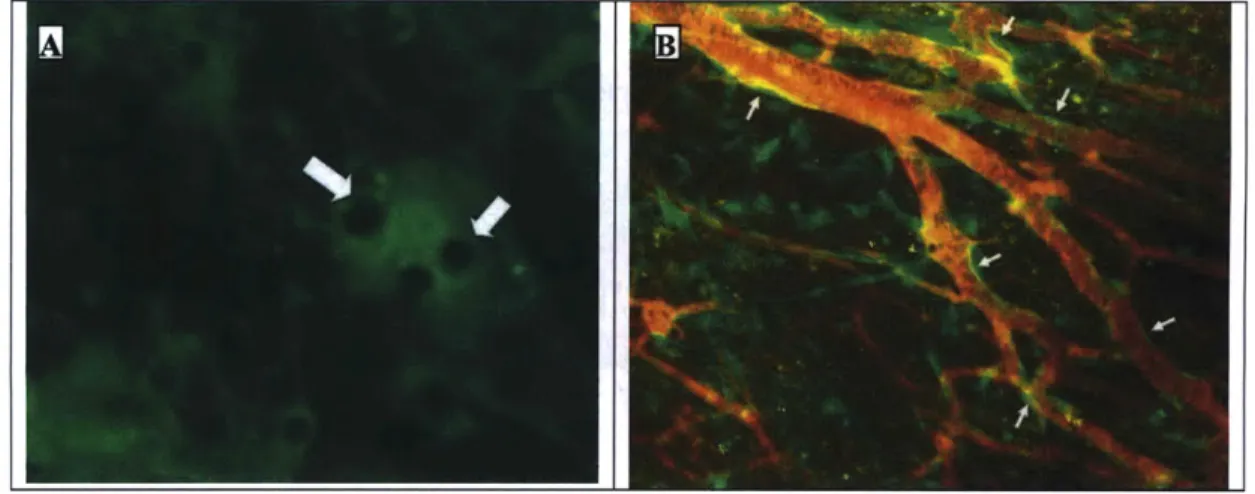
droplets began appearing in the implanted cells within 2 weeks (Figure 2-5A). Many of the new
vessels are lined with GFP-positive cells, indicating that endothelial cells within the ASC are
significant contributors to the new blood vessels (Figure 2-5B). These results show that cells in
the stromal vascular faction of fat tissue are responsible for the bulk of fat tissue's angiogenic
capabilities, and that ASC directly contributed to the formation of new vessels, presumably by
supplying endothelial and/or peri-vascular mural cells.
Fat
Kidn
Liver
Figure 2-3. Representative images of the angiogenic response at 1,2, and 4 weeks following the implantation of
different tissue types. Fat (n=7), kidney (n=4), and liver (n=5) tissue pieces (-8 mm3 each) from GFP-expressing mice (green) were implanted in dorsal skinfold chambers, then monitored over 4 weeks. Blood vessels were visualized by i.v. injection of tetramethylrhodamine labeled 200,000 MW dextran (red). Fat tissue induced a robust angiogenic response: numerous perfused vessels were visible throughout the fat tissues by 2 weeks after
Figure 2-4. Angiogenic response at time 0, 1, and 4 weeks following the implantation of adipocytes (n=3) or ASC (n=3) from GFP-expressing mice (green). Blood vessels were visualized by i.v. injection of tetramethylrhodamine labeled 200,000 MW dextran (red). ASC induced a robust angiogenic response, whereas no angiogenesis was observed after implantation of fat cells alone.
Figure 2-5. (A) High power view of structures resembling lipid droplets in the implanted ASC after 2 weeks (arrows). (B) expressing cells line new blood vessels (arrows) formed in response to the implantation of GFP-expressing ASC.
Chapter 3
:
Adipose Stromal Cells for Blood Vessel
Engineering
Data in this chapter have been collected in collaboration with Patrick Au.
Portions of this chapter have been taken from:
Au P, Tam J, Fukumura D, Jain RK. Bone marrow derived mesenchymal stem cells facilitate
engineering of long-lasting functional vasculature. Blood. 2008; 111(9): 4551-4558.
Au P, Tam J, Fukumura D, Jain RK. Small blood vessel engineering. Methods Mol Med. 2007;
140:183-95.Introduction
To date, almost all of the successfully engineered tissues/organs have relatively thin dimension
and/or avascular structures (e.g. skin
44, cartilage
45, bladder
46), where post-implantation
vascularization from the host is sufficient to meet the implant's demand for oxygen and nutrients.
Vascularization remains a critical obstacle impeding attempts to engineer thicker, metabolically
demanding organs such as the heart, brain and liver. The ability to produce long-lasting,
functional blood vessels would be a giant step forward for tissue engineering in order to repair
defective and/or damaged tissues/organs.
Our lab has developed a novel cell-based method to produce long-lasting, functional
microvessels
47. We hypothesized that peri-vascular mural cells, which have been largely
overlooked in tissue engineering attempts, play a crucial role in the survival and function of the
engineered vessels. We evaluated this hypothesis by comparing the process of vascular
development following the in vivo implantation of human umbilical-vein endothelial cells
(HUVECs), versus the vascular development that occurs after HUVECs were co-implanted with
10TI cells, a mesenchymal precursor cell line that is capable of differentiating into mural cells
through heterotypic interaction with endothelial cells. Blood vessels formed by HUVECs alone
showed minimal perfusion and lasted less than 60 days, whereas vessels formed by the
co-implantation of HUVECs and 10TI cells remained stable and functional more than one year
after the initial implantation.
^.-X~-ili~-LII1-~-While the results from these studies were encouraging, this model cannot be immediately
adapted for human use because HUVECs and 10T
2cells are not immunocompatible with
humans.
I have presented results in the last chapter showing that adipose stromal cells (ASC) are
responsible for the bulk of the angiogenic capability of adipose tissue. ASC is an attractive
alternative source for perivascular cells, due to the abundance, ease of harvest and low donor site
morbidity of fat tissue. A recent study also reported that ASC have protein expression and
functional characteristics that resemble pericytes
48. The experiments described in this chapter
were aimed at determining whether ASC can replace 10T 2 cells in our vascular tissue
engineering system.
Materials and Methods
Animal Models
Animal maintenance, husbandry, and experimental procedures were as described in Chapter 2.
Cell culture
Discarded human fat tissue from liposuction procedures was obtained from Dr. Michael J.
Yaremchuk (The Boston Center, Boston, MA). Fat cells and adipose stromal cells (ASC) were
separated and collected as described in Chapter 2. Cells used for in vivo imaging were implanted
within 3 passages. Cells used for differentiation assays were cultured in the corresponding
differentiation medium (described below). C3H10T1/2 (10T1/2) (American Type Culture
Collection, Manassas, VA) were grown and maintained in Dulbecco's modified Eagle's medium
(DMEM) (Mediatech, Herndon, VA) supplemented with 10% fetal bovine serum (FBS),
penicillin (100 units/ml) and streptomycin (100 mg/ml) (Life Technologies, Inc). Human
umbilical cord vein endothelial cells (HUVECs) were obtained from Center of Excellence in
Vascular Biology, Brigham and Women's Hospital (Boston, MA) and maintained in Endothelial
Growth Medium (EGM, Cambrex).
Cranial Windows
Cranial windows (CWs) were implanted in mice following a previously published protocol49, 50 The head of the animal was fixed by a stereotactic apparatus. The skin on top of the frontal and
parietal regions of the skull was cleaned with antimicrobial solution. A longitudinal incision of
the skin was made between the occiput and forehead. Then the skin was cut in a circular manner
on top of the skull, and the periosteum underneath was scraped off to the temporal crests. A
6-mm circle was drawn over the frontal and parietal regions of the skull bilaterally. Using a high
speed air-turbine drill (CH4201S; Champion Dental Products, Placentia, CA) with a burr-tip 0.5
mm in diameter, a groove was made on the margin of the drawn circle. This groove was made
thinner by cautious and continuous drilling of the groove until the bone flap becomes loose. Cold
saline was applied during the drilling process to avoid thermal injury of the cortical regions.
Using a malis dissector, the bone flap was separated from the dura mater underneath. After
removal of the bone flap, the gelfoam was placed on the cutting edge and the dura mater was
continuously kept moist with physiological saline. A nick was made close to the sagittal sinus,
and iris microscissors were passed through the nick. The dura and arachnoid membranes were
cut completely from the surface of both hemispheres, avoiding any damage to the sagittal sinus.
The window was sealed with an 8-mm cover glass which was glued to the bone with
histocompatible cyanoacrylate glue.
Preparation of the 3-D construct for tissue engineered blood vessels.
The engineered blood vessel model was prepared following a protocol previously established in
our lab4 7'51. 106 HUVECs and 2x10⁵ ASC; 106 HUVECs and 2x10⁵ 10T1/2 cells; or 106
HUVECs alone were suspended in 1 ml solution of rat-tail type 1 collagen (1.5 mg/ml) (BD
Biosciences, Bedford, MA) and human plasma fibronectin (90 mg/ml) (Sigma) in 25 mM Hepes
(Sigma) buffered EGM medium at 40C. pH was adjusted to 7.4 with IN NaOH (Fisher Science, NJ). The cell suspension was pipetted into 12 well plates (Falcon) and warmed to 37C for 30
min to allow polymerization of collagen. Each solidified gel construct was covered by 1 ml of
warmed EGM medium. After 1 day culture in 5% C02, a skin puncher was applied to create
circular disk-shape pieces of the construct (4-mm diameter). The circular piece of collagen gel
was then implanted into a cranial window of a severe combined immunodeficient (SCID) mouse.
The in vivo fate of the fluorescent protein-labeled endothelial cells was tracked by intravital
imaging with multiphoton laser scanning microscopy at various time points52. Image was taken
with 20x/0.50 NA water objective.
Differentiation assays
The ability of pluripotent cells to differentiate along different lineages was tested by culturing
cells with adipogenic and osteogenic differentiation media. Adipogenic differentiation media
consisted of Dulbecco's Modified Eagle's Medium (DMEM), 10% fetal bovine serum (FBS), 0.5mM 3-Isobutyl-1-methylxanthine, 1 uM dexamethasone, 10 uM insulin, 200 uM
indomethacin, and 1% penicillin/streptomycin (pen/strep). Osteogenic differentiation media
consisted of DMEM, 10% FBS, 0.1 jtM 0.1 uM dexamethasone, 50 uM
ascorbate-2-phosphate, 10 mM b-glyceroascorbate-2-phosphate, 1% pen/strep. All cells were cultured in a humidified
incubator, at 37C and 5% CO2.After 4-6 weeks in culture, aidpogenic differentiation was ~
confirmed by oil-red-O staining, while osteogenic differentiation was confirmed by alkaline
phosphatase staining.
Oil Red 0 staining protocol
Oil Red O stock solution was prepared by dissolving 0.03g of Oil Red O dye (Sigma-Aldrich, St.
Louis, MO) in 10 ml of 2-propanol. Undissolved solids were filtered out. Immediately prior to
use, a working Oil Red O stain was prepared by mixing 820 jpL of Oil Red O stock solution with
680 jpL of dH20. Cells were fixed in 4% paraformaldehyde at room temperature for 30 minutes, then washed with water. The working stain was applied to the fixed cells and incubated at room
temperature for 20 min. Afterwards the cells were washed with water, allowed to air dry, then
counterstained with hematoxylin.
Alkaline phosphatase staining protocol
Alkaline phosphatase staining was performed using the Vector Red Alkaline Phosphatase
Substrate Kit I (Vector Laboratories), following the manufacturer's protocol. Briefly, fixed cells
were incubated in the Vector Red substrate working solution at room temperature for 30 minutes
in the dark. The working solution was then removed, and the cells were incubated in 100%
ethanol for 5 minutes. The cells were then mounted in VECTASHIELD mounting medium with
DAPI (Vector Laboratories).
Results
Stromal Vascular Fraction cells isolated from human fat tissue are multipotent
ASC were isolated from human fat tissue by enzymatic digestion and their multipotency was
culture, adipogenic differentiation was confirmed by oil-red-O staining (Figure 3-1A), and
osteogenic differentiation was confirmed by alkaline phosphatase staining (Figure 3-1B). This
shows that ASC are able to differentiate along different specific lineages.
Figure 3-1. ASC from fat tissue are multipotent. (A) Adipogenic differentiation in ASC cultured in adipogenic
media was confirmed by oil-red-O staining (red), and counter-stained with hematoxylin (blue). (B) Osteogenic differentiation in ASC cultured in osteogenic media was confirmed by alkaline phosphatase staining (red). Nuclei were stained by DAPI (blue). ASC cultured in non-differentiating medium were used as negative controls (not
shown).
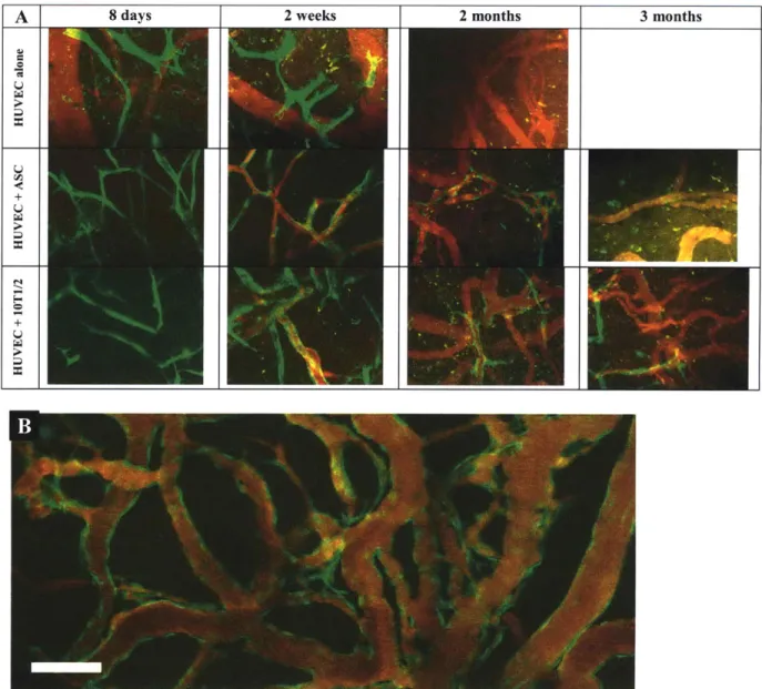
Ability of multipotent ASC to sustain engineered blood vessels
Multipotent ASC isolated from human adipose tissue were co-implanted with GFP-labeled
human umbilical vein endothelial cells (HUVECs) in collagen gels into cranial windows,
following a protocol previously established in our lab
4 7. Blood vessels formed in the implants
were monitored regularly by intravital microscopy. Gel implants with HUVECs alone, and those
with HUVECs and 10T1/2 cells, were used as negative and positive controls, respectively. The
implanted HUVECs formed tubular structures within a few days after implantation in all three
cases. However, the tubular structures formed by HUVECs alone never became perfused, and all
the GFP-labeled HUVECs disappeared within 2 months (Figure 3-2A). On the other hand, the
HUVECs+ASC and HUVECs+10T1/2 constructs formed perfused blood vessel networks within
2 weeks after implantation (Figure 3-2A). The vessels in the HUVECs+ASC construct remained
patent for 2 months, but by the 3 month they began to regress (Figure 3-2A), and eventually they
too disappeared. In contrast, vessels in HUVECs+I OT1/2 constructs typically remain stable and
functional for over a year47
In a parallel experiment, we investigated whether human bone marrow-derived mesenchymal
stem cells (hMSC) could be a suitable replacement for 10T1/2 cells. hMSC were able to stabilize
HUVECs to form blood vessels that remained patent and functional for more than 4 months in
vivo (Figure 3-2B)53. Results from this experiment show that while multipotent ASC from human
fat tissue are able to stabilize HUVECs to form patent engineered blood vessels for a time, this
effect is transient, and ASC may not be an ideal candidate cell source for replacing 10T1/2 cells
in our tissue engineering system. One potential reason for this is the fact that ASC have a
tendency to accumulate lipid (Figure 2-5A), and has been shown to spontaneously differentiate
into adipocytes in vivo54, 55. Our tissue engineering system relies on the ability of mesenchymal
stem cells to differentiate into perivascular mural cells, which will in turn stabilize the
endothelial cells. The tendency of ASC to differentiate into adipocytes may undermine this
ability.
Given the unique advantages of ASC as a cell source for tissue engineering (ease of harvest, minimal donor site morbidity), it may be worthwhile to investigate whether inhibiting the
differentiation of these cells into adipocytes, e.g. by using dominant negative gene constructs42,
makes them more suitable for tissue engineering applications.
Figure 3-2. Tissue engineering of blood vessels in mice. Tissue constructs were produced and implanted into mice baring cranial windows, as previously described47. HUVECs labeled by GFP protein (green), functional blood vessels visualized by rhodamine-dextran (red). (A) Consistent with our previous experience, implants with HUVECs
alone formed cord-like structures but were never perfused, and by 2 weeks post-implantation most of the green cells had disappeared. When HUVECs were combined with multi-potent ASC, functional blood vessels were formed by 2 weeks post-implantation. These engineered blood vessels persisted for about 2 months, but most of them receded by the 3rd month. The period of stabilization provided by ASC is substantially less than 10T1/2 cells4 7 or bone
marrow-derived mesenchymal stem cells53
. (B) Vessels formed by combining HUVECs and bone marrow-derived mesenchymal stem cells remained patent more than 130 days after implantation53.
Chapter 4 : Dynamic measurement of hepatocyte function by
two-photon microscopy
Data in this chapter have been collected in collaboration with Patrick Au.
Portions of this chapter have been taken from:
Au P*, Tam J", Kwok J, Fukumura D, Jain RK. Dynamic measurement of hepatic tissue
Introduction
Tissue assemblies greater than 100-200 micrometers (the limit of oxygen diffusion) cannot
survive for long without a perfused vascular bed to supply nutrients and to remove waste
products and metabolic intermediates
56. Besides their crucial role in providing oxygen and
nutrients, there is also a growing body of evidence showing that vascular networks play an
integral role in the development, organization, and function of a wide range of tissue types.
Blood vessel endothelium has been shown to play requisite roles in early liver
57and pancreas
5 8development, and liver regeneration following partial hepatectomy is dependent on
angiogenesis
59. Soluble factors secreted by endothelial cells stimulate neural stem cell
self-renewal and neuron production
60. Co-culture of cardiac myocytes and endothelial cells reduced
cardiac myocyte apoptosis and necrosis, and promoted cardiac myocyte reorganization and
function (synchronized contraction and connexin expression)
61.
These examples suggest that a
long-lasting, functional vascular network will be required not only for adequate perfusion of the
tissue, but also for the proper cellular organization, development, and function of any
regenerating and/or engineered solid tissue construct.
Our laboratory has previously established a method for producing long-lasting functional
microvascular networks4 7 51, 53, 62.In order to determine if microvascular networks produced by
this method could be used to support parenchymal cells, we investigated the possibility of adding
parenchymal cells to our tissue engineering constructs, and monitoring the survival and function
of the parenchymal cells following in vivo implantation. After studying the literature and some
preliminary experiments, we decided to use hepatocytes as the parenchymal cell type. However
one major obstacle had to be overcome before we could include hepatocytes in our system
-currently there is no established method to assess the functional status of in vivo engineered liver
tissues in real-time without extraction of the tissues. The most common way to assess hepatocyte
phenotype is to measure the production of characteristic proteins such as albumin and urea.
However, protein synthesis by engineered tissues is difficult to measure reliably in vivo, due to
the confounding effects of host production. To circumvent this problem, investigators often have
to resort to either extracting and destroying the engineered tissue after some time in vivo to
evaluate the mRNA or protein expression of characteristic markers, or performing hepatectomy
in animals prior to implantation of liver constructs, resulting in high mortality rates
63. Other
methods focus on functional characteristics such as cytochrome P450 and glucuronidation
activities 64. However most of these methods were developed for cells in culture, requiring
analysis of conditioned media or cell lysates. To apply these methods in engineered liver tissue
implants again necessitates the extraction of the implants. This common requirement for
extraction of tissue implants restricts the experiment to only one measurable time point, and thus
severely limits the ability to evaluate the long-term in vivo viability and function of engineered
liver tissues.
To overcome this limitation, we have developed a novel imaging technique for measuring the
metabolic function of engineered liver tissues. We utilized multi-photon laser scanning
microscopy to interrogate, in real-time, the cytochrome P450 function of engineered liver tissues.
This method is non-invasive and does not require the extraction of tissues, thus it can potentially
be used for the long-term continuous monitoring of implanted liver tissue constructs.
Material and Method
Materials
3-cyano-7-ethoxycoumarin (CEC) and 3-cyano-7-hydroxycoumarin (CHC) were purchased
from Invitrogen (Carlsbad, CA). CEC and CHC were dissolved in DMSO (Sigma, St. Louis,
MO) to a concentration of 30mM stock solution, stored at -20
0C, and protected from light until
use.
Hepatocyte isolation and culture
Hepatocyte isolation was performed following a previously published protocol
65, with minor
adjustments. Neonatal (1-3 days old) mice were sacrificed by decapitation. The livers were
quickly removed, minced, then digested in 0.3% collagenase (type 2, Worthington Biochemical
Corp., Lakewood, NJ) for 30 minutes at 37
0C on a shaker. The cells were than passed through a
cell strainer (70 gm, BD Falcon, Bedford, MA), and washed twice by centrifugation. The cells
were then plated onto collagen 1 coated cell culture plates (BD BioCoat, BD Labware, Bedford,
MA), and cultured in Dulbecco's Modified Eagle Medium with high glucose, 10% fetal bovine
serum, 0.5 U/mL insulin, 7 ng/mL glucagons, 7.5 [lg/mL hydrocortisone, and 1%
penicillin-streptomycin
66Spectroscopic evaluation of CEC and CHC
The emission spectra of CEC and CHC were measured with a fluorescence spectrometer (Ocean
Optics, Dunedin FL). For the excitation spectra, the fluorescence emission was recorded and
averaged for 5s by a computer-controlled photon counter (Stanford Research Systems, Model
SR400). The excitation wavelength was tuned from 720 to 990nm with 20 nm resolution and the
power of the incident bean was kept constant with the means of a power meter. Two-photon
excitation of the dyes was confirmed by intensity test in order to demonstrate the power-squared
dependence of the fluorescence. The integrated fluorescence was measured at various excitation
intensities by a photon counter. The results were then plotted on a log-log scale and fit to a line.
The slope of the trend line yielded the power-dependence exponent.
Functional evaluation of hepatocytes grown in monolayer and in three-dimension in vitro
Neonatal hepatocytes were cultured on Type I collagen coated dish (BD Biosciece). In
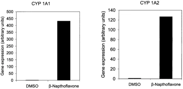
experiments with induction of cytochrome P450, 30M of P-Napthoflavone was exposed to the
cells for 72 hours. On the day of experiment, the media were changed and replaced with DMEM
containing 30M of CEC. Cytochrome P450 activity was measured by submerging a 20x
objective (Zeiss Aeroplane) into the media. The cells were maintained at 37'C at all time by
placing the cell culture dish on a heating pad. CHC in the media was two-photon excited at 810
nm wavelength and emission was filtered through a 470nm/40 filter set. The fluorescence
emission was detected by a photomultiplier tube (Hamamatsu, Hamamatsu City, Japan) that was
fed to a gated photon counter (SR400, Stanford Research Systems, Sunnyvale, CA). The
concentration of CHC in the medium was determined by comparing the photon count to a
standard curve of CHC prepared in DMEM.
To grow hepatocytes in three-dimensional culture, neonatal hepatocytes were suspended in a
fibronectin/type I collage matrix as previously described
47' 53. Briefly, 300,000 hepatocytes were
suspended in 1 ml solution of rat-tail type 1 collagen (1.5 mg/ml) (BD Biosciences, Bedford, MA)
and human plasma fibronectin (90 pg/ml) (Sigma) in 25 mM Hepes (Sigma) buffered DMEM
medium at 4
0C. pH was adjusted to 7.4 by using IN NaOH (Fisher Science, NJ). The cell
suspension was pipetted into a single well of a 12 well plates and warmed to 37'C for 30 min to
allow polymerization of collagen. Once the collagen gel had solidifed, one ml of warmed
DMEM medium was added into the well and the cell culture plate was then placed in an
incubator maintained at 37'C and 5% CO2. After 4 days in culture, the hepatocytes-laded gel was
exposed to 30pM of 3-Napthoflavone for 3 days to induced expression of P4501A2. A skin
puncher (4-mm diameter) was then applied to the collagen gel construct to excise a circular piece.
CytochromP450 activity was measured directly on the excised piece of hepatocytes-lade gel
using technique similarly to that of the hepatocyte monolayer. 30pM of CEC in DMEM was
applied to the collagen gel and a 20x water immersion objective piece was then lowered to make
contact with it. The collagen gel was imaged at various timepoints and flourescence intensity
was quantified.
Gene expression analysis
Gene expression was measured by real-time quantitative PCR (qRT-PCR). Total RNA was
isolated from neonatal hepatocytes using the RNeasy Mini Kit (Qiagen, Valencia, CA). Quantity
and purity of RNA were determined by ultraviolet absorbance at 260 and 280 nm. cDNA was
synthesized using TaqMan Reverse Transcription Reagents (Applied Biosystems, Foster City, CA). qRT-PCR was performed using the 7300 Real-Time PCR System and Power SYBR Green
PCR Master Mix (Applied Biosystems). Primers were designed using Primer Express (Applied
Biosystems) or Primer3 (Whitehead Institute, Cambridge, MA), and purchased from Integrated
DNA Technologies (Coralville, IA). Primer sequences are listed in Table 4-1. Primer specificity
for each gene of interest was confirmed by comparison to known sequences in the BLAST
database (National Center for Biotechnology Information). Primer efficiency was validated using
,,~~;-~--standard curves generated from 10-fold dilutions of mouse liver cDNA. Samples were analyzed in triplicates, and the gene expression level for each sample was normalized to the corresponding glyceraldehydes-3-phosphate dehydrogenase (GAPDH) expression level, to control for loading differences. Negative controls were performed for each sample using non-reverse-transcribed RNA.
Table 4-1. Primer sequences for qRT-PCR
Gene Forward Primer (5'-3') Reverse Primer (5'-3')
CYP1Al CCACCTGCTGAGGCTAAACAG TGCCCCCCACATGCA
CYP1A2 CTGTCCAGGAGCACTACCAAGA TGAACAGGGCACTTGTGATGTC
GAPDH AAGAAGGTGGTGAAGCAGGCA TGCTGTTGAAGTCGCAGGAGA
CYPlA1, cytochrome P450, family 1, subfamily a, polypeptide 1;CYP1A2, cytochrome P450, family 1, subfamily a, polypeptide 2; GAPDH, glyceraldehydes-3-phosphate dehydrogenase
Results
Spectroscopic property of CEC and CHC
We investigated the spectroscopic property of 3-cyano-ethoxycoumarin (CEC) and 3-cyan-hydroxycoumarin (CHC)6 7. The dealkylation reaction of CEC to CHC is mediated by
cytochrome P450 1A2 in the endoplasmic reticulum of a hepatocyte68 (Figure 4-1A). We first measured the emission spectra of CEC and CHC. The de-ethylation reaction yields a blue fluorescent product with a maximum emission at 450nm, while the maximum emission of the substrate, CEC, is at 430nm (Figure 4-1B). Next, we determined whether CEC and CHC could be excited by 2-photon using near-infrared laser. Knowledge of the 2-photon excitation cross-section spectra allows for a better signal to noise ratio and minimizes the potential for photo-damage. The fluorescence signal was measured as the excitation wavelength was tuned from 720 to 990nm with 20 nm resolution (Figure 4-1C). The 2-photon excitation peak was 740 nm and
810 nm for CEC and CHC, respectively. This result in is in accordance with the expectation that
the 2-photon excitation peak should be approximately doubled and slightly blue-shifted relative
to that of the single-photon excitation peak. We found that at 810nm, CHC was maximally
excited with minimal excitation of CEC. We decided to use this excitation wavelength for
subsequent in vitro and in vivo measurements.
A.
,CN
CH3CH
20
0
0
3-cyano-7ethoxycoumarin
Cytochrome P450 1A2_;Q7
CN
* HO' ON3-cyano-7hydroxycoumarin
CEC & CHC Normalized Emission Spectra
-cHC 0.02 0.015 0 .01 500000 450000 400000 350000 300000 250000 200000 150000 100000 50000 0-710 -CHC -- CEC 760 810 860 910 960 Excitation Wavelength (nm)
Figure 4-1. (A) Chemical structures of 3-cyano-7-ethoxycoumarin (CEC) and 3-cyano-7-hydroxycoumarin (CHC). Cytochrome P4501A2 mediates the dealkylation of CEC into CHC. (B) Plot of fluorescent intensity versus emission wavelength of CEC and CHC. CEC was dissolved in PBS at a concentration of 30pM and excited at 740nm. CHC was dissolved in PBS at 500nM and excited at 810nm. (C) Plot of photon count versus excitation wavelength of CEC and CHC tunes from 710 to 990nm.