31P Nuclear Magnetic Resonance Spectroscopy Studies of Cardiac Energetics and Function in the Perfused Rat Heart
by
Richard Glenn Stevens Spencer
B.A., University of California at Berkeley (1977)
M.A., University of California at Berkeley (1981)
SUBMITTED TO THE DEPARTMENT OF
MEDICAL ENGINEERING AND MEDICAL PHYSICS
IN PARTIAL FULFILLMENT OF THE REQUIREMENTS FOR THE DEGREE OF
DOCTOR OF PHILOSOPHY IN
MEDICAL PHYSICS
at the
MASSACHUSETTS INSTITUTE OF TECHNOLOGY April 1987
Richard G. S. Spencer 1987
The author hereby grants to M.I.T. permission to reproduce and to distribute copies of this thesis document in whole or in part.
Signature of Author
Department of Medical Engineering and Medical Physics April 1987
Certified by
Associate Profeior Joanne S. Ingwall
___ Thesis Supervisor
Accepted by
U
Co-Director of the iealth
Professor Roger G. Mark Harvard-MIT Division of Sciences and Technology
--DEDICATION
To. My Parents
For Their Unlimited Love and Support Throughout the Years
and
To My Wife
Habe nun, ach! Philosophie, Juristerei und Medizin
Und leider auch Theologie
Durchaus studiert, mit heifem BemUhn. Da steh ich nun, ich armer Tor,
Und bin so klug als wie zuvor! Heine Magister, heine Doktor gar Und ziehe schon an die zehen Jahr Herauf, herab und quer und krumm Meine Schiler an der Nase
herum--Und sehe, dab wir nichts wissen knnen! Johann Wolfgang Goethe Faust, Erster Teil, Nacht
I have pursued, alas, philosophy, Jurisprudence, and medicine,
And, help me God, theology,
With fervent zeal through thick and thin, And here, poor fool, I stand once more, No wiser than I was before.
They call me Magister, Doctor, no less, And for some ten years, I would guess, Through ups and downs and tos and fros
I have led my pupils by the nose--And see there is nothing we can know!
Johann Wolfgang Goethe Faust, Part One, Night
ACKNOWLEDGEMENTS
It has been my very good fortune to carry out this research in the Harvard Medical School Nuclear Magnetic Resonance Laboratory, under the direction of Joanne Ingwall. To her I offer my sincere thanks, for her careful guidance of my work, her willingness to move in new directions, and her
(almost) infinite patience.
Scientific cooperation and personal camaraderie among denizens of the NMR lab have contributed in no small measure to making my tenure there enjoyable and productive. I would particularly like to thank Jim Balschi and John Bittl for
invaluable assistance and discussion.
I am grateful to my Thesis Committee, W. Grossman, M. Kushmerick, L. Neuringer, and F. Villars, for their reading of my thesis and for their comments. Productive collaboration with James Scheuer and Peter Buttrick is also acknowledged.
I would also like to acknowledge three of my early mentors, Chun-Ming Leung, then of the National Radio Astronomy Observatory in Charlottesville, Virginia, Jon Arons, in the Department of Astronomy at U.C. Berkeley, and especially Allan Kaufman, in the Department of Physics at Berkeley. These individuals' impeccable scientific standards and personal integrity provided me with the most favorable possible start in research.
My home computer was tamed largely through the tireless efforts of Herb Gordon; the hours he devoted to this task are much appreciated.
My wife, Christine, has put up with a variety of privations as a result of this work. To her go my thanks and my love.
TABLE OF CONTENTS
Abstract...
Introduction...
Chapter 1. Double Saturation Transfer Measurements of ATP Synthesis and Degradation in the Perfused Rat
Heart at High Workload ... Page 20
Chapter 2. NMR Saturation Factors in the Presence of
Chemical Exchange ... Page 59
Chapter 3, Part 1. The Hemodynamic and Metabolic Response to Hypoxia of the Normal and Hypertrophied Rat Heart...
Chapter 3, Part 2. The Hemodynamic and Metabolic Response to Hypoxia of the Rat Heart Hypertrophied Secondary to Chronic Exercise ...
Page 138
Page 190
Chapter 3, Part 3. Saturation Transfer and Biochemical Analyses of the Creatine Kinase System in the Trained
Rat Heart ... Page 237
Chapter 4. Attenuation of Hypoxic Contracture by
Acidosis in the Perfused Rat Heart ... Page 264 ... Page 6
31 NMR Spectroscopy Studies of
Cardiac Energetics and Function in the Perfused Rat Heart by
Richard Glenn Stevens Spencer
31P nuclear magnetic resonance (NMR) spectroscopy provides the ability to measure the concentration and turnover of high-energy phosphate compounds in the heart while simultaneously assessing hemodynamic function. Thus, relationships between cardiac function and metabolism may be investigated directly.
ATP is the dominant substrate for energy-dependent processes in the heart, while creatine phosphate (CP), through the creatine kinase (CK) reaction, serves the function of an energy buffer, allowing ATP levels to be maintained at times of acute stress. In terms of phosphorus flux, the CK reaction is the dominant reaction in which ATP participates. Measurements of flux through the CK reaction in the isolated perfused heart using the NMR technique of saturation transfer have shown that the flux towards ATP synthesis is greater than that towards CP synthesis. Explanations for this apparent violation of detailed balance include metabolic compartmentation, and the participation of ATP in reactions besides ones catalyzed by CK. Direct tests of these hypotheses have been difficult, because the conventional magnetization transfer techniques widely employed in NMR spectroscopy to assess reaction kinetics cannot be applied to systems of coupled reactions. Accordingly, an extension of this technique, double saturation transfer, has been developed which can be applied to systems with competing reactions. Applied to the CK system, we find that ATP homeostasis can be directly demonstrated by using our double saturation technique to properly account for myofibrillar ATPase and mitochondrial ATP synthesis reactions. In addition, our results indicate that exchange between CP and NMR-invisible, saturable, ATP is not a dominant component of the CK reactions in the heart; thus, we may expect that overall ATP concentration as measured by NMR reflects the functional energetic status of the heart.
In order to achieve quantitatively reliable, high-time-resolution measurements of physiologic samples, signal-to-noise (S/N) considerations are paramount. One common method of increasing S/N is to allow partial saturation of spectral resonances during data acquisition. This partial saturation can be accounted for using saturation factors to obtain correct abundances only if the measured metabolites are not undergoing chemical exchange. In general, this is not the case in organ physiology. Accordingly, we have developed the theory of saturation factors in the presence of chemical exchange. We find that the error which results from neglect
of exchange depends on: 1) the changes in the ratio of metabolite abundances, 2) the reaction rate, 3) the difference in Tl's of the exchanging metabolites, and 4) the interpulse delay and flip angle used.
Based on the preceding considerations, we have used 31p NMR spectroscopy to study two important problems of modern cardiac physiology: cardiac hypertrophy and diastolic function. Recent clinical studies indicate that diastolic dysfunction, which is often a prominent feature of hypertrophy, may be as important as systolic dysfunction as a cause of congestive heart failure.
Cardiac hypertrophy may represent a state of diminished cardiac reserve rather than a state of inability to function under normal conditions. Accordingly, we compared the response to prolonged global hypoxia of normal rat hearts and rat hearts which had hypertrophied secondary to chronic hypertension. During hypoxia, [ATP] was an excellent predictor of diastolic (but not systolic) dysfunction, while [CP] was an excellent predictor of systolic (but not diastolic) dysfunction. Further, the additional diastolic stiffening due to hypertrophy could be accounted for by increased muscle mass, without the need to invoke a defect in high-energy phosphate metabolism. This is consistent with the parallel replication of normal sarcomeres which may occur in pressure-overload hypertrophy.
Cardiac hypertrophy also occurs secondary to exercise. If a diastolic defect proportional to increased muscle mass occurs in this setting as well, it would be strong evidence for a maladaptation of exercised hearts. Instead, we found that hearts from animals exercised by chronic swimming exhibited a marked preservation of diastolic function during prolonged global hypoxia. This result could not be explained by preservation of metabolic energy stores or by the series replication of sarcomeres which may occur in volume-overload hypertrophy. To test other possible mechanisms, we also performed kinetic studies using saturation transfer, and biochemical analyses of cardiac enzymes, in this model of exercise hypertrophy. Although we found small increases in the rate constant of the CK reaction and in mitochondrial CK activity, these changes may be too small to adequately account for the improved diastolic performance of the hearts from exercised animals.
These studies showed a direct relationship between loss of ATP and impaired diastolic function during hypoxia. However, the rate of ATP depletion was unchanged for either type of hypertrophy, so that we could not directly demonstrate that attenuation of hypoxic diastolic dysfunction accompanies preservation of ATP. By superimposing a negative inotropic intervention (mild acidosis) on the hypoxic state, we were able to demonstrate this.
Thesis Supervisor: Dr. Joanne Ingwall, Associate Professor of Physiology and Biophysics in the Department of Medicine, Harvard Medical School
Cardiac biochemistry encompasses an exceedingly wide range of processes. These range in scale from the organ level, where the overall neurohormonal regulation of cardiac function is exerted by the intact organism, to the submolecular level of myosin light-chain control of contractile protein ATPase activity. However, despite the complexity of cardiac function, the ultimate purpose of the heart is to pump, and the sine qua non of this activity is energy. Chemical energy is provided to tissues in a variety of forms. For example, the heart preferentially metabolizes circulating free fatty acids, but it has considerable adaptability with respect to substrate. However, in spite of this vast potential for heterogeneity of metabolic input, it is part of the general plan of metabolic processes to produce a single class of compounds, the high-energy phosphates, whose hydrolysis provides energy for active cellular
function.
Of the various high-energy phosphate compounds found in the heart, ATP is the metabolite which is most directly
related to function. The production and utilization of this compound are both highly regulated and highly sensitive to cardiac functional demands. Phosphocreatine (PCr) is the next-most important high-energy phosphate in the heart. At the least, it serves as an energy buffer, allowing ATP levels to be maintained during times of acute stress through the
creatine kinase (CK) reaction
ADP + PCr ATP + Cr
between ATP and PCr. In terms of phosphorus flux, the CK reaction is, moreover, the dominant reaction in which ATP participates. Thus, an understanding of the role of PCr and of the properties of the CK reaction is an essential part of the understanding of cardiac biochemistry.
In recent years, 31p nuclear magnetic resonance (NMR) spectroscopy has proved to be an extraordinarily versatile
technique for studying cardiac physiology, primarily because it provides the ability to monitor phosphate metabolites of physiologic significance while simultaneously assessing hemodynamic function. Thus, relationships between cardiac function and metabolism may be investigated directly. In this context, it is worth commenting on the exceptional nature of the relationship between function and metabolism in the heart. In no other organ can these two aspects of overall activity be so readily distinguished. For example,
in the kidney, liver, or brain, function and biochemistry are largely one and the same, i.e., the main function of the organ is biochemical. In the heart, on the other hand, biochemical activity is to a great extent subservient to mechanical activity; the purpose of cardiac biochemistry is to provide energy for cardiac function. It is precisely this fact that lends to cardiac physiology its exceptional richness and fascination. It is also a prime reason that NMR studies of the heart, allowing simultaneous assessment of function and metabolism, can be so illuminating.
NMR spectroscopy has the capability of providing insight into metabolic processes at two distinctly different levels, through the use of kinetic and steady-state techniques.
Kinetic experiments may be performed to study reaction rates and metabolite fluxes through reactions in steady-state systems. In effect, this constitutes an assessment of the instantaneous behavior of a dynamic system. NMR is unique in its ability to provide such kinetic data on specific, enzyme-catalyzed reactions in whole organs and living systems. Chemical fluxes can thereby be directly related to the physiologic state of the preparation. On the other hand, standard biochemical techniques for studying isolated reactions in vitro, involving, for example, radioactive tracer uptake, generally use homogeneous tissue preparations as samples. Thus, biochemical studies are highly controlled and versatile, but lack the physiologic context of
whole-organ studies. Therefore, these two general types of kinetic studies, NMR and biochemical, are complementary.
Steady-state NMR can be used to provide measurements of net metabolite abundances. In general terms, this may be thought of as an integration with respect to time of kinetic measurements. However, it is more than that, because the system need not be in a steady state for these types of studies. The time evolution of metabolite abundances can therefore be followed after pharmacologic or other interventions. In this role, NMR is not unique, since such serial measurements of metabolites have been performed for decades using standard biochemical techniques. However, NMR still provides tremendous advantages in certain instances. While standard biochemical techniques require the destruction of the sample prior to measurement, NMR measurements are non-invasive, so that the same preparation may be followed throughout the length of the experiment. With NMR, then, not only does each sample serve as its own control, but it also becomes practical to accumulate measurements with reasonably high temporal resolution since each sample can be studied for the duration of the protocol. This eliminates the need to prepare separate samples for each time point. Consider, for example, an experiment in which a control measurement is followed by measurements every three minutes, for 24 minutes, after an intervention. Such an experiment is described in Chapter 3, Part 1, in which measurements are performed on a total of 14 hearts using NMR. To accumulate the equivalent data using a destructive technique such as freeze clamping, 126 hearts, as opposed to the 14 actually used, would be required. This ability to obtain high temporal resolution with NMR is especially important in relating function to metabolism, and so can be used to great advantage in studies of the heart. Finally, we mention that NMR provides the ability to monitor non-invasively true intracellular pH during the course of an experiment; again, this capability is of considerable importance in studies of cardiac physiology.
Next, we briefly summarize the methods used in this Thesis. The NMR system consists of an Oxford Instruments 360 wide-bore superconducting magnet (field strength 8.45 Tesla) interfaced with a Nicolet 1280 spectrometer. The phosphorus spectra obtained allow the measurement of relative abundances of ATP, PCr, and inorganic phosphate. In addition, intracellular pH is monitored by measurement of the chemical shift between phosphocreatine and inorganic phosphate.
Hearts are perfused retrograde through the aorta by phosphate-free Krebs-Henseleit buffer, gassed with a mixture of 02, N2, and C02, depending on the oxygenation and pH desired. The system may be configured for either constant-pressure or constant-flow-rate perfusion. A water-filled latex balloon is inserted into the left ventricle of the perfused heart through an incision in the left atrium, via the mitral valve, to permit hemodynamic measurements. These measurements are obtained via a water-filled tube running from the intraventricular balloon to a Stratham pressure transducer, producing a continuous record of systolic and end-diastolic pressure and heart rate, which are recorded on a Hewlett-Packard chart recorder. Coronary flows are measured either by timed collection of coronary effluent or with an in-line flow gauge. See Figure 1.
We now describe the specific applications we have made of 31p NMR spectroscopy to further elucidate the relationship of cardiac function to high-energy phosphate metabolism.
In measurements of flux through the CK reaction in the isolated perfused heart using the NMR technique of saturation transfer, it has been consistently found that the flux towards ATP synthesis is greater than that towards PCr synthesis. This result is in apparent violation of detailed balance. Plausible explanations for this apparent paradox include certain types of metabolic compartmentation, in which ATP exists in two (or more) separate pools in the myocyte which have different exchange properties with PCr, and the participation of ATP in many other reactions besides the one
catalyzed by CK. Direct tests of these hypotheses have been difficult, because the conventional magnetization transfer techniques widely employed in NMR spectroscopy to assess reaction kinetics can only be applied to systems of coupled reactions in certain restrictive circumstances. To overcome this limitation, we have developed a technique, called double saturation transfer, which can be applied to systems with competing reactions. This is described in Chapter 1. Applied to the CK system, we found that ATP homeostasis could be directly demonstrated using our double saturation technique to properly account for myofibrillar ATPase and mitochondrial ATP synthesis reactions.
The ability to describe ATP homeostasis in the heart in this way provides an important step in the validation of steady-state NMR measurements of metabolite concentrations. If exchange between PCr and an NMR-invisible pool of ATP were a dominant feature of the kinetics of ATP reactions, then the interpretation of overall abundance measurements would be complicated enormously. Our results provide support for the notion that overall ATP concentration measurements do indeed reflect the functional energetic status of the heart. Thus, we may make steady-state measurements with some confidence in their relevance to cardiac energetics.
In order to make high-quality steady-state measurements of physiologic samples, such as the perfused hearts used in this Thesis, signal-to-noise (S/N) considerations are paramount. Reasons for this include, first of all, the fact that the preparations themselves are always more or less unstable, so that speed in obtaining the required measurements is essential. In addition, for experiments which follow the time course of metabolic changes following an intervention (such as hypoxia), high time-resolution is desirable. This is important in the hypoxia studies we will
describe, for example, because of our interest in developing explicit functional relationships between cardiac mechanical performance and metabolic state during hypoxia. These
relationships can be constructed only from data having high temporal resolution, so that data points do not represent simply the time-average of very different physiologic states. Finally, to establish such quantitative relationships, it is obviously essential to have quantitatively reliable data, and high S/N facilitates this as well.
One way in which S/N is commonly increased is to allow partial saturation of spectral resonances during data acquisition. This occurs when the interpulse delay time is insufficient to allow complete relaxation of nuclear spins between pulses. Partial saturation can be accounted for by using saturation factors to obtain correct abundances only if the measured metabolites are not undergoing chemical exchange, as has been assumed in previous treatments of saturation factors. While this may be the case for many NMR applications in organic chemistry and solution biochemistry, it is in general not true for intact organ physiology.
Accordingly, we have developed the theory of saturation factors in the presence of chemical exchange, as described in Chapter 2. This treatment extends the powerful S/N enhancement technique of saturation factors so that it may be properly applied to systems with exchange. We found that chemical exchange alters the fundamental character of the problem, in that it causes the magnitude of saturation factors to be dependent on metabolite abundances. Neglect of this fact results in an error in abundance calculations, whose magnitude depends on the reaction rate, on the difference in T's of the exchanging metabolites, on the changes in the ratio of metabolite abundances, and on the interpulse delay and flip angle used. In contrast, our development allows for a rigorous treatment of saturation factors so that abundances may be determined accurately.
Two of the outstanding problems of modern cardiac physiology are the understanding of cardiac hypertrophy and diastolic function. While the hypertrophy which accompanies certain pathologic states has long been recognized as an
extremely important cardiac adaptation, with both beneficial and deleterious aspects, the importance of diastolic function has come to be appreciated only recently. In fact, recent clinical studies suggest that diastolic dysfunction may be as important a factor as systolic dysfunction in the etiology of congestive heart failure. Furthermore, the problem of hypertrophy is linked to this by the fact that such diastolic dysfunction is very often found in the setting of cardiac hypertrophy.
Hypoxia is an appropriate setting to study the function of hypertrophied hearts because cardiac hypertrophy may represent a state of diminished cardiac reserve, rather than a state of inability to function under normal conditions. In fact, it is likely that the clinically important increased susceptibility of pathologically hypertrophied hearts to ischemic injury is a manifestation of a variety of cellular and organ-level alterations which accompany the hypertrophy process. To study this, we performed experiments, described in Chapter 3, Part 1, comparing the response to prolonged global hypoxia of normal rat hearts and rat hearts which had hypertrophied secondary to chronic hypertension. In this investigation, we were especially interested in alterations in the relationship between metabolic and functional deficits which may occur in the state of mild compensated hypertrophy, with particular emphasis on diastolic function. We found that during hypoxia, for both control and hypertrophied hearts, [ATP] was an excellent predictor of diastolic (but not systolic) dysfunction, while [PCr] was an excellent predictor of systolic (but not diastolic) dysfunction. In addition, we found that the additional diastolic stiffening exhibited by the hypertrophied hearts could be accounted for by their increased muscle mass, without the need to invoke a defect in high-energy phosphate metabolism. This is consistent with the parallel replication of normal sarcomeres which may occur in pressure-overload hypertrophy.
Cardiac hypertrophy occurs not only in pathologic states, but also secondary to exercise. Because the diastolic defect seen in the pathologically hypertrophied rat hearts appears to be a direct result of their increased mass, we examined the response to hypoxia of hypertrophied hearts from rats which had been trained by chronic swimming. These experiments are described in Chapter 3, Part 2. If we had again found a diastolic defect proportional to increased muscle mass, this would be strong evidence for a maladaptation of exercised hearts. Instead, we found that the hearts from exercised animals exhibited a marked preservation of diastolic function during prolonged global hypoxia, as compared with controls. This result was independent of mass normalization, and so could not be explained by the series replication of sarcomeres which may occur in volume-overload hypertrophy. To test other possible mechanisms, we performed kinetic studies using the NMR technique of saturation transfer, and biochemical analyses of cardiac enzymes, in this model of exercise hypertrophy, as discussed in Chapter 3, Part 3. Although we found a small increase in the rate constant of the CK reaction and in mitochondrial CK activity, these changes may be too small to adequately account for the improved diastolic performance of the hearts from exercised animals.
These studies showed a direct relationship between loss of ATP and impaired diastolic function during hypoxia. However, the rate of ATP depletion was unchanged for either type of hypertrophy, so that we were unable to directly demonstrate that attenuation of hypoxic diastolic dysfunction accompanies preservation of ATP. In Chapter 4, we describe experiments in which we did demonstrate this by superimposing a negative inotropic intervention (mild acidosis) on the hypoxic state. In addition, we find that the hearts subjected to mild acidosis during hypoxia exhibit markedly improved recovery of function during reoxygenation.
In summary, in this Thesis we have considered the relationship between cardiac function and high-energy phosphate metabolism by using 31p NMR spectroscopy to assess both instantaneous metabolite turnover and time-dependent changes in metabolite concentrations. The problem of detailed balance for reactions involving ATP as a substrate was addressed by developing an extension of standard NMR kinetic techniques. Correctly accounting for chemical exchange was also the underlying motivation for our development of the theory of saturation factors in the presence of exchange. Finally, the ability of NMR spectroscopy to make measurements of metabolite abundances with high temporal resolution was used to develop explicit relationships between functional and metabolic parameters for normal rat hearts and for rat hearts which had undergone either pathologic or physiologic hypertrophy.
Figure 1. Schematic diagram of the isolated heart perfusion apparatus used for this Thesis. The composition of both the buffer in the temperature-controlled reservoir and of the gas with which it is bubbled may be altered to suit the requirements of a particular experimental protocol. Outflow from the reservoir can be either under a constant gravitational pressure head or maintained at a constant flow rate by a variable-speed peristaltic pump. The inflow line to the heart is water-jacketed to maintain the desired temperature. The hearts are perfused directly through the aorta in a retrograde fashion against a competent aortic valve, allowing for perfusion of the coronary arteries and coronary vasculature. A water-filled catheter runs from the left ventricle to a pressure transducer through a side-port, permitting measurement of pressures and heart rate. A glass NMR tube is placed around the heart as shown.
LLJ a: 0 n-WCL LUQ LLJ D X> _ t Q.. OC: LLJ cr 19 i cl a) 00 I
Chapter 1
Double Saturation Transfer Measurements of ATP Synthesis and Degradation in the Perfused Rat Heart at High Workload
Abstract
A limitation of magnetization transfer techniques for studying enzyme kinetics with NMR has been the difficulty of treating systems with more than two exchanging species. This problem was addressed in the original papers on saturation transfer, and since then a number of approaches have been taken to work with these complex situations. Here, we present a simplified method based on the transient saturation transfer experiment in which spin-lattice relaxation time constants and reaction rates are obtained from the same data, and which is particularly suitable for biological samples. We apply the method to evaluate flux balance in the three-site linear exchange network comprised of creatine phosphate, adenosine triphosphate, and inorganic phosphate in the heart.
Introduction
Saturation transfer methods can directly yield kinetic information characterizing two-site exchange when applied to a system in which there are only two reactants, or in which reactions between all but two species may be neglected. In addition, one can by this method obtain the spin-lattice relaxation times, T1, of both species. However, biologic and other complex chemical systems are frequently characterized by the presence of multiple, competing, reactions. In these cases, application of standard saturation transfer techniques leads to a problem of complicated and ambiguous data analysis in which competing reactions can only be clearly accounted for in certain limiting cases. If competing reactions are significant, moreover, approaches which neglect them can lead to results which are inconsistent with known facts about the system under study. In particular, one may obtain apparent violations of the principle of detailed balance in spite of constant metabolite levels.
Thus, an extension of usual saturation transfer methods is required to account properly for competing reactions. This problem was recognized by Forsen and Hoffmanl, who presented a direct extension of their original theory2 , 3 to
the N-site system, allowing for simultaneous exchange between each pair of sites. Since then, other work4,5 which has
appeared on the multisite exchange problem has centered on steady-state saturation techniques in which spin-lattice relaxation times are determined in one set of experiments, and then used along with the results of a separate protocol to derive kinetic constants. Both the transient method of Fors6n and Hoffman and these steady-state methods extend the single-resonance saturation technique of standard saturation-transfer experiments by saturating two resonances simultaneously. UgurbilS,6, adapting the steady-state approach, was the first to apply a multiple saturation technique to an in vivo system, the creatine kinase (CK)
reaction between creatine phosphate (CP) and adenosine triphosphate (ATP) in the perfused rat heart:
kf
CP ATP
kr
The subscripts f and r denote respectively the forward and reverse directions of the reaction with respect to ATP synthesis. This reaction scheme as written may be referred to as the two-site model for phosphorus exchange.
This particular enzyme system has been the subject of a number of recent studies, out of which an apparent paradox has emerged. Nunnally and Hollis7, Matthews et al.8, and Bittl and Ingwall9 all performed saturation transfer experiments in which r-ATP or CP was saturated, and all three
investigations found that the flux towards ATP synthesis, Ff = [CP]kf, was greater than the flux towards CP synthesis, Fr = [ATP]kr. This result is inconsistent with the known steady-state of these metabolites in the well-oxygenated perfused heart. Because CP participates only in the CK reaction, saturation transfer provides, in principle, a reliable measure of CP breakdown. However, ATP is known to participate in a number of other reactions, the fluxes through which depend on the physiologic state of the heart (e.g., membrane transport and contractile demands). These competing reactions invalidate straightforward saturation transfer measurements of flux from ATP to CP. Therefore, the value obtained for Ff by saturating CP could differ from the value obtained for Fr by saturating r-ATP, to a degree dependent upon the relative flux of ATP through competing reactions.
Matthews et al.8 suggested that participation of ATP in processes besides the CK and ATPase reactions leads to this
apparent inequality. However, the method they employed did not allow them to explicitly treat ATPase flux.
Alternatively, Nunnally and Hollis7 attributed the discrepancy to intracellular compartmentation of ATP.
Ugurbil's work has provided support for the notion that, when treating the CK system in a manner which can explicitly account for competing reactions, the three-site reaction network for phosphorus
kf kr '
--- > --- >
CP ATP Pi,
<--- <
---kr kf '
which explicitly includes flux through ATPases, is in fact adequate to describe CK flux measurements.
In this Chapter, we study this same enzymatic system using a variation of the original Fors6n and Hoffman transient saturation transfer method, which has two important advantages over steady-state methods for in vivo studies. First, in our transient experiments, the reaction rates and spin-lattice relaxation times are derived from the same data set rather than from data produced in independent experiments. This insures that both quantities reflect the same state of the sample. This is a nontrivial issue, given the inherent instability of most biological samples and the duration of a saturation-transfer experiment. Second, as discussed by Koretsky and Weiner1 0, there is a fundamental difference between the T's obtained by the two methods if one of the reactants is composed of two NMR-visible pools, one of which is in exchange and another which is not. In this case, as we will show in the next section, steady-state methods will necessarily yield a T1 which is a hybrid of the Tl's of the two pools, while the reaction rate itself can be derived only if the relative magnitudes of the exchanging and nonexchanging pools are known or assumed, and if the T of the exchanging pool is known. Transient methods, on the other hand, allow for the determination of the true T1 of the
exchanging pool and of the rate constant of the exchange, uncontaminated by the effects of the nonexchanging pool.
Using a transient saturation technique, we obtain results that are in overall agreement with Ugurbil's, that the three-site reaction scheme above is adequate to describe NMR-derived CK fluxes in the perfused rat heart.
Theory
We will first describe in some detail the steady-state and transient saturation-transfer methods mentioned above, in order to provide motivation for our choice of method. Consider a system of two exchanging species, A and B:
kB A
1) A B
kAB
where kAB and kBA denote pseudo-first order rate constants. The Bloch equations for the evolution of this system following a perturbation are
2) dM(A)/dt = (M0(A) - M(A))/T1(A) - kABM(A) + kAM(B) 3) dM(B)/dt = (Mo(B) - M(B))/T1(B) - kBAM(B) + kABM(A)
where T1(S) denotes the longitudinal relaxation time, M(S) is the equilibrium magnetization, and M(S) is the instantaneous value of the magnetization, for species S. Saturation transfer methods allow one to avoid fitting data
to the solution of these coupled equations, which would require data of much higher quality than is generally
available.
In the steady-state form of saturation transfer, a long-time saturation is applied at, for example, B, so that the magnetization at A reaches a new steady-state, which will, from Eq. 2), be given by
4) Mss(A)/Mo(A) = ((A)/Tl(A))
where (A)-l = T1(A)-1 + kAB.
l(A) is then determined in a separate experiment, such as an inversion recovery protocol during which B must be continuously saturated so that M(A) evolves according to a
simplified form of Eq. 2) in which the coupling to Eq. 3) has been eliminated:
5) dM(A)/dt = (M0(A) - M(A))/Tl(A) - kABM(A).
The appropriate initial condition for an inversion-recovery experiment is M(A)(t=O) = -Mo(A), and the solution to Eq. 5) is then
6) M(A)(t)
= M(A)[T(A)/Ti(A) - ((A)/Tl(A) + )exp(-t/(A))]
where t is the recovery time for M(A) between the inversion and the observation pulses. Variable recovery-time data for A can be fit to the monoexponential Eq. 6) to give the time constant (A), and so, with (A)/Ti(A) obtained by using Eq. 4), kAB and T(A) can be determined.
In the transient saturation-transfer experiment, closer to the one introduced by Forsen and Hoffmanl,2,3, B is saturated for different lengths of time, during which A again evolves according to Eq. 5), and M(A) is measured at the end of each saturation period. kAB and Ti(A) may now be obtained from a two-parameter fit of the data to the solution of Eq. 5) with the appropriate initial condition M(A)(t=O) = M(A):
7) M(A)(t) = Mo(A)(1-kAB(A)(1-exp(-t/t(A))))
where t is the duration of saturation. Fors6n and Hoffman described essentially just this method. They proposed obtaining (A) by writing 7) as
8) (M(A) - ((A)/Ti(A))MO(A)) = (1 - (A)/Tl(A))exp(-t/T(A)) and fitting the saturation data to a plot of
log(M(A) - ((A)/Tl(A))MO(A)) to obtain (A). (A)/Ti(A) is then obtained as above from Eq. 4) following a long-time
saturation at site B. However, the two-parameter fit described above obviates the need to observe this steady-state, resulting in a considerable savings in experimental time.
Both of the saturation-transfer methods described are symmetric in A and B, so that, just as saturation at B gives
kAB and T(A), saturation at A gives kBA and T(B). kAB and
kBA contain redundant information for the system 1) , as they are related by the principle of detailed balance:
9) kAB/kBA = M(B)/Me(A).
However, independent derivation of kAB and kBA provides an important check on the validity of the assumed exchange network 1), or, in cases in which the network may be considered known, on the quality of the data. We will consider this point further, in the Discussion.
In order to correctly account for an exchange network more complex than 1), we have developed a simple modification of the transient experiment described above, which resolves the paradox of unequal forward and reverse fluxes through the CK reaction in the perfused heart, and which can be generalized to treat a variety of systems of competing reactions. The experimental method is based on simultaneous saturation transfer from two peaks, and may be described as follows.
Consider the following simple system of competing reactions: kAB kB C _...._ _ > --- > 10) A B C <--- <---kB A kc B
In the model of cardiac energetics we are concerned with, we will take species A = CP, B = ATP, and C = Pi, so that the reaction between A and B is the CK reaction, and the reaction
between B and C represents a variety of ATPases. The equations of motion are:
11) dM(A)/dt = (Mo(A) - M(A))/Tl(A) - kABM(A) + kBAM(B) 12) dM(B)/dt = (M0(B) - M(B))/Tl(B) - (kBA + kBc)M(B)
+ kABM(A) + kCBM(C)
13) dM(C)/dt = (Me(C) - M(C))/Ti(C) - kcsM(C) + kBcM(B)
First, a conventional saturation transfer experiment may be performed on this system in which the magnetization of species B is nulled. While M(B) = 0, the evolutions of M(A) and M(C) are governed by:
14) dM(A)/dt = M(A)/Tl(A) - M(A)/r(A) 15) dM(C)/dt = M0(C)/T1(C) - M(C)/T(C)
where 1/t(A) = 1/Tl(A) + kAB and 1/(C) = 1/T1(C) + kcs. Eqs. 14) and 15) are of the same form as Eq. 5) and have the
solution
16) M(S)(t)/Mo(S) = sksBexp(-t/rs) + s/T1 (S),
where the species label S = A or B. This solution could also be written in the same form as Eq. 7), of course.
Obtaining measurements for a sequence of saturation times t yields a curve of M(S)(t)/Mo(S) vs t, which is parameterized by the constants kss and T1(S). As discussed above, a two-parameter fit of the data to Eq. 16) then yields the spin-lattice relaxation times Ti(S) and the rate constants ksB. Defining the flux from species S to species R by FsR = ksRMO (S), we see that from the ks B ' s and measurements of Mo(S) in the absence of saturation, we may obtain the magnetization fluxes FAB = kABM0(A) from A to B and FCB = kCBMO (C) from C to B.
If we now invoke the steady-state conditions FBA = FAB, FBC = FCB, we need go no further in investigating the fluxes
out of B into A and C. Using detailed balance, the remaining rate constants may be derived from the two already obtained. However, this ignores the possibility that the reaction scheme as written is incomplete, which is an important part of the question being addressed. Therefore, we desire a direct measurement of the total flux out of B, FBA + FBC =
(kBA + kBc)M0(B), for comparison with the total flux into B,
FAB + FCB. In order to draw an analogy, consider again the two-site model of the CK system discussed in the Introduction. Recall that when a direct saturation-transfer measurement of the forward (saturating r-ATP) and reverse
(saturating CP) CK fluxes is made, a discrepancy is found. Obviously, this discrepancy could not occur if the flux had been measured in only a single direction, with the reverse flux assumed to be equal (to satisfy detailed balance).
Returning to the three-site model Eq. 10), and repeating the procedure which led to Eqs. 14) and 15) with saturation at A rather than at B, we obtain the following modified version of Eq. 12):
17) dM(B)/dt = (M0(B) - M(B))/Ti(B)
- (kBA + kBc)M(B) + kcBM(C).
To proceed most simply, the exchange between B and C must be approximated as zero, which is equivalent to setting
18) kBc = kcB = 0.
The resulting equation
19) dM(B)/dt = M(A)/Ti(B) - (kBA + 1/Ti(B))M(B)
is simple to solve in the same way as Eqs. 14) and 15), but is only applicable under the assumption used in deriving it, namely, Eq. 18). Thus, we expect the flux FBA obtained from the solution of Eq. 19) to be equal to the value of FAB
obtained from the solution of Eq. 14) only when exchange between B and C is negligible. This is consistent with the analysis of the CK system in Ref. 9, in which the forward and reverse fluxes through CK become disparate in situations when one expects a relatively significant flux through the competing (ATPase) reactions (i.e. at high workload).
We can now examine the outcome if the results of a single saturation at A of the network Eq. 10) were used to estimate either kBA or (kBA + kBC). Consideration of the steady-state saturation-transfer experiment allows us to show that this would lead to an underestimate of these quantities, and hence of FBA or of (FBA + FBC).
For the network 10), a long-time saturation of A yields the pair of algebraic equations
20) 0 = (Mo(B) - M(B))/T1(B) - (kBA + kBC)M(B) + kABM(A)
+ kBM(C)
21) 0 = (Mo(C) - M(C))/Ti(C) - kcBM(C) + kBcM(B).
Solving simultaneously for the steady-state measured value of M(B), one finds readily
22) Ms. (B)/Mo(B) =
(1 + kBcT1(B) + kBTi(C))/(1 + (kBA + kBc)T1(B) + kCBT1 (C) + kBAkCBT1 (B)T1 (C)).
Now suppose that one interprets Eq. 22) as reflecting the kBA obtained in the two-site model Eq. 1) with resonance A saturated. Assuming T(B) is known, the derived k', which may be regarded as an estimate of kBA or of (kBA + kBC), would satisfy
23) Mss(B)/Me(B) = 1/(1 + T(B)k').
Solving Eqs. 22) and 23) for k', we find
24) k' = kBA[1 - {kBcTI(B)/(1 + kBTl(C) + kBcTi(B))} .
Note that this k' satisfies k'< kBA < kBA + kc. Thus, such a measurement will underestimate the flux out of B by an amount depending on the relative magnitudes of kBcTl(B) and kcBTl (C).
Hence, we are still left with the problem of achieving an independent measurement of flux into and out of ATP in order to show whether a proper treatment of the three-site model is capable of accounting for the apparent discrepancy in forward and reverse CK fluxes at high cardiac workload.
This can be done in the following fashion. Consider the three-site model Eqs. 10)-13). By simultaneously saturating sites A and C for a time t, we obtain, for the evolution of M(B) during the saturation,
25) dM(B)/dt = M0(B)/Ti(B) - (kBA + kc + 1/T1(B))M0(B).
This is an equation in a single variable, of the same form as Eq. 5). Eq. 25) is similar to Eq. 19), except that in the latter the assumption kBc = 0 was required. Eq. 25) can be solved readily to yield a solution identical in form to the solution of Eq. 5):
26) M(B)(t) = M(B){1-(kBA + kBc)t(B)(1 - exp(-t/z(B)))}
where 1/T(B) = 1/T1(B) + (ksA + kBc). The two-parameter fit to Eq. 26) of experimental double-saturation data now gives (kBA + kBc) and T(B). From the former, we can calculate the total magnetization flux out of B, FBA + FBc = (kBA + kB C)M0 (B).
If the three-site exchange network shown in Eq. 10) is adequate to describe NMR-observable fluxes between species A, B, and C, we expect that the sum of FAB (derived from fitting data to the solution of Eq. 14)) and FCB (derived from fitting data to the solution of Eq. 15)) will be equal to
(FBA + FBC) (derived from fitting data to the solution of Eq.
25)):
27) FAB + FCB = FBA + FBC.
We will apply just this approach to study flux balance in the CK system.
We now turn to a direct comparison of the steady-state and transient saturation-transfer techniques. As stated earlier, transient methods have two significant advantages in biological NMR. First, biological preparations are generally deteriorating to a greater or lesser extent throughout the duration of an experiment. In addition, many other experimental factors, such as the temperature of the preparation or effectiveness of oxygenation of the perfusate, may vary somewhat over time. As a result, if T1 and k values are obtained from separate protocols performed necessarily at separate times, as in the steady-state experiment, there is no assurance that they reflect the same physiologic state of the preparation. However, in the transient experiment, these quantities are obtained from the same data set, so that this concern is not as severe.
The second and more important advantage of the transient experiment is particularly of interest in the CK/ATPase system, due to the possible compartmentation of intracellular ATP into a cytosolic pool in fast exchange with CP, and a mitochondrial pool which is in slow exchange. To illustrate, consider a modification of Eq. 1) in which A is composed of two pools, one, Ae, exchanging with B, and another, An, not exchanging: kBAe 28) Ae B kA e B An 33
The appropriate Bloch equations for this system are
29) dM(Ae)/dt = (Me(Ae) - M(Ae))/T1(Ae) - kAeBM(Ae)
+ kBAeM(B)
30) dM(An)/dt = (Me(An) - M(An)/T1(An)
31) dM(B)/dt = (M0(B) - M(B))/T1 (B) - kAeM(B) + kAeBM(Ae). With saturation at B, these reduce to
32) dM(Ae)/dt = M(Ae)/Ti(Ae) - M(Ae)/z(Ae) 33) dM(An)/dt = (M0(An) - M(An) )/Tl(An) where 1/(Ae) = 1/T1 (Ae) + kAeB.
Consider first a determination of T1 by recovery experiment, performed with saturation the appropriate initial conditions for Eqs. 32)
M(Ae)(t=o) = - M(Ae),
an inversion-at B. Here, and 33) are M(An)(t=O) = - M(An).
Solving Eqs. 32) and 33) with these initial conditions and adding the solutions, one obtains for the time course of recovery of the observed pool of A after inversion,
34) M(A)(t) = M(Ae)(t) + M(An)(t) Me(Ae){ z(Ae)/T1(Ae)
- ((Ae)/T1(Ae)) + 1)exp(-t/z(Ae) + M(An){ 1 - 2exp(-t/T1 (An) }. This is of the form
35) M(A)(t) = K1 - K2exp(-t/z(Ae)) - K3exp(-t/T1 (An),
showing explicitly that, in general, measuring this recovery from inversion cannot give (Ae) by itself.
Further, consider a long-time saturation of B, so that A reaches steady-state; then from Eqs. 32) and 33) we have
36) Mss(A)(t) = M(An) + M(Ae)(z(Ae)/T1(Ae)).
Even supposing (Ae) were known, it is clear that the contributions of the two pools cannot be separated to obtain information about T(Ae) or kAeB unless an assumption is made about the relative magnitudes of Mo(Ae) and M(An).
However, now consider a transient saturation-transfer experiment performed on this system as described above. The appropriate initial conditions are
M(Ae)(t=O) = M(Ae), M(An)(t=O) = M(An).
Adding the solutions of Eqs. 32) and 33) with these initial conditions, one obtains for the time course of A after saturation at B
37) M(A)(t) = M(Ae)(t) + M(An)(t)
= M0(Ae){ (Ae)kAeBexp(-t/z-(Ae)) + (Ae)/T1(Ae) } + M(An).
Thus, the presence of the nonexchanging pool merely results in an offset, and saturation data fitted to Eq. 37) will yield values for kAeB and r(Ae), and hence for Ti(Ae) as well.
Therefore, in situations in which metabolite compartmentation of the type described above may be important, the results of a transient experiment are more relevant and much simpler to interpret than are the results obtained from steady-state saturation. In the case of the CK/ATPase system, we consider the possibility that part of the ATP is in a very slowly exchanging, NMR-visible mitochondrial compartment, while the balance is cytosolic, and in rapid exchange with CP and Pi. In terms of the
preceding discussion, we would like to possibility
Ae = cytosolic ATP An = mitochondrial ATP B = cytosolic Pi and CP.
Accordingly, we have applied the foregoing theory of the transient saturation transfer experiment to the CK/ATPase system in the perfused rat heart to determine whether the
simplest three-site model for phosphorus exchange
kf kr'
--- > --- >
38) CP ATP Pi
<--- <
---kr kf'
with the associated Bloch equations
39) dM(CP)/dt = (M0(CP) - M(CP))/Ti(CP) - kf M(CP) + kr M(r-ATP)
40) dM(r-ATP)/dt = (M0(r-ATP) - M(r-ATP))/T1(r-ATP)
- (kr + kr ')M(r-ATP) + kfM(CP) + kf 'M(Pi) 41) dM(Pi)/dt = (M0(Pi) - M(Pi))/T1(Pi)
- kf 'M(Pi) + kr 'M(r-ATP)
is adequate to describe NMR-observable fluxes of phosphorus in this system. We have in fact found this three-site network to be adequate, and conclude that the apparent inequality of fluxes through the CK reaction as found in Refs. 1-3 is caused by the application of Eqs. 18) and 19) to a case in which they do not apply, namely, significant flux through the ATPase reaction, that is, significant Ff' = kf '[Pi] and Fr ' = kr '[ATP]).
36
Methods
Heart Perfusion
Male Sprague-Dawley rats weighing 350-400 grams were anesthetized with an intraperitoneal injection of 40 mg sodium pentobarbital. Hearts, with some adjacent tissue, were then rapidly excised and immersed in ice cold buffer (see below). The aorta was then dissected free and the heart perfused retrograde through the aorta by 37o phosphate-free Krebs-Henseleit buffer gassed with a mixture of 95% 02 and 5% CO2 (pH 7.4), as previously described1 l. The buffer was
composed of (mM) NaCl (118), KC1 (4.7), EDTA (.5), MgSO4 (1.2), CaC12 (1.75), NaHCO3 (25), and glucose (11). A constant-pressure system was used, providing a perfusion pressure of 100 mm Hg under gravity feed.
Hemodynamic measurements
A water-filled latex balloon was inserted into the left ventricle through an incision into the left atrium, via the
mitral valve. The volume of this balloon was adjustable, so that preload, and hence the rate-pressure product (RPP), could be adjusted in all hearts. Pressures were obtained via a water-filled tube running from the latex balloon to a pressure transducer, connected to a Hewlett-Packard chart recorder. Systolic and diastolic pressures were monitored continuously throughout the experiment. By adjusting preload, we created two groups of hearts, with RPP of 22,000 and 32,000 mm Hg/min, respectively. Hearts were excluded if their rate-pressure products could not be closely adjusted to one of these two values.
NMR measurements
The perfused hearts were placed into a 20 mm NMR sample tube and inserted into the bore of an Oxford Instruments 360 wide-bore magnet (field strength, 8.46 Tesla, operating at 145.75 MHz for phosphorus), interfaced with a Nicolet 1280
computer.
Experiments were performed with 600 broad-band observation pulses with a minimum interpulse delay of 3 seconds. Each spectrum was the Fourier transform of the average of 76 free-induction decays, with an applied line broadening of 20 Hz. Low-power saturations were applied either at r-ATP or at Pi and CP for 0, .3, .6, 1.2, 2.4, and 4.8 seconds for each set of kinetic data acquisition. Loss of intensity due to saturation transfer for each time-point was assessed by measurement of peak heights (after fitting the spectral lines to a Lorentzian form).
Spillover to the adjacent CP peak during single saturation at r-ATP was measured by moving the saturating frequency upfield of CP by an amount equal to the frequency separation between r-ATP and CP. Measured in this way, direct spillover saturation of CP during r-ATP saturation was found to be less than 10%.
For the simultaneous saturation at Pi and CP, we mixed the output of an audio frequency oscillator with a low-power narrowband radio frequency carrier signal from the spectrometer. The carrier signal was placed halfway between the Pi and CP resonances, and the audio frequency mixed was equal to one-half of this spacing. The spectrum of the mixer output during the double-saturation configuration is shown in Fig. 1, and a typical spectrum is shown in Fig. 2. The two central peaks (1 and 1') in Fig. 1 are at the appropriate frequency to saturate Pi and CP when the carrier frequency is located halfway between Pi and CP, position CF2 in Fig. 2, while the smaller peaks in Fig. 1 are higher-order harmonics. r-ATP is located midway between the mixer output peaks labeled 1' and 2' in Fig. 1, so that, judging from the power output spectrum of the mixer, little spillover is to be expected at r-ATP. To assess this more directly, we first moved the spectrometer carrier frequency to a position CF2 downfield of r-ATP by an amount equal to the distance the carrier frequency was located upfield of r-ATP during a
double-saturation experiment (that is, position CF1 of Fig. 2). However, the frequency separation between Pi and CP is almost exactly twice that between CP and F-ATP. Consideration of this fact and of the overtone spectrum of the mixer shows that positioning the carrier at CF2 results in direct saturation of CP by the large first sideband labeled 2 in Fig. 1; this saturation at CP was carried to r-ATP through the CK reaction, rendering impossible such an
assessment of direct "spillover" saturation of r-ATP during simultaneous saturation at Pi and CP. Instead, this effect was estimated by placing the carrier at position CF3 in Fig. 2, upfield of -ATP by an amount equal to the upfield distance of the carrier from r-ATP during double saturation. Assessed in this way, spillover, as averaged from such baseline saturations of seven hearts, was found to be 10% during the double saturation experiments.
Hearts were excluded from the study if they showed metabolic instability (defined as a 10% or greater change in NMR-measured metabolite levels) or hemodynamic instability (defined as a 10% or greater change in RPP) during the course of each protocol. Both single (r-ATP) and double (Pi and CP) saturation measurements were obtained on each heart included in the study.
Data analysis
The results of each transient saturation transfer protocol comprised of the full sequence of saturation times were fitted to the form
f(t;a,b,c) = a-exp(-t/b) + c
by the nonlinear least-squares routine of the RS/1 software of BBN Research Systems, Cambridge, Mass. From the best-fit values of a,b, and c, the corresponding k and T1 for each of the two saturation protocols on each heart were calculated
according to
T1 = [(a + c)b/c]
and
k = a/[(a + c)b].
These T 's and k's were then averaged; errors are given as the standard error of the mean (SEM) of this average.
Fluxes were derived from the k's and from relative metabolite levels obtained by integrating the Pi, -ATP, and CP peaks of each heart, with the integral of -ATP arbitrarily set to 100 magnetization units. In each heart, the metabolite levels were measured independently at the beginning and end of each of the two protocols to confirm metabolic stability.
All errors are presented as + SEM. Final values of the fluxes + SEM were derived under the assumption that the error in the k's and in the metabolite levels were independent. Thus, the formula for the derived error1 2 in a function f of two variables x and y becomes
af = (df/dx)2ax2 + (df/dy)2ay 2
where of denotes the standard deviation of the derived quantity f(x,y), and ax and ay are the standard deviations of the independent variables x and y. For flux,
F = Mk, this formula becomes
aF = 4(MO) 2ak 2 + k2 rM02
Thus, F + F = kM0 + (Me)2ok2 + k2aM02. Similarly, the error in the ratio R of two quantities x and y was derived from the formula
aR = Cax 2 /y2 + x2 y2/y4 .
Results
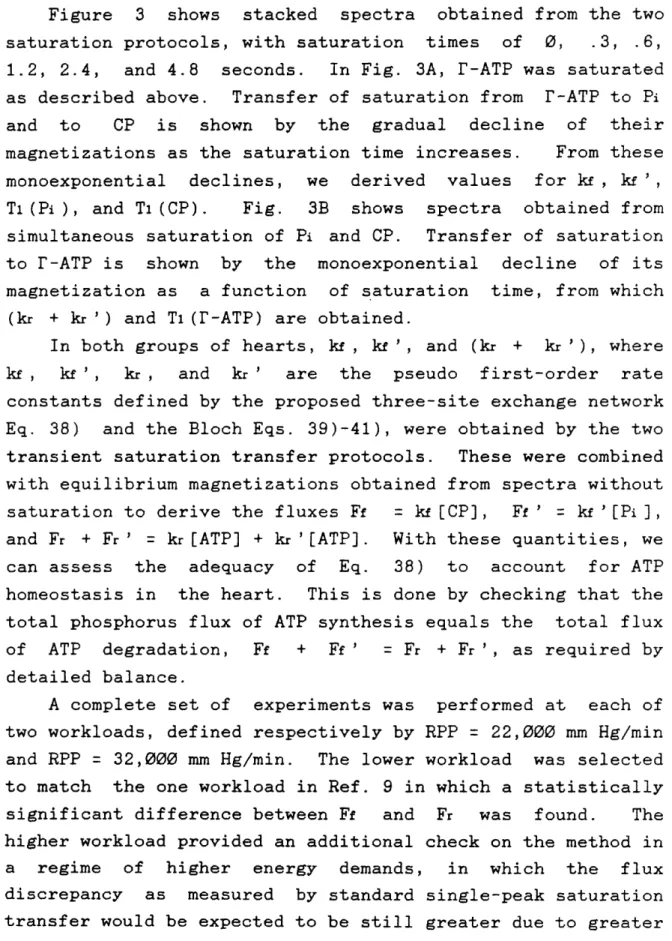
Figure 3 shows stacked spectra obtained from the two saturation protocols, with saturation times of 0, .3, .6, 1.2, 2.4, and 4.8 seconds. In Fig. 3A, r-ATP was saturated as described above. Transfer of saturation from r-ATP to Pi and to CP is shown by the gradual decline of their magnetizations as the saturation time increases. From these monoexponential declines, we derived values for k , kf ', T1(Pi), and T(CP). Fig. 3B shows spectra obtained from simultaneous saturation of Pi and CP. Transfer of saturation to r-ATP is shown by the monoexponential decline of its magnetization as a function of saturation time, from which
(kr + kr') and Tl(r-ATP) are obtained.
In both groups of hearts, kf , k ', and (kr + k '), where kf , kf ', kr , and kr ' are the pseudo first-order rate constants defined by the proposed three-site exchange network Eq. 38) and the Bloch Eqs. 39)-41), were obtained by the two transient saturation transfer protocols. These were combined with equilibrium magnetizations obtained from spectra without saturation to derive the fluxes Ff = [CP], Ff' = k '[Pi], and Fr + Fr' = kr[ATP] + kr '[ATP]. With these quantities, we can assess the adequacy of Eq. 38) to account for ATP homeostasis in the heart. This is done by checking that the total phosphorus flux of ATP synthesis equals the total flux of ATP degradation, Ff + Ff' = Fr + Fr', as required by detailed balance.
A complete set of experiments was performed at each of two workloads, defined respectively by RPP = 22,000 mm Hg/min and RPP = 32,000 mm Hg/min. The lower workload was selected to match the one workload in Ref. 9 in which a statistically significant difference between Ff and Fr was found. The higher workload provided an additional check on the method in a regime of higher energy demands, in which the flux discrepancy as measured by standard single-peak saturation transfer would be expected to be still greater due to greater
activity of ATPase systems.
The quantities measured in the NMR experiments at each workload were the relative concentrations of CP, r-ATP, and Pi, the spin-lattice relaxation times Ti(CP), Ti(r-ATP), and T1(Pi), and the kinetic constants kf, (kr + kr'), and kf'. Note that of these, the double-saturation technique is required to obtain correct values only for T(r-ATP) and (kr + k '), while standard saturation-transfer experiments suffice to obtain T(CP), T(Pi), kf, and kf'.
From the experimental data, we derived
Fayn = total ATP synthesis = kf [CP] + kf '[Pi], Fdeg = total ATP degradation = (kr + kr ')[r-ATP], FCK = flux through the CK reaction = kf [CP], and
FATPase = flux through ATPases = k '[Pi].
The values of the measured quantities and the fluxes are given in Table 1. There were no significant differences in any of the measured or derived quantities between the two workloads investigated. The derived ratio of phosphorus flux through the ATPase reaction to the flux through the CK reaction was greater by a factor of two at the higher workload, but the increase was not statistically significant due to the size of the errors.
The main result of this study is that the three-site model Eq. 38) is indeed adequate to show detailed balance in the combined CK-ATPase system of the heart, and suffices to demonstrate ATP homeostasis directly. As shown in Table 1, the ratio of the phosphorus flux of ATP synthesis to the flux of ATP degradation at each of the two workloads was indistinguishable from unity: at RPP = 22,000 mm Hg/min, Fsyn/Fdeg = .94 + .18, while at RPP = 32,000 mm Hg/min,
Fsyn/Fdeg = 1.18 + .18.
It is useful to make a direct comparison of our results to those of Ref. 9, in which a similar series of experiments was carried out but with the difference that the reverse CK reaction was analyzed with saturation applied only at CP, rather than at both CP and Pi, as in the present study.
In Fig. 4, the left-hand panel displays our values for the fluxes Fdeg and Fyn, derived from the spectroscopic data as discussed above. At both workloads, Fdeg Fsyn. To calculate Fsyn and Fdeg from the data of Ref. 9, we used Fsyn = k [CP] + kf'[Pi], as in the present study, and Fdeg =
kr[r-ATP]. The right-hand panel of Fig. 4 shows these fluxes. As discussed in the Theory section of this Chapter, the validity of this derivation of Fdeg rests on assumptions which may not be valid at these workloads. Here, we see that Fsyn and Fdeg are substantially different.
Fig. 5 shows a direct comparison between the double-saturation experiments of the present study and the results of Ref. 9, with respect to apparent balance between ATP synthesis and ATP degradation. As shown explicitly in the Theory section of this Chapter, single-saturation would be expected to underestimate the rate of ATP degradation. In fact, as seen in Fig. 5, at a workload of 22,000 mm Hg/min, the single-saturation experiment underestimated Fdeg to the extent that the apparent Fsyn/Fdeg = 1.51 + .25. In contrast, the double-saturation treatment of Fdeg lowers this to .94 + .18, a value indistinguishable from unity. At the higher workload of 32,000 mm Hg/min, the discrepancy between synthesis and degradation flux is still greater in Ref. 9, Fsyn/Fdeg = 1.69 + .33, while in the present study this ratio is Fsyn/Fdeg = 1.18 + .18, again indistinguishable from unity.
Discussion
At this point, it is worthwhile to reemphasize exactly what type of information one obtains in a saturation transfer experiment. The chemistry of a kinetic system in steady-state is fully characterized by two sets of relationships. First are the appropriate statements of the law of detailed balance, which must hold in steady state when all reactants are properly accounted for. Second, to interpret the saturation transfer data, an assumption must be made about the form of the exchange network. Since detailed balance is always true in the steady-state, and is also an underlying assumption of all present saturation transfer-techniques, the saturation transfer experiment itself provides no further information about that set of relationships. However, the relations of detailed balance themselves furnish internal checks on the kinetic constants derived from the data, and provide the ability to test the validity of an assumed reaction network.
To take the simplest example, consider again the simple two-site exchange, 1):
kB A
1) A B
kAB
Either steady-state or transient experiments may be performed to determine T(A) and kAB, and, in a separate experiment,
T1(B) and kBA. If the reaction network as written is correct, then
42) kAB = kBA(Me(B)/Me(A))
should be obtained. Provided that this relation is satisfied within experimental error, one may assume that departures from the scheme Eq. 1), such as enzymatic intermediates or