Printed in the United States of America
Sensory feedback to the
cerebral cortex during voluntary
movement in man
P. E. Roland
Department of Clinical Physiology, Bispebjerg Hospital, Bispebjerg Bakke 23 DK-2400, NV, Copenhagen, Denmark
Abstract: This article describes a series of experiments directed toward the following questions: (1) Do signals from musculotendinous
receptors reach consciousness? (2) Does feed-forward information of muscular force and expected extent of voluntary movement exist?
To answer these questions data from voluntary compression of springs and strain-gauge have been analysed in healthy young subjects
and in patients with unilateral focal lesions of the cerebral hemispheres.
By successive elimination of information from other sources it was possible to verify that receptors in muscles and tendons do signal
movement magnitude and muscular tension to the cerebral cortex, and that this information does indeed reach consciousness. There
also exists a feed-forward mechanism signalling parameters of voluntary contraction. However, it is unclear whether peripheral,
subcortical, or intracortical loops are directly involved. Perception of signals of muscular tension is abolished by lesions of the
contralateral cortex near the central sulcus. It is possible that there exist separate cortical projection areas for kinaesthetic signals from
muscles and from joints.
Keywords: consciousness, discrimination, kinaesthesia, motor feedback, muscle receptors, perception, voluntary movement
Are our muscles "insentient"? Do signals from
musculo-tendinous receptors remain unconscious? Or do signals from
receptors in muscles and tendons provide man with conscious
information about the force exerted and the extent of movement
during voluntary contraction? This article is an attempt to give a
consistent answer to these questions.
The motor system is the part of the nervous system that
con-trols contraction of striate muscles. The nervous impulses that
initiate voluntary muscular contractions are presumed to
originate in the cerebral cortex and thence to spread to
subcortical parts of the motor system. By way of the large (a)
mo-toneurones and smaller (y) momo-toneurones in the anterior horn of
the spinal cord, these impulses are transmitted through the
pe-ripheral nerves to the respective motor endplates. At the motor
endplates the nervous impulses release a chemical transmitter
(acetylcholine), which in turn excites the two types of muscular
fibers and induces contraction.
Voluntary contraction need not be accompanied by movement.
Under some circumstances, when the muscles are used to
op-pose external forces, there is no movement of the joints and the
length of the muscles remains constant (isometric contraction).
Voluntary movements, like all other movements of solid
ob-jects, consist of operations in time and space specified by
physical parameters. When, for example, a person lifts a cup of
tea to his lips, this voluntary movement is specified by the
trajec-tory of the teacup, the force vectors, the acceleration and velocity
at every point, the total length of the path, the locus of origin, and
the time of onset. Voluntary movements of this kind are normally
carried out quickly and precisely (by adults) and without
in-formation concerning the total mass of the cup or its contents.
There are two types of explanations of the often surprising
precision with which we move our limbs. One is that the motor
system calculates in advance the values of movement parameters
sufficiently accurately to assure successful performance. The
other type of explanation is based on the fact that every muscular
contraction inevitably changes the state of receptors in muscles
and tendons and normally also of those in joints and skin
(Fig-ure 1). These receptors are able to meas(Fig-ure the parameters of
voluntary contraction and transmit this information to the motor
system. The motor system is then thought to control voluntary
contraction under the guidance of sensory feedback from these
receptors. Although there is no contradiction between these two
explanations, physiologists disagree as to the relative importance
of specific motor commands versus sensory feedback.
Another and equally important dimension in the
understand-ing of the physiological organization of voluntary motor control is
the question of the levels in the central nervous system at which
the desired values of voluntary contraction parameters are
calcu-lated; or, in other words, the extent to which voluntary
contrac-tions are automatisms. Because man is able, consciously, to vary
the parameters of voluntary contraction in an infinite number of
ways, one might be tempted to believe that, at the moment they
are triggered, descending signals from the cerebral cortex
provide the desired values of the contraction parameters in
coded form. The task left for the subcortical part of the motor
system would then consist solely of compensating for
unex-pected events during the movement or contraction (for instance,
should the aforementioned teacup prove unexpectedly heavy).
In this model, sensory feedback has a dual role: partly to provide
concurrent sensory feedback to the subcortical parts of the motor
system about the parameters of contraction, and partly to update
cerebral cortical information concerning the consequences of
voluntary contraction in the peripheral field of action; the latter
would be for later use in the programming of further voluntary
contractions. The contrasting view is that coded motor output
from the cerebral cortex does not specify trajectories, force
vec-tors, velocity, acceleration, and so forth, but merely constitutes a
message for the subcortical motor systems concerning the
ulti-mate goal of the voluntary contraction. The rest of the motor
or-ganization of voluntary contraction would then be carried out
au-tomatically, with calculation and control of contraction
parameters left to the subcortical motor system; sensory
feed-JOINT
FINGER
A D Ui GOLGI INTRAFUSAL FIBERS' O R G A NN/ EXTRAFUSAL FIBERS —/ y PRIMARY ENDING
ANTERIOR ROOT
Figure 1. Simplified diagram of the sources of afferent information
dur-ing voluntary contraction. The muscles that move the fdur-inger are located in
the forearm. Muscles consist of two types of muscular fibers. Extrafusal
fibers do the muscular work and are innervated by a-motoneurones in the
anterior horn of the spinal cord. Each a-motoneurone innervates some
20-200 extrafusal fibers. The a-motoneurone and the extrafusal muscle fibers
it innervates are called a motor unit. The intrafusal fibres, components of
the muscle spindles are specially developed contractile sense organs
ar-ranged in parallel with the extrafusal fibers. Muscle spindles may be
further divided into two classes on a morphological basis: nuclear bag
fibers and nuclear chain fibers. Both have two kinds of receptors: the
primary endings, which mainly record muscular length and velocity; and
the secondary endings, whose function is still obscure. The functional
pat-tern of these two receptors is complicated because they are contractile and
can be activated by four different routes: (1) by a change in muscle length;
(2) as the result of impulses from the a-motoneurones (in other words,
dur-ing muscle contraction and relaxation); (3) directly from the
a-mo-toneurones (so-called /8-innervation); (4) by the y-moa-mo-toneurones that
in-nervate the muscular part of the receptors. The matter is further
compli-cated because there exist two types of y-motoneurones: y-static
andy-c/t/-namic fusimotor neurones. Each type has small separate contacts with the
intrafusal fibers, where a neurotransmitter substance is leleased, which in
turn excites the muscular part of the muscle spindle Electrical
stimula-tion of the y-dynamic neurones increases the sensitivity of the primary
endings to the velocity of a linear stretch of the muscle; stimulation of the
y-static neurones increases the number of impulses from the secondary
endings. Nuclear bag fibers are thought to receive mnervation from
they-dynamie fusimotor neurones while nuclear chain fibers are thought to be
innervated by y-static fibers (P. B. C, Matthews, 1972). In this way, the
muscle spindles can be "tuned" to give a very differentiated response
dur-ing voluntary contractions
The extrafusal fibers are likewise subdivided into two types:
slow-twitch fibers (or "red" fibers) and fast-slow-twitch fibers (or "white" fibers).
These names refer to the color and the velocity of contraction upon brisk
electrical stimulation
In series with the extrafusal fibers are the Golgi tendon organs, which
measure musculai tension and changes in tension. If a muscular
contrac-tion leads to a movement of the joints, then joint receptors signal the
change in position. In addition, mechanoreceptors in the skin and
subjacent tissue can signal changes at the skin surface as well as pressure
if the movement is opposed by some external object. To this afferent
in-formation must be added signals from tendon organs and primary and
secondary endings The sensory part of the peripheral nerve carries
af-ferent fibers from skin receptors, joint receptors, Golgi tendon organs
(groups //;-afferents), primary endings (group /rt-afferents) and
second-ary endings (group //-afferents) to the spinal cord Here the afferents
from the spindles activate a-motoneurones such that these neurones fire a
short burst of impulses that can once again reactivate the whole system.
back from receptors in muscles and tendons would be
transmit-ted exclusively to this part of the motor system. There would be
no need for sensory feedback from these receptors to the cerebral
cortex, because the latter could make no use of such afferent
in-formation, any more than a driver can use the magnitude of
pro-pulsive and restrictive force vectors from road and air friction in
order to change speed. Although this highly developed servo
system is supposed to compensate for all unexpected events
dur-ing contraction, the cerebral cortex needs some additional
in-formation as to whether the ultimate goal has indeed been
at-tained. Ordinary visual and auditory feedback, eventually in
combination with some information from the skin and the joints,
is usually sufficient.
These four explanations are neither topical physiological
models nor theories, therefore no references have been given.
Rather, they are four extreme viewpoints that provide
cornerstones for various existing theories concerning the role of
sensory feedback in voluntary motor control.
The vast majority of investigations of the afferent signals from
muscle spindles and tendon organs have been carried out on
anaesthetized animals whose muscles have been subjected to
various forms of stretch; hence little is known about the sensory
function of musculotendinous receptors during voluntary
contraction in man. The results from animal experiments of this
kind have gradually led to the view that one of the main
func-tions of the muscle spindle is in the servo control of muscular
contraction (Merton, 1951, 1953). A simplified diagram of the
ele-ments in the muscle servo is shown in Figure 2. The most
im-portant detector element in the loops is the muscle spindle. As
muscle spindles not only react to muscular contractions, but do
so in a way that is controlled by efferent impulses from
y-mo-toneurones, it is quite difficult to ascertain whether or not these
sense organs can really measure some of the parameters
pre-viously mentioned (extent of movement, velocity, acceleration).
This was exactly the type of argument raised by Rose and
Mount-castle (1960) when they stated that muscular receptors could not
measure joint angles or extent of movement. This more
theoretical rejection of man's faculty to perceive signals from
muscle spindles was supported by experiments of Chambers and
Gilliat (1954), Provins (1958), and Merton (1964). These authors
anaesthetized the skin and joints of the fingers in human beings
and found that this abolished sensation of passive finger
move-ment, while active movements of the joints continued to be made
with approximately the same positional accuracy as in the
unanaesthetized state (Merton, 1964). Apparently this was not
due to impulses from muscle spindles, because when
anaesthetized subjects were instructed to oppose passive
move-ment by muscular contraction, they showed no improvemove-ment in
perception of stationary joint angles (Provins, 1958).
Further experiments were set up to prove that it was
impossi-ble for man and animals to perceive signals from either muscle
spindles or from tendon organs. For example, Gelfan and Carter
(1967) conclude:
"The problem of'conscious proprioception,' whether there
is awareness of muscle length and tension changes, was
investigated in volunteer patients whose muscle tendons
available at the wrist and ankle were exposed under local
anesthesia restricted to the skin. All subjects tested uniformly
failed to detect length and tension changes when only the
muscles were stretched. The signals from the tension and stretch
receptors did not contribute to the kinesthetic sense when the
joints of the fingers, hand or foot were also moved. None of the
sensations experienced, including awareness of position and
movement of joints, were referable to signals from muscle
spindles or Golgi tendon organs. It was concluded that there is
no muscle sense in man."
In addition, Swett, Bourassa, and Inoue (1964) and Swett and
Bourassa (1967) showed that it is impossible to condition a cat to
respond (by pressing a bar) to electrical stimulation of afferents
from primary endings and Golgi tendon organs. On the basis of
these and other experiments (e.g., Brindley and Merton, 1960)
the conviction soon became widely adopted that it is impossible
to perceive signals of muscular tension and change in limb
posi-tion from musculotendinous receptors (e.g., Rose and
Mount-castle, 1960; Matthews, 1964; Gardner, 1967; Merton, 1964a,
1964b, 1970, 1972; Mountcastle and Darian-Smith, 1968;
Phillips, 1969).
130
THE BEHAVIORAL AND BRAIN SCIENCES (1978), 1 https://doi.org/10.1017/S0140525X00060386FEED BACK
-CONTROL
Figure 2. Schematic survey of interrelations of some components of the
"muscle servo." The main principle is this: If a-innervation is initially too
weak and extrafusal fiber tension too small to overcome the external load
(symbolized by the coil spring), the muscle is stretched over and above
the preprogrammed length during contraction and along with it are
stretched the parallel coupled muscle spindles. This releases increased
impulses in the spindle afferents (group la and II), which polysynaptically
increases the impulses from the already active a-motoneurones to the
ex-trafusal fibers and causes a recruitment of previously inactive
a-motoneurones; the muscle is thereby supplied by new active motor units.
The net effect depends, of course, on how the y-dynamic and y-static
neurones are biased from their control centers, and also on the time lag
between intra- and extrafusal activation during voluntary contraction.
These two questions and that of whether the a-motoneurones are preset to
a desired length or to a desired tension have generated some of the main
physiological controversies in the last twenty-five years. In addition, at the
spinal level the system is modulated by negative feedback from Renshaw
cells and, at very high tensions, by negative feedback from Golgi tendon
organs. The broken line symbolizes facilitating effects on the
a-mo-toneurones to the flexor muscles in the limbs. This facilitation is probably
mediated at supraspinal levels. (A pair of muscles with opposing effects on
a joint are called agonists and antagonists. If the movement is a flexion of a
joint, all flexor muscles are agonists, and all extensors are antagonists.)
Even when muscle and tendon receptors are excluded, there
are other sources that may supply the cerebral cortex with
in-formation about the parameters of voluntary contraction (Figure
1). It is well known that phasic and tonic receptors in the joint
capsules measure joint position (Goldscheider, 1898; Boyd and
Roberts, 1953; Skoglund, 1956) as well as length, velocity, and
direction of movement (Skoglund, 1956, 1973). In primates,
af-ferents from these receptors are known to project to the cerebral
cortex (Mountcastle and Powell, 1959; Werner and Mountcastle,
1963). In addition, it is possible that receptors in the connective
tissue around joints measure tension (Skoglund, 1956; Millar,
1972). However, sensory feedback from these receptors and from
others around joints cannot be the cause of the continued
ac-curacy of voluntary movements in Merton's subjects since the
signals from these receptors were all blocked by anaesthesia. As
the muscles were held to be insentient, it was necessary to
formulate a new hypothesis in terms of which the necessary
in-formation for a sense of position during anaesthesia was ascribed
to a central origin.
The feed-forward hypothesis
The physiological models that will be mentioned under this
de-signation all have the common feature that the cerebral cortex
is informed a priori about some of the physical parameters of
voluntary contractions (Figure 3). Gradually increasing insight
into the functions of alpha and gamma motoneurones and muscle
spindles has led to the common conviction that voluntary
move-ments are to a large extent automatic in character. It is widely
ac-cepted that voluntary contractions are, from the moment they
are triggered in the cerebral cortex, under the control of
subcortical and hence automatic mechanisms, and that
distur-bances are partially or entirely compensated by the muscle servo
mechanism. In the 1960s, afferent signals from muscular
recep-tors and Golgi-tendon organs were considered to be subjectively
inaccessible features of the internal working of the servo
mechanism. It was thought that if sensory feedback from Golgi
organs and muscle spindles reached consciousness this would
only serve to disturb the action of the muscle servo, because any
C
0
R
T
I
C
A
•~| MOTOR CORTEX |«f~| SENSORY C O R T E T
S
U
P
R
A
S
P
I
N
A
L
S
E
R
V
0
\f-\ SENS
S E
N S 0 R I U M COMPARATORRENSHAW
CONTROL
m-CONTROL
EXTRAFUSAL FIBRES1
JOINT U SKINREC MR
Figure 3. Block diagram of some of the main elements in the
feed-forward hypothesis. The blocks do not refer to anatomical entities. Before
it was known that group I and group II afferents from muscle spindles
projected to the cerebral cortex, it was supposed that every self-induced
muscular contraction was accompanied by corollary discharges from
mo-tor areas into sensory systems serving to prepare the latter for the expected
changes that would ordinarily result from the movement (the path ec in
the figure). After it became known that both group I and group II afferents
project to the cerebral cortex, this path (b) was mainly thought of as the
af-ferent link in a transcortical load compensation reflex. In addition, it is
possible that afferents from skin and joints can participate in the
trans-cortical load-compensation reflex. The comparator receives information
about descending motor impulses and this information is compared with
afferent feedback from the spindles and tendon organs. If any disturbance
occurs during contraction, it will be compensated by impulses from the
comparator to the Renshaw and ^-control, which in turn set the new bias
of the muscle servo. The afferent information signalling to the cortex that
contraction has been appropriately carried out comes from skin and joint
receptors and sometimes, in addition, from the eye and ear. Information
from these sources reaches the sensoriurn, that from other peripheral
sources information does not.
misalignment between desired and actual muscular length and
tension was already taken care of automatically by the muscle
servo (Merton, 1964a). The discovery that spindle afferents did
indeed project to the cerebal cortex (see below) only led to the
interpretation that these projections were the afferent path of a
transcortical servo loop (Phillips, 1969, Merton, 1970,1972). The
muscles were still held to be insentient.
On the basis of man's evident capacity to adjust his commands
to the motoneurones in proportion to the requirements of
ex-pected performance, Merton (1964a, 1964b) reintroduced the
concept "sense of effort." The sense of effort was a kind of
con-scious "feeling of innervation" that accompanied the departure
of voluntary impulses from the cortex (1964a, 1964b, 1970). In
the version of von Hoist (1954), an "efference copy" of the motor
command is stored, that is, the coded output from the cerebral
cortex "leaves a copy of itself somewhere in the central nervous
system to which the reafference (sensory feedback) of this
move-ment compares as the negative of a photograph compares to its
print." If there is no mismatch between the efference copy and
the sensory feedback, nothing further happens; if there is, the
difference will have certain effects according to the particular
or-ganization of the motor system for the species studied, which for
primates and man should mean that the muscle servo
com-pensates for the difference. Yet, it is not clear where this
ef-ference copy is stored and where the comparison takes place in
the central nervous system. In Sperry's (1950) version of the
same principle, "corollary discharges" issue from the motor
cortex to the sensory cortex to be compared with the sensory
feedback from the periphery.
The vagueness with which the feed-forward hypothesis is
expressed makes empirical testing difficult. The problem is that
the nature of the motor command from the cerebral cortex is not
specified. There is no evidence for the claim that replicas of
outgoing motor commands are stored somewhere in the central
nervous system or reach the sensory cortex. It is especially
un-clear whether feed-forward impulses contain any information
about the parameters of voluntary contraction. Yet Merton
(1964a) mentions "that we know through our sense of effort
which way our eyes are pointing in the dark, and in the thumb
experiment the subject knows through his sense of effort how far
he had moved the pointer." Gandevia and McCloskey (1977a)
use "sense of effort" in another way, to refer to centrally
generated sensations concerned with the estimation of weights
and tensions. Finally, Kennedy (1973) has stated that the relation
between the requisite contraction and control parameters is
genetically determined.
Thus, according to the feed-forward hypothesis, it is
postu-lated that the cerebral cortex or other parts of the central nervous
system receive information about the nature of outgoing motor
commands via intercortical connections or subcortical loops and
that this information reaches consciousness. Although the nature
of the descending motor commands from the cerebral cortex is
usually not specified, some authors have provided various hints
as to how such voluntary contraction parameters might be
avail-able to the subject a priori. These parameters are position,
mag-nitude of movement, force, and estimated load. In the pages that
follow, the validity of the feed-forward hypothesis with respect
to the first three of these parameters will be examined.
Specification of the hypothesis to be investigated
The hypothesis that I shall attempt to support with some recent
experimental results is the opposite of the one just presented. I
propose to show that information about tension and change in
limb position is supplied to the human cerebral cortex by
recep-tors in muscles and tendons during voluntary muscular
contrac-tion, and, further, that this information does indeed reach
con-sciousness.
Although consciousness is a diffuse and ill-defined concept, I
feel obliged to use it because this was the way the present
prob-lem was introduced by previous authors (e.g., Merton, 1964a,
1964b, 1970, 1972; Gelfan and Carter, 1967). To make my
hypothesis amenable to empirical testing, I shall use operational
criteria for consciousness. If the concept is to have any empirical
content, it must have something to do with the capacity to
perform discrimination. To discriminate is to be able, upon
verbal instruction, to distinguish between two physical inputs
and make a decision about the respective magnitudes of one or
more parameters. Similarly, matching of two physical inputs
along one or more parameters should be one of the distinctive
marks of consciousness.
To find out whether information about tension and movement
path length reaches consciousness, it is not sufficient to show
that potentials from group Ib and la afferents (see legend to
Figure I) can be recorded from the human cortex or scalp: such
projections may merely be the afferent link in a cortical
auto-matism such as the accommodation reflex in the visual system. But
if subjects can discriminate and match degrees of applied tension
and movement magnitude, this means that information about
tension and extent of movement is indeed conscious. Of course,
this need not in itself imply that such information is transmitted
from peripheral receptors, for this ability to discriminate could
also be based on feed-forward signals.
Consequently, three kinds of experimental evidence are called
for: first, a demonstration that information about tension and
extent of movement is conscious; second, evidence that
move-ment magnitude and tension signals are transmitted by the
receptors in muscles and tendons; third, data showing that
af-ferents from muscle spindles and tendon receptors project to the
human cerebral cortex.
As it is far from clear which of the muscular contraction
parameters are registered by the various receptors in skin, joints,
and muscles, it is necessary to introduce some sensory functions
that define the relation between contraction parameters and
sensation. Kinaesthesia means feeling of movement, but is often
used synonymously with "position sense." This has caused a
confusion among the static and dynamic proprioceptive
func-tions. Kinaesthesia is used in this article to mean perception of
change in the position of a limb due to muscular contraction.
Kinaesthesia would then be sensory feedback occurring only
during muscular contraction. The other term, position sense, or,
better, statognosia, is reserved for perception of the position of a
limb in space. In clinical neurology, position sense often refers
to sensation of passive movement. Sensations of passive
move-ment are, by definition, sensations, induced by external forces, of
change in the position of a passive limb in the absence of
mus-cular contraction. The term position sense will not be used here.
The expressions sense of tension and sense of force are not
synonyms, although both tension and force are measured in
Newtons. Provided the present hypothesis is correct, man has a
sense of tension. Since it is not possible to measure tension
directly in man without surgical intervention, the experiments
reported below approach the problem through a study of force
output during compression with thumb and index finger. The
measuring device is interposed between thumb and index
finger; dynamics of elastic tissues between measuring device
and muscles are ignored.
Kinaesthesia, statognosia, sense of passive movement, and
sense of tension are all sensory functions; by definition, this
means that the signals upon which these sensations are based
reach consciousness. Note that kinaesthesia involves four of the
physical parameters of voluntary contraction: direction, extent of
movement, velocity, and acceleration. Statognosia involves only
spatial position, while sense of tension involves force and its
time derivatives. Now the hypothesis can be reformulated to
state that kinaesthesia and sense of tension are based, wholly or
in part, on signals from receptors in muscles and tendons.
In summary, during voluntary contraction there may exist
sensory feedback of tension and kinaesthesia from
musculo-132
THE BEHAVIORAL AND BRAIN SCIENCES (1978), 1 https://doi.org/10.1017/S0140525X00060386tendinous receptors, joint receptors and cutaneous receptors
together with feed-forward information about voluntary force.
The relative significance of these sensory mechanisms will be
analysed below.
Experimental evidence for sensory feedback of tension from
musculotendinous receptors
The following evidence is based on recent experiments (Roland,
1975; Roland and Ladegaard-Pedersen, 1977) that will be
sum-marized here to an extent sufficient for detailed criticism. First,
we attempted to show that tension information was conscious by
having subjects (Ss) discriminate the strengths of coil springs. S
sat behind a curtain with arms unsupported and right hand
extended in front. In order to avoid visual cues, the springs,
en-capsulated by small cylinders (Figure 2), were never visible to S.
Likewise, great care was taken to exclude any unintended tactile
or auditory information.
S held the encapsulated spring between thumb and index
finger, the three ulnar fingers maximally flexed. Then, on
com-mand, the spring was compressed with the index finger. During a
two-alternative forced-choice discrimination of spring strength,
S's task was to compare two springs of slightly different strength
and to choose the stronger one (Figure 4).
When a coil spring is compressed, force at a given moment is
related to extent of compression (or movement path length) and
acceleration in the following way (Roland and
Ladegaard-Pedersen, 1977):
Pi = k
dt
2(1)
where P; is the compressing force at a given moment; k is the
spring strength in N/m; Sj is the extent of compression; m is the
mass of the upper cylinder plus one third of the spring mass; and
d
2s/dt
2is the acceleration. Formally, S's task is to solve two
equa-tions, identical with equation (1), in two unknowns (kx and k
2,
corresponding to the strength of the first and second spring).
This solution is possible only if S "knows" the applied force
(P), the path length of the compressing movement (s ) and the
acceleration d
2s/dt
2. In other words, it is not possible to
dis-criminate spring strengths without kinaesthetic as well as force
information. However, as is apparent from Figure 1, there are
various potential sources that could provide the necessary
in-formation: skin receptors, receptors in and around joints,
muscu-lotendinous receptors, and feed-forward signals. To clarify the
relative significance of these sources, information from skin
receptors, joint receptors, and feed-forward signals was
suc-cessively eliminated by anaesthesia and gallamine blockade (see
below).
Signals from skin receptors in thumb and index finger were
blocked by four dorsal injections of 1.5 ml of 2 percent (w/v)
lignocaine (a local anaesthetic) on either side of the proximal
phalanx, near the volar surface (see Figure 1). A rubber band was
tightened around the proximal phalanx 1.5 cm from the
metacarpophalangeal joint to prevent the local anaesthetic from
being removed by the circulation of the blood. During the
ex-periments, effectiveness of anaesthesia was checked several
times by having S attempt to discriminate a difference of over 20
N/cm
2in pressure applied to the fingertips. If S could not
distin-guish the greater pressure fix/m the lesser, anaesthesia was
considered sufficient.
Signals from joint receptors were likewise blocked by
lignocaine injections. The joint capsule and adjacent convective
tissue were infused with 1.5 - 2.5 ml of 2 percent lignocaine and
the same amount was injected into the joint space of the
inter-phalangeal joints, the metacarpointer-phalangeal joints of thumb and
index finger and the carpometacarpal joint. If S was unable to
de-tect an approximately 207sec passive movement of the joint with
muscles relaxed, anaesthesia was sufficient. Thus joint
anaes-thesia severely impaired the sense of passive movement.
At the start of the discrimination experiment, two springs of
different strengths were selected. S was told to compress
maximally the first spring, then to release it and compress and
release the second, and finally to decide which had been the
stronger. The first two springs were always easy to discriminate,
but S was told that the rest would be difficult. The rules for
two-alternative forced-choice discrimination were then explained
(see legend to Figure 4). Apart from this, S received no other
training or instructions. Discrimination limits (see legend to
Figure 4) for twenty-five different springs were measured under
three different conditions: without anaesthesia, with skin
anaesthesia, and with combined anaesthesia of skin and joints.
Under the first condition (no anaesthesia), muscular tension
and pressure against the fingertips yield the same information. In
addition, kinaesthesia might be signalled from three types of
receptors: cutaneous, articular, and muscular. With skin
anaesthetized, Ss spontaneously stated that, prior to compressing
it on command, they were not aware of any spring between their
fingers. Discrimination limits were unaffected by the lack of
in-formation from skin receptors (Figure 5). When skin and joints
were anaesthetized, there were likewise no changes in
discrimi-nation limits, despite the lack of information from skin and joints
(Figure 5). Under skin and joint anaesthesia, Ss are able to
dis-criminate spring strength only if they have conscious information
about the applied force (or tension), the path length of the
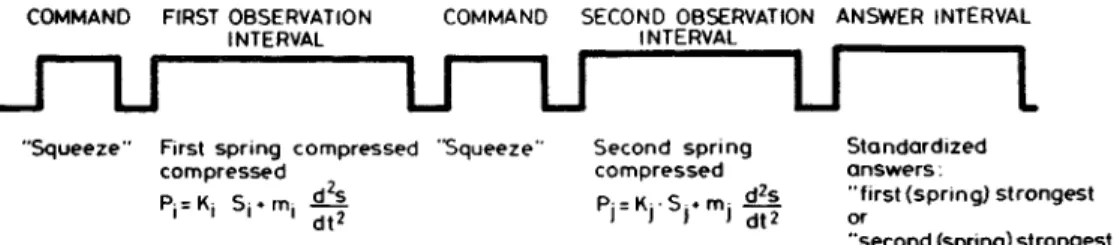
COMMAND FIRST OBSERVATION COMMAND SECOND OBSERVATION ANSWER INTERVAL
INTERVAL INTERVAL
_ru
i_r~u
u
i
"Squeeze" First spring compressed "Squeeze" compressed
Second spring
compressed
Figure 4. Two-alternative forced-choice discrimination. The figure
shows the events in a two-alternative forced-choice discrimination trial. In
this modification of the forced-choice procedure, no feedback is given
about the correctness of the answers. The order of presentation is always
randomized. Here the physical events during spring strength
discrimina-tion are used as examples. Ss were instructed to press the springs on the
command "Squeeze." They were told to respond either "first stronger" or
"last stronger." When trials are repeated with the same two springs (i and
j), the probability of a correct answer (pc) can be calculated as:
Standardized answers:
"first (spring) strongest or
"second (spring) strongest
in which aji = number of trials in which spring j is presented as the first
spring, a
u= number of trials in which spring i is presented as the first
spring, c ,, = number of correct answers when spring j is presented in the
first observation interval, b = number of incorrect answers when spring i
is presented in the first observation interval. Plotting kj - k,, for any given
kj, against the probability of correct answer yields a psychometric curve.
pc = [e
(kr
k'
)2/s]/[l + e
k^
l&]
The pc values run from 0.5 to 1.00. s is a parameter corresponding to the
standard deviation. The discrimination limit for ki is then the differential
quotient of the psychometric curve in the interval (0.5-1.0). The
discrimi-nation limit corresponds roughly to a probability correct level of 0.75.
N cm
SPRING STRENGTH
80
100
120
U0Ncm"1
Figure 5. Discrimination of spring strength: regression of discrimination
limits on spring strength. Each regression line is based on more than
20,000 observations. N: discrimination without anaesthesia; S:
discrimina-tion during skin anaesthesia; S + J: discriminadiscrimina-tion with skin and joints
anaesthetized. The hatching shows confidence limits of N. Eleven
sub-jects participated in each experiment. [From Roland &
Ladegaard-Pedersen, 1977, reprinted with the permission of the editor of Brain.]
compressing movement, and the acceleration (unless the latter is
zero); there being no other cues present it follows that force
in-formation is conscious. However, it cannot yet be concluded
from this that force information is signalled by
musculo-tendinous receptors. If the feed-forward hypothesis is correct, Ss
may be managing with only two types of sensory feedback, say,
extent of the movement and acceleration, with force signalled via
feed-forward loops.
To verify the existence of a sense of tension, the normal
rela-tion between motor signals and muscular force must be altered
or eliminated, with the magnitude of this change "unknown" to
S. The relation between nervous impulses in the motor system
and muscular force is most easily changed experimentally at the
motor endplate, where neuromuscular transmission can be
blocked wholly or in part by curare or curare-like drugs such
as gallamine triethiodide. A sphygmomanometer cuff was
tightened around the upper arm and inflated to a pressure of 300
mm mercury. Thereafter a retrograde injection of gallamine
solu-tion was administered in the forearm veins. After five minutes of
ischaemia (occlusion of blood flow), practically all gallamine was
attached to the receptor sites (motor endplates) in forearm and
hand, and release of the cuff caused only a slight diplopia
(dou-ble vision). The dose used did not cause total paralysis but
allowed S small voluntary contractions amounting to about seven
percent of normal maximum isometric force when the effect of
gallamine was greatest (Figure 6). The experiments stopped
when 75 percent of normal maximum force was restored (Figure
6). The block at the extrafusal motor endplate implies that
volun-tary contractions are possible only within a narrow range of force
in accordance with Figure 6. For example, seven minutes after
injection, a maximal voluntary contraction will only result in a
muscular force of one-fifth normal maximal voluntary force, but
the cortical motor command will still specify maximum force
output to the y-motoneurones and the servo-mechanism. If the
forward hypothesis is correct, and there is no sensory
feed-back of muscular tension, then S would not only fail to recognize
such a gallamine-induced paresis, but he would also greatly
overestimate the muscular force of the gallamine paretic hand.
The effect of gallamine blockade was therefore investigated in
four experiments in which afferent signals from the periphery
were restricted to those from receptors in muscles and tendons,
with skin and joints anaesthetized as described earlier (Figure
7): (1) compression magnitude matching and spring strength
dis-
10-
8-
.6- 4-2
Figure 6. Time course of gallamine induced paresis. At time = 0,
gallamine injection is terminated and experiment starts. Ordinate shows
average ratio of maximal voluntary force from 11 Ss. P
max,
0is maximal
voluntary force for compression with left thumb and index finger before
injection. P
max,t is maximum voluntary force at time t. Cuff is released after
five minutes. Effects of the ischaemiaon the peripheral nerves are thought
to subside one minute after cuff release; thus the total ischaemic period is
six minutes.
crimination without gallamine; (2) compression magnitude
matching and spring strength discrimination with gallamine
in-duced paresis of one arm; (3) force matching without gallamine;
and (4) force matching with gallamine paresis of one arm.
In the gallamine experiments, Ss spontaneously commented
upon their weakness within the first two minutes after injection,
although they were able to move their fingers. Results of the
dis-crimination and matching experiments were likewise
in-consistent with the predictions of the feed-forward hypothesis.
Under the feed-forward hypothesis, one would expect that the
subjective effort to compress the i-spring with the gallamine
paretic hand from 0 to S
twould be much greater than the effort to
compress the reference spring (j-spring) from 0 to Sj. In the
dis-crimination experiment, therefore, the probability of correct
answers should decrease significantly; and in the matching
ex-periment there should be a systematic overestimation of the
force of the weakened hand. As is apparent from the results in
Figure 7, there was no statistically significant decrease in the
probability of correct discrimination of spring strength and there
was no systematic error in the force-matching condition with
gallamine. However, the accuracy of force-matching decreased
after gallamine blockade of the motor endplates, as the measure
of uncertainty of matching (the RMS-value) increased.
In the types of voluntary contraction analysed so far, both
mus-cular tension and kinaesthetic parameters change during the
contraction. As the decrease in accuracy of force-matching
during gallamine blockade might be related to this type of
traction, it was decided to investigate force-matching during
con-ditions in which muscle length was constant (isometric
contrac-tion).
The experimental procedure was nearly the same, but instead
of compressing springs, S was required to press a strain gauge
with his gallamine paretic arm and then match the force of this
contraction by pressing the strain gauge with the other arm. Both
hands had skin and joints anaesthetized as before. A solution of
gallamine in physiological saline was injected into the left arm.
First, S pressed the strain gauge with the weakened left thumb
and index finger until a specified level of force, as indicated by
an audible stop signal, was reached. The actual force of this
contraction, called (P
set), was measured on a peak tension meter.
Then S was required to match this force by pressing the strain
gauge with the right (nonparetic) thumb and index finger. The
force obtained with the right hand is called (P
obt)-Before the experiment, Ss were told that they would receive an
injection of curare and that this would induce paresis. They were
informed that although they might notice that great effort was
re-quired to obtain any more or less forceful muscular contraction of
this limb, it was not in fact their efforts that were of interest but
the actual force produced by their left hand. Ss were told to
remember the force produced at the moment they heard the stop
signal and then to match this force as accurately as possible with
their other hand.
134 THE BEHAVIORAL AND BRAIN SCIENCES (1978). 1 https://doi.org/10.1017/S0140525X00060386
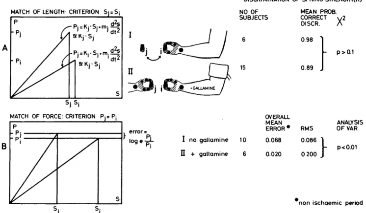
DISCRIMINATION OF SPRING STRENGTH(K)
MATCH OF LENGTH-CRITERION Sj = Sj
p
"
Pj
fr
~F
SS
NO OF
SUBJECTS
MEAN PROB.
CORRECT
V2
DISCR. A
II
15
0.98
0.89
- p > 0.1
S
j S jMATCH OF FORCE: CRITERION Pj= P,
B
p
p-pj y
s
error =
S:
S:
Figure 7 Force discrimination and force matching, with and without
gallamine blockade. A: In the first experiment (I) S matched compression
magnitudes. Indicator spring i (without insertion cylinder) was selected
among the twenty-five springs according to a table of random numbers.
Skin and joints of the hand were anaesthetized. S pressed the spring down
to the prescribed length Si (S! likewise determined from a table of random
numbers). Compression is slow, with a velocity of approximately 4
mm/sec. A stop signal indicated when S
4was reached. Subsequently Ss
matched S
tby compressing a spring of another strength (kj). After this
matching, S was asked which of the two springs was the stronger. Because
the velocity of compression is approximately constant, the second
expression on the right side of the equations vanishes. II: The experiment
was repeated, but now the motor endplates in one arm (the "indicator"
arm) were blocked by gallamine. Skin and joints of both hands were
OVERALL
MEAN
ERROR * RMS
I no gallamine 10
II + gallamine 6
0.068
0.020
0.086
0 200
ANALYSIS
OFVAR
p<0.01
non ischaemic period
anaesthetized. As before, S was asked to discriminate spring strengths (the
slopes of the curves) under a two-alternative forced-choice paradigm. B:
Matching of force. S pressed the first spring (i-spring) slowly down (2
mm/sec) to the prescribed target force (Pj). He then received a spring of
different strength (j-spring) to be matched with the first. His result (Pj)
was noted. The error is then log
e(Pj/Pi). Error was normally distributed.
RMS is the root-mean-square or mean value of the standard deviations
averaged over all trials. This is a measure of uncertainty of matching. The
overall mean error is a measure of systematic deviations from correct
matching. I: Matching of force with skin and joints anaesthetized but no
gallamine. Here the "reference" arm and the "indicator" arm are the
same. II: Matching of force with skin and joints of both hands
anaesthetized, but with the "indicator" arm paralyzed by gallamine.
As gallamine does not pass the blood/brain barrier (Cohen,
Hood, and Golling, 1968), it has no direct influence on outgoing
motor commands from the cerebral cortex; hence, the range
within which the sense of effort operates must be assumed to be
unchanged, even when the effective force range for voluntary
contraction is narrowed by gallamine blockade (Figure 6).
Feed-forward signals, which require force output, must therefore
increase in order to obtain a voluntary contraction of the same
strength with a gallamine paretic hand as with a nonparetic hand.
The most simple assumption is that these signals increase in
pro-portion to the degree ot paresis. Or that
PobtPmax.o ~ Pset/Pmax.t (2)
in which P
maX)o is the maximum force of the left arm before the
gallamine injection and P
max>t is the maximum isometric force in
the paretic left arm at time t. The expected error in isometric
force matching under the feed-forward hypothesis would then be
ee = loge(Pm a x,o/Pm a x,t) (3)
The natural logarithm of (P
obt/P
Set) is used as the error measure
because of its more normal distribution. Equation (2) involves
rather strong assumptions, such as the negligibility of the effects
of the muscle servo. As it seems reasonable that the effect of the
muscle servo should increase with increasing demands of force
output, the expected error predicted by equation (3) is too high.
How much too high depends on the gain of the servo and the
degree of blockade at the motor endplate, since even the muscle
servo has to exert its final effects through the a-motoneurones
and the extrafusal motor endplate.
Results are presented in Figure 8, which shows the mean error
and standard error of the mean for isometric force matching
performance. The mean error reflects systematic deviations from
correct matching. As is apparent from Figure 8, no such
devia-tions occurred during the nonischaemic period. The standard
ror of the mean reflects uncertainty of matching. The standard
er-ror was greater in the ischaemic period (first six minutes) than in
the nonischaemic period (p < 0.01), which stresses the
im-portance of peripheral information in matching. It is further
ap-parent from Figure 8, that Ss indeed perceive the actual force (or
tension) of their gallamine paretic arm, and do not rely on any
feed-forward signals of force. When anaesthesia is sufficient, Ss
have only two main sources of information: receptors in muscles
and tendons, and feed-forward signals of central origin. If the
muscle servo accounts for the greater part of the final force
output from the gallamine paretic hand, then feed-forward
signals need increase only slightly, and the assumption about the
expected error in equation (3) is no longer valid. But as a
conse-quence, it would not be possible to discriminate spring strength
by means of feed-forward information with even approximately
the same accuracy as with anaesthesia alone. On the other hand,
mechanism exists, however, is still unanswered. The assumption
of equation (2) likewise requires testing.
A small group of new Ss was accordingly instructed to attempt
to match with their nonparetic hand their efforts in pressing the
strain gauge with their gallamine-paretic hand. Skin and joints in
both hands were anaesthetized. Instructions and explanations
were analogous to those for the isometric force matching
experi-ment described above. Recall that the expected outcome of this
experiment would be that P
()bt= P
set(Pmax.o/Pmax.t); or, as shown
in Figure 9, that the logarithmic difference in force level
between the nonparetic and gallamine-paretic hand should be
equal to the logarithmic difference in maximum force at time t.
The results of this experiment are presented in Figure 9. In
general, the prediction holds; however, there is a slight but
al-most constant undershoot that is probably due to the additional
force from the muscle servo. So, apparently, man has both a
sense of tension and a sense of effort.
Figure 8. Isometric force matching. The "indicator" arm (Figure 7) is
paralyzed with gallamine. The "reference" arm is not. Both hands are skin
and joint anaesthetized. Ordinate: mean error = mean of log
e(P
ot>t/?«*)» in
which P
set is the prescribed peak force of compression of the strain-gauge
with the paralyzed arm ("indicator" arm). Standard error of the mean is
in-dicated. P
obt. is obtained peak force (or the result of the matching) with the
reference arm t: time from start of experiment (see Figure 6). Expected
er-ror (log
e=
Pmax.o/Pmax.t) is the performance expected on the hypothesis
that matching was based on corollary discharges or feed-forward signals
Note discrepancy between mean and expected error. N = 11. (For further
details, see text.) [From Roland & Ladegaard-Pedersen, 1977, reprinted
with the permission of the editor of Brain.]
if the muscle servo accounts for only a minor part of the final
force output from the gallamine-weakened hand, then the
pre-diction about the expected error holds, and its consequence is
that the performance depicted in Figure 8 would be impossible
unless Ss receive tension information from musculotendinous
receptors.
Some previously overlooked experiments
Some earlier experiments seem to have been overlooked when
Rose and Mountcastle (1960) and Gelfan and Carter (1967)
con-cluded that it was impossible for man to perceive signals about
tension and extent of movement from musculotendinous
recep-tors. Katz (1925) showed that it is possible to judge the elasticity
of an object placed between the teeth, and he believed that
receptors in the masticatory muscles were responsible for this
faculty (see also Roland, 1973).
Before him, von Frey (1914, 1915) studied the "Kraftsinn," or
"sense of force," which in his terminology referred to sensations
transmitted by receptor nerves from muscles and tendons. Ss
dis-criminated torque and moment of inertia with the right arm
cutaneous sensation reduced by a stiff case tightened around the
arm. Von Frey reports the discrimination threshold as being
about 5 percent of the stimulating torque, and about one-tenth of
this value for the moment of inertia.
Renqvist (1927) used the ergometer of Hill (1922) to
inves-tigate flexion-extension movements of the elbow joint. He
systematically changed mass, torque, velocity, and acceleration
parameters and found that two movements were perceived as
equally strong when physical forces were equal.
Evidence for feed-forward signalling of force
during voluntary movement
The experiments on force matching during gallamine block of
the motor endplates show that Ss do not rely on feed-forward
signals of force. The question of whether or not a feed-forward
Experimental evidence for feedback of kinaesthesia from
muscular receptors
In 1972, Goodwin et al., and Eklund independently and
si-multaneously published papers on distortion of statognosia in
man by vibration of the tendons. Eklund (1972) vibrated the
patellar tendon of one of S's legs and then had him track with the
other leg the extension induced by the tonic vibration reflex. He
found that Ss constantly underestimated the extension of the
vi-brated leg. They apparently did not rely on information from the
joints, but followed the information from muscle spindles, which
are strongly stimulated by vibration (Brown, Engberg, and
Mat-thews, 1967). The artificially high afferent inflow from the
spindles probably caused Ss to judge the quadriceps as stretched
more than it actually was.
Goodwin, McCloskey, and Matthews (1972) performed similar
experiments on the upper limb, but showed, in addition, that
when the biceps tendon was vibrated, the illusion of the arm's
being in the direction of stretch persisted during voluntary
contractions of low and medium strength. The illusion
disap-peared during voluntary contractions near maximum force,
however. When joints and skin of the hand were anaesthetized
by ischaemia from a pressure cuff around the wrist, sensations of
passive movement persisted in the fingers, but the prerequisite
for this effect was that the forearm muscles be tensed.
In the above experiments, no attempt could be made to
quantify the precision of muscular receptors as kinaesthetic
recorders because perceptual illusions were involved. Some
recent direct approaches to this quantification (Roland, 1975;
Ro-land and Ladegaard-Pedersen, 1977) will now be described.
Kinaesthetic discrimination was investigated with
encapsu-lated springs similar to those used in the experiments described
above but with the modification that insertion cylinders of
various heights were placed between the bottom of the lower
cylinder and the upper cylinder (Figure 10). In this way, the
magnitude of movement from top position to bottom could be
manipulated. The strengths of the springs were chosen such that
a force of 9.80 N gave maximal compression. Thus, the
mag-nitude of the compressing movement from top position to bottom
was the quantity to be discriminated during voluntary
com-pression. Under the two-alternative forced-choice paradigm
the task was to decide which of the two objects could be
compressed more. Discrimination limits were then investigated
under the same three conditions as before: no anaesthesia,
anaesthesia of the skin, and anaesthesia of skin and joints. Figure
10 shows that discrimination limits were slightly raised during
skin anaesthesia. This was partly due to a raised absolute
threshold for detection of movement per se, and partly to a raised
differential threshold. (The differential threshold or just
notice-able difference refers to the discrimination limit when
stimula-tion is well above absolute threshold.) During combined skin
and joint anaesthesia, sensation of passive movement
disap-136
THE BEHAVIORAL AND BRAIN SCIENCES (1978), 1https://doi.org/10.1017/S0140525X00060386
40- 36- 32- 282 4 - 20- 16- 12- 8- 4- 00-- 400-- 4 8 4 -EFFORT y ^ N. Expected error / " \ / \ CUFF REL 1 tmin 12 U 16 18 EFFORT Expected error 10 12 1A 16 18
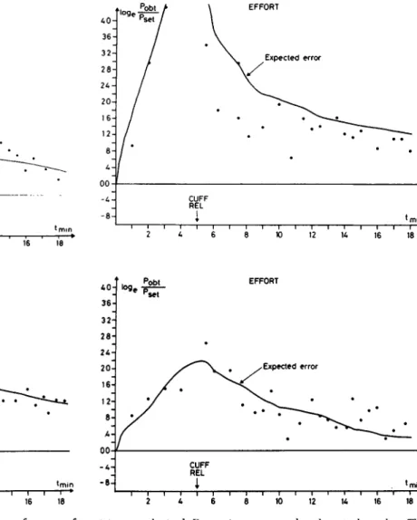
Figure 9. Matching of subjective effort. This shows the performance of
four Ss in matching the subjective effort of a press on a strain-gauge with a
gallamine paretic hand. P
set is the force of the gallamine paretic hand; P
Obt
is the force of the non-paretic "reference" hand. Both hands were skin and
A0- 36- 322 8 - 2A- 20- 16- 128 A -00 A 8 -loge / / / ' Pobt > Pset / / / / \ CUFF REL
i
EFFORT \ Expected error \ • * min A0- 36- 32- 28- 2A- 20- 16- 12- 8- A-00 A 8 -p set / j CUFF REL1
EFFORT y Expected error •min 2 A 6 8 10 12 1A 16 18joint anaesthetized. Data points correspond to the actual matches. The
curve is expected error with perfect effort matching through matching of
outgoing motor discharges. Note the almost constant undershoot, probably
due to the effect of the "muscle servo." (For further details, see text.)
peared, while kinaesthesia was preserved to the same degree as
during skin anaesthesia alone (Figure 10).
The effect on kinaesthesia of gallamine block of the motor
end-plates was also investigated in four experiments in which
af-ferent information from the periphery was restricted to signals
from musculotendinous receptors. Skin and joints were
anaesthetized as previously. The experiments were: force
match-ing and compression magnitude discrimination and matchmatch-ing, all
with and without gallamine block. Figure 11 summarizes the
ex-perimental procedures and the results. Ss exhibited a clear
im-pairment in discriminating voluntary movement magnitude
dur-ing gallamine block. Matchdur-ing capacity was generally poor in
both groups. In both cases, with and without gallamine, the
mag-nitude of error in kinaesthetic matching was correlated with the
ratio of the spring strengths (Figure 12). During compression of
weak springs, voluntary movement magnitude was generally
underestimated, while during compression of strong springs, it
was overestimated. Gallamine blockade caused no further
decrease in kinaesthetic matching capacity, but ischaemia
reduced this capacity considerably (p < 0.01).
In summary, these experiments have shown that muscular
receptors do measure extent of voluntary movement, and that
this information does reach consciousness, because Ss are still
able to perform kinaesthetic discrimination when information
from skin and joint receptors has been excluded by anaesthesia.
However, while muscular receptors are able to measure
move-ment magnitude, they will not signal equality in the path lengths
of two equally long voluntary movements executed with
dif-ferent contraction strengths. On the other hand, skin receptors
also seem to play a role in kinaesthesia, probably directly in the
initial phase of movement, in which the skin is in contact with
the object to be moved.
Some critical remarks about the experiments
Although these experiments clearly show that signals from
mus-culotendinous receptors do contribute to kinaesthesia and
un-derlie the sense of tension, there are some peculiarities in the
action of gallamine that may change the responses of these
receptors in a rather unpredictable way. At lower dosages,
gallamine seems almost exclusively to block the motor endplates
of extrafusal fibers. Under this condition, extrafusal fibers may
hence be paralyzed, with intrafusal fibers affected only slightly
or not at all (Granit, Homma, and Matthews, 1959; Bessou,
Emonet-Denand, and Laporte, 1965; Emonet-Denand and
Houk, 1968). During a normal voluntary contraction, there is
coactivation of a- and -y-motoneurones, such that these cells, and
hence the muscle spindles, increase their firing rate in concert at
the beginning and during the ascending phase of contraction.
(Vallbo, 1971, 1973, 1974). In consequence, afferent input to the
a-motoneurones increases during the same period, despite the
mechanical unloading effect on the spindles. During weak
voluntary isometric contractions, the impulse frequencies of
DISCRIMINATION
/95V./5-confidence limits
LENGTH OF VOLUNTARY MOVEMENT 2 k 6 8 10 12 14 16 18 20 22 24 26 27.5 mm